Identification of an Antimicrobial Agent
Effective against Methicillin-Resistant
Staphylococcus aureus
Persisters Using a
Fluorescence-Based Screening Strategy
Wooseong Kim1, Annie L. Conery2, Rajmohan Rajamuthiah1,2, Beth Burgwyn Fuchs1,2, Frederick M. Ausubel2, Eleftherios Mylonakis1*
1Division of Infectious Diseases, Rhode Island Hospital, Alpert Medical School of Brown University, Providence, Rhode Island, United States of America,2Department of Molecular Biology, Massachusetts General Hospital, Harvard Medical School, Boston, Massachusetts, United States of America
Abstract
Persisters are a subpopulation of normal bacterial cells that show tolerance to conventional antibiotics. Persister cells are responsible for recalcitrant chronic infections and new antibi-otics effective against persisters would be a major development in the treatment of these in-fections. Using the reporter dye SYTOX Green that only stains cells with permeabilized membranes, we developed a fluorescence-based screening assay in a 384-well format for identifying compounds that can kill methicillin-resistantStaphylococcus aureus(MRSA) persisters. The assay proved robust and suitable for high throughput screening (Z`-factor:
>0.7). In screening a library of hits from a previous screen, which identified compounds that
had the ability to block killing of the nematodeCaenorhabditisby MRSA, we discovered that the low molecular weight compound NH125, a bacterial histidine kinase inhibitor, kills MRSA persisters by causing cell membrane permeabilization, and that 5μg/mL of the com-pound can kill all cells to the limit of detection in a 108CFU/mL culture of MRSA persisters within 3h. Furthermore, NH125 disrupts 50% of established MRSA biofilms at 20μg/mL and completely eradicates biofilms at 160μg/mL. Our results suggest that the SYTOX Green screening assay is suitable for large-scale projects to identify small molecules effective against MRSA persisters and should be easily adaptable to a broad range of pathogens that form persisters. Since NH125 has strong bactericidal properties against MRSA persist-ers and high selectivity to bacteria, we believe NH125 is a good anti-MRSA candidate drug that should be further evaluated.
Introduction
A significant challenge in the treatment of bacterial infections has been the appearance of anti-biotic-resistant strains as a consequence of mutation or the acquisition of antibiotic resistance
OPEN ACCESS
Citation:Kim W, Conery AL, Rajamuthiah R, Fuchs BB, Ausubel FM, Mylonakis E (2015) Identification of an Antimicrobial Agent Effective against Methicillin-ResistantStaphylococcus aureusPersisters Using a Fluorescence-Based Screening Strategy. PLoS ONE 10(6): e0127640. doi:10.1371/journal.pone.0127640
Academic Editor:Gunnar F Kaufmann, The Scripps Research Institute and Sorrento Therapeutics, Inc., UNITED STATES
Received:January 19, 2015
Accepted:April 17, 2015
Published:June 3, 2015
Copyright:© 2015 Kim et al. This is an open access article distributed under the terms of theCreative Commons Attribution License, which permits unrestricted use, distribution, and reproduction in any medium, provided the original author and source are credited.
Data Availability Statement:All relevant data are within the paper and its Supporting Information files.
Funding:This study was supported by National Institutes of Health grant P01 AI083214 to EM and FMA and grant U54 AI057159 to NERCE. The funders had no role in study design, data collection and analysis, decision to publish, or preparation of the manuscript.
genes through horizontal gene transfer, as well as the transient reversible selection of antibiot-ic-tolerant persister cells during antibiotic therapy in individual patients. Most current antibi-otics target essential biosynthetic processes such as DNA replication, protein synthesis, or cell wall synthesis that occur during bacterial growth [1,2]. Antibiotic resistance can be caused by enzymes that degrade or modify the antibiotic, efflux pumps that export the antibiotic, or mu-tations that modify antibiotic targets [1]. A well-known example of antibiotic resistance is methicillin-resistantStaphylococcus aureus(MRSA), which was first identified in the 1960s as a hospital-acquired infection [3], but in recent years has been increasingly prevalent in the gener-al population (community-associated MRSA) [4].S.aureuscauses approximately 10,800 deaths per year in the United States and approximately 50% of these are due to MRSA [5]. Moreover, although vancomycin is currently used to treat MRSA as an antibiotic of last resort, vancomycin-resistantS.aureus(VRSA) strains have started to emerge, motivating the urgent development of new antibiotics effective against antibiotic-resistantS.aureus[6].
In contrast to antibiotic-resistant bacteria such as MRSA, antibiotic-tolerant bacteria, known as persisters, are phenotypic variants that exist as a subpopulation of normal cells. Per-sisters are non-growing dormant bacteria where the targets for most conventional antibiotics are inactive [7,8]. Persisters were first identified by Bigger in 1944 [9], but the molecular mech-anisms underlying persister formation are still only partially understood. Recent studies have shown that toxin-antitoxin (TA) modules play an important role in persister formation [10]. Under specific stresses, antitoxins are degraded and the resulting active toxins inhibit cellular processes, which eventually leads to persister formation [8]. Recent studies have shown that persisters are involved in chronic infections and are responsible for the recalcitrance of chronic infections to antibiotic chemotherapy [11,12]. Importantly, persisters are also responsible for the antibiotic tolerance of biofilms [13], surface-associated microbial communities encapsulat-ed by a self-producencapsulat-ed extracellular polymeric matrix, that are involvencapsulat-ed in up to 65% of bacterial infections in developed countries [14].
The bacterial cell envelope consisting of the bacterial membrane and cell wall is a promising target for novel antibiotics that would potentially be effective against both normal and persister cells. The bacterial cell envelope is essential for cell survival and contains about 30% of bacterial proteins, many of which are essential for survival [15–17]. Indeed, many types of antibiotics that target the cell envelope, including proteins, peptides, and small molecules, have been shown to be efficacious againstS.aureus[17–22]. For instance, lysostaphin and endolysins kill S.aureusby hydrolyzing peptidoglycan, which results in membrane permeabilization [18–20]. Nisin A, daptomycin and telavancin killS.aureusby inducing membrane permeabilization and depolarization [17,22]. Although their specific modes of action are different, the common feature of these agents is that each directly or indirectly induces rapid membrane permeabiliza-tion, which correlates with bactericidal activity [20,22,23].
Materials and Methods
Bacterial strains, growth conditions, and persister isolation
Community-acquired methicillin-resistanceS.aureus(CA-MRSA) strain MW2 BAA-170 was obtained from ATCC (Manassas, VA, USA). To isolate persister cells, overnight cultures of MW2 grown in tryptic soy broth (TSB) (BD) at 37°C were treated with 10X MIC (20μg/mL)
gentamicin for 4 h [26].
Antimicrobial agents and chemicals
All antibiotics except for NH125 were purchased from Sigma-Aldrich. NH125 was purchased from Tocris Bioscience. 10 mg/mL stock solutions of all antibiotics except for nisin A were made in DMSO or ddH2O. As described in a previous study [27], nisin A (hereafter nisin)
stock solutions were prepared in 0.02N HCl at a concentration of 250μg/ml.
Lysostaphin-treated samples were supplemented with 0.1% bovine serum albumin (BSA) to prevent non-specific adherence to plastic surfaces [28].
Persister membrane permeability assay
Black, clear-bottom 96-well plates (Corning no. 3904) were filled with 50μL of PBS/well
con-taining the indicated concentration of antibiotics. To isolate MRSA persisters, 25 mL of an S.aureusMW2 culture was grown to stationary phase and then treated with gentamicin at 20μg/mL for 4 h, as described above. Then bacteria were washed 3 times with the same volume
of phosphate buffered saline (PBS). The washed cells were diluted to OD600= 0.4 (~2 x 10⁸
CFU/mL) with PBS. SYTOX Green (Molecular Probes) was added to 10 mL of the diluted per-sister suspension to a final concentration of 5μM and incubated for 30 min at room temperature
in the dark. 50μL of the persister/SYTOX Green mixture was added to each well of 96-well plates
containing antibiotics and fluorescence was measured at room temperature for up to 4 h using a spectrophotometer (SpectraMax M3, Molecular Devices) with excitation and emission wave-lengths of 485 and 525 nm, respectively. All experiments were conducted in triplicate.
Time course assay
Persister cells were prepared by treating 25 mL of anS.aureusMW2 culture in stationary phase with gentamicin at 20μg/mL for 4 h, as described above. The isolated persisters were
washed 3 times with PBS and diluted to OD6000.4 (~2 x 10⁸CFU/mL) with the same buffer.
1 mL of the persister suspension containing 10X MICs of indicated antibiotics was added to the wells of a 2 mL deep well assay block (Corning Costar 3960) and incubated at 37°C, with shaking at 225 rpm. At specific times, 50μL samples were removed, serially diluted, and
spot-plated on tryptic soy agar (TSA, BD) plates to enumerate the number of persister cells. These experiments were also conducted in triplicate.
Compound screening
compound from the library. 20μL of the MRSA persister/SYTOX Green mixture prepared as
described above was added into each well of the 384-well plate containing compounds. 0.1% di-methyl sulfoxide (DMSO) or 1.25μg/mL lysostaphin were included in two columns as negative
and positive controls, respectively. Fluorescence was measured as described above. To identify hits, a Z-score was calculated from the fluorescence intensity data; Z-score = (x-μ)/σwhere x is
the fluorescence intensity of each sample,μis the mean of the sample population, andσis the
standard deviation of the sample population [29]. Samples with a Z-score>3 were
considered hits.
Z
’
-factor evaluation for assay quality
The robustness of the screening assay was evaluated based on the Z’-factor [30]. Z’-factor = 1-((3σp+3σn)/|μp-μn|) whereσpandσnare the standard deviations of the positive and negative
controls, respectively andμpandμnare the means of the positive and negative controls,
respec-tively [30]. 1>Z’-factor0.5 indicates a robust assay [30]. Fluorescence intensity data from a 384-well plate where half of the wells were filled with 0.1% DMSO (negative control) and the remaining wells contained 1.25μg/mL lysostaphin or 80μg/mL nisin (positive controls) were
used to calculate the Z’-factor.
Minimal inhibitory concentration (MIC) assay
The MICs of antibiotics were determined by the standard micro-dilution method recom-mended by the Clinical and Laboratory Standards Institute [31]. The assay was conducted in triplicate.
Biofilm persister viability assay
An overnight culture of cells was diluted 1:200 with TSB supplemented with 0.2% glucose and 3% NaCl [32]. A 13 mm diameter Millipore mixed cellulose ester membrane was placed at the bottom of each well of a 12-well plate (Falcon 353043). 1 mL of the diluted culture was added to each well and incubated statically at 37°C for 24 h. To remove planktonic cells, the mem-branes were washed 3-times with PBS and transferred to a new 12-well plate. 1 mL of PBS with 10X MICs of antibiotics was added to each well and the plate was incubated statically at 37°C for 24 h. The membranes were washed 3 times with PBS, placed in 1 mL PBS, and sonicated in an ultrasonic bath (Fisher Scientific FS 30) for 10 min. The sonicated samples were serially di-luted with PBS in a 96-well plate. The didi-luted samples were spot-plated on TSA plates and in-cubated at 37°C overnight. The experiment was conducted in triplicate.
Biofilm disassembly assay
An overnight culture ofS.aureusMW2 was diluted 1:200 with TSB supplemented with 0.2% glucose and 3% NaCl [32]. 100μL of the diluted culture was added to each well of a
U-bot-tomed 96-well microtiter plate (Falcon 353077). After 48 h of incubation at 37°C, the microtiter plate was washed 3 times with sterile water. 100μL of PBS including the indicated
concentra-tions of antibiotics were added to each well and incubated at 37°C for 24 h. After washing 3 times with water, the biofilm in each well was stained with 0.1% crystal violet (Sigma) for 15 min at room temperature. The plate was washed 3 times with water and then dried. The crystal violet stain was solubilized with 125μL of 95% ethanol for 15 min. 100μL of solubilized
crystal violet from each sample well was transferred to a new flat-bottomed 96-well microtiter plate and the amount of biofilm was determined by measuring the OD590 nmwith a
Results
Isolation of MRSA persisters
In previous studies, persisters have been isolated from antibiotic-susceptible cell populations by treating stationary phase cultures with a large dose of an antibiotic for 4 h [26,33]. To gener-ate MRSA persisters,S.aureusMW2 was grown to stationary phase and then treated with 20μg/mL (10X MIC) gentamicin for 4 h [26]. The concentration of cells in stationary phase
was ~1010CFU/mL and, after treating with 20μg/mL gentamicin for 4 h, the cell viability was
not decreased (Fig 1). To determine whether these cells are tolerant to other antibiotics in addi-tion to gentamicin, we treated the gentamicin-tolerant cells with an addiaddi-tional dose of 10X MIC gentamicin, or with ciprofloxacin (DNA synthesis inhibitor) or 10X MIC vancomycin (cell wall synthesis inhibitor) for an additional 4 h. No decrease in viability was observed after treating the gentamicin-tolerant cells with any of these antibiotics (Fig 1). Moreover, these gen-tamicin-tolerant cells do not show a further decrease in viability after treating with 100X MIC gentamicin, ciprofloxacin or vancomycin for 4 h (S1 Fig). These results indicated that essential-ly all of theS.aureusMW2 cells in a stationary phase culture are in a persistent state, which is consistent with previous studies showing that stationary-phaseS.aureuscells are persisters [26,33,34].
SYTOX Green assay to identify compounds that kill persister cells
Several potent antibiotics such as lysostaphin, nisin, and HT61 confer antimicrobial activity by damaging the cell wall or membrane, which both directly and indirectly causes membrane per-meabilization [20,22,35]. Because both actively dividing cells and persisters depend on an intact cell envelope for viability, as noted above, we reasoned that antimicrobial compounds that cause membrane permeabilization would be good candidates for potential drugs effective against MRSA persisters. In order to identify such compounds, we developed an assay using Fig 1. Isolation of MRSA persisters.An MRSA overnight culture was treated with 10X MIC (20μg/mL) gentamicin for 4 h and the titer of viable cells was determined. After the 4 h treatment with gentamicin, the culture was treated with additional antibiotics at the indicated concentrations (10X MIC) for an additional 4 h, followed by once again determining the titer of viable cells. Results are shown as means±s.d.; n = 3. Gm:gentamicin, Cipro: ciprofloxacin, Van: vancomycin.
the reporter dye SYTOX Green that is only taken up by cells with permeabilized membranes and shows>500-fold signal enhancement upon binding to DNA [24].
As a proof-of-concept, we measured membrane permeability and viability of MRSA persist-er cells aftpersist-er treatment with lysostaphin or nisin. As expected, both lysostaphin and nisin did not only induce membrane permeabilization, but also killed persisters, as measured by CFU counts, at a rate that was directly proportional to their concentration (Fig 2andS2 Fig). In con-trast, traditional antibiotics such as gentamicin, vancomycin, and ciprofloxacin at 10X MIC did not cause membrane permeabilization or cell death (S3 Fig). In addition, we found a strong correlation between membrane permeability as measured by the SYTOX Green assay and via-bility (Fig 2andS2 Fig). A correlation between membrane permeability and cell death was also observed with an exponential-phaseS.aureusculture treated with lysostaphin [20] or nisin [36], suggesting that their bactericidal activities correlate with membrane permeabilization and are the same in both growing and persister cells. These results support the hypothesis that anti-microbial agents that kill MRSA persisters by inducing membrane permeabilization can be identified using SYTOX Green.
High-throughput MRSA persister cell screen
The assay for identifying anti-persister drugs using SYTOX Green was adapted for HTS in 384-well microtiter plate format. The volume for the assay was adjusted to 40μL per well, and
fluorescence intensity was measured 1 h after compounds were added, based on the expecta-tion that effective compounds would induce rapid membrane permeabilizaexpecta-tion (Fig 2B and 2C). To evaluate the robustness and reproducibility of the assay, we determined the Z’-factor, a statistical parameter of assay quality as noted in the Materials and Methods [30]. The Z’-factor was calculated from fluorescence intensity data from a 384-well plate in which half of the wells were filled with 0.1% DMSO (negative control) and the remaining wells contained 10X MIC of lysostaphin or 10X MIC of nisin (positive controls). The Z’-factors calculated using lysostaphin and nisin were 0.767 and 0.712, respectively (Fig 3), which indicated that this assay is robust and suitable for large-scale, high throughput screening.
Pilot screen and identification of a compound that kills MRSA persisters
We previously screened 85,000 compounds obtained from the Institute of Chemistry and Cell Biology (ICCB), Harvard Medical School, using aC.elegans-MRSA assay [25] (manuscript in preparation) in order to identify anti-MRSA agents that are able to prolong the lifespan ofC.Fig 2. Lysostaphin and nisin kill MRSA persisters by inducing membrane permeabilization.MRSA persisters were treated with 0.1% DMSO (A), 10X MIC lysostaphin (B), or 10X MIC nisin (C). Membrane permeabilization (open circles) was measured spectrophotometrically by monitoring the uptake of SYTOX Green (excitation wavelength of 485 nm and an emission wavelength of 525 nm). Colony forming unit counts of persisters (solid circles) were measured by serial dilution and plating on TSA plates. The data points on the x-axis are below the level of detection (2x10²CFU/mL). Results are shown as means±s.d.; n = 3.
elegansinfected with MRSA. One of the advantages of thisC.elegans-based screening strategy is the ability to simultaneously assess toxicity and efficacy [25,37–40]. 101 anti-MRSA agents that prolonged the life of MRSA-infected nematodes, identified using theC.elegans-MRSA assay, were screened at 10μg/mL utilizing the SYTOX Green permeability assay. To identify
hits, Z-scores were calculated from the fluorescence intensity data.
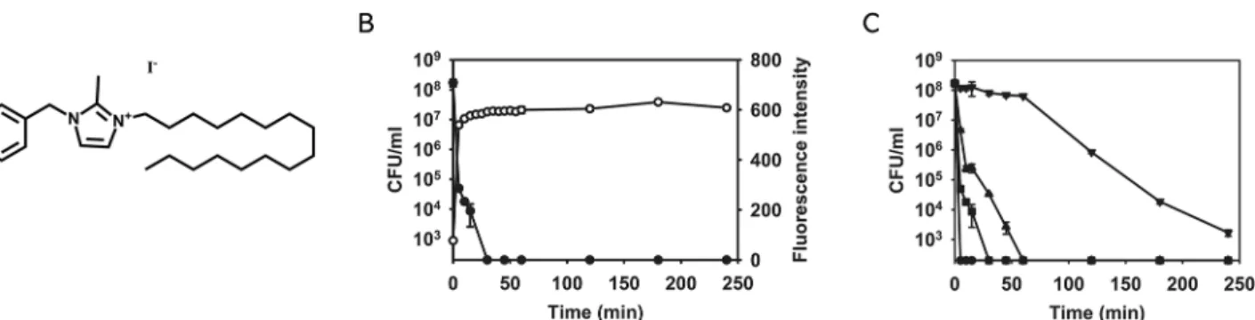
Among the 101 compounds, NH125 (1-Hexadecyl-2-methyl-3-(phenylmethyl)-1H-imida-zolium iodide) was identified as a hit with a Z-score of 10.61 (Fig 4A). NH125 is known to be an antibiotic and has been shown to inhibit bacterial histidine kinases and eukaryotic elonga-tion factor 2 kinase [41–43]. Consistent with previous work [41,42], the MIC of NH125 against S.aureusMW2 is ~2μg/mL (Table 1). As shown inFig 4B, 10μg/mL NH125 not only induced
rapid membrane permeabilization, but also resulted in a dramatic decrease in the viability of MRSA persisters. To confirm the correlation between membrane permeabilization and viabili-ty, we tested 5 sample compounds also identified in theC.elegans—MRSA screen that did not have activity in the SYTOX Green assay and found that they did not decrease CFU counts Fig 3. Validation of SYTOX Green assay robustness.To test the robustness of the SYTOX Green assay, the Z’-factor was calculated from fluorescence intensity data from a 384-well plate where half of the wells contained 0.1% DMSO (negative control, open circles) and the remaining wells contained 10X MIC lysostaphin (A) or 10X MIC nisin (B) (positive controls, solid circles). Fluorescence was measured with an excitation wavelength of 485 nm and an emission wavelength of 525 nm after incubation in the dark for 1 h. The Z’-factors for each assay were 0.767 (A) and 0.712 (B).
doi:10.1371/journal.pone.0127640.g003
Fig 4. NH125 kills MRSA persisters by inducing membrane permeabilization.(A) The chemical structure of NH125. (B) MRSA persisters were treated with 10μg/mL NH125. Membrane permeabilization (open circles) was measured spectrophotometrically by monitoring the uptake of SYTOX Green (excitation wavelength of 485 nm and an emission wavelength of 525 nm). Colony forming unit counts of persisters (solid circles) was measured by serial dilution and plating on TSA plates. (C) MRSA persisters were treated with several concentrations of NH125: 10X MIC (20μg/mL, circles), 5X MIC (10μg/mL, squares), 2.5X MIC (5μg/mL, triangles), and 1X MIC (2μg/mL, inverted triangles). Colony forming unit counts of persisters was measured by serial dilution and plating on TSA plates. The data points on the x-axis are below the level of detection (2x10²CFU/mL). Results are shown as means±s.d.; n = 3.
(data not shown). Next, we assessed the killing efficiency of NH125 on MRSA persisters at vari-ous NH125 concentrations. 2.5X MIC (5μg/mL) NH125 was sufficient to completely eradicate
MRSA persisters within 1 h, and 1X MIC (2μg/mL) killed 99.999% of MRSA persisters within
4 h (Fig 4C).
One of the advantages of the SYTOX Green screening method is that positive hits that kill persister cells by permeabilizing the membrane should also be able to kill non-persister cells. Like lysostaphin [20] and nisin [22], the correlation between viability and membrane perme-ability was also observed in growing MRSA treated with 10μg/ml NH125 (S4 Fig).
NH125 kills and disrupts MRSA biofilms
Since persisters in biofilms are known to be responsible for antibiotic tolerance of biofilms [44,45], we reasoned that NH125 would be effective in eradicating biofilms. First, we assessed the ability of NH125 to kill MRSA cells in biofilms. For these experiments, MRSA biofilms stat-ically cultured on 13 mm mixed cellulose membranes for 24 h at 37°C were treated with vanco-mycin or NH125 at 10X MIC for 24 h. Individual cells were freed from the biofilm matrix by sonication and cell viability was measured with CFU counts. As with planktonic persisters, vancomycin was unable to kill MRSA cells in biofilms (Fig 5A). In contrast, NH125 killed over 99% of MRSA biofilm cells at 20μg/mL (Fig 5A).
In addition to killing cells within biofilms, we assessed the ability of NH125 to disassemble biofilm biomass. MRSA biofilms formed in a 96-well microtiter plate for 48 h at 37°C were treated with various concentrations of vancomycin or NH125 (5X MIC to 160X MIC) for 24 h. The entire biofilm biomass was quantified using crystal violet, a cationic dye that stains all components of biofilms including cells and EPS [46,47]. Up to 160X MIC of vancomycin was unable to disrupt MRSA biofilms (Fig 5B). However, 10X MIC of NH125 removed 50% of bio-film biomass, and 80X MIC of NH125 completely disassembled the biobio-film (Fig 5B). These re-sults indicate that NH125 is able to effectively penetrate the EPS matrix, a protective“shield”
around biofilms, and kill persister cells as well as disrupting established biofilms.
Discussion
S.aureusis one of the most dangerous Gram-positive pathogens in the context of human health. Up to 30% of individuals are carriers ofS.aureus, which can cause a range of infectious diseases from acute to chronic infections in both healthy individuals and immunocompro-mised patients such as those with cancer and AIDS [4,48–54]. MRSA exhibiting resistance to commonly prescribed beta-lactam antibiotics is increasingly prevalent in hospitals as well as the community at large and has become an important public health problem [55]. Since MRSA can form persisters and is associated with chronic infection [56], development of drugs against MRSA persisters would be a significant advance in the treatment of MRSA infections.
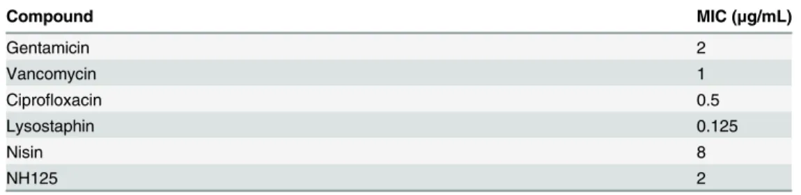
Table 1. Minimal inhibitory concentration (MIC) againstS.aureusMW2.
Compound MIC (μg/mL)
Gentamicin 2
Vancomycin 1
Ciprofloxacin 0.5
Lysostaphin 0.125
Nisin 8
NH125 2
To date, a variety of strategies have been used to kill persisters formed byS.aureusand MRSA. The first strategy was to facilitate the uptake of aminoglycosides into MRSA persisters. Although most biosynthetic processes that are targets for antibiotics are minimized in persist-ers, proteins are synthesized at a low level, and therefore, aminoglycosides can be effective against persisters [26,57]. However, the uptake of aminoglycosides is minimal in persisters due to the inactive state of transport mechanisms [26,57,58]. Allisonet al. reported that metabolites such as glucose, mannitol, or fructose can makeS.aureuspersisters aminoglycoside-susceptible by increasing the proton motive force of persister cell membranes [26]. Schmidtet al. engi-neered tobramycin by attachment of 12 amino acids, which promotes the uptake of tobramycin and subsequent killing ofS.aureuspersisters [57]. A second strategy was to induce protein deg-radation in persister cells by activating a protease. Conlonet al. identified ADEP4 that kills MRSA persisters by activating the ClpP protease, which subsequently leads to non-specific deg-radation of over 400 proteins including several that are essential for bacterial survival [34]. The third strategy was to directly attack structural components of the cell envelope such as the membrane or cell wall. As reviewed in Hurdleet al., many membrane active agents such as HT61 are known to killS.aureuspersisters [17]. In addition, Gutiérrezet al. reported that the phage endolysin LysH5 is able to eradicateS.aureuspersisters by hydrolyzing peptidoglycan [59]. However, a screening method for systematically identifying drugs targeting MRSA per-sisters has not previously been developed.
Based on the fact that cell envelope-targeting agents can directly and indirectly cause mem-brane permeabilization, which is correlated with bactericidal activity [20,22,23], we devised a HTS using SYTOX Green to discover new antimicrobial agents that are effective against MRSA persisters. Statistical evaluation of assay robustness showed that the assay is suitable for a large-scale screen. The SYTOX Green assay should also be broadly applicable for screens for drugs Fig 5. NH125 eradicates MRSA biofilms.(A) MRSA biofilms formed on 13 mm cellulose ester membranes for 24 hours were treated with 10X MIC of vancomycin (Van) or NH125 for 24 h. Survival was measured by comparing the number of viable cells in biofilms between non-treated and treated samples. (B) MRSA biofilms grown in a 96-well microtiter plate for 48 h were treated with the indicated concentration of vancomycin or NH125 for 24 h. The remaining biofilms were stained with 0.1% crystal violet dissolved with 95% ethanol and OD590 nmwas measured. Results are shown as means±s.d.; n = 3.
effective against persisters of other multidrug-resistant pathogens since SYTOX Green shows selective permeability in both Gram-positive and Gram-negative bacteria [24]. For example, we found a correlation between membrane permeability and viability ofE.colipersisters after treatment with polymyxin B [60], a membrane active antibiotic effective against Gram-negative bacteria (S6B Fig). By conducting a pilot screen with 101 anti-MRSA agents from a previously conductedC.elegans-MRSA screen for compounds that block the ability of MRSA to kill the nematodes, we identified NH125, which has low toxicity but strong antimicrobial property against MRSA persisters.
In our“proof of principle”screen of candidate antibiotics, we identified NH125 as a com-pound that is able to kill MRSA persisters. NH125 is already known to have antimicrobial properties with antibiotic activity (MIC 0.39–3.12μg/mL) against drug-resistant Gram-positive
bacteria, such as oxacillin-resistantS.aureus, penicillin G-resistantStreptococcus pneumoniae, and vancomycin-resistantEnterococcus faecalis[41]. A recent study revealed that NH125 hibits histidine kinases of bacterial two component signal transduction systems (TCS) by in-ducing non-specific aggregation of the histidine kinases [61]. NH125 has also been investigated as a potential antineoplastic drug with activity against eukaryotic elongation factor 2 kinase (eEF2K), which was shown to be due to nonspecific colloidal aggregation [62]. Based on the known kinase aggregation activity of NH125 [61,62], a possible mechanism by which NH125 kills persister cells may be by binding and aggregating kinases on the MRSA cell membrane, causing structural changes that result in membrane permeabilization and death.
Although many membrane-damaging agents have excellent anti-MRSA properties, includ-ing a low MIC, rapid killinclud-ing rate, and low probability for developinclud-ing resistance, they have a ten-dency to cause toxicity in mammals [63,64]. However, our starting library of 101 anti-MRSA agents identified using theC.elegans-MRSA HTS was expected to be enriched for nontoxic compounds, since the endpoint of the assay is enhanced survival of the nematodes in the pres-ence of MRSA. In fact, the invertebrate nematodeC.eleganshas been used as a model for as-sessing toxicity of many chemicals, including heavy metals, environmental pollutants, organic solvents, and neurotoxins [65–67]. Moreover, many studies have shown a strong correlation in toxicity betweenC.elegansand rodents [68,69]. In theC.elegans-MRSA HTS, 7.5μg/mL
NH125 kills MRSA but shows no toxicity toC.elegans(S7 Fig). Consistent with this result, up to 30μM (15.8μg/mL) NH125 has been reported to be nontoxic to sea urchin eggs [70].
Be-cause of low toxicity, the kinase inhibitory activity of NH125 has been intensively studied in human cells and mice [43,71–74]. Considering 2μg/mL of NH125 (equivalent to 1X MIC) kills
99.999% of MRSA persister cells within 4 h, we believe NH125 is a good candidate drug that warrants further studies as a therapeutic against MRSA persisters.
In summary, we have devised a fluorescence-based HTS for identifying drugs that eradicate MRSA. Using this screening method, we identified NH125, a compound that effectively kills MRSA persisters by inducing rapid membrane permeabilization but has selectivity to bacteria. Furthermore, NH125 kills MRSA persisters in biofilms and eradicates established MRSA bio-films. The screening method we developed can be used as a large-scale screening platform for antibiotic drug discovery against persisters of a broad range of pathogens. NH125, and/or de-rivatives of this molecule, warrant further evaluation as antibiotics for treatment of persistent or chronic infections.
Supporting Information
S1 Fig. Isolated MRSA persisters are tolerant to 100X MIC conventional antibiotics.A MRSA overnight culture was treated with 10X MIC (20μg/mL) gentamicin for 4 h and the
treated with additional antibiotics at the indicated concentrations (100X MIC) for an addition-al 4 h, followed by once again determining the titer of viable cells. Results are shown as means ± s.d.; n = 3. Gm: gentamicin, Cipro: ciprofloxacin, Van: vancomycin.
(TIFF)
S2 Fig. Lysostaphin and nisin kill MRSA persisters by inducing membrane permeabiliza-tion in a dose-dependent manner.MRSA persisters were treated with 5X MIC lysostaphin (A), 2.5X MIC lysostaphin (B),1X MIC lysostaphin (C), 5X MIC nisin (D), 2.5X MIC nisin (E), or 1X MIC nisin (F). Membrane permeabilization (open circles) was measured spectrophoto-metrically by monitoring the uptake of SYTOX Green (excitation wavelength of 485 nm and an emission wavelength of 525 nm). Colony forming unit counts of persisters (solid circles) were measured by serial dilution and plating on TSA plates. The data points on the x-axis are below the level of detection (2x10²CFU/mL). Results are shown as means ± s.d.; n = 3. (TIFF)
S3 Fig. Conventional antibiotics do not kill MRSA persisters or induce membrane permea-bilization.MRSA persisters were treated with 10X MIC (20μg/mL) gentamicin, 10X MIC
(10μg/mL) vancomycin (B), or 10X MIC (5μg/mL) ciprofloxacin (C). Membrane
permeabili-zation (open circles) was measured spectrophotometrically by monitoring the uptake of SYTOX Green (excitation wavelength of 485 nm and an emission wavelength of 525 nm). Col-ony forming unit counts of persisters (solid circles) was measured by serial dilution and plating TSA plates. Results are shown as means ± s.d.; n = 3.
(TIFF)
S4 Fig. NH125 kills growing MRSA by inducing membrane permeabilization.Growing MRSA was treated with 10μg/ml NH125. Membrane permeabilization (open circles) was
mea-sured spectrophotometrically by monitoring the uptake of SYTOX Green (excitation wave-length of 485 nm and an emission wavewave-length of 525 nm). Colony forming unit counts of persisters (solid circles) were measured by serial dilution and plating on TSA plates. The data points on the x-axis are below the level of detection (2x10²CFU/mL). Results are shown as means ± s.d.; n = 3.
(TIFF)
S5 Fig. Increase of ciprofloxacin concentration up to 1000X MIC does not affect the viabili-ty ofE.colipersisters.AnE.coliMG1655 overnight culture was treated with 10X MIC (0.3μg/mL), 100X MIC (3μg/ml), or 1000X MIC (30μg/ml) ciprofloxacin for 4 h and the titer
of viable cells was determined. Results are shown as means ± s.d.; n = 3. Gm: gentamicin, Cipro: ciprofloxacin, Van: vancomycin.
(TIFF)
S6 Fig. Polymyxin B killsE.colipersisters by inducing membrane permeabilization.E.coli MG1655 persisters were treated with 0.1% DMSO (A), 10X MIC (20μg/ml) polymyxin B (B),
10X MIC (0.3μg/mL) ciprofloxacin (C), 10X MIC (160μg/mL) ampicillin (D), or 10X MIC
(40μg/mL) gentamicin (E). Membrane permeabilization (open circles) was measured
spectro-photometrically by monitoring the uptake of SYTOX Green (excitation wavelength of 485 nm and an emission wavelength of 525 nm). Colony forming unit counts of persisters (solid cir-cles) were measured by serial dilution and plating on TSA plates. The data points on the x-axis are below the level of detection (2x10²CFU/mL). Results are shown as means ± s.d.; n = 3. (TIFF)
70% M9 buffer, 19% sheath solution (Union Biometrica Part no. 300-5101-000), 10% TSB, and 1% DMSO. The bacterial concentration in each well was adjusted to OD6000.04, and the final
concentration of drugs was 7.5μg/mL. After incubation in a humidified chamber at 25°C for 5
days, the worms were washed 8-times with M9 buffer and stained with 0.7μM SYTOX Orange
for staining dead worms. The plates were imaged using an Image Xpress Micro automated mi-croscope (Molecular Devices), capturing both transmitted light and TRITC (535 nm excitation, 610 nm emission) fluorescent images with a 2X objective.
(TIFF)
Acknowledgments
We thank ICCB-Longwood, Harvard Medical School for providing a chemical collection.
Author Contributions
Conceived and designed the experiments: WK EM. Performed the experiments: WK RR BBF. Analyzed the data: WK ALC. Contributed reagents/materials/analysis tools: ALC BBF. Wrote the paper: WK ALC FMA EM.
References
1. Wright GD. The antibiotic resistome: the nexus of chemical and genetic diversity. Nat Rev Microbiol. 2007; 5:175–186. PMID:17277795
2. Kohanski MA, Dwyer DJ, Collins JJ. How antibiotics kill bacteria: from targets to networks. Nat Rev Microbiol. 2010; 8:423–435. doi:10.1038/nrmicro2333PMID:20440275
3. Barrett FF, McGehee RF Jr., Finland M. Methicillin-resistantStaphylococcus aureusat Boston City Hospital. N Engl J Med. 1968; 279:441–448. PMID:4232865
4. David MZ, Daum RS. Community-associated methicillin-resistantStaphylococcus aureus: epidemiolo-gy and clinical consequences of an emerging epidemic. Clin Microbiol Rev. 2010; 23:616–687. doi:10. 1128/CMR.00081-09PMID:20610826
5. Klein E, Smith DL, Laxminarayan R. Hospitalizations and deaths caused by methicillin-resistant Staph-ylococcus aureus, United States, 1999–2005. Emerg Infect Dis. 2007; 13:1840–1846. doi:10.3201/ eid1312.070629PMID:18258033
6. Howden BP, Davies JK, Johnson PDR, Stinear TP, Grayson ML. Reduced vancomycin susceptibility in Staphylococcus aureus, including vancomycin-intermediate and heterogeneous vancomycin-interme-diate strains: resistance mechanisms, laboratory detection, and clinical implications. Clin Microbiol Rev. 2010; 23:99–139. doi:10.1128/CMR.00042-09PMID:20065327
7. Lewis K. Persister cells. Annu Rev Microbiol. 2010; 64:357–372. doi:10.1146/annurev.micro.112408. 134306PMID:20528688
8. Helaine S, Kugelberg E. Bacterial persisters: formation, eradication, and experimental systems. Trends Microbiol. 2014; 22:417–424. doi:10.1016/j.tim.2014.03.008PMID:24768561
9. Bigger JW. Treatment ofstaphylococcalinfections with penicillin by intermittent sterilisation. The Lan-cet. 1944; 244:497–500.
10. Gerdes K, Maisonneuve E. Bacterial persistence and toxin-antitoxin loci. Annu Rev Microbiol. 2012; 66:103–123. doi:10.1146/annurev-micro-092611-150159PMID:22994490
11. LaFleur MD, Qi Q, Lewis K. Patients with long-term oral carriage harbor high-persister mutants of Can-dida albicans. Antimicrob Agents Chemother. 2010; 54:39–44. doi:10.1128/AAC.00860-09PMID: 19841146
12. Mulcahy LR, Burns JL, Lory S, Lewis K. Emergence ofPseudomonas aeruginosastrains producing high levels of persister cells in patients with cystic fibrosis. J Bacteriol. 2010; 192:6191–6199. doi:10. 1128/JB.01651-09PMID:20935098
13. Spoering AL, Lewis K. Biofilms and planktonic cells ofPseudomonas aeruginosahave similar resis-tance to killing by antimicrobials. J Bacteriol. 2001; 183:6746–6751. PMID:11698361
15. Van Bambeke F, Mingeot-Leclercq MP, Struelens MJ, Tulkens PM. The bacterial envelope as a target for novel anti-MRSA antibiotics. Trends Pharmacol Sci. 2008; 29:124–134. doi:10.1016/j.tips.2007.12. 004PMID:18262289
16. Szweda P, Schielmann M, Kotlowski R, Gorczyca G, Zalewska M, Milewski S. Peptidoglycan hydro-lases-potential weapons againstStaphylococcus aureus. Appl Microbiol Biotechnol. 2012; 96:1157–
1174. doi:10.1007/s00253-012-4484-3PMID:23076591
17. Hurdle JG, O'Neill AJ, Chopra I, Lee RE. Targeting bacterial membrane function: an underexploited mechanism for treating persistent infections. Nat Rev Microbiol. 2011; 9:62–75. doi:10.1038/ nrmicro2474PMID:21164535
18. Francius G, Domenech O, Mingeot-Leclercq MP, Dufrêne YF. Direct observation ofStaphylococcus aureuscell wall digestion by lysostaphin. J Bacteriol. 2008; 190:7904–7909. doi:10.1128/JB.01116-08 PMID:18835985
19. Borysowski J, Weber-Dabrowska B, Górski A. Bacteriophage endolysins as a novel class of antibacte-rial agents. Exp Biol Med. 2006; 231:366–377. PMID:16565432
20. Lu X, Wang M, Qi J, Wang H, Li X, Gupta D, et al. Peptidoglycan recognition proteins are a new class of human bactericidal proteins. J Biol Chem. 2006; 281:5895–5907. PMID:16354652
21. Zou H, Koh J-J, Li J, Qiu S, Aung TT, Lin H, et al. Design and synthesis of amphiphilic xanthone-based, membrane-targeting antimicrobials with improved membrane selectivity. J Med Chem. 2013; 56:2359–
2373. doi:10.1021/jm301683jPMID:23441632
22. Zendo T, Yoneyama F, Sonomoto K. Lactococcal membrane-permeabilizing antimicrobial peptides. Appl Microbiol Biotechnol. 2010; 88:1–9. doi:10.1007/s00253-010-2764-3PMID:20645082 23. Silverman JA, Perlmutter NG, Shapiro HM. Correlation of daptomycin bactericidal activity and
mem-brane depolarization inStaphylococcus aureus. Antimicrob Agents Chemother. 2003; 47:2538–2544. PMID:12878516
24. Roth BL, Poot M, Yue ST, Millard PJ. Bacterial viability and antibiotic susceptibility testing with SYTOX green nucleic acid stain. Appl Environ Microbiol. 1997; 63:2421–2431. PMID:9172364
25. Rajamuthiah R, Fuchs BB, Jayamani E, Kim Y, Larkins-Ford J, Conery A, et al. Whole animal automat-ed platform for drug discovery against multi-drug resistantStaphylococcus aureus. PLoS ONE. 2014; 9:e89189. doi:10.1371/journal.pone.0089189PMID:24586584
26. Allison KR, Brynildsen MP, Collins JJ. Metabolite-enabled eradication of bacterial persisters by amino-glycosides. Nature. 2011; 473:216–220. doi:10.1038/nature10069PMID:21562562
27. Brumfitt W, Salton MRJ, Hamilton-Miller JMT. Nisin, alone and combined with peptidoglycan-modulat-ing antibiotics: activity against methicillin-resistantStaphylococcus aureusand vancomycin-resistant enterococci. J Antimicrob Chemother. 2002; 50:731–734. PMID:12407132
28. Climo MW, Patron RL, Goldstein BP, Archer GL. Lysostaphin treatment of experimental methicillin-re-sistantStaphylococcus aureusaortic valve endocarditis. Antimicrob Agents Chemother. 1998; 42:1355–1360. PMID:9624475
29. Malo N, Hanley JA, Cerquozzi S, Pelletier J, Nadon R. Statistical practice in high-throughput screening data analysis. Nat Biotechnol. 2006; 24:167–175. PMID:16465162
30. Zhang J, Chung T, Oldenburg K. A simple statistical parameter for use in evaluation and validation of high throughput screening assays. J Biomol Screen. 1999; 4:67–73. PMID:10838414
31. Clinical and Laboratory Standards Institute. Methods for dilution antimicrobial susceptibility tests for bacteria that grow aerobically; approved standard-ninth edition. CLSI document M07-A9. Wayne, PA; 2012.
32. Cassat JE, Smeltzer MS, Lee CY. Investigation of biofilm formation in clinical isolates of Staphylococ-cus aureus. Methods Mol Biol. 2014; 1085:195–211. doi:10.1007/978-1-62703-664-1_12PMID: 24085698
33. Keren I, Kaldalu N, Spoering A, Wang Y, Lewis K. Persister cells and tolerance to antimicrobials. FEMS Microbiol Lett. 2004; 230:13–18. PMID:14734160
34. Conlon BP, Nakayasu ES, Fleck LE, LaFleur MD, Isabella VM, Coleman K, et al. Activated ClpP kills persisters and eradicates a chronic biofilm infection. Nature. 2013; 503:365–370. doi:10.1038/ nature12790PMID:24226776
35. Hu Y, Shamaei-Tousi A, Liu Y, Coates A. A new approach for the discovery of antibiotics by targeting non-multiplying bacteria: a novel topical antibiotic forStaphylococcalinfections. PLoS ONE. 2010; 5: e11818. doi:10.1371/journal.pone.0011818PMID:20676403
37. Moy TI, Ball AR, Anklesaria Z, Casadei G, Lewis K, Ausubel FM. Identification of novel antimicrobials using a live-animal infection model. Proc Natl Acad Sci U S A. 2006; 103:10414–10419. PMID: 16801562
38. Kirienko NV, Kirienko DR, Larkins-Ford J, Wählby C, Ruvkun G, Ausubel FM.Pseudomonas aerugi-nosadisrupts Caenorhabditis elegans iron homeostasis, causing a hypoxic response and death. Cell Host and Microbe. 2013; 13:406–416. doi:10.1016/j.chom.2013.03.003PMID:23601103
39. Dolla NK, Chen C, Larkins-Ford J, Rajamuthiah R, Jagadeesan S, Conery AL, et al. On the mechanism of berberine–INF55 (5-Nitro-2-phenylindole) hybrid antibacterials. Aust J Chem. 2014; 67:1471–1480. 40. Jayamani E, Rajamuthiah R, Larkins-Ford J, Fuchs BB, Conery AL, Vilcinskas A, et al. Insect-derived
cecropins display activity againstAcinetobacter baumanniiin a whole-animal high-throughput Caenor-habditis elegansmodel. Antimicrob Agents Chemother. 2015; 59:1728–1737. doi:10.1128/AAC. 04198-14PMID:25583713
41. Yamamoto K, Kitayama T, Ishida N, Watanabe T, Tanabe H, Takatani M, et al. Identification and char-acterization of a potent antibacterial agent, NH125 against drug-resistant bacteria. Biosci Biotechnol Biochem. 2000; 64:919–923. PMID:10830522
42. Yamamoto K, Kitayama T, Minagawa S, Watanabe T, Sawada S, Okamoto T, et al. Antibacterial agents that inhibit histidine protein kinase YycG ofBacillus subtilis. Biosci Biotechnol Biochem. 2001; 65:2306–2310. PMID:11758928
43. Arora S, Yang J-M, Kinzy TG, Utsumi R, Okamoto T, Kitayama T, et al. Identification and characteriza-tion of an inhibitor of eukaryotic elongacharacteriza-tion factor 2 kinase against human cancer cell lines. Cancer Res. 2003; 63:6894–6899. PMID:14583488
44. Costerton JW, Stewart PS, Greenberg EP. Bacterial biofilms: a common cause of persistent infections. Science. 1999; 284:1318–1322. PMID:10334980
45. Lewis K. Riddle of biofilm resistance. Antimicrob Agents Chemother. 2001; 45:999–1007. PMID: 11257008
46. Bauer J, Siala W, Tulkens PM, Van Bambeke F. A combined pharmacodynamic quantitative and quali-tative model reveals the potent activity of daptomycin and delafloxacin againstStaphylococcus aureus biofilms. Antimicrob Agents Chemother. 2013; 57:2726–2737. doi:10.1128/AAC.00181-13PMID: 23571532
47. Lu J, Turnbull L, Burke CM, Liu M, Carter DA, Schlothauer RC, et al. Manuka-type honeys can eradi-cate biofilms produced byStaphylococcus aureusstrains with different biofilm-forming abilities. PeerJ. 2014; 2:e326. doi:10.7717/peerj.326PMID:24711974
48. Archer GL.Staphylococcus aureus: a well-armed pathogen. Clin Infect Dis. 1998; 26:1179–1181. PMID:9597249
49. Wertheim HF, Melles DC, Vos MC, van Leeuwen W, van Belkum A, Verbrugh HA, et al. The role of nasal carriage inStaphylococcus aureusinfections. Lancet Infect Dis. 2005; 5:751–762. PMID: 16310147
50. Ziakas PD, Pliakos EE, Zervou FN, Knoll BM, Rice LB, Mylonakis E. MRSA and VRE colonization in solid organ transplantation: a meta-analysis of published studies. Am J Transplant. 2014; 14:1887–
1894. PMID:25040438
51. Zervou FN, Zacharioudakis IM, Ziakas PD, Rich JD, Mylonakis E. Prevalence of and risk factors for methicillin-resistantStaphylococcus aureuscolonization in HIV infection: a meta-analysis. Clin Infect Dis. 2014; 59:1302–1311. doi:10.1093/cid/ciu559PMID:25031291
52. Zacharioudakis IM, Zervou FN, Ziakas PD, Mylonakis E. Meta-analysis of methicillin-resistant Staphy-lococcus aureuscolonization and risk of infection in dialysis patients. J Am Soc Nephrol. 2014; 25:2131–2141. doi:10.1681/ASN.2013091028PMID:24652802
53. Zervou FN, Zacharioudakis IM, Ziakas PD, Mylonakis E. MRSA colonization and risk of infection in the neonatal and pediatric ICU: a meta-analysis. Pediatrics. 2014; 133:e1015–23. doi: 10.1542/peds.2013-3413PMID:24616358
54. Ziakas PD, Anagnostou T, Mylonakis E. The prevalence and significance of methicillin-resistant Staph-ylococcus aureuscolonization at admission in the general ICU Setting: a meta-analysis of published studies. Crit Care Med. 2014; 42:433–444. doi:10.1097/CCM.0b013e3182a66bb8PMID:24145849 55. Fischbach MA, Walsh CT. Antibiotics for emerging pathogens. Science. 2009; 325:1089–1093. doi:10.
1126/science.1176667PMID:19713519
56. Goss CH, Muhlebach MS. Review:Staphylococcus aureusand MRSA in cystic fibrosis. J Cyst Fibros. 2011; 10:298–306. doi:10.1016/j.jcf.2011.06.002PMID:21719362
58. Taber HW, Mueller JP, Miller PF, Arrow AS. Bacterial uptake of aminoglycoside antibiotics. Microbiol Rev. 1987; 51:439–457. PMID:3325794
59. Gutiérrez D, Ruas-Madiedo P, Martínez B, Rodríguez A, García P. Effective removal of staphylococcal biofilms by the endolysin LysH5. PLoS ONE. 2014; 9:e107307. doi:10.1371/journal.pone.0107307 PMID:25203125
60. Daugelavicius R, Bakiene E, Bamford DH. Stages of polymyxin B interaction with theEscherichia coli cell envelope. Antimicrob Agents Chemother. 2000; 44:2969–2978. PMID:11036008
61. Francis S, Wilke KE, Brown DE, Carlson EE. Mechanistic insight into inhibition of two-component sys-tem signaling. MedChemComm. 2013; 4:269–277. PMID:23336064
62. Devkota AK, Tavares CDJ, Warthaka M, Abramczyk O, Marshall KD, Kaoud TS, et al. Investigating the kinetic mechanism of inhibition of elongation factor 2 kinase by NH125: evidence of a common in vitro artifact. Biochemistry. 2012; 51:2100–2112. doi:10.1021/bi201787pPMID:22352903
63. Koh J- J, Qiu S, Zou H, Lakshminarayanan R, Li J, Zhou X, et al. Rapid bactericidal action of alpha-mangostin against MRSA as an outcome of membrane targeting. Biochim Biophys Acta. 2013; 1828:834–844. doi:10.1016/j.bbamem.2012.09.004PMID:22982495
64. Liu D, Choi S, Chen B, Doerksen RJ, Clements DJ, Winkler JD, et al. Nontoxic membrane-active anti-microbial arylamide oligomers. Angew Chem Int Ed Engl. 2004; 43:1158–1162. PMID:14983462 65. Boyd WA, McBride SJ, Rice JR, Snyder DW, Freedman JH. A high-throughput method for assessing
chemical toxicity using aCaenorhabditis elegansreproduction assay. Toxicol Appl Pharmacol. 2010; 245:153–159. doi:10.1016/j.taap.2010.02.014PMID:20206647
66. Boyd WA, Smith MV, Kissling GE, Freedman JH. Medium- and high-throughput screening of neurotoxi-cants usingC.elegans. Neurotoxicol Teratol. 2010; 32:68–73. doi:10.1016/j.ntt.2008.12.004PMID: 19166924
67. Sochová I, Hofman J, Holoubek I. Using nematodes in soil ecotoxicology. Environment International. 2006; 32:374–383. PMID:16213020
68. Williams PL, Dusenbery DB. Using the nematodeCaenorhabditis elegansto predict mammalian acute lethality to metallic salts. Toxicol Ind Health. 1988; 4:469–478. PMID:3188044
69. Cole RD, Anderson GL, Williams PL. The nematodeCaenorhabditis elegansas a model of organo-phosphate-induced mammalian neurotoxicity. Toxicol Appl Pharmacol. 2004; 194:248–256. PMID: 14761681
70. Bellé R, Pluchon P-F, Cormier P, Mulner-Lorillon O. Identification of a new isoform of eEF2 whose phosphorylation is required for completion of cell division in sea urchin embryos. Dev Biol. 2011; 350:476–483. doi:10.1016/j.ydbio.2010.12.015PMID:21167828
71. Rose AJ, Alsted TJ, Jensen TE, KobberøJB, Maarbjerg SJ, Jensen J, et al. A Ca2+
–calmodulin–
eEF2K–eEF2 signalling cascade, but not AMPK, contributes to the suppression of skeletal muscle pro-tein synthesis during contractions. J Physiol. 2009; 587:1547–1563. doi:10.1113/jphysiol.2008. 167528PMID:19188248
72. Liu X-Y, Zhang L, Wu J, Zhou L, Ren Y-J, Yang W-Q, et al. Inhibition of elongation factor-2 kinase aug-ments the antitumor activity of temozolomide against glioma. PLoS ONE. 2013; 8:e81345. doi:10. 1371/journal.pone.0081345PMID:24303044
73. Arora S, Yang J-M, Utsumi R, Okamoto T, Kitayama T, Hait WN. P-glycoprotein mediates resistance to histidine kinase inhibitors. Mol Pharmacol. 2004; 66:460–467. PMID:15322237