Eugenol: A Phyto-Compound Effective
against Resistant and
Methicillin-Sensitive
Staphylococcus aureus
Clinical
Strain Biofilms
Mukesh Kumar Yadav1,2, Sung-Won Chae2, Gi Jung Im2, Jae-Woo Chung3, Jae-Jun Song2*
1Institute for Medical Device Clinical Trials, Korea University College of Medicine, Seoul, South Korea,2 Department of Otolaryngology—Head and Neck Surgery, Korea University College of Medicine, Seoul, South Korea,3Laboratory of Medicine, Dongguk University Ilsan Hospital, Goyang, Gyeonggi, South Korea
Abstract
Background
Inhibition and eradication ofStaphylococcus aureusbiofilms with conventional antibiotic is difficult, and the treatment is further complicated by the rise of antibiotic resistance among staphylococci. Consequently, there is a need for novel antimicrobials that can treat biofilm-related infections and decrease antibiotics burden. Natural compounds such as eugenol with anti-microbial properties are attractive agents that could reduce the use of conventional antibiotics. In this study we evaluated the effect of eugenol on MRSA and MSSA biofilmsin vitroand bacterial colonizationin vivo.
Methods and Results
Effect of eugenol onin vitrobiofilm andin vivocolonization were studied using microtiter plate assay and otitis media-rat model respectively. The architecture ofin vitrobiofilms and in vivocolonization of bacteria was viewed with SEM. Real-time RT-PCR was used to study gene expression. Check board method was used to study the synergistic effects of eugenol and carvacrol on established biofilms. Eugenol significantly inhibited biofilms growth of MRSA and MSSAin vitroin a concentration-dependent manner. Eugenol at MIC or 2×MIC effectively eradicated the pre-established biofilms of MRSA and MSSA clinical strains.In vivo, sub-MIC of eugenol significantly decreased 88%S.aureuscolonization in rat middle ear. Eugenol was observed to damage the cell-membrane and cause a leakage of the cell contents. At sub-inhibitory concentration, it decreases the expression of biofilm-and entero-toxin-related genes. Eugenol showed a synergistic effect with carvacrol on the eradication of pre-established biofilms.
OPEN ACCESS
Citation:Yadav MK, Chae S-W, Im GJ, Chung J-W, Song J-J (2015) Eugenol: A Phyto-Compound Effective against Resistant and Methicillin-SensitiveStaphylococcus aureusClinical Strain Biofilms. PLoS ONE 10(3): e0119564. doi:10.1371/ journal.pone.0119564
Academic Editor:Christophe Beloin, Institut Pasteur, FRANCE
Received:July 23, 2014
Accepted:January 14, 2015
Published:March 17, 2015
Copyright:© 2015 Yadav et al. This is an open access article distributed under the terms of the
Creative Commons Attribution License, which permits unrestricted use, distribution, and reproduction in any medium, provided the original author and source are credited.
Data Availability Statement:All relevant data are within the paper and its Supporting Information files.
Conclusion/Major Finding
This study demonstrated that eugenol exhibits notable activity against MRSA and MSSA clinical strains biofilms. Eugenol inhibited biofilm formation, disrupted the cell-to-cell con-nections, detached the existing biofilms, and killed the bacteria in biofilms of both MRSA and MSSA with equal effectiveness. Therefore, eugenol may be used to control or eradicate S.aureusbiofilm-related infections.
Introduction
Staphylococcus aureusis one of the major human pathogens responsible for the infections of skin, soft tissue, respiratory tissue and bone joints, causing endovascular infections such as bac-teremia, endocarditis, sepsis, and toxic shock syndrome [1]. It is one of the most common pathogens implicated in a wide variety of biofilm-related infections, including infections in-volving implanted medical devices such as bloodline catheters and heart implants, and infec-tions associated with cystic fibrosis, wounds, superficial skin infecinfec-tions, and otitis media [2,3,4,5,6]. In the adhesive state, bacteria attach to the surface of biomaterials or tissues and form a multilayered structure consisting of slow-growing bacteria enclosed in an extracellular polymeric matrix [7]. The physiology of cells in biofilms is different from that of planktonic cells, and the expression levels of a number of genes are reported to be enhanced exclusively in biofilm formation. Staphylococcal biofilm accumulation is mediated by polysaccharide inter-cellular adhesion (PIA), modulated by gene products encoded byicaoperon [8,9]. However, many researchers reported thatS.aureuscan formica-independent biofilm, mediated by pro-teins binding to propro-teins of the extracellular matrix [10,11]. SimilarlysarAgene expression has been reported to promote biofilm formation inS.aureus[12,13].
The bacteria in biofilms are slow-growing and were found to be particularly resistant to an-tibiotic treatment [14]. As a result conventional antibiotic therapy is ineffective in inhibition and eradication of biofilms infections. The treatment is further complicated by the rise of anti-biotic resistance amongstaphylococci. Consequently, there is a need for novel antimicrobials that can treat biofilm-related infections, are safe to use, and limit the emergence of antibiotic resistant bacteria. One of the emerging concepts in infection treatment is the disruption of the bacterial membrane bilayer or the proteins integral to membrane function in dormant bacteria of biofilms [15].
Only few antimicrobials are known to be capable of disrupting the cell membrane and are effective againstS.aureusbiofilm-related infection. Daptomycin and telavancin are the two an-tibiotics clinically approved in 2003 and 2009, respectively, for use againstS.aureusbiofilm in-fection [16]. However, it was reported that daptomycin may not be able to eradicate all types of
staphylococcalbiofilm infections in a mouse model [17]. Various phyto-compounds such as 220D-F2 derived fromRubus ulmifoliusplant, carvacrol (constituent of oregano), tea tree oil (Melaleuca alternifolia), and magnolol have been reported to be effective againstS.aureus bio-films [18,19,20,21,22].
Eugenol (4-allyl-2-methoxyphenol) is a major component of clove oil and has been used as a flavoring agent in food and cosmetic products. Studies have shown that eugenol exhibits a number of potentially beneficial biological properties, including antimicrobial, antioxidant, anti-inflammatory, anticarminative, and antispasmodic activities [23,24,25,26]. Recently, we demonstrated biofilm inhibition and eradication properties of eugenol againststreptococci
[27]. Previous studies have also demonstrated that eugenol can disrupt the cell membrane of
Salmonella typhi, and decreases toxin-related gene expression ofS.aureusat sub-inhibitory concentrations [28,29]. However, the effect of eugenol on resistant and methicillin-sensitiveS.aureus(MSSA and MRSA respectively) clinical strain biofilms has not been studied. In this study, we aimed to evaluate the effect of eugenol on MRSA and MSSA biofilmsin vitro
and on bacterial colonization usingin vivootitis media (OM) model.
We demonstrated here that eugenol exhibits notable activity against MRSA and MSSA clini-cal strain biofilms. Eugenol inhibited biofilm formation, disrupted the cell-to-cell connections, detached the existing biofilms, and killed the bacteria in biofilms of both MRSA and MSSA with equal effectiveness. It decreased the gene expression of biofilm and enterotoxin genes, and decreased bacterial colonization in the middle ear. Therefore, eugenol alone or in combination with carvacrol may be used to control or eradicateS.aureusbiofilm-related infections.
Materials and Methods
Ethics statement
All animal experiments were carried out in accordance with the guidelines of the Animal Re-search Committee Dongguk University Ilsan Hospital. The animal experiments were approved by Animal Research Committee Dongguk University Ilsan Hospital, Gyeonggi, South Korea. The bacterial experimental protocol was approved by the Institutional Review Board Dongguk University Ilsan Hospital, Gyeonggi, South Korea (Permit Number: 2013-50). Bacterial strains were isolated from clinical samples collected from patients visiting Dongguk University Ilsan Hospital (June 2013-January 2014). And all patients provided written informed consent for the collection of samples and subsequent analysis.
Bacterial strains and culture conditions
MSSA strain ATCC 29213 was obtained from the American Type Culture Collection (ATCC, USA). Seventy twoS.aureusclinical strains (43 MRSA and 29 MSSA) were isolated and identi-fied at the department of clinical microbiology, Dongguk University Ilsan Hospital, Goyang, South Korea by using standard procedures. These clinical strains were isolated from the follow-ing samples: blood (n= 10); sputum (n= 10); wound infections (n= 10); pus (n= 10); ear dis-charge (n= 10); nasal swab (n= 7); urine (n= 4); and other sites, such as catheters (n= 11). Five MRSA strains (CCARM 3903, CCARM 3108, CCARM 3912, CCARM 3967, and CCARM 3807) resistant to oxacillin (128.0μg/mL), erythromycin (128.0μg/mL), clindamycin (128.0μg/mL), and gentamicin (32–128.0μg/mL) were purchased from Culture Collection of Antimicrobial Resistant Microbes (CCARM), Seoul, Korea (S1 Table). Mueller-Hinton broth (MHB), tryptic soya broth (TSB), and BHI agar were purchased from BD Biosciences, Inc. (Sparks, MD, USA). Eugenol (oil) was purchased from Sigma-Aldrich (St. Louis, MO, USA), and stock solutions were prepared in dimethyl sulfoxide (DMSO) (Sigma-Aldrich).
Determinations of minimum inhibitory concentration (MIC) and minimum
bactericidal concentration (MBC)
was added to each well to give a final concentration of 1–5×10⁵colony-forming units (cfu)/mL, as confirmed by viable counts. In each MIC and MBC experiment wells without added bacteria (media only) were used as negative controls. The plates were incubated for 24 h at 37°C and growth was assessed by measuring the optical density at 600 nm (OD600) by using a
spectro-photometer. Aliquots (10μL) obtained from the wells showing no visible growths were plated on BHI agar and the number of colonies was counted following overnight incubation at 37°C. The MIC was defined as the lowest eugenol concentration with no visible growth, and the MBC was defined as the lowest concentration that was observed to reduce the initial inoculum by99.9%. The experiments were performed in triplicates and repeated 3 times.
Effect of eugenol on biofilm growth
The biofilm assay was performed using 24- or 96-well polystyrene flat-bottom microtiter plates (BD falcon, Sparks, MD, USA) in a static model, using a previously described procedure [31]. Overnight cultures ofS.aureuswere diluted 1:200 with fresh sterile TSB medium containing 0.5% glucose. For the screening of test compounds, 200-μl aliquots of the diluted cultures were transferred to the wells of 96-well microtiter plates with eugenol or DMSO controls performed in triplicate. Eugenol diluted by serial 2-fold dilutions (final concentration ranging from 0.01% to 0.08%) were added to the cell suspensions. Cell suspensions (200μL) were inoculated in 96-well microtiter plate and incubated for 24 h at 37°C. After incubation, the medium was dis-carded, and plates were gently washed three times with sterile PBS. Thereafter, plates were stained with 50μL of crystal violet (0.1%) for 15 min. Excess stain was removed by washing with PBS. The crystal violet (CV) attached to the biofilm samples were dissolved with 200μL of ethanol. The absorbance at 570 nm (OD570) was measured using a spectrophotometer
(Mole-cule Probe) as the value of biofilm formation. Inhibition of CV staining by the drug treatment was calculated as a percentage of the mean of DMSO-treated control samples. Experiments were repeated three times to obtain the mean and standard deviation of means of biofilm for-mation under each treatment.
Effect of eugenol on established biofilms
The biofilm formation ability of all 72 clinical strains (43 MRSA and 29 MSSA) was evaluated by CV-microtitter plate assay as described above in separate experiments (S1 Fig.). High bio-film-forming clinical isolates (n= 26) were selected for further study. To test the effect of euge-nol on established biofilms, biofilms that had been pre-formed for 24 h were washed with PBS to eliminate planktonic bacteria and exposed to indicated concentrations of eugenol for 6 h at 37°C. The biofilms were then washed again with PBS, dispersed by sonication, and dissolved in 100μL PBS. The dispersed biofilm cells were used to determine viable cfu per mL from diluted samples plated and grown on BHI agar. The biomass of the biofilm was detected by the CV-mi-crotiter plate method, as described above.
Determination of rate of kill bacteria within biofilms
Scanning electron microscopic analysis of biofilm
Scanning electron microscopy (SEM) was used to observe the architecture ofS.aureusbiofilms grown in the presence of eugenol (0.5 × MIC concentration) and to evaluate the eradication of established biofilms treated with eugenol. The cell suspension was prepared as described above and 1.5 mL cell suspensions were inoculated in a 24-well microtiter plate and incubated at 37°C for 24 h. For SEM analysis of biofilm eradication, the media of pre-established biofilms were replaced with fresh media containing eugenol (0.04%), and further incubated for 6 h. The plate was gently washed twice with sterile PBS to remove the planktonic cells and preserved in SEM solution. The samples were pre-fixed by immersion in 2% glutaraldehyde in 0.1 M phos-phate buffer, and post-fixed for 2 h in 1% osmic acid dissolved in PBS. The samples were treat-ed in a gradtreat-ed series of ethanol and t-butyl alcohol, dritreat-ed in a freeze dryer (ES-2030, Hitachi, Tokyo, Japan), platinum-coated using an ion coater (IB-5, Eiko, Kanagawa, Japan), and ob-served under a Field Emission-Scanning Electron Microscope (FE-SEM; Hitachi, S-4700, Tokyo, Japan).
In vivo
colonization using OM rat model
Twenty seven pathogen-free Sprague-Dawley rats weighing 150–200 g were obtained from Ori-ent Bio (Gyeonggi, South Korea). All animals were examined for middle ear abnormalities prior to the experiment and were kept isolated in an infection-free zone for 2 weeks. The rats were assigned randomly to groups that received bacteria only (n= 8), bacteria with 0.5×MIC eugenol (n= 8), media with DMSO (DMSO control;n= 7) or no procedure (n= 4). The bacte-rial cell suspension was prepared in TSB medium supplied with DMSO at the final concentra-tion of 0.1% to dissolve eugenol. The cytotoxicity of DMSO on human middle ear epithelium cell (HMEEC) line andin vivowas evaluated separately by CCK-8 kit (Dojindo, Maryland, USA) and histological analysis. And no significant effect of 0.1% DMSO was detected on HMEEC line andin vivo(S2&S3 Fig.). Cell suspension aliquots (50μL) containing 3×10⁷cfu
S.aureus(ATCC 29213) were injected into the middle ear cavity through the tympanic mem-brane of the right ear using a tuberculin syringe and a 27-gauge needle [32]. The animals were euthanized using a 1:1 combination of ZoletilH (tiletamine-zolazepam, Virbac, Carros, France) and RompunH (xylazine-hydrochloride, Bayer, Leverkusen, Germany) 1 week after inocula-tion, and the bulla was aseptically acquired. The tympanic membrane was removed and ears were irrigated to remove the planktonic bacteria. The bulla from each group were dissected and cleaned, and the visualized middle ear was photographed. The bulla were homogenized, se-rially diluted, and plated on blood agar plates (BAP) to obtained cfu counts. For SEM analysis the bulla were preserved in SEM solution and analyzed as described above.
viable cells in the established biofilm. The term MIC is replaced by MBEC for the calculations of FICI and FICs.
Crystal violet absorption
The alteration in membrane permeability was detected by the crystal violet assay [28]. Suspen-sions of the ATCC 29213 strain were prepared in LB broth. Cells were harvested by centrifuga-tion at 4500×g for 5 min at 4°C. The cells were washed twice and resuspended in PBS (pH-7.4). Eugenol and oxacillin at MIC and 2×MIC (MIC values for eugenol and oxacillin were 0.04% and 3.2μL/mL, respectively) were added to the cell suspension and incubated at 37°C for 30 min. Control samples were prepared similarly without treatment with eugenol. The cells were har-vested at 9300×g for 5 min. After that the cells were resuspended in PBS containing 10μL/mL of crystal violet. The cell suspension was then incubated for 10 min at 37°C. The suspension was then centrifuged at 13,400×g for 15 min and the OD590of the supernatant was measured in
spec-trophotometer. The OD value of the crystal violet solution, which was originally used in the assay, was taken and it was considered as 100%. The percentage of crystal violet uptake of all the samples was calculated using the following formula:
OD value of the sample
OD value of the crystal violet solution100
Quantification of gene expressions using Real-time RT-PCR
S.aureus(ATCC 29213) biofilms were grown for 24, 36,or 48 h in 12-well plate as described above at 37°C either with eugenol (at 0.5 × MIC) or without eugenol. RNA was prepared as de-scribed by Qiu et al [29]. Briefly, medium was discarded, biofilms were washed three times with PBS and cells were harvested by centrifugation (5,000 ×gfor 5 min at 4°C). The cell pellet was dissolved in 100μL (100μg/mL) of lysostaphin (Sigma-Aldrich) for 10 min at 37°C. Total bacterial RNA was isolated using Qiagen RNeasy kit (Qiagen, Hilden, Germany) in accordance with the manufacturer’s instructions. The contaminating DNA was removed using the optional on-column RNase-free DNase I step (Qiagen). RNA concentrations were determined by OD260
measurements and the quality of RNA was checked on an RNase-free 2% agarose gel. cDNA synthesis was carried out using the ImProm-II Reverse Transcriptase Kit (Promega, Madison, WI, USA) according to the manufacturer’s instructions. Briefly, tailing of RNA with a random hexamer primer was performed at 70°C for 5 min, annealing at 25°C for 5 min, extension at 37°C for 1 h and inactivation of samples at 70°C for 15 min. The primer pairs used in real-time RT-PCR are listed inTable 1. Real-time RT-PCR was carried out in a total volume of 20μL, consisting of 10μL 2X SYBR Green PCR Master Mix (Roche Applied Science, Indianapolis, IN, USA), 2.5 pmol of forward and reverse primers, and 2μL of cDNA. PCR conditions includ-ed initial denaturation at 95°C for 10 min, followinclud-ed by 45 cycles of denaturation (95°C for 15 sec), annealing (56°C for 15 sec), extension (72°C for 15 sec) followed by final extension (72°C for 5 min) and melting curve analysis from 60–95°C. Negative controls containing nucle-ase-free water instead of c-DNA were run concomitantly to confirm that the samples were free from contamination. To verify the absence of contaminating genomic DNA, each RT-PCR ex-periment included a no reverse transcriptase control. The relative gene expression was analyzed using the 2−ΔΔCTmethod [34]. The reference gene was 16S r-RNA and the standard condition
Statistical analysis
Data were calculated as the means of individual experiments performed in triplicate and com-pared with those of the control groups. Statistical analysis was performed using 2-tailed Stu-dent’st-test and one-way analysis of variance (ANOVA). Statistical significance was set at a p-value of less than 0.05.
Results
MIC and MBC of eugenol
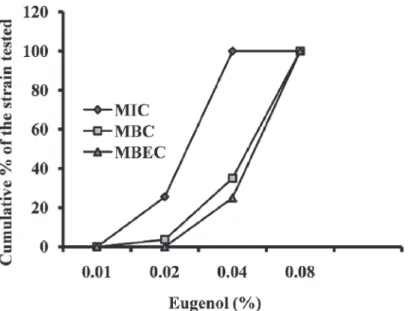
The MIC values of eugenol forS.aureusranged from 0.01% to 0.04%, with the MBC measured to be approximately double the MIC. No difference was observed in MIC values between the MRSA and MSSA isolates. The MIC, MBC, and MBEC of eugenol are presented inFig. 1. No significant growth inhibitory activity of 0.1% DMSO was detected onS.aureus(S2 Fig.).
Table 1. List of primers used for gene expression study.
Gene name Sequence Base pair Amplicon size (base pair)
icaD F-CGTGTTGCTTTAAACATTGAAAAT 24 126
R- TCTTCCTCTCTGCCATTTTTG 21
seA F- ATGGTGCTTATTATGGTTATC 21 163
R- CGTTTCCAAAGGTACTGTATT 21
sarA F-TTTCTCTTTGTTTTCGCTGATG 22 124
R- TTGCTTTGAGTTGTTATCAATGG 23
16S F- GCTGCCCTTTGTATTGTC 18 178
R-AGATGTTGGGTTAAGTCCC 19
doi:10.1371/journal.pone.0119564.t001
Fig 1. Cumulative minimum inhibitory concentration (MIC), minimum bactericidal concentration (MBC) and minimum biofilm eradication concentration (MBEC) of eugenol againstStaphylococcus aureusexpressed as a percentage of tested strains.MIC and MBC,n= 78; MBEC,n= 32.
Eugenol inhibits biofilm growth
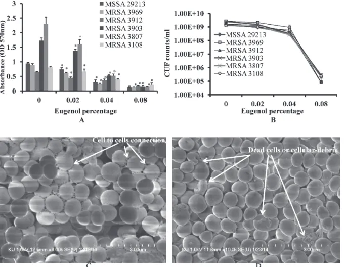
Eugenol significantly decreased MRSA and MSSA biofilm growthin vitro. At low concentra-tions, the decrease in biofilm growth was dose-dependent, reaching complete inhibition of bio-film formation at higher concentrations. Eugenol at concentration of 0.5×MIC decreased the biomass of MRSA (CCARM) and MSSA (ATCC 29213) isolates biofilms by more than 50% (Fig. 2A). The cfu of eugenol-supplemented biofilms were found to be significantly decreased approximately two log10 steps (Fig. 2B).
The architecture ofS.aureus(ATCC 29213) biofilms grown with and without eugenol (at 0.5×MIC) were analyzed by SEM. The SEM analysis revealed significantly decreased biofilm growth in eugenol-supplemented sample in comparison to the untreated samples (Fig. 2C&D). The biofilms grown without eugenol were thick, organized in 3-dimensional structure, with the cells connected to each other. Conversely, the biofilms grown in the presence of eugenol were not well organized, with the cells scattered and remaining attached to the bottom of the micro-titer plate. The results of the micromicro-titer plate assay and cfu count indicates that eugenol kills or inhibits the bacterial growth consequently with low cells decreased biofilms were formed. As a
Fig 2. Inhibition of MRSA and MSSA biofilm growthin vitrowith different concentrations of eugenol.(A) Decreased biofilm biomass detected using the microtiter plate assay. (B) Decreased cell viability within biofilms, detected by cfu counts. The error bars represent standard deviation from the mean value. (C) Representative SEM image ofS.aureus(ATCC 29213) biofilm grown without eugenol supplement. (D) Representative SEM image of biofilms grown with 0.5×MIC eugenol. The SEM image scale bar corresponds to 10μm.
result of low biofilms, the cell aggregation and cell-to-cell connections were prevented (as shown by SEM images), resulting in loosely arranged cell that can be easy disrupted.
Eugenol eradicates established biofilms
Eugenol demonstrated strong anti-biofilm properties against both MRSA and MSSA clinical strains (Table 2). The biomasses of already established biofilms were significantly decreased upon eugenol treatment (Fig. 3A). Similarly, the number of viable bacteria were significantly decreased in eugenol-treated biofilms (Fig. 3B). Eugenol at MIC significantly decreased the
Table 2. Effect of eugenol on the eradication of established biofilms of MRSA and MSSA clinical strains.
S. aureusstrains Mean OD570(CV) of biofilms detected by microtiter plate assay
0% (Control) SD 0.02% SDc 0.04% SD 0.08% SD
MSSA ATCCb29213 0.9430 ±0.02 0.7485 ±0.04 0.3072 ±0.07 0.1233 ±0.007 MRSA CCARMa3969 0.8896 ±0.05 0.6172 ±0.01 0.2412 ±0.02 0.1270 ±0.007
MRSA CCARM 3912 0.6565 ±0.01 0.4565 ±0.02 0.3602 ±0.05 0.1710 ±0.016 MRSA CCARM 3903 1.7317 ±0.08 1.3803 ±0.05 0.5279 ±0.02 0.1452 ±0.042 MRSA CCARM 3807 2.2991 ±0.13 1.6092 ±0.12 0.5256 ±0.05 0.1435 ±0.009
MRSA CCARM 3108 0.8122 ±0.02 0.6678 ±0.08 0.3991 ±0.01 0.1098 ±0.035
MRSA (Wound) 0.9122 ±0.04 0.7942 ±0.07 0.5913 ±0.09 0.1468 ±0.002 MRSA (Nasal cavity) 1.356 ±0.18 0.9549 ±0.04 0.5150 ±0.06 0.1443 ±0.001 MRSA (Nasal cavity) 1.266 ±0.12 0.9566 ±0.05 0.5177 ±0.16 0.1479 ±0.003
MRSA (Wound) 0.9443 ±0.08 0.5191 ±0.03 0.4883 ±0.03 0.1271 ±0.016
MRSA (catheter) 0.9075 ±0.04 0.6906 ±0.05 0.4997 ±0.04 0.1087 ±0.015 MRSA (Wound) 0.9603 ±0.05 0.7073 ±0.06 0.5251 ±0.02 0.1107 ±0.012 MRSA (Wound) 1.0737 ±0.07 0.6989 ±0.04 0.5251 ±0.02 0.1131 ±0.011
MSSA (Blood) 1.523 ±0.14 0.8527 ±0.07 0.6448 ±0.06 0.1547 ±0.002
MSSA (Blood) 0.8546 ±0.09 0.5329 ±0.07 0.4521 ±0.01 0.1236 ±0.023 MRSA (Wound) 0.9657 ±0.16 0.7506 ±0.07 0.5402 ±0.09 0.1505 ±0.008 MSSA (Pus) 1.2114 ±0.15 0.8946 ±0.05 0.6665 ±0.01 0.1273 ±0.004
MSSA (Pus) 0.8612 ±0.11 0.5628 ±0.03 0.3428 ±0.08 0.1262 ±0.003
MRSA (Wound) 0.9651 ±0.12 0.5884 ±0.06 0.4552 ±0.07 0.1383 ±0.007 MRSA (Wound) 0.9121 ±0.06 0.5384 ±0.03 0.4218 ±0.08 0.1324 ±0.006 MSSA (Ear discharge) 0.8621 ±0.05 0.6747 ±0.02 0.4678 ±0.05 0.1274 ±0.012
MRSA (Blood) 0.9062 ±0.05 0.6751 ±0.07 0.4553 ±0.06 0.1648 ±0.034
MRSA (Ear discharge) 1.1972 ±0.19 0.7403 ±0.006 0.4654 ±0.06 0.1467 ±0.035 MSSA (Ear discharge) 0.9331 ±0.07 0.7581 ±0.09 0.5686 ±0.11 0.1254 ±0.010 MRSA (Urine) 1.2712 ±0.25 0.7011 ±0.03 0.4452 ±0.05 0.1143 ±0.003
MSSA (Ear discharge) 1.1241 ±0.11 0.8771 ±0.02 0.7200 ±0.05 0.1379 ±0.014
MSSA (blood) 1.0931 ±0.11 0.6723 ±0.05 0.4743 ±0.16 0.1464 ±0.034 MSSA (Blood) 0.987 ±0.15 0.7075 ±0.01 0.5273 ±0.05 0.1289 ±0.019
MRSA (Pus) 1.062 ±0.08 0.6963 ±0.01 0.527 ±0.02 0.1328 ±0.013
MRSA (Catheter) 0.921 ±0.07 0.6769 ±0.09 0.5219 ±0.02 0.1206 ±0.018
MSSA (Pus) 1.1926 ±0.21 0.7383 ±0.02 0.5216 ±0.01 0.119 ±0.009
MSSA (Urine) 1.0713 ±0.15 0.6547 ±0.07 0.4899 ±0.03 0.1164 ±0.016
CCARMaCulture Collection of Antimicrobial Resistant Microbes, Seoul, Korea ATCCbAmerican Type Culture Collection
SDcStandard Deviation
biomass of already established biofilms by more than 50%. A decrease of four log 10 steps in the number of viable cells was observed in biofilm treated with 2×MIC eugenol. The MBEC of eugenol against biofilms from MRSA and MSSA clinical strains was found to be twice the MIC value.
The SEM analysis ofS. aureusbiofilm treated with eugenol and control (untreated) biofilm are shown inFig. 3C and D. In untreated samples, the biofilm was very organized in structure, with intact cell-to-cell connections (pointed by the arrow). The morphology of untreatedS. au-reuscells was smooth and regular, with an intact cell membrane. Conversely, the bacteria in eu-genol-treated biofilms lost cell-to-cell connections. The organized structures of the biofilm were also disrupted in the eugenol-treated samples. From the rough and shrunken appearance of the cells, it is apparent that the eugenol-treated bacterial cells lost their normal morphology. The completely dead cells or cellular debris were identifiable by the pores in cell membrane (pointed by arrow). These results indicate that the biofilm-eradication potential of eugenol may be due to (1) disruption of the cell-to-cell connections and (2) cell lysis.
Fig 3. Eradication of established biofilms of MRSA and MSSA by a range of concentrations of eugenol.(A) Decreased biomasses of established biofilms, detected by microtiter plate assay. (B) Decreased cell viability of biofilm, detected by cfu counts. The error bars represent standard deviation from the mean value. (C) Representative SEM image of established untreated biofilm ofS.aureus(29213). (D) Representative SEM image of established biofilm treated with eugenol (MIC). SEM images scale bar corresponds to 3μm.
Effect of eugenol on rate of kill of
S. aureus
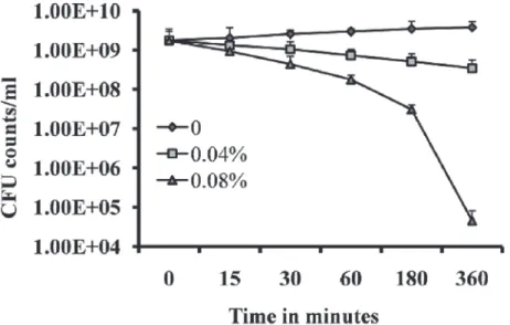
in preformed biofilms
Time-kill analysis was performed to determine the rapidity and duration of antibacterial activi-ty of eugenol onS. aureuswithin the biofilms. Treatment of pre-established biofilms with 2 × MIC exhibited a strong bactericidal effect onS. aureuswithin biofilms. Within 3 and 6 h of treatment, counts of viable bacterial cells were decreased by more than two log 10 steps and four log 10 steps (Fig. 4). This observation suggests that the strong bactericidal activity of euge-nol onS. aureuswithin biofilms reaches maximal effect within 3–6 h of incubation.
Eugenol reduced
in vivo
colonization of
S. aureus
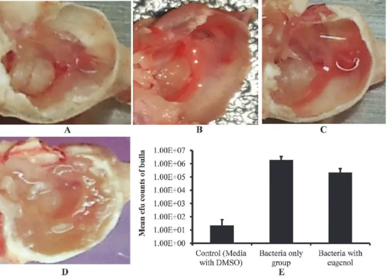
After one week of inoculation, rats treated with bacteria alone, bacteria with eugenol (0.5× MIC), DMSO control or no procedure control were sacrificed and their bulla were collected and photographed (Fig. 5 A, B, C and D). The bulla of rats inoculated with bacteria only were completely filled with biofilm matrix, cell debris, and obstructed with swelling mucosa. Con-versely, the bulla of rats inoculated with bacteria and eugenol were clear with no visible biofilm, matrix, or cell debris. Similarly the DMSO control and no procedure control bulla were also clean with no swelling mucosa.
The cfu counts of whole bulla lysates showed significantly lower number ofS. aureus recov-ered from the bulla of rats inoculated with bacteria and eugenol in comparison to the counts from the group treated with bacteria alone. The mean cfu counts of group treated with bacteria only was 1.96 ×10⁶(SD= 1,493,569), while cfu from group treated with bacteria and eugenol was 2.23 × 10⁵(SD= 189,661.1). No bacteria were detected in most of the DMSO control bulla, however few bacteria 2.2 ×101(SD= 38), were observed in some bulla, which may be normal body flora. A significant (p<0.003) 88% decrease inS. aureuscolonization was observed in the middle ear of rat in the presence of eugenol (Fig. 5E).
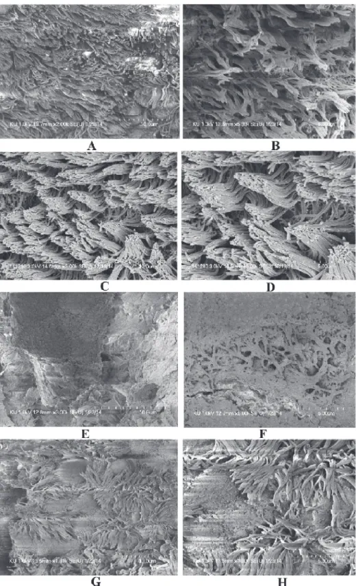
The SEM images of middle ears of rats inoculated with bacteria only, bacteria with eugenol, DMSO control or no procedure controls are shown inFig. 6. In the untreated (Figs. 6 A & B) and DMSO control middle ear (Fig. 6 C & D), the ciliated epithelium in the hypotympanum
area and Eustachian tube orifice area are intact. In the group treated with bacteria only, the whole middle ear was covered with bacterial biofilms (Fig. 6 E & F). In these samples, cell de-bris and exopolysaccharide (EPS) of biofilms were deposited on the tip of the cilia, making the cilia invisible. Conversely, in bulla of rats treated with bacteria and eugenol, there was no visible biofilms formation or detectable cell debris in the middle ear. There was no deposition of debris or EPS on the cilia of the epithelium, although the cilia of the epithelium appeared conglomer-ated (Fig. 6 G & H).
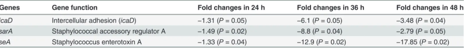
Eugenol decreases gene expression of biofilm and enterotoxin related
genes
The quantification oficaD,sarAandseAgene expression in biofilm grown with eugenol in compare to control detected a significantly (p<0.05) more 2-fold changes (Table 3). The inter-cellular adhesion (icaD) gene was significantly down regulated by 1.3, 6.1 and 3.4 folds in 24, 36 and 48 h biofilms. The expression of the staphylococcus enterotoxin A gene (seA)was sig-nificantly decreased by 1.3, 12.9 and 17.8 folds in 24, 36 and 48 h biofilms. Similarly, thesarA Fig 5. Effect of eugenol on colonization ofS. aureus(ATCC 29213)in vivoin the middle ear of rat.(A) Image of no procedure control rat middle ear. (B) Image of rat middle ear inoculated with DMSO (0.1%). (C) Image of rat middle ear inoculated with bacteria and eugenol. (D) Image of rat middle ear
inoculated with bacteria only. (E) Mean cfu counts of rat bullas inoculated with DMSO, bacteria only and bacteria with eugenol groups. The values are means of two independent experiments. The statistical significance was calculated using one-way analysis of variance (ANOVA) (P<0.003).
Fig 6. The effect of eugenol on colonization ofS. aureus(ATCC 29213) in middle ear of ratin vivo. (A&B) Representative SEM images of no procedure control rat middle ear. (C & D) Representative SEM images of middle ear of rat inoculated with DMSO. (E & F) Representative SEM images of middle ear of rat inoculated with bacteria only. (G & H) Representative SEM images of middle ear of rat inoculated with bacteria and eugenol. Images scale bars correspond to 5 and 10μm.
gene expression was also significantly decreased in biofilms grown over 24, 36 and 48 h by 1.5, 8.8, and 2.9-folds respectively. The down regulation oficaD,sarAandseAindicates that euge-nol at sub-inhibitory concentration may decrease biofilm growth by reducing the expression of biofilm-related genes.
Eugenol increases crystal violet absorption
The results of the crystal violet absorption assay indicate that eugenol alters membrane perme-ability ofS. aureusand increases crystal violet absorption. The uptake of crystal violet byS. au-reuswas 42% in the absence of eugenol, but increased to 90% and 92% after MIC and 2×MIC eugenol treatments respectively (Fig. 7). Oxacillin used as a negative control elicited no detect-able effect, indicating that it does not alter the membrane permeability.
Table 3. Relative expression levels oficaD,seaandsarAinS. aureus(ATCC 29213) biofilms grown with eugenol at 0.5×MIC for 24, 36 and 48 h.
Genes Gene function Fold changes in 24 h Fold changes in 36 h Fold changes in 48 h
icaD Intercellular adhesion (icaD) −1.31 (P= 0.05) −6.1 (P= 0.05) −3.48 (P= 0.04)
sarA Staphylococcal accessory regulator A −1.49 (P= 0.02) −8.8 (P= 0.04) −2.79 (P= 0.05)
seA Staphylococcus enterotoxin A −1.33 (P= 0.04) −12.9 (P= 0.02) −17.85 (P= 0.02)
doi:10.1371/journal.pone.0119564.t003
Fig 7. Crystal violet uptake of eugenol and oxacillin-treatedS. aureus(ATCC 29213).The bacteria were treated with 2×and 4×MIC of eugenol or oxacillin and crystal violet absorption was assayed as show in the methods section. The mean±SD for three replicates are presented.
Eugenol acts synergistically with carvacrol to eradicate biofilm
The combination of eugenol with carvacrol exhibited a synergistic effect against established MRSA and MSSA biofilms. MBEC of eugenol and carvacrol was determined to be 2×MIC (0.08% and 0.04%). Treatment of established biofilms with 0.25×MBEC (0.02%) of eugenol alone decreased the cfu counts by 25%. Similarly, 0.25×MBEC (0.01%) carvacrol decreased the viable bacteria by 40% in established biofilms. The combination of the two compounds (0.02% eugenol and 0.01% carvacrol) significantly decreased the bacteria in already established bio-films by 99%. The cfu counts of MRSA and MSSA biobio-films were significantly decreased from 2.79×10⁹and 1.32×10⁹to 2.91×10⁷and 1.07×10⁷respectively (Fig. 8). When eugenol and car-vacrol were used in combination, the MBEC was reduced4-fold producing a synergistic ef-fect as defined by FICI0.5.
Discussion
The paradigm of biofilm resistance presents a major hurdle for the treatment of a number of infectious disease types, increasing the economic burden, prolonging hospital stays, leading to recurrent infections, and increasing fatalities in the most recalcitrant cases [35]. The classical antibiotics target actively growing bacteria and are ineffective against the quiescent bacteria within biofilms [36]. Many phyto-compounds, including eugenol, have been reported to dis-rupt the bacterial membrane bilayer, cause membrane depolarization, and increase the mem-brane permeability in bacteria [28,37,38,39]. In the present study, we investigated the potential effects of eugenol on MRSA and MSSA clinical strains biofilmsin vitroand bacterial coloniza-tion using the OM modelin vivo.
Our results demonstrate significant inhibitory activity of eugenol on MRSA and MSSA bio-filmsin vitro. Eugenol can effectively eradicate pre-established biofilms, and decreases the ex-pression of biofilm- and enterotoxin-related genes. It predominantly damages the cell
Fig 8. Synergistic effect of eugenol with carvacrol for eradication of preformed biofilm.The MRSA and MSSA pre-established biofilms were treated with different concentrations of eugenol or carvacrol alone or in combination, and the cell viability was determined by cfu counts. The experiment was performed in triplicates and repeated two times.
membrane and causes the leakage of the cell contents. It significantly decreases the colonization ofS. aureusin rat middle earin vivo(Fig. 9).Staphylococcus aureusforms biofilms by two path-ways;ica-dependent andica-independent. Theica-dependent biofilm formation (in MSSA) is mediated by the polysaccharide intercellular adhesion or poly-N-acetylglucosamine (PIA/ PNAG), which is synthesized by enzymes encoded byica,while the ica-independent biofilm formation (in MRSA) is mediated by proteins binding to proteins of the extracellular matrix [10,11]. In this study eugenol is equally effective against MRSA and MSSA biofilms; therefore we have not considered this point. However, the biofilm experiments were carried-out in TSB medium supplied with 0.5% glucose, and it was previously reported that glucose can induce
ica-independent biofilm formation in MRSA, probably mediated by proteins [10]. Therefore, it can be assumed that eugenol may be equally effective againstica-independent biofilm.
Eugenol significantly inhibits the growth of MRSA and MSSA biofilms in a concentration-dependent manner. At sub-MIC concentrations,>50% biofilm biomass was reduced in euge-nol-treated samples, as compared to the control samples. The decreased cfu in eugeeuge-nol-treated biofilms indicate that eugenol prevents cell aggregation and disrupts the organization of bio-films. These results were further supported by the SEM images, in which low numbers of ad-herent cells were visible. The inhibitory effect of eugenol on planktonic cell was low, as compared to biofilm inhibition. This is in line with other studies, reporting that phenol-con-taining phyto-compounds such as eugenol decrease biofilms growthin vitro[40].
Using the OM rat model, we analyzed the colonization ofS. aureusin the middle ear of rats. The digital images of bulla of rats inoculated with bacteria and eugenol or DMSO only were clear in comparison to the bulla of rats treated with bacteria only. The bulla of rats treated with bacteria only were full of matrix, exhibited swollen mucosa, and significant presence of cell de-bris. The significantly decreased cfu counts and the tissue morphology in SEM images further support our results, showing that eugenol decreasesS. aureuscolonization in rat middle ear. Our results ofin-vivoexperiments demonstrate that eugenol possesses notable activity against colonization ofS. aureusin middle ear. However, further study with suitable OM model is needed to elucidate the pre-established biofilm eradication activity of eugenol in the middle ear. Eugenol, the major phenolic compound and antimicrobial component of clove oil, has
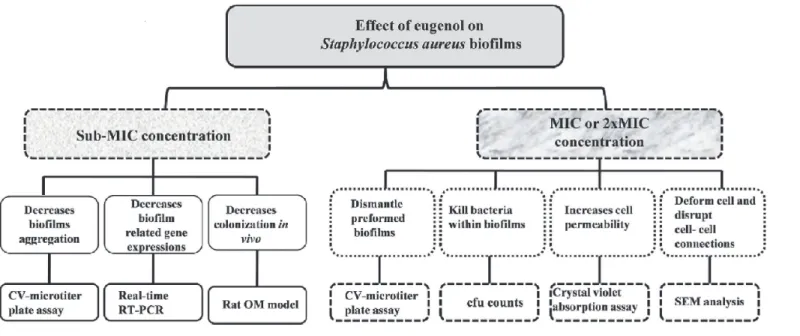
Fig 9. Systematic diagram showing effect of eugenol onStaphylococcus aureusbiofilms.
been registered in the European Union (EU) for use as a flavoring ingredient in foodstuffs and is considered to pose no risk to consumer health [41]. The eugenol structure or eugenol com-bined with other adjuvants can therefore be used againstS. aureusbiofilm infections. In partic-ular, it may have potential for the treatments of OM where the liquid antibiotics are not permeable through the tympanic membrane. Previously it was reported that the vapors of es-sential oils can diffuse through the tympanic membrane and reach the middle ear to produce an antimicrobial effect on acute OM [42,43].
To elucidate the mechanism underlying the observed decreased biofilm growth in the pres-ence of eugenol, we analyzed the gene expression of biofilm-relatedsarAandicaDgenes. The staphylococcal accessory regulatory (sarA) locus encodes a DNA-binding protein (SarA) and has a global impact on gene expression and biofilm formation inS. aureus [17,44,45]. Similarly, theicaDis an important biofilm candidate gene. It regulates synthesis of an intercellular adhe-sion polysaccharide that supports cell-to-cell bacterial contacts by means of a multilayered bio-film [8,9]. In our current study, we detected a low expression of thesarAandicaDgenes in biofilms grown with eugenol. This result indicates that eugenol interferes in the expression of these biofilm-related genes, resulting in decreased accumulation of polysaccharides and de-creased adhesion of cells in biofilmsin vitro. The low expression ofseAgene in eugenol-treated biofilms indicates that eugenol is effective in decreasing the enterotoxins. Previously, Que et al. have reported down regulation ofsarA,ica, andseAgene expression levels in the presence of eugenol in a stationary culture ofS. aureus[29]. Similarly the decreased expression ofsarA
gene has been shown to contribute to low inflammation and colonization in biofilm formation
in vivo[46].
Our results demonstrate significant biofilm-eradicating property of eugenol on MRSA and MSSA clinical strains. Eugenol treatment at concentration twice of MIC significantly decreased biomass and cell viability of established biofilms. This activity is maximal within 6 h of inocula-tion. The decrease in biomasses and cell viability indicates that eugenol eradicatesS. aureus
biofilm by two mechanisms; primarily it causes a lysis of bacteria within biofilms, and, second-ary, it disrupts cell-to-cell connections and dismantles the biofilm organization. Eugenol is a phenolic compound, hydrophobic in nature, which primarily acts by disrupting the cyto-plasmic membrane, resulting in the loss of normal shape of the bacteria, (resulting in shrunken appearance) and disruption of cell-cell connections [23]. These cell-to-cell connections are im-portant for the formation of organized biofilm and bacterial colonization [47] and the disrup-tion of these structures may result in the detachment of cells within biofilm, allowing them to be easily washed away. As a lipophilic compound, eugenol can pass through the cell wall and cytoplasmic membrane, disrupting the structure of different layers of polysaccharides, fatty acids, and phospholipids, permeabilizing the cell membrane, and resulting in cell lysis [48]. In the current study and others reports [38] depolarization and increased permeability were dem-onstrated in eugenol-treated bacteria. The severity of effect of eugenol onS. aureusbiofilms re-sults from the disruption of cell-to-cell connection, increased cell permeability causing leakage of internal cell contents to complete cell lysis, as seen in SEM analysis. Loss of membrane integ-rity and cell surface damage indicate that the bactericidal action of eugenol againstS. aureusis elicited through the mechanism of membrane disruption and blocking of cell growth [28,41].
Carvacrol is a phenolic monoterpenoid and a major constituent of oregano, which has been reported to disintegrate the outer membrane of bacteria [49,50]. In current study, we detected a synergistic effect of eugenol with carvacrol againstS. aureusbiofilms. The MBEC was reduced
due to the structure differences, the two compounds interact in distinct ways with the cell membrane to enhance the biofilm eradication activity [52]. Further study is warranted to deter-mine the exact mechanism underlying the synergistic effect of these two compounds onS. aureus.
Conclusion
In conclusion, this study demonstrated that eugenol has excellent antibacterial activity, either alone or in combination with carvacrol, against MRSA and MSSA clinical strains biofilms. Eu-genol was able to inhibit biofilm formation, disrupt the cell-to-cell connections, detach existing biofilms, and kill bacteria in biofilms of both MRSA and MSSA with equal effectiveness. It sig-nificantly decreased the expression of biofilm- and enterotoxin-related genes, and decreased bacterial colonization in the middle ear. Eugenol is safe to use and approved by EU and FDA as a food preservative. Therefore, eugenol administered alone or in combination with carvacrol could be used againstS. aureusbiofilm infections.
Supporting Information
S1 Fig.In-vitrobiofilm formation capability ofS. aureusclinical strains.(A)In-vitrobiofilm formation ability of 72 clinical strains (43 MRSA and 29 MSSA) was detected by CV-microtiter plate assay. (B) 72 clinical strains divided into High, intermediate and low biofilm producer on basis of biofilm growthin-vitro
(TIF)
S2 Fig. Effect of DMSO onStaphylococcus aureusand on Human middle ear epithelium cell line (HMEEC).(A) Optical density ofStaphylococcus aureusATCC 29213 strain grown with different concentration of DMSO for 24 h. (B) Absorbance at 450nm (detected by Cell Counting Kit-8) of HMEEC line grown with different concentrations of DMSO.
(TIF)
S3 Fig. Hematoxylin and Eosin (H&E) histology of middle ear of rat inoculated with DMSO.Fig. A, showing images of the middle ear mucosa of no procedure control group. In no procedure control middle ear mucosa is composed of single layered epithelium and sub-epithe-lial tissue attached to bony bulla. Fig. B, C and D, showing images of the middle ears inoculated with DMSO. No difference in morphology or any widen space is visible between epithelium layer and bony bulla of DMSO treated group, indicating no cytotoxicity on middle
ear epithelium. (TIF)
S1 Table. CCARM MRSA strains used in this study with antibiotic resistance profile. (DOCX)
Acknowledgments
The authors thank Dr Bo Hae Kim, Department of Otorhinolaryngology-Head and Neck Sur-gery, Dongguk University Ilsan Hospital for helping in animal experiments.
Author Contributions
References
1. Lowy FD. Staphylococcus aureus Infections. N Engl J Med. 1998; 339: 520–532. PMID:9709046 2. Agarwal A, Singh KP, Jain A. Medical significance and management of staphylococcal biofilm. FEMS
Immunol Med Microbiol. 2010; 58: 147–160. doi:10.1111/j.1574-695X.2009.00601.xPMID:19793317 3. Costerton JW, Stewart PS, Greenberg EP. Bacterial Biofilms: A Common Cause of Persistent
Infec-tions. Science. 1999; 284: 1318–1322. PMID:10334980
4. Maki DG, Kluger DM, Crnich CJ. The Risk of Bloodstream Infection in Adults With Different Intravascu-lar Devices: A Systematic Review of 200 Published Prospective Studies. Mayo Clin Proc. 2006; 81: 1159–1171. PMID:16970212
5. Otto M. Looking toward basic science for potential drug discovery targets against community-associat-ed MRSA. Mcommunity-associat-ed Res Rev. 2010; 30: 1–22. doi:10.1002/med.20160PMID:19399829
6. Thornton RB, Wiertsema SP, Kirkham L-AS, Rigby PJ, Vijayasekaran S, Coates HL, et al. Neutrophil Extracellular Traps and Bacterial Biofilms in Middle Ear Effusion of Children with Recurrent Acute Otitis Media—A Potential Treatment Target. PLoS One. 2013; 8: e53837. doi:10.1371/journal.pone. 0053837PMID:23393551
7. Donlan RM. Biofilms: microbial life on surfaces. Emerg Infect Dis. 2002; 8: 881–890. PMID:12194761 8. Cramton SE, Gerke C, Schnell NF, Nichols WW, Götz F. The Intercellular Adhesion (ica) Locus Is
Pres-ent in Staphylococcus aureus and Is Required for Biofilm Formation. Infect Immun. 1999; 67: 5427– 5433. PMID:10496925
9. Mack D, Fischer W, Krokotsch A, Leopold K, Hartmann R, Egge H, et al. The intercellular adhesin in-volved in biofilm accumulation of Staphylococcus epidermidis is a linear beta-1,6-linked glucosamino-glycan: purification and structural analysis. J Bacteriol. 1996; 178: 175–183. PMID:8550413
10. O'Neill E, Pozzi C, Houston P, Smyth D, Humphreys H, Robinson DA, et al. Association between Methi-cillin Susceptibility and Biofilm Regulation in Staphylococcus aureus Isolates from Device-Related In-fections. J Clin Microbiol. 2007; 45: 1379–1388. PMID:17329452
11. Vergara-Irigaray M, Valle J, Merino N, Latasa C, García B, Ruiz-de LM, et al. Relevant Role of Fibro-nectin-Binding Proteins in Staphylococcus aureus Biofilm-Associated Foreign-Body Infections. Infect Immun. 2009; 77: 3978–3991. doi:10.1128/IAI.00616-09PMID:19581398
12. Beenken KE, Blevins JS, Smeltzer MS Mutation of sarA in Staphylococcus aureus Limits Biofilm For-mation. Infect Immun. 2003; 71: 4206–4211. PMID:12819120
13. Valle J, Toledo-Arana A, Berasain C, Ghigo J-M, Amorena B, Penades JR, et al. SarA and notσB is es-sential for biofilm development by Staphylococcus aureus. Mol Microbiol. 2003; 48: 1075–1087. PMID: 12753197
14. Davies D. Understanding biofilm resistance to antibacterial agents. Nat Rev Drug Discov. 2003; 2: 114–122. PMID:12563302
15. Hurdle JG, O'Neill AJ, Chopra I, Lee RE. Targeting bacterial membrane function: an underexploited mechanism for treating persistent infections. Nat Rev Microbiol. 2011; 9: 62–75. doi:10.1038/ nrmicro2474PMID:21164535
16. Guskey MT, Tsuji BT. A Comparative Review of the Lipoglycopeptides: Oritavancin, Dalbavancin, and Telavancin. Pharmacotherapy. 2010; 30: 80–94. doi:10.1592/phco.30.1.80PMID:20030476 17. Weiss EC, Zielinska A, Beenken KE, Spencer HJ, Daily SJ, Smeltzer MS. Impact of sarA on
Daptomy-cin Susceptibility of Staphylococcus aureus Biofilms In Vivo. Antimicrob Agents Chemother. 2009; 53: 4096–4102. doi:10.1128/AAC.00484-09PMID:19651914
18. Burt SA, Ojo-Fakunle VTA, Woertman J, Veldhuizen EJA. The Natural Antimicrobial Carvacrol Inhibits Quorum Sensing inChromobacterium violaceumand Reduces Bacterial Biofilm Formation at Sub-Le-thal Concentrations. PLoS One. 2014; 9: e93414. doi:10.1371/journal.pone.0093414PMID: 24691035
19. Kwieciński J, Eick S, Wójcik K. Effects of tea tree (Melaleuca alternifolia) oil onStaphylococcus aureus in biofilms and stationary growth phase. Int J Antimicrob Agents. 2009; 33: 343–347. doi:10.1016/j. ijantimicag.2008.08.028PMID:19095413
20. Ma Y, Xu Y, Yestrepsky BD, Sorenson RJ, Chen M, Larson SD, et al. Novel Inhibitors of Staphylococ-cus aureus Virulence Gene Expression and Biofilm Formation. PLoS One. 2012; 7: e47255. doi:10. 1371/journal.pone.0047255PMID:23077578
22. van Alphen LB, Burt SA, Veenendaal AKJ, Bleumink-Pluym NMC, van Putten JPM The Natural Antimi-crobial Carvacrol InhibitsCampylobacter jejuniMotility and Infection of Epithelial Cells. PLoS One. 2012; 7: e45343. doi:10.1371/journal.pone.0045343PMID:23049787
23. Gill AO, Holley RA. Mechanisms of Bactericidal Action of Cinnamaldehyde against Listeria monocyto-genes and of Eugenol against L. monocytomonocyto-genes and Lactobacillus sakei. Appl Environ Microbiol. 2004; 70: 5750–5755. PMID:15466510
24. Hashimoto S, Uchiyama K, Maeda M, Ishitsuka K, Furumoto K, Nakamura Y.In vivoandin vitroEffects of Zinc Oxide-Eugenol (ZOE) on Biosynthesis of Cyclo-oxygenase Products in Rat Dental Pulp. J Dent Res. 1988; 67: 1092–1096. PMID:3165402
25. Mohammed MJ, Al-Bayati FA. Isolation and identification of antibacterial compounds from Thymus kotschyanus aerial parts and Dianthus caryophyllus flower buds. Phytomedicine. 2009; 16: 632–637. doi:10.1016/j.phymed.2008.12.026PMID:19200700
26. Moon S-E, Kim H-Y, Cha J-D. Synergistic effect between clove oil and its major compounds and antibi-otics against oral bacteria. Arch Oral Biol. 2011; 56: 907–916. doi:10.1016/j.archoralbio.2011.02.005 PMID:21397894
27. Yadav MK, Park S-W, Chae S-W, Song J-J, Kim HC. Antimicrobial activities of Eugenia caryophyllata extract and its major chemical constituent eugenol against Streptococcus pneumoniae. APMIS. 2013; 121: 1198–1206. doi:10.1111/apm.12067PMID:23594212
28. Devi KP, Nisha SA, Sakthivel R, Pandian SK. Eugenol (an essential oil of clove) acts as an antibacterial agent against Salmonella typhi by disrupting the cellular membrane. J Ethnopharmacol. 2010; 130: 107–115. doi:10.1016/j.jep.2010.04.025PMID:20435121
29. Qiu J, Feng H, Lu J, Xiang H, Wang D, Dong J, et al. Eugenol Reduces the Expression of Virulence-Re-lated Exoproteins in Staphylococcus aureus. Appl Environ Microbiol. 2010; 76: 5846–5851. doi:10. 1128/AEM.00704-10PMID:20639367
30. Clinical Laboratoty Standards Institute. Performance standards for antimicrobial susceptibility testing; Fifteenth informational supplement. CLSI/NCCLS document M100-S15, CLSI; 2015, Wayne, PA. 31. Christensen GD, Simpson WA, Younger JJ, Baddour LM, Barrett FF, Melton DM, et al. Adherence of
coagulase-negative staphylococci to plastic tissue culture plates: a quantitative model for the adher-ence of staphylococci to medical devices. J Clin Microbiol. 1985; 22: 996–1006. PMID:3905855 32. Yadav MK, Chae S-W, Song J-J.In VitroStreptococcus pneumoniae Biofilm Formation and In Vivo
Middle Ear Mucosal Biofilm in a Rat Model of Acute Otitis Induced by S. pneumoniae. Clin Exp Otorhi-nolaryngol. 2012; 5: 139–144. doi:10.3342/ceo.2012.5.3.139PMID:22977710
33. Petersen PJ, Labthavikul P, Jones CH, Bradford PA.In vitroantibacterial activities of tigecycline in combination with other antimicrobial agents determined by chequerboard and time-kill kinetic analysis. J Antimicrob Chemother. 2006; 57: 573–576. PMID:16431863
34. Livak KJ, Schmittgen TD. Analysis of Relative Gene Expression Data Using Real-Time Quantitative PCR and the 2−ΔΔCTMethod. Methods. 2001; 25: 402–408. PMID:11846609
35. Thomas JG, Litton I, Rinde H. Economic impact of biofilms on treatment costs. In: Pace JL, Rupp ME, Finch RG, editors. Biofilms, Infection and Antimicrobial Therapy. Boca Raton, FL: CRC Press. Taylor and Francis; 2006, pp. 21–37.
36. Chopra I, Hesse L, O'Neill AJ. Exploiting current understanding of antibiotic action for discovery of new drugs. J Appl Microbiol. 2002; 92: 4S–15S. PMID:12000608
37. Bakkali F, Averbeck S, Averbeck D, Idaomar M. Biological effects of essential oils—A review. Food Chem Toxicol. 2008; 46: 446–475. PMID:17996351
38. Hammer KA, Heel KA. Use of multiparameter flow cytometry to determine the effects of monoterpe-noids and phenylpropamonoterpe-noids on membrane polarity and permeability in staphylococci and enterococci. Int J Antimicrob Agents. 2012; 40: 239–245. doi:10.1016/j.ijantimicag.2012.05.015PMID:22795655 39. Nostro A, Cellini L, Zimbalatti V, Blanco AR, Marino A, Pizzimenti F, et al. Enhanced activity of carvacrol
against biofilm of Staphylococcus aureus and Staphylococcus epidermidis in an acidic environment. APMIS. 2012; 120: 967–973. doi:10.1111/j.1600-0463.2012.02928.xPMID:23030501
40. Nostro A, Roccaro AS, Bisignano G, Marino A, Cannatelli MA, Pizzimenti FC, et al. Effects of oregano, carvacrol and thymol on Staphylococcus aureus and Staphylococcus epidermidis biofilms. J Med Microbiol. 2007; 56: 519–523. PMID:17374894
41. Hemaiswarya S, Doble M. Synergistic interaction of eugenol with antibiotics against Gram negative bacteria. Phytomedicine. 2009; 16: 997–1005. doi:10.1016/j.phymed.2009.04.006PMID:19540744 42. Inouye S, Takizawa T, Yamaguchi H. Antibacterial activity of essential oils and their major constituents
43. Kristinsson KG, Magnusdottir AB, Petersen H, Hermansson A. Effective Treatment of Experimental Acute Otitis Media by Application of Volatile Fluids into the Ear Canal. J Infect Dis. 2005; 191: 1876– 1880. PMID:15871121
44. Beenken KE, Dunman PM, McAleese F, Macapagal D, Murphy E, Projan SJ, et al. Global Gene Ex-pression in Staphylococcus aureus Biofilms. J Bacteriol. 2004; 186: 4665–4684. PMID:15231800 45. Tsang LH, Cassat JE, Shaw LN, Beenken KE, Smeltzer MS. Factors Contributing to the
Biofilm-Defi-cient Phenotype of Staphylococcus aureus sarA Mutants. PLoS One. 2008; 3: e3361. doi:10.1371/ journal.pone.0003361PMID:18846215
46. Snowden JN, Beaver M, Beenken K, Smeltzer M, Horswill AR, Kielian T.Staphylococcus aureus sarA Regulates Inflammation and Colonization during Central Nervous System Biofilm Formation. PLoS One. 2013; 8: e84089. doi:10.1371/journal.pone.0084089PMID:24386336
47. Mandlik A, Swierczynski A, Das A, Ton-That H. Pili in Gram-positive bacteria: assembly, involvement in colonization and biofilm development. Trends Microbiol. 2008; 16: 33–40. PMID:18083568
48. Burt S. Essential oils: their antibacterial properties and potential applications in foods—a review. Int J Food Microbiol. 2004; 94: 223–253. PMID:15246235
49. Helander IM, Alakomi H-L, Latva-Kala K, Mattila-Sandholm T, Pol I, Smid DJ, et al. Characterization of the Action of Selected Essential Oil Components on Gram-Negative Bacteria. J Agri Food Chem. 1998; 46: 3590–3595.
50. La Storia A, Ercolini D, Marinello F, Di Pasqua R, Villani F, Mauriello G. Atomic force microscopy analy-sis shows surface structure changes in carvacrol-treated bacterial cells. Res Microbiol. 2011; 162: 164–172. doi:10.1016/j.resmic.2010.11.006PMID:21168481
51. Ultee A, Kets EPW, Smid EJ. Mechanisms of Action of Carvacrol on the Food-Borne Pathogen Bacillus cereus. Appl Environ Microbiol. 1999; 65: 4606–4610. PMID:10508096