www.reumatologia.com.br
REVISTA BRASILEIRA DE
REUMATOLOGIA
* Corresponding author.
E-mail: i [email protected] (S.C.M.S. Fialho).
0482-5004/$ - see front matter. © 2013 Elsevier Editora Ltda. All rights reserved.
Original article
Subclinical atherosclerosis in ankylosing spondylitis: is there
a role for infl ammation?
Renato Leandro Mattar Valente
a, Jamil Mattar Valente
b, Gláucio Ricardo Werner de Castro
c,
Adriana Fontes Zimmermann
c, Sonia Cristina de Magalhães Souza Fialho
c,*,
Ivânio Alves Pereira
ca Internal Medicine Division, Hospital Universitário, Universidade Federal de Santa Catarina, Florianópolis, SC, Brasil b Cardiology Division, Hospital Universitário, Universidade Federal de Santa Catarina, Florianópolis, SC, Brasil c Rheumatology Division, Hospital Universitário, Universidade Federal de Santa Catarina, Florianópolis, SC, Brasil
a r t i c l e i n f o
Article history:
Received 1 September 2012 Accepted 13 December 2012
Keywords:
Ankylosing spondylitis Subclinical atherosclerosis Cardiovascular risk
a b s t r a c t
Objectives: To evaluate the prevalence of subclinical atherosclerosis in patients with
anky-losing spondylitis (AS) in comparison to controls with similar cardiovascular risk factors.
Methods: Forty-two consecutive patients with AS and 42 controls matched for age (43.3 ±
11.7 vs. 43.7 ± 11.3, P = 0.89), gender, smoking, diabetes mellitus and arterial hypertension were enrolled. Participants were excluded if a personal cardiovascular disease (CV) history was present. A questionnaire recording demographic data, medical and medication history was fuli lled. Blood pressure, abdominal circumference, height and weight were measured. Lipid proi le was determined in a 12-hour fastened blood sample. Ultrasound analysis of the common carotid artery was performed by one blind observer. The distance between the lumen-intima interface and the leading edge of the media-adventitia interface (IMT) was measured and participants were also evaluated for the presence of plaques.
Results: The comparative analysis of demographic and cardiovascular risk factors between
AS patients and controls did not reveal statistically signii cant differences. Also, no signii -cant differences between groups were observed for TC, HDL-C, T-C/HDL-C, LDL-C, triglycer-ides, or dyslipidemia frequency. IMT measures were not different in AS and controls (0.62 ± 0.09 vs. 0.61 ± 0.09, P = 0.39) as well as plaques frequencies (19% vs. 17%, P = 0.78).
Conclusions: Subclinical atherosclerosis assessed through carotid ultrasound imaging was
not more prevalent in the AS group when compared to controls with similar cardiovascular risks. Our observations may imply that CV risk factors may have more inl uence on the CV system than AS itself. These i ndings should be coni rmed in a larger population with a prospective study design.
Aterosclerose subclínica em pacientes com espondilite anquilosante: há um papel para a infl amação?
Palavras-chave:
Espondilite anquilosante Aterosclerose subclínica Risco cardiovascular
r e s u m o
Objetivos: Avaliar a prevalência de aterosclerose subclínica em pacientes com espondilite
anquilosante (EA) em comparação com controles com fatores de risco cardiovasculares similares.
Métodos: Foram recrutados 42 pacientes consecutivos com EA e 42 controles equiparados
para idade (43,3 ± 11,7 vs. 43,7 ± 11,3, P = 0,89), gênero, tabagismo, diabetes mellitus e hi-pertensão arterial. Qualquer participante seria excluído se estivesse presente uma história pessoal de doença cardiovascular (CV). Foi preenchido um questionário registrando dados demográi cos e histórias médica e de medicação. Foram determinados: pressão arterial, circunferência abdominal, altura e peso. O peri l lipídico foi determinado em uma amostra de sangue com 12 horas em jejum. Foi realizada uma análise ultrassonográi ca da artéria carótida comum por um observador desconhecedor da pesquisa. Foi medida a distância entre a interface lúmen-íntima e a borda de ataque da interface média-adventícia (EIM) e os participantes também foram avaliados para presença de placas.
Resultados: A análise comparativa dos fatores de risco demográi cos e cardiovasculares
en-tre pacientes com EA e controles não revelou diferenças estatisticamente signii cativas. Também não foram observadas diferenças signii cativas entre grupos para TC, HDL-C, T-C/ HDL-C, LDL-C, triglicerídeos ou frequência de dislipidemia. As medidas de EIM não foram diferentes em EA e controles (0,62 ± 0,09 vs. 0,61 ± 0,09, P = 0,39) e nem as frequências de placas (19% vs. 17%, P = 0,78).
Conclusões: A aterosclerose subclínica avaliada por meio de imagens ultrassonográi cas da
carótida não foi mais prevalente no grupo EA, em comparação com os controles com riscos cardiovasculares similares. Nossas observações podem implicar que os fatores de risco CV podem ter mais inl uência no sistema CV versus a própria EA. Esses achados devem ser coni rmados em uma população maior, por meio de um estudo prospectivo.
© 2013 Elsevier Editora Ltda. Todos os direitos reservados.
Introduction
Ankylosing spondylitis (AS) is a chronic, inl ammatory rheu-matic disease. Its musculoskeletal manifestations include both inl ammation and structural damage. Characteristic extra-articular manifestations include aortitis, cardiac con-duction defects, pulmonary i brosis and inl ammatory bowel disease indicating that AS is a systemic disease.
AS is known primarily for causing a lifetime of pain, im-paired physical function, work disability, and decreased quality of life, rather than for shortening life itself. However, patients with AS also experience premature mortality.1 The
standardized mortality rates (SMR) associated with AS are ap-proximately 50% higher than in the general population.2,3 The
four non-inception cohort studies published to date quote SMRs of 1.33,4,5 1.56 and 1.8.7
Increased mortality is largely attributable to cardiovascu-lar diseases (CV).3 A recent large population based study has
shown more ischemic heart disease (prevalence ratio 1.2), pe-ripheral vascular disease (ratio 1.6), atherosclerosis (ratio 1.5), congestive heart failure (1.8) and more cardiovascular risk factors (prevalence ratios between 1.3 and 1.7) in AS patients compared to healthy controls.8
It is unclear whether the increased cardiovascular risk of AS patients could be explained by traditional cardiovascular risk factors alone. In fact, there is increasing evidence that
the underlying inl ammatory process in chronic inl ammato-ry conditions resembles the chronic inl ammatoammato-ry processes that contribute to various stages of atherothrombosis, from early atheroma formation to plaque instability and thrombus formation.9 However, it is unknown whether AS patients
with-out CV disease risk factors show early signs of large artery damage compared to controls, and if so, what the determi-nants of such large-vessel abnormalities are. This knowledge could prove useful for development of risk stratii cation, in-tervention strategies and for a better disease understanding.
High-resolution ultrasonography can be used to measure the intima-media thickness (IMT) as well as vascular elas-ticity of the carotid artery. An increased carotid IMT rel ects the atherosclerotic burden and predicts the development of (clinically apparent) CV disease in the general population.10,11
Hence, this study was designed to determine whether signs of subclinical atherosclerosis are more prominent in a sample of AS patients compared to controls without the disease but with similar cardiovascular risks. Other studies assessing IMT in AS patients and controls have been published but results were contradictory12-18 and will be discussed further.
Methods
Federal University of Santa Catarina, Brazil. All patients ful-i lled the modful-iful-i ed New York dful-iagnostful-ic crful-iterful-ia for AS.19
Forty-two volunteers (hospital staff or patients who attended the General Clinic for the University employees) matched for age, sex, smoking (current or in the last i ve years), diabetes melli-tus and systemic arterial hypertension served as controls. All participants gave written informed consent and the institu-tional ethics committees of the University Hospital approved the study protocol.
Patients and controls were excluded if a personal CV dis-ease history was present (myocardial infarction, percutane-ous transluminal coronary angioplasty, surgery for ischemic heart disease, stroke, transient ischemic attack, carotid end-arterectomy, peripheral arterial reconstructive surgery, or limb amputation).
Patients and controls were examined by a research phy-sician. A questionnaire recording demographic data, medical and medication history was fuli lled. Blood pressure, abdomi-nal circumference, height and weight were measured. Body mass index (BMI) was calculated as the ratio of weight and height squared. We considered the patient/control as diabetic if they referred hypoglycemic drug use or in the presence of least two glycemic tests higher than 126 mg/dL. We consid-ered the patient/control as hypertensive if they referred an-ti-hypertensive drugs use or a systolic blood pressure > 140 mmHg or diastolic blood pressure > 90 mmHg measured in two different occasions. We considered the patient/control as dyslipidemic if they referred hypolipemiants drugs use or if they presented at least one of the following: LDL-C (low den-sity lipoprotein) > 130 mg/dL; triglycerides > 150 mg/dL; HDL-C (high-density lipoprotein) < 40 mg/dL.
Laboratory variables were determined in a 12-hour fas-tened blood sample and included: TC (total cholesterol), HDL-C, LDL-C, triglycerides (all analyzed by enzymatic tech-niques); C-reactive protein (CRP by nephelometry method), and erythrocyte sedimentation rate (ESR by Westergreen method).
Arterial measurements were conducted in a quiet room after 15 minutes of rest, with the subjects in supine position. Ultrasound analysis of the common carotid artery (bilater-al) was performed by a cardiologist who were unaware of the participants’ clinical or laboratory characteristics. Mea-surements were performed using a B mode high resolution ultrasound ATL HDI 3000 (Phillips Bothel, WA, USA) with a 5-12MHz linear probe. The distance between the lumen-in-tima interface and the leading edge of the media-adventitia interface of the far wall corresponds with IMT. After localiza-tion of the common carotid artery, cross-seclocaliza-tional measure-ments were performed 10 mm proximal from the carotid bulb. Sites with mural atherosclerotic plaque were excluded while measuring. Three measurements were performed at each side. A mean of each side (right or left) was calculated and i nally the mean of both sides (mean) was achieved. We dei ned plaques as focal widening of the vessel wall of 50% relative to adjacent segments with protrusion into the lu-men or a IMT > 1.5 mm.
The distribution of each continuous variable was exam-ined graphically and statistically for normality. Numerical data are summarized as the mean and standard deviation (SD). Variables not normally distributed were compared
us-ing the Wilcoxon nonparametric test for differences. Vari-ables normally distributed were compared using students’ tests. Categorical data among groups were compared by the chi-square or the Fischer exact test statistics when appropri-ate. Some results were evaluated according to the established normal values and were subsequently ranked as elevated or depressed. A statistical signii cance was set at P < 0.05. All sta-tistical analyses were performed using NCSS software.
Results
Comparative analysis of demographic and cardiovascular risk factors between AS patients and controls did not reveal sta-tistically signii cant differences as demonstrated in table 1. AS mean age was 43.3 ± 11.7 years-old and the mean time of disease duration was 15.9 years.
Also, no signii cant differences between groups were ob-served for TC, HDL-C, T-C/HDL-C, LDL-C, triglycerides, or dys-lipidemia frequency as showed in table 2. AS patients had a signii cant elevation in CRP (38.1% vs. 14.3%, P = 0.01), but not in the ESR.
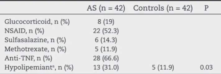
In table 3 we demonstrate medications use in both groups. 31% against 11.9% of AS patients were on hypolipemiants (P = 0.03). Anti-TNF was used in 66.6% of all AS patients.
There was no difference in IMT measures in AS and con-trols (0.62 ± 0.09 vs. 0.61 ± 0.09, P = 0.39). Also no difference was observed in the frequency of plaques (table 4).
Table 1 – Clinical and demographic features in AS and controls.
AS (n = 42) Controls (n = 42) P
Age (years) ± SD 43.3 ± 11.7 43.7 ± 11.3 0.89
Male, n (%) 26 (61.9) 26 (61.9) 1.0
Caucasian, n (%) 35 (83.3) 40 (95.2) 0.08
Smokers, n (%) 8 (19.04) 8 (19.04) 1.0
CV FH, n (%) 14 (33.3) 12 (28.57) 0.64
AH, n (%) 15 (35.7) 11 (26.2) 0.35
DM, n (%) 3 (7.1) 3 (7.1) 1.0
BMI (kg/m2) ± SD 26.5 ± 3.9 27.6 ± 4.5 0.25
AC (cm) ± SD 90.1 ± 12.3 91.6 ± 12.8 0.58
AS, ankylosing spondylitis; SD, standard deviation; CV FH, cardiovascular family history; AH, arterial hypertension; DM, diabetes mellitus; BMI, body mass index; AC, abdominal circumference.
Table 2 – Laboratory proi le in AS and controls. AS
(n = 42)
Controls (n = 42)
P
Total-cholesterol (mg/dL) ± SD 194.8 ± 36.0 196.8±36.4 0.81
HDL-C (mg/dl) ± SD 52.7 ± 10.9 52.1±14.9 0.82
LDL-C (mg/dl) ± SD 120.1 ± 32.4 119.3 ± 35.1 0.92
Tryglicerides (mg/dL) ± SD 109.7 ± 49.9 126.8 ± 103.7 0.88
CT/HDL ± SD 3.8 ± 0.9 4.0 ± 1.3 0.36
Dyslipidemia, n (%) 25 (59.5) 26 (62) 0.82
Elevated CRP, n (%) 16 (38.1) 6 (14.3) 0.01
ESR (mm/1st hour) ± SD 22.2 ± 17.7 20.3 ± 16.8 0.29
Discussion
Given that inl ammation has increasingly been acknowledged as the reason rheumatic patients bear elevated CV risk,20 we
selected a study population of AS patients matched for i ve major CV risk factors in order to better assign for this issue.
Our study revealed that CV risk among AS patients as in-dicated by IMT is not different from controls without the dis-ease. On the opposite, Peters at al found a greater IMT in AS patients in comparison with controls.12 However, the authors
also found a high CV risk factor proi le in patients with AS, and some of these risk factors (lipids and BMI) were associated with a greater carotid IMT and increased arterial stiffness. No association between large-vessel properties and higher Bath AS indices or CRP values were found. Also, Mathieu et al.14
found signii cantly increased IMT in the AS group compared with healthy controls. However, after adjustment for con-founding factors, only an underlying trend towards increased IMT was present. IMT was positively correlated with tobacco use and blood pressure but not correlated with CRP level or mSASS. In the AS group, IMT was correlated with traditional risk factors, such as smoking and systolic blood pressure.
Although the cross-sectional study design does not permit a good estimate of the cumulative inl ammatory burden and the small series of patients are associated with a low statistical pow-er, their results suggest that an adverse CV risk proi le may cause, at least partly, the greater IMT found by them and there is no sufi cient evidence to support a role of biological inl ammation.
Gonzalez et al., recruited 64 AS patients and 64 matched controls with no cardiovascular morbid. Patients with AS ex-hibited greater carotid IMT than did matched controls (mean ± SD, 0.74 ± 0.21 mm vs. 0.67 ± 0.14 mm; P = 0.01; differences of means, 0.077; 95% coni dence interval [CI], 0.016-0.139). In this case, although the best predictors for carotid plaques in patients with AS were erythrocyte sedimentation rate (ESR) at time of disease diagnosis (odds ratio [OR], 1.18; 95% CI, 1.04-1.33; P = 0.01) and duration of disease (OR, 1.39; 95% CI, 1.01-1.92; p = 0.05); there was no signii cant correlation between carotid IMT and either ESR or C-reactive protein.13
Indeed, many of the traditional risk factors for cardio-vascular disease are present in the AS versus the general population, including a higher incidence of hypertension, elevated lipids, increased i brinogen and CRP levels, and poorer physical activity levels. BMI and total cholesterol and triglycerides have been positively correlated with IMT and/ or arterial stiffness.21
In accordance with our i ndings, Choe et al. found that ca-rotid IMT and parameters related with arterial elastic proper-ties in young AS patients without clinically evident cardiovas-cular risk factors were not different from those of sex- and age-matched healthy controls. Serum levels of TNF-a, IL-6, and MCP-1 did not rel ect the degree of carotid subclinical atherosclerosis.15 In addition, recently, Capkin et al. evaluated
a total of 67 AS patients, and age, sex, body mass index (BMI) smoking status, lipid proi les and blood pressure-matched healthy control subjects (n = 34). They also found no differ-ence in IMT-C between groups.
Our study has some limitations. No disease activity index was analyzed. However, the isolated disease activity mea-sure would not allow drawing any conclusions about the in-l ammatory burden possibin-ly associated with atheroscin-lerosis. Also, 66.6% of patients were on anti-TNF treatment. A sub-analysis comparing AS patients suggested that the group on anti-TNF treatment (n = 28) was not different from the group treated with non-biologic drugs (n = 14) when it comes to the IMT, however, they had numerically less plaques (10.7% vs. 35.7%, P = 0.09). Although this study was not designed to assign for this exact issue, our i nding raises the ques-tion about the anti-TNF playing a part to a better cardiovas-cular outcome. Studies on ischemic heart disease related mortality and morbidity following anti-TNF therapy have shown mixed results. Ferrante et al.22 observed a signii cant
decrease of carotid IMT in anti-TNF-treated RA patients af-ter two years but not in the group treated with methotrex-ate alone, although signii cant improvements were seen in measures of disease activity, CRP and i brinogen levels with both type of treatments. It is thought though that anti-TNF treatment has the potential not only to reduce inl amma-tion but also to modify tradiamma-tional cardiovascular risk factors and endothelial dysfunction in RA.23,24 Additionally, one third
of the AS patients were more frequently on hypolipemiants (31% vs. 11,9%), although dyslipidemia (as dei ned by this study) was not more prevalent in AS patients. However, it well known that statins’s vascular improvement is indepen-dent of statins’ cholesterol-lowering actions, fact that has been associated with their anti-inl ammatory and immu-nomodulatory properties.25 Similarly to the anti-TNF
treat-ment, because of the study design we cannot be conclusive about statins contribution for a possible better cardiovascu-lar outcome on these AS cases.
In conclusion, our observations are in agreement with the i ndings of others and may imply that CV risk factors have more inl uence on the CV system than AS itself. However, we could not be conclusive because of the anti-TNF and hypoli-pemiants use. These i ndings should be coni rmed in a larger population with a prospective study design. Further research concerning the pathogenesis of increased cardiovascular risk in AS patients should be high priority, as many risk factors are likely to be modii able.
Table 3 – Medications use in AS and controls.
AS (n = 42) Controls (n = 42) P
Glucocorticoid, n (%) 8 (19)
NSAID, n (%) 22 (52.3)
Sulfasalazine, n (%) 6 (14.3)
Methotrexate, n (%) 5 (11.9)
Anti-TNF, n (%) 28 (66.6)
Hypolipemianta, n (%) 13 (31.0) 5 (11.9) 0.03
AS, ankylosing spondylitis; NSAID, non-steroidal anti-inl ammatory drugs.
aAll hypolipemiants used were from the statins group
Table 4 – Carotid ultrasound in AS and controls. AS (n = 42) Controls (n = 42) P
IMT (mm) ± SD 0.62 ± 0.09 0.61 ± 0.09 0.39
Plaques, n (%) 8 (19.0) 7 (17.0) 0.78
Confl icts of interest
The authors declare no conl icts of interest.
R E F E R E N C E S
1. Braun J, Pincus T. Mortality, course of disease and prognosis of patients with ankylosing spondylitis. Clin Exp Rheumatol. 2002;20(Suppl. 28):S16-S22.
2. Sokka T, Abelson B, Pincus T. Mortality in rheumatoid arthritis: 2008 update. Clin Exp Rheumatol.
2008;26(Suppl.51):S35-S61.
3. Zochling J, Braun J. Mortality in ankylosing spondylitis. Clin Exp Rheumatol. 2008;26(Suppl.51):S80-S84.
4. Kaprover E, Little AH, Graham DC, Rosen PS. Ankylosing spondylitis: survival in men with and without radiotherapy. Arthritis Rheum. 1980;23:57-61.
5. Khan MA, Khan MK, Kushner I. Survival among patients with ankylosing spondylitis: a life-table analysis. J Rheumatol. 1981;8:86-90.
6. LehtinenK. Mortality and causes of death in 398 patients admitted to hospital with ankylosing spondylitis. Ann Rheum Dis. 1993;52:174-6.
7. Radford EP, Doll R, Smith PG. Mortality among patients with ankylosing spondylitis not given x-ray therapy. N Engl J Med. 1977;297:572-6.
8. Han C, Robinson DW Jr, Hackett MV, Paramore LC, Fraeman KH, Bala MV. Cardiovascular disease and risk factors in patients with rheumatoid arthritis, psoriatic arthritis, and ankylosing spondylitis. J Rheumatol. 2006;33:2167-72. 9. Libby P. Role of inl ammation in atherosclerosis associated
with rheumatoid arthritis. Am J Med. 2008;121Suppl:S21-S31. 10. O’Leary DH, Polak JF. Intima-media thickness: a tool for
atherosclerosis imaging and event prediction. Am J Cardiol. 2002;90:18L-21L.
11. Bots ML, Dijk JM, Oren A, Grobbee DE. Carotid intima-media thickness, arterial stiffness and risk of cardiovascular disease: current evidence. J Hypertens. 2002;20:2317-25. 12. Peters MJ, van Eijk IC, Smulders YM, Serne E, Dijkmans BA, van der Horst-Bruinsma IE, et al. Signs of accelerated preclinical atherosclerosis in patients with ankylosing spondylitis. J Rheumatol. 2010;37(1):161-6.
13. Gonzalez-Juanatey C, Vazquez-Rodriguez TR, Miranda-Filloy JA, Dierssen T, Vaqueiro I, Blanco R, et al. The high prevalence of subclinical atherosclerosis in patients with ankylosing spondylitis without clinically
evident cardiovascular disease. Medicine (Baltimore). 2009;88(6):358-65.
14. Mathieu S, Joly H, Baron G, Tournadre A, Dubost J, Ristori JM, et al. Trend towards increased arterial stiffness or intima-media thickness in ankylosing spondylitis patients without clinically evident cardiovascular disease. Rheumatology. 2008;47(8):1203-7.
15. Choe JY, Lee MY, Rheem I, Rhee MY, Park SH, Kim SK. No differences of carotid intima-media thickness between young patients with ankylosing spondylitis and healthy controls. Joint Bone Spine. 2008;75:548-53.
16. Malesci D, Niglio A, Mennillo GA, Buono R, Valentini G, La MG. High prevalence of metabolic syndrome in patients with ankylosing spondylitis. Clin Rheumatol. 2007;26:710-4. 17. Sari I, Okan T, Akar S, Cece H, Altay C, Secil M, et al. Impaired
endothelial function in patients with ankylosing spondylitis. Rheumatology. 2006;45:283-6
18. Capkin E, Kiris A, Karkucak M, Durmus I, Gokmen F, Cansu A, et al. Investigation of effects of different treatment modalities on structural and functional vessel wall properties in patients with ankylosing spondylitis. Joint Bone Spine. 2011;78(4):378-82. 19. van der Linden S, Valkenburg HA, Cats A. Evaluation of
diagnostic criteria for ankylosing spondylitis. A proposal for modii cation of the New York criteria. Arthritis Rheum 1984;27(4):361-8.
20. Sattar N, McCarey DW, Capell H, McInnes IB. Explaining low “high-grade” systemic inl ammation accelerates vascular risk in rheumatoid arthritis. Circulation. 2003;108:2957-63. 21. Malesci D, Niglio A, Mennillo GA, Buono R, Valentini G, La
MG. High prevalence of metabolic syndrome in patients with ankylosing spondylitis. Clin Rheumatol. 2007;26:710-4. 22. Ferrante A, GiardinaAR, Ciccia F, Parrinello G, LicataG, Giardina
E, et al. Long-term anti-tumor necrosis factor therapy reverses the progression of carotid intima-media thickness in female patients with active rheumatoid arthritis. Rheumatol Int. 2009;30(2):193-8.
23. Spanakis E, Sidiropoulos P, Papadakis J, Ganotakis E, Katsikas G, Karvounaris S, et al. Modest but sustained increase of serum high density lipoprotein cholesterol levels in patients with inl ammatory arthritides treated with inl iximab. J Rheumatol. 2006;33:2440-6.
24. Hürlimann D, Forster A, Noll G, Enseleit F, Chenevard R, Distler O, et al. Anti−Tumor necrosis factor- treatment improves endothelial function in patients with rheumatoid arthritis. Circulation. 2002;106:2184-7.