Evaluation of medical devices in thoracic
radiograms in intensive care unit - time to pay
attention!
INTRODUCTION
horacic radiograms are one of the main auxiliary diagnostic exams performed in intensive care units (ICU).(1) However, according to the American College of Radiology,(2) the daily use of thoracic radiograms is not appropriate, except when there are changes in the patient’s medical condition or following the placement of a medical device. horacic radiograms should be performed after the placement of medical devices because there are potential complications associated with this practice, and the irst element that should be evaluated is the absence or presence and correct positioning of medical devices.(3)
Ana Sofia Linhares Moreira1, Maria da Graça
Alves Afonso1, Mónica Ribeiro dos Santos
Alves Dinis1, Maria Cristina Granja Teixeira dos
Santos2,3
1. Department of Radiology, Centro Hospitalar do Algarve - Faro, Portugal.
2. Department of Emergency and Intensive Care, Centro Hospitalar do Algarve - Faro, Portugal. 3. Department of Biomedical Sciences and Medicine, Universidade do Algarve - Faro, Portugal.
Objective: To identify and evaluate the correct positioning of the most commonly used medical devices as visualized in thoracic radiograms of patients in the intensive care unit of our center.
Methods: A literature search was conducted for the criteria used to evaluate the correct positioning of medical devices on thoracic radiograms. All the thoracic radiograms performed in the intensive care unit of our center over an 18-month period were analyzed. All admissions in which at least one thoracic radiogram was performed in the intensive care unit and in which at least one medical device was identiiable in the thoracic radiogram were included. One radiogram per admission was selected for analysis. he radiograms were evaluated by an independent observer.
Results: Out of the 2,312 thoracic radiograms analyzed, 568 were included in this study. Several medical devices
Conflicts of interest: None
Submitted on April 4, 2016 Accepted on June 16, 2016
Corresponding author:
Ana Sofia Linhares Moreira
Departamento de Radiologia do Centro Hospitalar do Algarve
Rua Leão Penedo Faro Faro 8000-386 Portugal
E-mail: [email protected]
Responsible editor: Pedro Póvoa
Avaliação de dispositivos médicos nas radiograias de tórax em
unidades de terapia intensiva - tempo de prestar atenção!
ABSTRACT
Keywords: Radiography, thoracic; Central venous catheters/utilization; Intubation, intratracheal/instrumentation; Equipment and supplies; Intensive care units
were identiied, including monitoring leads, endotracheal and tracheostomy tubes, central venous catheters, pacemakers and prosthetic cardiac valves. Of the central venous catheters that were identiied, 33.6% of the subclavian and 23.8% of the jugular were malpositioned. Of the endotracheal tubes, 19.9% were malpositioned, while all the tracheostomy tubes were correctly positioned.
Conclusion: Malpositioning of central venous catheters and endotracheal tubes is frequently identiied in radiograms of patients in an intensive care unit. his is relevant because malpositioned devices may be related to adverse events. In future studies, an association between malpositioning and adverse events should be investigated.
Erroneous positioning of medical devices has been shown to be related to certain adverse events.(2,3) Possible adverse events related to the incorrect placement of medical devices may be diagnosed on thoracic radiograms and can include pneumothorax or hemothorax caused by central venous catheters (CVC),(4,5) more commonly by subclavian catheters,(2) cardiac arrhythmias or myocardial rupture that could be caused by low placement of CVC with the tip in the right atrium,(4,5) lung or lobar atelectasis caused by a low placement of an endotracheal tube with bronchial intubation, and an increased risk of extubation when the endotracheal tube tip is placed too high.(4,6)
here are several technical limitations in the performance of radiograms in ICU, namely, lack of cooperation from patients in terms of their positioning, making it only possible the execution of antero-posterior incidences in the majority of patients.(3)
he aim of this study was to evaluate the types of medical devices commonly used in patients in intensive care units that can be identiied on thoracic radiograms and to evaluate their correct positioning, when feasible.
METHODS
We conducted a literature search for criteria used to evaluate the correct positioning of medical devices on thoracic radiograms, the results of which are summarized in table 1(4-11) and were used for the analysis of the positioning of the devices identiied in our study. his study was approved by the Ethics Committee of the Centro Hospitalar do Algarve (CHA), under nº 2264/2015, and there was no need for consent signature.
We reviewed all thoracic radiograms that were performed in the ICU of the Faro Pole of CHA over a period of 18 months (01/01/2014 to 30/06/2015), and all the patients (admissions) who had a thoracic radiogram during their stay in the ICU were included in this study (Table 2). he evaluation was performed by ICU admission and not by patient, as a small number of patients had more than one admission over the 18-month period studied.
One radiogram per patient was selected for analysis because some patients had multiple thoracic radiograms during their stay in the ICU. he selection criteria used was the irst radiogram performed that presented the highest number of identiiable medical devices to avoid
any potential selection bias toward selecting radiograms with malpositioned versus correctly positioned devices.
he absence of identiiable medical devices on the radiogram was an exclusion criterion (Table 2).
Selection and analysis of the radiograms was performed by one independent observer from the ICU. When doubt arouse about the classiication of the placement of devices, the radiogram in question was evaluated by three of the authors to counteract any potential biases.
We analyzed the type of devices that were more frequently identiied and, when possible, the positioning of these devices. When a suboptimal radiogram was obtained for evaluating the medical devices, in particular for endotracheal tubes, a 2cm range was used to account for the patient’s head position.
RESULTS
During the 18 months of the study, there were 755 patients admitted to the ICU (313 females and 442 males), and 2,312 thoracic radiograms were performed on 572 of the admitted patients. All 2,312 radiograms were reviewed to select the irst radiogram from each patient (572 radiograms) that had the highest number of identiiable medical devices. After applying the exclusion criteria to the selected radiograms, 4 were excluded: 3 because of an absence of identiiable medical devices and 1 due to the patient’s severe scoliosis, which prevented the accurate evaluation of the medical device placements.
A total of 568 radiograms were analyzed for the presence of medical devices, and their positioning was evaluated when possible.
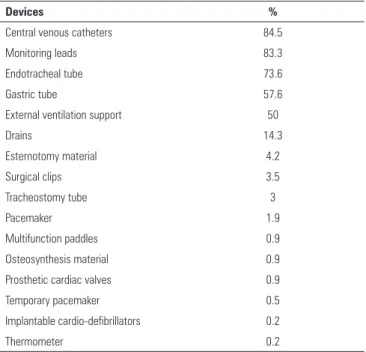
he multitude of medical devices identiied on the radiograms is reported in table 3 along with the relative frequency of each device.
In the current study, all of the radiograms were acquired with an antero-posterior incidence. herefore, the evaluation of the correct positioning of some of the identiied devices was not feasible, namely, for pacemakers. Consequently, we were only able to analyze the accuracy of the positioning of CVC, endotracheal tubes and tracheostomy tubes.
Table 1 - Criteria for the correct positioning of medical devices as visualized via thoracic radiograms, based on the literature search. These criteria were applied to assess the accuracy of the positioning of devices in the present study
Endotracheal tube 5cm above the carina or immediately above the aortic arch, with the head in a neutral position
If the carina is not visible, the distal end of the tube should be at the level of the medial borders of the claviculae
Tracheostomy tube Equidistant between the stoma and the carina (generally D3)
Nasogastric and feeding tubes Cross the mediastinum, in intra-esophageal topography
The nasogastric tube passes the cardia and the side holes should be at the level of the gastric antrum The feeding tubes should have their distal end in the second portion of the duodenum
Central venous catheters Ideally positioned in the superior vena cava
Inferior to the junction of the brachiocephalic veins, at the level of the first intercostal space Superior to the cavo-atrial junction, at the level of the inferior border of the intermediate bronchia Prosthetic cardiac valves Requires at least 2 incidences to evaluate
Pacemakers
Implantable cardioverter defibrillators
The accurate evaluation of the lead position requires at least 2 incidences
Thoracostomy tubes (drains) Tube should be at the surface of the expanded lung, between the 2 pleuras (for correct visualization 2 incidences are needed)
Fourth intercostal space, at the level of the anterior or medium axillary line Side hole should always be medial to the internal margin of the ribs Antero-superior orientation - for pneumothoraxes
Postero-inferior orientation - for liquid collections
Table 2 - Inclusion and exclusion criteria used to select the radiograms for analysis of the medical device placement
Inclusion criteria Admission with at least one thoracic radiogram performed in the intensive care unit One radiogram was selected for each patient admission
When multiple radiograms were performed per admission, the first radiogram that had the highest number of medical devices was chosen
Exclusion criteria Absence of identifiable medical devices on the thoracic radiogram
Table 3 - Type of devices identified and their relative frequency
Devices %
Central venous catheters 84.5
Monitoring leads 83.3
Endotracheal tube 73.6
Gastric tube 57.6
External ventilation support 50
Drains 14.3
Esternotomy material 4.2
Surgical clips 3.5
Tracheostomy tube 3
Pacemaker 1.9
Multifunction paddles 0.9
Osteosynthesis material 0.9
Prosthetic cardiac valves 0.9
Temporary pacemaker 0.5
Implantable cardio-defibrillators 0.2
Thermometer 0.2
Figure 1 - Distribution of the percentages of the types of positions of central venous catheters.
Figure 2 - Distribution of the percentages of the types of positions of endotracheal tubes.
DISCUSSION
A thoracic radiogram is an essential tool for the evaluation of medical devices immediately after placement, especially in patients in the ICU. A physician should request a thoracic radiogram when the expected indings, either positive or negative, could alter the approach to treating the patient.(12-18) In this context, a positive inding would be that a device is malpositioned, which would merit repositioning the device to prevent the development of adverse events, while a negative inding would be that a device is positioned correctly.
his is the irst study to analyze medical devices in the ICU of our institution, and its strengths are that it had a substantial sample size: 2,312 thoracic radiograms were reviewed, and 568 of these were analyzed. Furthermore, the evaluations were performed by professionals from outside of the ICU, which decreased the risk of potential bias.
he study also had some limitations. For example, the study was retrospective. It also involved imperfect technical conditions that are inherent to exams performed on patients in the ICU, such as the antero-posterior incidence, the lack of cooperation from and inappropriate positioning of the patient, and the impossibility of achieving a neutral head position.
Due to the aforementioned technical diiculties, all radiograms were performed using an antero-posterior incidence, which limited the ability to investigate the medical devices. For instance, some of the identiied devices require at least two incidences to accurately determine their location, namely, prosthetic cardiac valves, pacemakers, implantable cardioverter deibrillators and thoracic drains. In addition, there is a need for complementary radiograms
to accurately assess whether the distal extremity of these tubes was in the stomach or in the duodenum, nor was it possible to assess whether the proximal extremity was in the mouth or in the nose.
Due to these limitations, it was only possible to analyze the correct positioning of CVC, endotracheal tubes and tracheostomy tubes.
We found a relatively high frequency of malpositioning, with incorrectly placed CVC found in 30.6% of cases (with some series recording from 10% up to 40% misplaced CVC)(2,5,6) and with incorrectly placed endotracheal tubes identiied in 20% of analyzed cases (some records report values from 15% up to 28 - 46%).(2,5,6)
he association between these indings and possible related adverse events was not evaluated in the present study and should be investigated in future studies. In addition, no risk factors for the malpositioning of devices were sought or evaluated, but this should also be examined in the future.
CONCLUSION
In an intensive care unit context in our study, only central venous catheters, endotracheal tubes and tracheostomy tubes could be evaluated due to the limitations stated above.
An increased awareness of the use of radiograms as a method to identify and diagnose malpositioned medical devices could prevent the development of adverse events. hus, the knowledge of correct and incorrect positions of medical devices as visualized via radiograms is essential for making diagnoses of correct medical device positioning, which is crucial because of the relatively high prevalence of incorrectly positioned devices in an intensive care unit population (30% of central venous catheters and 20% of endotracheal tubes were malpositioned in this sample).
In future studies, an association between malpositioning and adverse events should be investigated.
Authors’ contribution
Objetivo: Identiicar e avaliar o posicionamento correto dos dispositivos médicos mais comumente utilizados, observados nas radiograias de tórax de pacientes durante a permanência em unidade de terapia intensiva de nosso centro.
Métodos: Foi realizada uma pesquisa bibliográica quanto aos critérios utilizados para avaliar o posicionamento correto dos dispositivos médicos nas radiograias de tórax. Avaliamos todas as radiograias de tórax realizadas na unidade de terapia intensiva de nosso centro durante um período de 18 meses. In-cluíram-se todas as admissões nas quais foi realizada uma radio-graia do tórax na unidade de terapia intensiva, nas quais fosse identiicável a presença de pelo menos um dispositivo médico. Para análise, selecionou-se uma radiograia por admissão. As radiograias foram avaliadas por um observador independente.
Resultados: De um total de 2.312 radiograias analisadas, 568 foram incluídas neste estudo. Identiicaram-se diversos
dispositivos médicos, incluindo eletrodos de monitoramento, tubos endotraqueais, cânulas de traqueostomia, cateteres venosos centrais, marca-passos e próteses valvares cardíacas. Dentre os cateteres venosos centrais identiicados, 33,6% dos subclávios e 23,8% dos jugulares estavam mal posicionados. Dentre os tubos endotraqueais, 19,9% estavam mal posicionados, enquanto todas as cânulas de traqueostomia tinham posicionamento correto.
Conclusão: Frequentemente se identiicam, na radiograia de tórax realizada em pacientes na unidade de terapia intensiva, cateteres venosos e tubos endotraqueais mal posicionados. Isso é importante, pois dispositivos mal posicionados podem se relacionar a eventos adversos. Estudos futuros devem investigar possíveis associações entre o mau posicionamento dos dispositivos e eventos adversos.
RESUMO
Descritores: Radiograia torácica; Cateteres venosos cen-trais/utilização; Intubação intratraqueal/instrumentação; Equi-pamentos e provisões; Unidades de terapia intensiva
REFERENCES
1. Trotman-Dickenson B. Radiology in the intensive care unit (Part I). J Intensive Care Med. 2003;18(4):198-210.
2. Amorosa JK, Bramwit MP, Mohammed TLH, Reddy GP, Brown K, Dyer DS, et al. ACR appropriateness criteria routine chest radiographs in intensive care unit patients. J Am Coll Radiol. 2013;10(3):170-4.
3. Savoca CJ, Gamsu G, Rohlfing BM. Chest radiography in intensive care units. West J Med. 1978;129(6):469-74.
4. Hunter TB, Taljanovic MS, Tsau PH, Berger WG, Standen JR. Medical devices of the chest. Radiographics. 2004;24(6):1725-46.
5. Godoy MC, Leitman BS, de Groot PM, Vlahos I, Naidich DP. Chest radiography in the ICU: Part 2, Evaluation of cardiovascular lines and other devices. AJR Am J Roentgenol. 2012;198(3):572-81.
6. Godoy MC, Leitman BS, de Groot PM, Vlahos I, Naidich DP. Chest radiography in the ICU: Part 1, Evaluation of airway, enteric, and pleural tubes. AJR Am J Roentgenol. 2012;198(3):563-71.
7. Jain SN. A pictorial essay: Radiology of lines and tubes in the intensive care unit. Indian J Radiol Imaging. 2011;21(3):182-90.
8. Torres-Ayala SC, Santacana-Laffitte G, Maldonado J. Radiography of cardiac conduction devices: a pictorial review of pacemakers and implantable cardioverter defibrillators. J Clin Imaging Sci. 2014;4:74. 9. Costelloe CM, Murphy WA Jr, Gladish GW, Rozner MA. Radiography
of pacemakers and implantable cardioverter defibrillators. AJR Am J Roentgenol. 2012;199(6):1252-8.
10. Johnston AJ, Bishop SM, Martin L, See TC, Streater CT. Defining peripherally inserted central catheter tip position and an evaluation of insertions in one unit. Anaesthesia. 2013;68(5):484-91.
11. Ryu HG, Bahk JH, Kim JT, Lee JH. Bedside prediction of the central venous catheter insertion depth. Br J Anaesth. 2007;98(2):225-7.
12. Ruza GC, Moritz RD, Machado FO. Routine chest radiography in intensive care: impact on decision-making. Rev Bras Ter Intensiva. 2012;24(3):252-7. 13. Ganapathy A, Adhikari NK, Spiegelman J, Scales DC. Routine chest x-rays
in intensive care units: a systematic review and meta-analysis. Crit Care. 2012;16(2):R68.
14. Tolsma M, van der Voort PH, van der Meer NJ. Why intensivists want chest radiographs. Crit Care. 2015;19:100
15. Cruz J, Ferra M, Kasarabada A, Gasperino J, Zigmund B. Evaluation of the clinical utility of routine daily chest radiography in intensive care unit patients with tracheostomy tubes: a retrospective review. J Intensive Care Med. 2016;31(5):333-7.
16. Tolsma M, Rijpstra TA, Schultz MJ, Mulder PG, van der Meer NJ. Significant changes in the practice of chest radiography in Dutch intensive care units: a web-based survey. Ann Intensive Care. 2014;4(1):10.
17. Lotano R, Gerber D, Aseron C, Santarelli R, Pratter M. Utility of postintubation chest radiographs in the intensive care unit. Crit Care. 2000;4(1):50-3.