rev bras ortop. 2013;48(4):377-380
www.rbo.org.br
0102-3616/$–see front matter © 2013 Sociedade Brasileira de Ortopedia e Traumatologia. Publicado pela Elsevier Editora Ltda. Todos os direitos reservados. «Work performed in the Department of Orthopedics and Traumatology, Faculdade de Medicina de Marília, Marília, SP, Brazil
*Corresponding author at: Faculdade de Medicina de Marília, Departamento de Ortopedia e Traumatologia, Avenida Monte Carmelo, 800, Fragata, Marília, SP, Brazil. CEP: 17519-030.
E-mail: [email protected] (E.P. Palacio).
Case Report
Severe ocular alteration associated with fracture of the
scapula. Case report
«
Evandro Pereira Palacio,
a,* Missa Takasaka,
bTatiana Rocha Bastos,
bAndrea Alonso Negrini,
cKauan Simões Val,
cPatrícia da Silva Fernandes,
cRoberto Ryuiti Mizobuchi,
dand Alcides Durigam Júnior
eaPhD; Assistant Professor in the Department of Orthopedics and Traumatology, Faculdade de Medicina de Marília, Marília, SP, Brazil bResident Physician in the Department of Orthopedics and Traumatology, Faculdade de Medicina de Marília, Marília, SP, Brazil cUndergraduate Medical Student, Faculdade de Medicina de Marília, SP, Brazil
dPhD; Assistant Professor and Head of the Orthopedics and Traumatology Service, Faculdade de Medicina de Marília, Marília, SP, Brazil ePhD; Assistant Professor and Head of the Shoulder Group, Department of Orthopedics and Traumatology, Faculdade de Medicina de
Marília, Marília, SP, Brazil
doi:
a b s t r a c t
Objective: Scapula fractures are rare entities and occur along with severe respiratory complications, usually associated with lung tissue injuries. The authors describe a rare case of eye arteriolar thromboembolism due to scapular body fracture in a patient victim of a car accident.
© 2013 Sociedade Brasileira de Ortopedia e Traumatologia. Published by Elsevier Editora Ltda. All rights reserved. A RT I C L E I N F O
Article history:
Received on June 25, 2012 Accepted on September 12, 2012
Keywords: Retinal diseases Scapula
378
rev bras ortop. 2013;48(4):377-380Alteração ocular grave associada à fratura de escápula. Relato de caso
Introduction
Scapular fractures are rare entities that represent 0.4% to 1% of all fractures1,2 and are associated with direct and indirect
high-energy trauma. They may not be diagnosed immediately, and this may often culminate in severe consequences for such patients, given that this trauma may be associated with severe lung tissue contusions.3,4
The clinical condition is characteristic, with functional impotence and the upper limb adducted against the chest wall, along with high-intensity pain on palpation of the scapular region. Comolli’s sign, i.e. triangular edema on the posterior face of the thorax, over the scapula, is frequently present.5
Radiographic examination is mandatory. Radiographs in true anteroposterior, scapular lateral and axillary lateral views are usually sufficient for reaching a diagnostic conclusion of fracture, when performed properly. Computed tomography also has a role, especially when there is a suspicion of involvement of the glenoid joint surface or for surgical planning.6
Although the scapula is a bone without a true medullary canal, it is perfectly capable of releasing fatty emboli when fractured, like any long bone, and may cause the same fatal consequences.
Case report
This study received approval from the Research Ethics Committee (CAAE: 04181612.1.0000.5413).
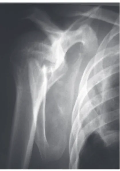
The patient was a 43-year-old single male farm worker, and was a car accident victim in which the car overturned. He was taken to the emergency service of Hospital de Clínicas, Marília Medical School, with a report of intense pain in the region of the right scapular belt and in the ipsilateral hemithorax. There were no alterations with regard to the ATLS protocol. During the orthopedic examination, only the initial complaints of pain were confirmed. In radiological and tomographic examinations, a multifragmentary extra-articular fracture of the scapular body was presented (AO/OTA: 09-A2.2) (Figs. 1 and 2). Nonsurgical treatment was indicated and the patient was immobilized using a shoulder suspender and placed under observation for 24 hours.
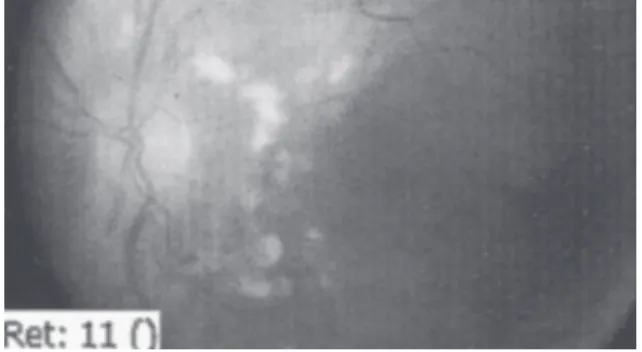
On the second day of the hospital stay, hours before discharge, the patient complained of ocular pain and diminished visual acuity bilaterally. He then started to present a condition of subconjunctival hyperemia (Fig. 3), with even greater reduction of visual acuity. An emergency ophthalmological assessment was requested, which showed retinal edema and hemorrhaging through ocular fundus examination. Retinography (Fig. 4) and fluorescein angiography (Fig. 5) were then performed by ophthalmologists, and these showed cotton-wool (whitened) patches, pre-capillary occlusions and peripapillary hemorrhagic foci. These findings were compatible with the obstructive ophthalmological condition known as Purtscher’s retinopathy.
Prednisone (60 mg/day, orally) was prescribed for eight weeks and the patient evolved with good but not total recovery of visual acuity. He is continuing to attend six-monthly ophthalmological follow-ups on an outpatient basis, without any prediction of discharge.
The scapular fracture has now satisfactorily consolidated.
Figure 1 - Scapular lateral radiographic image in which fracturing of the body of the scapula is shown.
r e s u m o
Objetivo: As fraturas de escápula são entidades raras e que frequentemente cursam com complicações respiratórias graves, geralmente associadas a contusões do tecido pulmonar. Os autores descrevem um caso raro de tromboembolismo arteriolar ocular causado por fratura multifragmentária do corpo escapular em paciente vítima de acidente automobilístico.
© 2013 Sociedade Brasileira de Ortopedia e Traumatologia. Publicado pela Elsevier Editora Ltda. Todos os direitos reservados. Palavras-chave:
Doenças retinianas Escápula
rev bras ortop. 2013;48(4):377-380
379
Discussion
Scapular fractures have low incidence, probably because of the anatomical characteristics of the scapulae: thick borders, great mobility and capacity for retraction, and location between muscle layers. Trauma of considerable energy is needed for this fracturing to occur.
In the case in question, in addition to the clinical complaints of localized pain, the functional deficit and proof of multifragmentary fracturing from imaging examinations, our attention was drawn to the sudden complaint of ocular pain in association with diminished visual acuity, in a patient who was healthy apart from the scapular fracture.
Although any bone is technically capable of releasing fatty emboli when fractured, the great majority of such fractures rarely have any true clinical repercussions. Orthopedic professionals who are accustomed to the routines of emergency rooms are aware of the high rates of occurrence of fatty thromboembolism that are associated with cases of fractures of long bones like the femur or tibia. These occurrences are real and should be foreseen. However, it is rare to think of this situation in treating a scapular fracture.
In the case in question, another point of great importance that diverges from the patterns described in the literature also has to be taken into consideration: the site of occurrence of the obstruction. In orthopedic practice, occurrences of pulmonary and cerebral vascular occlusions are very common. However, we did not identify any reports on obstruction of the retinal arterioles due to fractures, in a variety of osteomuscular databases of Brazilian origin. Thus, this report is the first case described.
Purtscher’s retinopathy is a disease with few descriptions, even within the field of ophthalmology, and there is controversy in relation to its physiopathological mechanism. Although its association with fatty thromboembolism is irrefutable, this disease was initially correlated with vascular lesions due to severe cranial trauma. However, more recently, it has been described in association with injuries due to air bags and the practice of bungee jumping.7
From ocular fundoscopy, the retina is characterized by cotton-wool stains, edema and macula associated with superficial hemorrhage.7,8 The consensus is that fatty emboli
are formed and lead to arteriolar occlusion and consequently to infarction of the microvasculature in the retinal nerve fiber layer. The patients’ main complaints are ocular pain and loss of visual acuity: both of them at varying intensities. These may arise a few hours or even a few days after the initial trauma.
It should be emphasized that history-taking and physical examination, when well conducted, are sufficient for affirming the likely diagnosis. It is presumptuous and unnecessary to indicate treatment with platelet antiaggregants for each and every fracture, regardless of whether they are in the scapula, sternum or head of the radius, with the aim of preventing such situations. Nonetheless, orthopedists should not avoid placing value on their patients’ complaints, even if they relate to other medical specialties.
Figure 2 - 3D tomographic reconstruction image in which multifragmentary fracturing of the body of the scapula is shown.
Figure 3 - Image of bilateral subconjunctival effusion, 20 hours after scapular fracturing.
Figure 4 -Retinography examination showing cotton-wool whitened images that are typical of Purtscher’s retinopathy.
380
rev bras ortop. 2013;48(4):377-380Conflicts of interest
The authors declare that there were no conflicts of interest in conducting this work, in accordance with Resolution No. 1595/2000 of the Federal Medical Council.
R E F E R E N C E S
1. Wilson PD. Experience in the management of fractures and dislocations (based on an analysis of 4390 cases) by staff of the Fracture Service MGH, Boston. Philadelphia: JB Lippincott; 1938.
2. Newell ED. Review of over 2,000 fractures in the seven years. South Med J. 1927;20:644-8.
3. Fischer RP, Flynn TC, Miller PW. Scapular fractures and associated major ipsilateral upper-torso injuries. Curr Concepts Trauma Care. 1985;1(1):14-6.
4. Thompson DA, Flynn TC, Miller PW, Fischer RP. The
significance of scapular fractures. J Trauma. 1985;25(10):974-7. 5. Landi A, Schoenhuber R, Funicello R, Rasio G, Esposito
M. Compartment syndrome of the scapula. Definition on clinical, neurophysiological, and magnetic resonance data. Ann Chir Main Memb Super. 1992;11(5):383-8.
6. McAdams TR, Blevins FT, Martin TP, DeCoster TA. The role of plain films and computed tomography in the evaluation of scapular neck fractures. J Orthop Trauma. 2002;16(1):7-11. 7. Medeiros HAG, Medeiros JEG, Caliari LC, Silva JF. Retinopatia
de Purtscher e Purtscher-like. Rev Bras Oftalmol. 2009;68(2):114-19.