CLINICAL SCIENCE
Prevalence of eye disease in Brazilian patients with
psoriatic arthritis
Fernanda B. F. de Lima, Maria Fernanda Abalem, Danilo G. Ruiz, Beatriz de A. F. Gomes, Ma´rio N. L. de Azevedo, Haroldo V. Moraes, Jr., Ariyah Seth Yeskel, Newton Kara-Junior
Federal University of Rio de Janeiro, Ophthalmology, Rio de Janeiro/RJ, Brazil.
OBJECTIVES: The aim of this study was to report the type and frequency of ocular manifestations in Brazilian psoriatic arthritis patients.
METHODS:We conducted a cross-sectional study in a Brazilian tertiary hospital. The test group included 40 patients who had psoriatic arthritis according to the Classification Criteria for Psoriatic Arthritis. A control group of 40 individuals was matched for age and gender. All of the patients underwent ophthalmic evaluation, which included best-corrected visual acuity, slit lamp and fundus examinations, and dry eye diagnostic tests (Schirmer I, tear break-up time and rose bengal). Demographic parameters were also evaluated.
RESULTS:The mean age of the patients was 53.9¡13.1 years; the mean disease duration was 8¡10.5 years. Most of the patients were women (60%), and the majority had polyarticular disease (57.5%). Several ocular abnormalities were found, including punctate keratitis, pinguecula, blepharitis, pterygium, cataract, glaucoma, uveitis, and retinal microvascular abnormalities. There were no significant differences in the rates of these abnormalities compared with the control group, however. The Keratoconjunctivitis sicca and dry eye diagnostic tests were more often positive in the patients with psoriatic arthritis than in the control group.
CONCLUSIONS:In this study, keratoconjunctivitis sicca was the most common ocular finding related to psoriatic arthritis. Therefore, we recommend early ophthalmologic evaluations for all psoriatic arthritis patients who complain of eye symptoms.
KEYWORDS: Psoriatic Arthritis; Eye; Keratoconjunctivitis Sicca; Ocular Findings.
Lima FB, Abalem MF, Ruiz DG, Gomes BA, Azevedo MN, Moraes-Junior HV, et al. Prevalence of eye disease in Brazilian patients with psoriatic arthritis. Clinics. 2012;67(3):249-253.
Received for publication onNovember 24, 2011;First review completed onNovember 27, 2011;Accepted for publication onJanuary 10, 2012 E-mail: [email protected]
Tel.:+55 21 2491 6351
INTRODUCTION
Psoriatic arthritis (PsA) is an inflammatory arthritis associated with psoriasis. Skin lesions are typically the first signs of the disease, followed by the articular manifesta-tions. The articular manifestations may occur first in rare cases, however (1).
Psoriasis is a chronic, immunologically mediated disease with both genetic and environmental risk factors. It affects 1 to 3% of the world’s population. Arthritis occurs in up to 30% of the patients with psoriasis, and it is diagnosed based on clinical and radiological features (2,3). The inflammatory arthritis associated with psoriasis is typically seronegative for rheumatoid factor and exhibits a variety of disease patterns, including oligoarticular disease, polyarticular disease, distal
interphalangeal disease (IFD), arthritis mutilans, and axial disease (4).
The seronegative spondyloarthropathies, including PsA, are a group of disorders that share many clinical and pathologic features (5). The HLA-B27 antigen is found in some patients with PsA, and this antigen can be used as a prognostic marker for clinical disease progression (6). HLA-B27 is associated with earlier onset of psoriatic arthritis and bilateral sacroiliitis and is more common in men.
An ocular inflammatory reaction is commonly found in many rheumatologic diseases (7), and there are various ocular disorders associated with psoriatic arthritis. These disorders include abnormalities of the conjunctiva, cornea, sclera, uvea, and lens. To date, there are no significant publications exploring the prevalence of eye disease in patients with psoriatic arthritis (8).
The aim of this study was to report the ocular manifesta-tions of the psoriatic arthritis patients seen at a university hospital in Brazil.
METHODS
This cross-sectional study included outpatients with PsA seen at the university hospital of the Federal University of
Copyrightß2012CLINICS– This is an Open Access article distributed under
the terms of the Creative Commons Attribution Non-Commercial License (http:// creativecommons.org/licenses/by-nc/3.0/) which permits unrestricted non-commercial use, distribution, and reproduction in any medium, provided the original work is properly cited.
Rio de Janeiro, Brazil, from March 2010 to October 2010. This study was approved by the Medical Ethics Committee of the hospital. Before enrollment, all of the patients pro-vided informed consent in accordance with the Helsinki Declaration.
The test group consisted of 40 patients who had been diagnosed with PsA according to the Classification Criteria for Psoriatic Arthritis (CASPAR) and who were being treated by the Rheumatology Unit (9).
The control group consisted of 40 patients without PsA who were recruited from the Primary Care Unit. To avoid clinical and statistical bias, these patients were selected and matched for age and gender.
In both groups, we excluded the patients younger than 18 years of age and those with inflammatory connective tissue diseases, nephropathy, lung and heart disease, gastroenter-ological disease/inflammatory bowel disease, neurgastroenter-ological disease, neoplasia, metabolic bone disease, skin diseases (except psoriasis), infections, hematological disease, liver disease, previous ocular surgery, active eye infection, active ocular allergy, and evidence of an abnormal eyelid move-ment disorder.
We also recorded other patient characteristics, including age, gender, history of systemic hypertension or diabetes, and use of topical eye medications, for the cases and controls. The disease duration and use of systemic PsA medications were also recorded for the patients in the case group.
The patients were not permitted to use artificial tears within 2 hours of the screening evaluation.
One ophthalmologist performed all of the ophthalmic exams in sequential order.
First, the best-corrected visual acuity (BCVA) was measured using the Snellen chart.
Second, a slit lamp examination was used to evaluate the cornea for the presence of active inflammation and structural changes at a magnification of 10X to 16X. A cataract evaluation was performed according to the Lens Opacities Classification System III (10).
Third, the tear break-up time (TBUT), Schirmer I and rose bengal staining tests were used sequentially to detect dry eyes. To perform the TBUT, 1% fluorescein (Fludiag, Oftalmopharma, Sao Paulo, Brazil) was applied to the eye, and the average of three consecutive breakup times (manually determined with a stopwatch) was calculated. A time of ,10 seconds was considered abnormal. The Schirmer I test was performed by applying a 5635 mm paper strip (Schirmer strips, Ophthalmos, Sao Paulo, Brazil) without anesthesia to the lower temporal lid margin; values of,10 mm in 5 minutes were considered to be abnormal. These cutoff values were based on the normal reference values described in the literature (11). The inferior bulbar conjunctiva was touched with a rose bengal strip (rose bengal strips, Ophthalmos, Sa˜o Paulo, Brazil) to assess staining. The reaction was classified according to the van Bijsterveld Scoring System and values.3 were considered to be abnormal (12). For each sign, the most extreme measurement was used in the analysis.
The Japanese criteria for dry eyes, which include symptom evaluation, the Schirmer I or TBUT tests, and rose bengal staining, were applied (13). For a definitive diagnosis, the patient needed to have had symptoms and two positive tests. The patients who met only two criteria were classified as having probable dry eye.
The Ocular Surface Disease Index (OSDI) questionnaire was administered to assess the ocular dry eye symptoms (14).
Intraocular pressure was measured using Goldmann applanation tonometry. Values .21 mmHg were consid-ered to be abnormal. A diagnosis of open-angle glaucoma was based on visual field, gonioscopic, and optic nerve abnormalities (15).
Fundoscopy was performed using indirect ophthalmo-scopy with a 20-diopter Volk lens.
Statistical Analysis
Due to the need for consistency, patients (rather than affected eyes) were matched when choosing the control group. The descriptive data are presented in tables and are expressed by frequency (n) and percentage (%) for categorical data and the mean ¡ SD (or median ¡ IQR) for numerical data.
To compare the numerical data, we used the Student’s t-test for normally distributed data or the Mann-Whitney t-test for non-normally distributed data. We used thex2 test or
Fisher’s exact test for comparisons of categorical data. The nonparametric method was used because some variables did not have a normal (Gaussian) distribution due to large dispersion (the hypothesis of normality was rejected in the Kolmogorov-Smirnov test).
The significance level was set atp,0.05. Statistical analyses were performed using the SAS H version 6.11 statistical software (SAS Institute Inc., Cary, North Carolina).
RESULTS
In total, 40 PsA patients were evaluated. The mean age of the PsA patients was 53.9¡13.1 years, and 24 (60%) were female. The mean disease duration was 8¡10.5 years. The demographic data are shown in Table 1. Polyarticular disease was the most common disease characteristic (57.5%). Majority of patients were taking medications for PsA: 27 patients (67.5%) were using methotrexate, 17 patients (42.5%) were using oral corticosteroids, 13 patients (32.5%) were using tumor necrosis factor alpha receptor antagonists (TNF), and three patients (7.5%) were using anti-malarial medications.
Of the four patients with glaucoma, three were using timolol maleate 0.5%, and one was using brimonidine tartrate 0.2%. In addition, four other patients were using artificial tears.
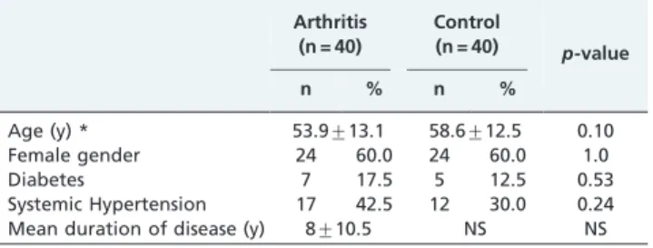
Table 1 -Demographic data by group.
Arthritis (n = 40)
Control
(n = 40) p-value
n % n %
Age (y) * 53.9¡13.1 58.6¡12.5 0.10
Female gender 24 60.0 24 60.0 1.0
Diabetes 7 17.5 5 12.5 0.53
Systemic Hypertension 17 42.5 12 30.0 0.24 Mean duration of disease (y) 8¡10.5 NS NS
The majority of the patients (90% in the study group and 85% the control group) presented with a BCVA of 0.66 or better. The remaining patients had BCVAs of 0.25 or worse in at least one eye. The low visual acuity was bilateral and was caused by nuclear cataracts in all of these patients.
Several ocular abnormalities were found upon biomicro-scopy and fundobiomicro-scopy evaluation. Using the data from our control group, we were able to correlate the large number of ocular abnormalities with PsA. With the exception of pterygium and uveitis, the frequency and type of ophthal-mologic findings (glaucoma, cataract, blepharitis, pingue-cula, punctate keratitis, and retinal microvascular abnormalities) did not differ significantly between the case and control groups. There were not enough cases of pterygium and uveitis to make comparisons between the groups. These findings are summarized in Table 2.
It is also important to note that all of the cataracts were age related, the glaucoma cases were all classified as primary open-angle glaucoma and all of the patients who presented with retinal microvascular abnormalities also presented with systemic hypertension.
All of the uveitis cases appeared to be anterior uveitis in which there were posterior and anterior synechias and corneal endothelium changes but no fundal involvement. Both of the uveitis cases were observed in patients with oligoarticular arthritis.
The mean intra-ocular pressures for all of the patients, including those with glaucoma, were 13.0¡1.7 in the right eyes and 13.1¡1.8 in the left eyes. These relatively normal values were most likely due to the patients with glaucoma taking medications.
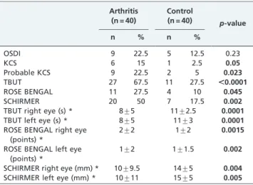
Ocular tests were used to test for KCS. In the PsA group, we found 27 patients (67.5%) with abnormal TBUTs, 20 patients (50%) with abnormal Schirmer I tests, and 11 patients (27.5%) with abnormal rose bengal tests; all of these tests were significantly more likely to be positive in the PsA group. Nine patients (22.5%) in the PsA group were symptomatic and tested positive on the OSDI. However, these results were not significantly different from those of the control group. According to the criteria adopted in this study, 6 PsA patients (15%) had a definitive diagnosis and 9 (22.5%) had a probable diagnosis of KCS (p,0.05 for both). In addition, the mean TBUT, Schirmer and rose bengal
values were calculated and described in Table 3. Most of the patients in the definitive and probable KCS groups presented with oligoarthritis and/or polyarticular disease, and the majority were using methotrexate for disease control.
The demographic parameters are shown in Table 1. In the control group, three patients were using timolol maleate 0.5%, and two patients were using artificial tears.
DISCUSSION
This study represents the largest group of Brazilian psoriatic arthritis patients who have been screened for associated ocular manifestations.
Polyarthritis was the most common PsA presentation in this study. Some researchers have concluded that oligoar-thritis is the most common manifestation of PsA, whereas some have claimed that polyarthritis is more common (16). Overlapping PsA articular subtypes are common over time, and the distributions may vary as different definitions are applied. Similarly, patterns change over time due to increases in disease duration and changes in natural history that may lead patients to develop a polyarticular pattern (17).
According to the results presented here, PsA patients frequently develop ocular inflammatory conditions. Previous studies have shown that PsA patients may present with conjunctivitis, uveitis, and KCS (18). We did not find any patients with conjunctivitis because we specifically selected patients who visited the department of rheumatology for treatment.
Uveitis is an important ocular abnormality that is described in PsA patients (19). The frequency of uveitis associated with psoriatic arthritis ranges from 0.4 to 18% (20-22). In this study, 5% of the patients exhibited signs of uveitis, which was classified as anterior, unilateral and acute. In general, these signs are classically associated with spondyloarthropathies and are not specifically related to PsA. (23). It is important to emphasize that these patients Table 2 -Ocular findings by group.
Arthritis (n = 40)
Control
(n = 40) p-value
n % n %
Visual Acuity$0.66 36 90.0 34 85.0 0.49
Glaucoma 4 10.0 3 7.5 0.50
Cataract 4 10.0 8 20.0 0.21
Blepharitis 5 12.5 9 22.5 0.23
Punctate keratitis 9 22.5 4 10.0 0.13
Pinguecula 8 20.0 9 22.5 0.78
Pterygium 2 5.0 2 5.0 fc
Uveitis 2 5.0 0 0.0 fc
Microvascular abnormalities 6 15 4 10 0.49 IOP right eye (mmHg) * 13.0¡1.7 13.2¡2.2 0.73 IOP left eye (mmHg) * 13.1¡1.8 13.0¡2.1 0.86
fc: fewer than 5 cases.
*IOP was expressed as the mean¡SD and analyzed using the Student’s t-test for independent samples. Thex2 test or Fisher’s exact test was used to compare proportions (categorical data).
Table 3 -KCS features by group.
Arthritis (n = 40)
Control (n = 40)
p-value
n % n %
OSDI 9 22.5 5 12.5 0.23
KCS 6 15 1 2.5 0.05
Probable KCS 9 22.5 2 5 0.023
TBUT 27 67.5 11 27.5 ,0.0001
ROSE BENGAL 11 27.5 4 10 0.045
SCHIRMER 20 50 7 17.5 0.002
TBUT right eye (s) * 8¡5 11¡2.5 0.0001
TBUT left eye (s) * 8¡5 11¡3 0.0001
ROSE BENGAL right eye (points) *
2¡2 1¡2 0.0015
ROSE BENGAL left eye (points) *
1¡2 1¡1.5 0.002
SCHIRMER right eye (mm) * 10¡9.5 14¡5 0.004
SCHIRMER left eye (mm) * 10¡11 15¡5 0.005
*The numerical KCS measurements were expressed as the median¡IQR and analyzed using the Mann-Whitney (nonparametric) test.
Thex2 test or Fisher’s exact test were used to compare proportions (categorical data).
were not using anti-TNF-alpha medications because these agents may induce paradoxical uveitis in patients with rheumatic diseases (24,25).
In this study, we found a high number of abnormal dry eye tests in the PsA patients. Many of these patients met the European classification criteria for Sjogren’s syndrome (26), and we believe that these results demonstrate the presence of important ocular SS signs in this study group. The presence of SS in patients with spondyloarthropathies has been corroborated by the literature (27).
We also found that 15% of the PsA patients met the criteria for KCS and that another 22.5% had probable KCS. This rate may be underestimated because we used rigid diagnostic criteria. Furthermore, the disease may have been misclassified, particularly in the early stages when the typical signs and symptoms are less clear. There is a poor relationship between the signs and symptoms of dry eye (28). Therefore, these data are important because KCS is well recognized in other rheumatologic diseases, such as rheumatoid arthritis (29,30).
Blepharitis, pterygium, pinguecula, and keratitis are ophthalmic conditions commonly found in the general population (31,32,33). Our study showed a high frequency of these findings in the PsA patients. However, these rates did not differ markedly from the data in the literature or from the control group.
Most of the patients in this study presented with good visual acuity. Cataracts were responsible for most of the visual impairment. All of these patients presented with the age-related nuclear cataract subtype, which differs from cataracts caused by uveitis or steroid use (34).
Our study group had a higher prevalence (10%) of primary open-angle glaucoma than that of the general population (0.8 a 3.0%) (15). We observed no signs of glaucoma secondary to uveitis or steroid use, which is the type of glaucoma more commonly found in rheumatologic diseases (35). Moreover, there were no significant differ-ences between the two groups in the number of glaucoma diagnoses or average intraocular pressure values.
In this study, 15% of the patients had micro-vascular retinal abnormalities that were indistinguishable from those related to systemic hypertension. There were no significant differences between the two groups (36,37).
High levels of TNF-alpha are found in the joint fluid and tissue of patients with psoriatic arthritis. TNF-alpha induces the production of other pro-inflammatory cytokines that mediate multiple biological processes responsible for joint damage (38). A clinical trial of etanercept provided strong evidence that this medication is effective for psoriatic arthritis (39). The efficacy of anti-TNF agents for patients with uveitis and scleritis has been observed in a limited study, but anti-TNF treatment has caused both improve-ment and exacerbation of inflammatory eye disease (40). Another author has shown that Infliximab was effective for suppressing intraocular inflammation; the clinical symp-toms improved significantly, and anterior chamber cells decreased rapidly (41). These findings may explain why the patients in this study who were using anti-TNF agents presented without uveitis or scleritis. The inflammatory conditions that they presented with were not severe (blepharitis and keratitis).
The retinal toxicity of antimalarial drugs, such as chloroquine and hydroxychloroquine, has been recognized
for many years (42). No retinopathy was found in any of the patients in this study.
The clinical efficacy of methotrexate for PsA is well known, and the agent has been a mainstay of treatment for many years. Methotrexate is associated severe adverse events, of which liver toxicity is the most common. The patients in our study maintained regular follow-ups with their rheumatologists; at the conclusion of the study, our patients did not experience any significant adverse events (43).
In conclusion, many ocular abnormalities were found in the PsA patients, including cataracts, glaucoma, and blepharitis. These findings were found in similar rates in the control group. However, the increased rate of Keratoconjunctivitis sicca in the PsA patients was statisti-cally significant. This finding is important for understand-ing the disease and for clinical management. Although uveitis was not significantly more common in the PsA group, it must be monitored in PsA patients due to its clinical relevance.
AUTHOR CONTRIBUTIONS
Lima FB examined all patients, obtained ethics approval, wrote and submitted the manuscript. Abalem MF and Gomes BA wrote the manuscript. Ruiz DG recruited the patients. Yeskel AS translated the manuscript. Azevedo MN and Moraes-Junior HV directed and defined the research. Kara-Junior N directed the research.
REFERENCES
1. Moll JM, Wright V. Psoriatic arthritis. Semin Arthritis Rheum. 1973;3(1):55-78, http://dx.doi.org/10.1016/0049-0172(73)90035-8. 2. Gladman DD. Psoriatic arthritis. Dermatol Ther. 2009;22(1):40-55, http://
dx.doi.org/10.1111/j.1529-8019.2008.01215.x.
3. Jamshidi F, Bouzari N, Seirafi H, Farnaghi F, Firooz A. The prevalence of psoriatic arthritis in psoriatic patients in Tehran, Iran. Arch Iran Med. 2008;11(2):162-5.
4. Zachariae H. Prevalence of Joint Disease in Patients with Psoriasis: Implications for Therapy. Am J Clin Dermatol. 2003;4(7):441-7, http:// dx.doi.org/10.2165/00128071-200304070-00001.
5. Gladman DD, Antoni C, Mease P, Clegg DO, Nash P. Psoriatic arthritis: epidemiology, clinical features, course, and outcome. Ann Rheum Dis. 2005;64 Suppl 2ii14-7.
6. Queiro R, Sarasqueta C, Belzunegui J, Gonzales C, Figueroa M, Torre-Alonso JC. Psoriatic Spondyloarthropathy: A comparative study between HLA-B27 positive and HLA-B27 negative disease. Semin Arthiritis Rheum. 2002;31(6):413-8, http://dx.doi.org/10.1053/ sarh.2002.33470.
7. Bana˜res A, Herna´ndez-Garcia C, Ferna´ndez-Gutie´rrez B, Jover JA. Eye involvement in the Spondyloarthropathies. Rheum Dis Clin North Am. 1998;24(4):771-84,ix, http://dx.doi.org/10.1016/S0889-857X(05)70041-7. 8. Lambert JR, Wright V. Eye inflammation in psoriatic arthritis. Ann
Rheum Dis. 1976;35(4):354-6, http://dx.doi.org/10.1136/ard.35.4.354. 9. Taylor W, Gladman D, Helliwell P, Marchesoni A, Mease P, Mielants H.
CASPAR Study Group Classification Criteria for Psoriatic Arthritis: development of new criteria from a large internacional study. Arthritis Rheum. 2006;54(8):2265-673.
10. Chylack LT, Wolfe JK, Singer DM, Leske MC, Bullimore MA, Bailey IL, et al. The lens Opacities Classification System III.The Longitudinal Study of Cataract Study Group. Arch Ophthalmol. 1993;111(6):831-6, http:// dx.doi.org/10.1001/archopht.1993.01090060119035.
11. Sullivan BD, Whitmer D, Nichols KK, Tomlinson A, Foulks GN, Geerling G, et al. An Objective Approach to Dry Eye Disease Severity. Invest Ophthalmol Vis Sci. 2010;51(12):6125-30, http://dx.doi.org/10.1167/ iovs.10-5390.
12. Van Bijsterveld OP. Diagnostic tests in the Sicca syndrome. Arch Ophthalmol. 1969;82(1):10-4, http://dx.doi.org/10.1001/archopht.1969. 00990020012003.
13. Subcommittee of the international dry eye workshop. Methodologies to diagnose and monitor dry eye disease: report of the diagnostic methodology subcommittee of the international dry eye workshop. Ocul Surf. 2007;5(2):108-52.
15. Wolfs RC, Borges PH, Ramrattan RS, Klaver CC, Hulsman CA, Holfman A, et al. Changing Views on Open-Angle Glaucoma: Definitions and Prevalence- The Rotterdam Study. Invest Ophthalmol Vis Sci. 2000; 41(11):3309-21.
16. Reich K, Kruger K, Mossner R, Augustin M. Epidemiological and clinical pattern of psoriatic arthritis in Germany: a prospective interdisciplinary epidemiological study of 1511 patients with plaque-type psoriasis. Br J Dermatol. 2009;160(5):1040-7, http://dx.doi.org/10.1111/j.1365-21 33.2008.09023.x.
17. Mahugh NJ, Balachrishnan C, Jones SM. Progression of peripheral joint disease in psoriatic arthritis: a 5-yr prospective study. Rheumatology. 2003;42(6):778-83, http://dx.doi.org/10.1093/rheumatology/keg217. 18. Paiva ES, Macaluso DC, Edwards A, Rosenbaum JT. Characterization of
uveitis in patients with psoriatic arthritis. Ann Rheum Dis. 2000;59(1):67-70, http://dx.doi.org/10.1136/ard.59.1.67.
19. Sampaio-Barros P, Conde RA, Bonfiglioli R, Be´rtolo MB, Samara AM. Characterization and outcome of uveitis in 350 patients with spondy-loarthropathies. Rheumatol Int. 2006;26(12):1143-6, http://dx.doi.org/ 10.1007/s00296-006-0203-7.
20. Gouveia EB, Yamamoto JH, Abdalla M, Hirata CE, Kubo P, Olivalves E. Causes of uveitis in a tertiary center in Sao Paulo city, Brazil. Arq Bras Ophthalmol. 2004;67:139-45, http://dx.doi.org/10.1590/S0004-2749200 4000100025.
21. Queiro R, Torre JC, Belzunegui J, Gonzales C, Dios JR, Unanue F, et al. Clinical Features and Predictive Factors in Psoriatic Arthritis- Related uveitis. Semin Arthritis Rheum. 2002;31(4):264-70, http://dx.doi.org/ 10.1053/sarh.2002.28798.
22. Zeboulon N, Dougados M, Gossec L. Prevalence and characteristics of uveitis in the spondyloarthropathies: a systematic literature review. Ann Rheum Dis. 2008;67(7):955-9.
23. Cantini F, Niccoli L, Nannini C, Kaloudi O, Bertoni M, Cassara E. Psoriatic arthritis: a systematic review. In J Rheum Dis. 2010;13(4):300–17. doi: 10.1111/j.1756-185X.2010.01540.x.
24. Lim LL, Fraunfelder FW, Rosenbaum JT. Do tumor necrosis factor inhibitors cause uveitis? A registry-based study. Arthritis Rheum. 2007;56(10):3248-52.
25. Kakkassery V, Mergler S, Pleyer U. Anti-TNF-alpha treatment: a possible promoter in endogenous uveitis? observational report on six patients: occurrence of uveitis following etanercept treatment. Curr Eye Res. 2010;35(8):751-6, http://dx.doi.org/10.3109/02713683.2010.486520. 26. Vitali C, Bombardieiri S, Jonnson R, Moutsopoulos HM, Alexander EL,
Carsons SE, et al. Classification criteria for Sjogren’s syndrome: a revised version of the European criteria proposed by the American-European Consensus Group. Ann Rheum Dis. 2002;61(6):554-8, http://dx.doi.org/ 10.1136/ard.61.6.554.
27. Scotto di Fazano CS, Grilo RM, Vergne P, Coyral D, Inoui R, Bonnet C, et al. Is the relationship between spondyloarthropathies and Sjogren’s syndrome in women coincidental? a study of 13 cases. Joint Bone Spine. 2002;69(4):383-7, http://dx.doi.org/10.1016/S1297-319X(02)00414-1. 28. Nichols KK, Nichols JJ, Mitchell GL. The Lack of Association Between
Signs and Symptoms in Patients With Dry Eye Disease. Cornea. 2004;23(8):762-70, http://dx.doi.org/10.1097/01.ico.0000133997.07144.9e.
29. Lemp MA. Dry Eye (Keratoconjunctivitis Sicca), Rheumatoid Arthritis, and Sjo¨gren’s Syndrome. Am J Ophthalmol. 2005;140(5):898-9, http:// dx.doi.org/10.1016/j.ajo.2005.06.031.
30. Fujita M, Igarashi T, Kurai T, Sakane M, Yoshino S, Takahashi H. Correlation between dry eye and rheumatoid arthritis activity. Am J Ophthalmol. 2005;140(5):808-13, http://dx.doi.org/10.1016/ j.ajo.2005.05.025.
31. Lemp MA, Nichols KK. Blepharitis in the United States 2009: a survey-based perspective on prevalence and treatment. Ocul Surf. 2009:7(2 Suppl):S1-S14.
32. Paula JS, Thorn F, Cruz AA. Prevalence of pterygium and cataract in indigenous populations of the Brazilian Amazon rain forest. Eye. 2006;20(5):533-6, http://dx.doi.org/10.1038/sj.eye.6701917.
33. Sharma S. Keratitis. Biosci Rep. 2001;21(4):419-44, http://dx.doi.org/ 10.1023/A:1017939725776.
34. James ER. The etiology of steroid cataract. J Ocul Pharmacol Ther. 2007;23(5):403-20, http://dx.doi.org/10.1089/jop.2006.0067.
35. Hamideh F, Prete PE. Ophthalmologic Manifestations of Rheumatic Diseases. Semin Arthritis Rheum. 2001;30(4):217-41, http://dx.doi.org/ 10.1053/sarh.2001.16639.
36. Klein R, Klein BE, Moss SE, Wang Q. Hypertension and retinopathy,arter-iolar narrowing, and arteriovenous nicking in a population. Arch Ophthalmol. 1994;112(1):92–8, http://dx.doi.org/10.1001/archopht.1994. 01090130102026.
37. Wong TY, Mitchell P. The eye in hypertension. Lancet. 2007;3;369 (9559):425-35.
38. Mease PJ. Tumor Necrosis Factor (TNF) in psoriatic arthritis: pathophy-siology and treatment with TNF inhibitors. Ann Rheum Dis. 2002;61(4):298-304, http://dx.doi.org/10.1136/ard.61.4.298.
39. Mease PJ, Goffe BS, Metz J, Vanderstoep A, Finck B, Burge DJ. Etanercept in the treatment of psoriatic arthritis and psoriasis: a randomized trial. The Lancet. 2000;356(9227):385-90, http://dx.doi.org/10.1016/S0140-6736(00)02530-7.
40. Smith JR, Levinson RD, Holland GN, Jabs DA, Robinson MR, Whitcup SM, et al. Differential efficacy of Tumor Necrosis Factor Inhibition in the management of inflammatory eye disease and associated rheumatic disease. Arthritis Rheum. 2001;45(3):252-7, http://dx.doi.org/10.1002/ 1529-0131(200106)45:3,252::AID-ART257.3.0.CO;2-5.
41. El-Shabrawi Y, Hermann J. Anti-tumor Necrosis Factor-alpha Therapy with Infliximab as an alternative to corticosteroids in the treatment of human leukocyte antigen B27-associated acute anterior uveitis. Ophthalmology. 2002;109(12):2342-6, http://dx.doi.org/10.1016/S0161-6420(02)01292-7.
42. Elman A, Gullberg R, Nilsson E, Rendahl I, Wachtmeister L. Cloroquine retinopathy in patients with rheumatoid arthritis. Scand J Rheumatol. 1976; 5(3):161-6, http://dx.doi.org/10.3109/03009747609165456.