CLINICAL SCIENCE
Division of Vascular Surgery, Hospital Regional Sul – São Paulo/SP, Brazil.
Email: [email protected]
Received for publication on April 03, 2008 Accepted for publication on May 29, 2008
A PRACTICAL PROTOCOL TO MEASURE COMMON
CAROTID ARTERY INTIMA-MEDIA THICKNESS
Ivan Benaduce Casella,Calógero Presti,Rina Maria Pereira Porta,Cláudio Rogério Donmarco Sabbag,Maria Alice Bosch,Yumiko Yamazaki
doi: 10.1590/S1807-59322008000400017
Casella IB, Presti C, Porta RMP, Sabbag CRD, Bosch MA, Yamazaki Y. A practical protocol to measure common carotid artery intima-media thickness. clinics. 2008;63:515-20.
OBJECTIVE: To describe and test a practical protocol to measure common carotid intima-media thickness that uses the combined values of two longitudinal examination angles to increase sensitivity.
METHOD: Between February and September 2005, 206 patients underwent duplex scan examination of carotid vessels, and the intima-media thickness of 407 common carotids were measured in three angles: transversal, longitudinal posterolateral, and antero-lateral, with three intima-media thickness measurements for each near and far wall. In addition to numbers obtained from the three angles of measurement, a fourth visual perspective was obtained by combining the intima-media thickness results of posterolateral and anterolateral longitudinal views and considering the thickest wall measurement.
RESULTS: Two hundred seventy (66.3%) carotid arteries had an intima-media thickness thicker than 1mm. The mean intima-media thickness values achieved by the different incidences were 1.26±0.6mm (transversal), 1.17±0.54mm (longitudinal anterolateral), and 1.18±0.58mm (longitudinal posterolateral). A significant difference in intima-media thickness measurement values was observed when the three angles of examination plus the combined positive results of both longitudinal angles were compared by ANOVA (P=0.005). The LSD Post-Hoc test determined that the combined longitudinal view results were similar to the transversal views (P=0.28) and had greater intima-media thickness means than isolated anterolateral or posterolateral longitudinal views (P=0.02 and 0.05, respectively).
CONCLUSIONS: The protocol presented is a practical method for obtaining common carotid artery intima-media thickness measurements. The combined longitudinal posterolateral and anterolateral longitudinal views provide a more sensitive evaluation of the inner layers of the carotid walls than isolated longitudinal views.
KEYWORDS: Intima-media thickness. Carotid artery. Protocol. Atherosclerosis. Duplex scan.
INTRODUCTION
The measurement of the common carotid artery intima and media combined layers as determined by high-resolution B-mode ultrasonography is now widely recognized as a useful tool for early identification for systemic atherosclerosis. Intima-media thickness (IMT) evaluation can identify individuals at risk for future cardiovascular events,1-7 and also evaluate the effectiveness of different
therapy modalities for controlling morbidities that lead to the development of atherosclerotic-based diseases by identifying regression in IMT values after such interventions.8-13
IMT measurement methods have become more complex since the initial reports. The necessity of same-patient serial measurements in prospective cohort studies demanded a homogenization of the technique in order to avoid differences between repeated exams due to technical reproducibility difficulties as opposed to real IMT changes. Also, a uniform technique would prevent significant measurement differences between different observers. However, complex models require longer examination periods, are more expensive, and require auxiliary devices and additional human resources.
the combined values of two longitudinal examination angles to increase its sensitivity.
METHOD
Between February and September 2005, 210 patients underwent duplex scan examination of carotid vessels at our institution. Generally, the indication for carotid investigation was the occurrence of a neurological event (stroke, TIA, or both), assessment of cardiovascular status in patients with risk factors for atherosclerosis, and preoperative evaluation for vascular surgeries. Four patients performed examinations due to carotid trauma, and were excluded. The remaining 206 patients were evaluated in a cross-sectional study where conventional carotid and vertebral duplex investigation was complimented by IMT measurements taken from both common carotid arteries (CCA), except in five cases due to unilateral CCA occlusion. All examinations were performed by a single physician with certified skills in duplex scan diagnostic procedures. The exams were initially performed with an ATL HDI 1500 ultrasound system (Phillips Medical Systems, Bothel, WA, USA) using a 10-12 MHz linear transducer. In the latter included patients, a Logiq 5 ultrasound system (GE Healthcare, Milwaukee, WI, USA) and a 7-10 MHz linear transducer were used.
Technique Description
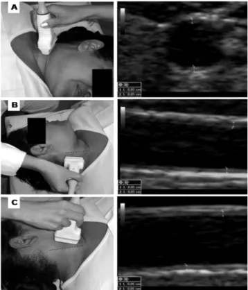
IMT measurements were obtained with the patient lying in the supine position and with the neck rotated to the opposite side of examination. CCA images were obtained to measure IMT by using three different angle views for each vessel (Figure 1). Initially, a transversal scanning view of the ACC was performed in the longest extension possible from the base of the neck to the carotid bulb. At least three IMT points were measured in the near and far walls in the most thickened area of each vessel. Also, lateral wall measurements were taken when both thickening was evident and accurate images were possible. Subsequently, the vessel was scanned by two longitudinal views: posterolateral (PL), with the transducer positioned parallel to the posterior border of the sternocleidomastoid muscle, and anterolateral (AL), with the transducer positioned parallel to the anterior border of the sternocleidomastoid muscle; at least three IMT measurements were obtained for each near and far wall. Optimal B-mode settings of gain, depth, focal zone placement, and compression were individually adjusted for each vessel to enhance arterial wall structures and image quality. IMT was measured by manual technique using electronic calipers, similar to the method of Sidhu and Desai.14 The maximum IMT value was selected for each
angle. An IMT superior to 1.0mm was considered to be an abnormal finding.15
Quantitative IMT measurement differences between the different angles were calculated by ANOVA with the LSD post-hoc test. Qualitative differences were calculated by McNemar’s test. A level of significance was defined as a P< .05.
RESULTS
The population studied had a mean age of 60.8±12.9 years, and 106 (51.5%) of the individuals were male. Four hundred seven CCAs were evaluated. Reliable measurement images could be achieved in 406 vessels in the transversal angle view, 404 in the longitudinal posterior view, and 398 in the longitudinal anterior angle view. The mean time necessary to take measurements was 8.1±1.24 minutes.
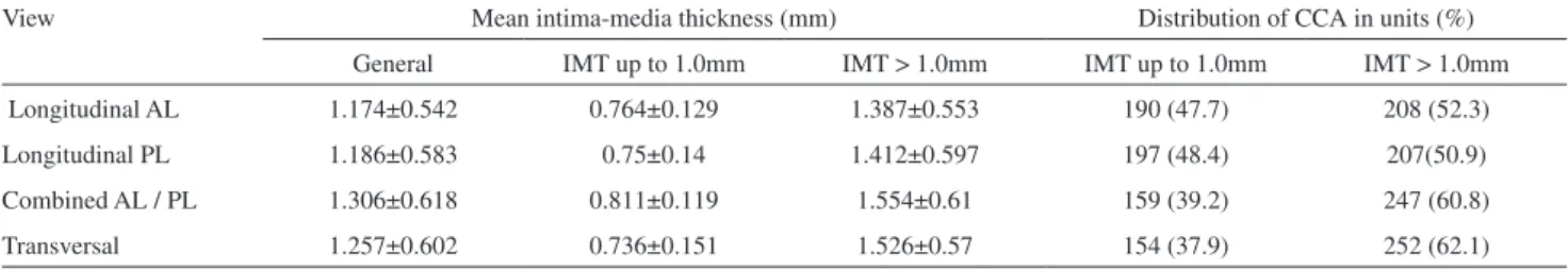
Comparing the thickest measurement obtained from all views, 270 (66.3%) carotid arteries had an IMT greater than 1mm. The mean IMT values achieved with the different incidences were 1.26±0.6mm (transversal), 1.17±0.54mm (longitudinal AL), and 1.18±0.58mm (longitudinal PL). Detailed data of mean IMT for each angle view are presented in Table 1.
A significant difference in IMT measurement values
Figure 1 - Observation angles for intima-media thickness measurement: A,
was observed when the three angles of examination plus the combined positive results of both longitudinal angles were compared by ANOVA (P=0.005). The LSD Post-Hoc test identified that the combined results of longitudinal views had similar results to transversal views (P=0.28) and higher IMT means than isolated AL or PL longitudinal views (P=0.02 and 0.05, respectively).
A direct comparison of the different angles of measurement was obtained with McNemar’s test. No statistical difference was observed in comparison between AL and PL longitudinal angles (P=0.63). The transversal view identified a greater number of arteries with increased IMT in comparison to the anterior or posterior longitudinal examination angles (P<0.001 for both). The combined results of the two longitudinal angles show superior results over isolated longitudinal views (P<0.001 for both) and similar results to transversal measurements (P=0.52). These data are detailed in Table 2.
DISCUSSION
IMT measurement is a simple, feasible, and cost-effective method for assessing systemic atherosclerosis and cardiovascular risk in adults.1 Also, is somewhat easy
to provide specific training for professionals with previous vascular ultrasound experience to take IMT measurements that meet good quality standards after a short period of practice.14 In this study, we proposed an objective and
practical method to evaluate IMT with the intention of improving the measurement quality by a short increase in the time required for carotid examination while maintaining its characteristic simplicity.
Recent protocols for determining carotid IMT include a series of sophisticated features. Specific software16-20 has
been developed for automatic IMT evaluation that minimizes the subjectivity of direct eye observation. Computer-assisted IMT measurements are widely used in other countries for longitudinal cohort studies and are encouraged by latest
consensus.21-22 However, such an option demands complex
human and material resources. Although software can perform more than a hundred measurements from each segment in a short time, the results obtained were not proven to be more accurate than manual measurements.23
Another improvement is the concomitant use of ECG24
during examination, which allows the observer to take measurements only during systolic activity, preventing small differences that could appear during the diastolic period when the inner arterial layers are less compressed. Also, external devices that measure head rotation and transducer position in degrees, such as the Meijer Arc,make it possible to reproduce the same positions in repeated examinations. These improvements bring many advantages and raise the quality of the data obtained, but such procedures also require more time24 and are more expensive. The application of such
protocols should be preceded by a careful cost-benefit analysis that depends on the objectives of each investigation.
We also avoided the use of external devices for probe positioning. Only the sternocleidomastoid and the carotid bulb were used, which represent simple anatomical references that remain virtually unchanged over time.
Table 1 - General measurement data showing mean values of intima-media thickness (IMT) and the distribution of common carotid arteries (CCA) divided by angle of view and groups of normal and thickened vessels. Combined views represent the thickest measurement obtained from the two longitudinal views
View Mean intima-media thickness (mm) Distribution of CCA in units (%)
General IMT up to 1.0mm IMT > 1.0mm IMT up to 1.0mm IMT > 1.0mm
Longitudinal AL 1.174±0.542 0.764±0.129 1.387±0.553 190 (47.7) 208 (52.3)
Longitudinal PL 1.186±0.583 0.75±0.14 1.412±0.597 197 (48.4) 207(50.9)
Combined AL / PL 1.306±0.618 0.811±0.119 1.554±0.61 159 (39.2) 247 (60.8)
Transversal 1.257±0.602 0.736±0.151 1.526±0.57 154 (37.9) 252 (62.1)
AL, anterolateral; PL, posterolateral.
Table 2 - A direct comparison of the accuracy of the com-bined positive results of longitudinal views with the accuracy of the isolated transversal and longitudinal measurement angles– number of common carotid arteries with increased intima-media thickness (>1mm)
Combined positive results of longitudinal views
P
Normal Increased IMT Longitudinal
anterolateral Normal Increased IMT
155 00
34 208
<0.001 Longitudinal
posterolateral Normal Increased IMT
158 00
39 207
<0.001
Transversal Normal Increased IMT
137 22
17 229
The transversal angle view provided a panoramic observation of the CCA, thus allowing the observer to identify and measure the thickest IMT points, even of the side walls. However, measurements taken from the transversal view may result in poor quality images, which are subject to many artifacts that are mainly the result of refraction from the side walls. Rather than being a proper measurement angle, the transversal view is useful as a screening image to guide longitudinal view measurements.
Previous authors have performed IMT measurements from a single longitudinal view,14,25-26 which can provide
limited observation of the vessel walls, leading to the underestimation of early atherosclerotic processes. We avoided directly comparing our findings with these reports due to the methodological differences of each study. Instead, we used the numbers obtained in our investigation to compare the combined results of IMT measured from the two longitudinal views with results from each single longitudinal view. As shown in the results, isolated longitudinal views had poorer sensitivity on identifying areas of increased IMT. The longitudinal views obtained in this investigation provided at least 12 different measurement points (three from each wall), covering the whole extension of the CCA in perpendicular angles to generate virtual tri-dimensional information about carotid wall thickness.
Although some authors believe that the near wall images are not accurate enough for IMT measurements,27-28 we
decided to consider these values. In many cases, the near wall presents layers that are far thicker than those in the far wall, which may even have normal values. Thus, ignoring these findings would underestimate the real state of the carotid artery atherosclerotic process.29-30
Recent studies revealed a tendency to use the mean maximum thickness of the CCA, carotid bulbs, and internal carotid artery (ICA).31 In this investigation, we avoided taking
measurements from the carotid bulb or ICA for the following reasons: First, even though bulb and ICA measurements may be worthwhile, they present limitations: as opposed to
CCA, they can be measured only in limited angles that may not reflect the thickest portions of vessel wall.32-33 Also, up
to 52% of images can fail to obtain accurate bulb and ICA measurements.34 The variance of results of bulb and ICA
IMT amongst repeated examinations and different physicians is higher than CCA measurements, making longitudinal comparisons more unreliable.35-37 Finally, CCA, bulb, and
ICA have morphological differences that make direct IMT comparison among them inaccurate.21 Yet, CCA / bulb / ICA
measurements do not appear to be superior to CCA IMT alone.38
We believe that the suggested protocol is an inexpensive method to measure IMT. We avoided expressing costs in numbers because values may vary from country to country and from private to public healthcare models. Thus, the eventual cost calculation in our institution may not reflect the reality for many readers.
Although we did not show numbers to confirm our impression, we judged that the presented technique is less expensive because it does not uses complimentary devices and does not depend on external software to analyze the obtained images. Human resources would be required to deal with ancillary equipment. The acquisition and maintenance of the necessary devices would certainly impact the final costs of examinations. The mean time expended in taking all measurements was about 8 minutes. This value is shorter than other models suggested,24 thus representing less of an
impact in terms of physician human resources than more complex models.
CONCLUSION
The protocol presented is a fast and practical method for obtaining common carotid artery intima-media thickness measurements. The combined values of anterolateral and posterolateral longitudinal views provide a more sensitive evaluation of the inner layers of the carotid walls than isolated longitudinal views.
REFERENCES
1. Baldassarre D, Amato M, Bondioli A, Sirtori CR, Tremoli E. Carotid artery intima-media thickness measured by ultrasonography in normal clinical practice correlates well with atherosclerosis risk factors. Stroke. 2000;31:2426-30.
2. Zureik M, Ducimetière P, Touboul PJ, Courbon D, Bonithon-Kopp C, Berr C, et al. Common carotid intima-media thickness predicts occurrence of carotid atherosclerotic plaques: longitudinal results from the Aging Vascular Study (EVA) study. Arterioscler Thromb Vasc Biol. 2000;20:1622-9.
3. Rosfors S, Hallerstam S, Jensen-Urstad K, Zetterling M, Carlström C. Relationship between intima-media thickness in the common carotid artery and atherosclerosis in the carotid bifurcation. Stroke. 1998;29:1378-82.
5. Burke GL, Evans GW, Riley WA, Sharrett AR, Howard G, Barnes RW, et al. Arterial wall thickness is associated with prevalent cardiovascular disease in middle aged adults: the Atherosclerosis Risk in Communities (ARIC) Study. Stroke. 1995;26:386-91.
6. Allan PL, Mowbray PI, Lee AJ, Fowkes FGR. Relationship between carotid intima-media thickness and symptomatic and asymptomatic peripheral arterial disease: the Edinburgh Artery Study. Stroke. 1997;28:348-53.
7. Holaj R, Spacil J, Petrasek J, Malik J, Haas T, Aschermann M. Intima-media thickness of the common carotid artery is the significant predictor of angiographically proven coronary artery disease. Can J Cardiol. 2003;19:670-6.
8. Boutouyrie P, Bussy C, Hayoz D, Hengstler J, Dartois N, Laloux B, et al. Local pulse pressure and regression of arterial wall hypertrophy during long-term antihypertensive treatment. Circulation. 2000;101:2601-6. 9. van Tits LJ, Smilde TJ, van Wissen S, de Graaf J, Kastelein JJ, Stalenhoef
AF. Effects of atorvastatin and simvastatin on low-density lipoprotein subfraction profile, low-density lipoprotein oxidizability, and antibodies to oxidized low-density lipoprotein in relation to carotid intima media thickness in familial hypercholesterolemia. J Investig Med. 2004;52:177-84.
10. Nagasaki T, Inaba M, Henmi Y, Kumeda Y, Ueda M, Tahara H, et al. Decrease in carotid intima-media thickness in hypothyroid patients after normalization of thyroid function. Clin Endocrinol (Oxf). 2003;59:607-12.
11. Markus RA, Mack WJ, Azen SP, Hodis HN. Influence of lifestyle modification on atherosclerotic progression determined by ultrasonographic change in the common carotid intima-media thickness. Am J Clin Nutr. 1997;65:1000-4.
12. Stanton AV, Chapman JN, Mayet J, Sever PS, Poulter NR, Hughes AD, et al. Effects of blood pressure lowering with amlodipine or lisinopril on vascular structure of the common carotid artery. Clin Sci (Lond). 2001;101:455-64.
13. Simon A, Gariepy J, Moyse D, Levenson J. Differential effects of nifedipine and co-amilozide on the progression of early carotid wall changes. Circulation. 2001;103:2949-54.
14. Sidhu PS, Desai SR. A simple and reproducible method for assessing intimal-medial thickness of the common carotid artery. Br J Radiol. 1997;70:85-9.
15. Kanters SD, Algra A, van Leeuwen MS, Banga JD. Reproducibility of in vivo carotid intima-media thickness measurements: a review. Stroke. 1997;28:665-71.
16. Seçil M, Altay C, Gülcü A, Ceçe H, Göktay AY, Dicle O. Automated measurement of intima-media thickness of carotid arteries in ultrasonography by computer software. Diagn Interv Radiol. 2005;11:105-8.
17. Selzer RH, Hodis HN, Kwong-Fu H, Mack WJ, Lee PL, Liu CR, et al. Evaluation of computerized edge tracking for quantifying intima-media thickness of the common carotid artery from B-mode ultrasound images. Atherosclerosis. 1994;111:1-11.
18. Selzer RH, Mack WJ, Lee PL, Kwong-Fu H, Hodis HN. Improved common carotid elasticity and intima-media thickness measurements from computer analysis of sequential ultrasound frames. Atherosclerosis. 2001;154:185-93.
19. Wendelhag I, Liang Q, Gustavsson T, Wikstrand J. A new automated computerized analyzing system simplifies readings and reduces the variability in ultrasound measurement of intima-media thickness. Stroke. 1997;28:2195-200.
20. Liang Q, Wendelhag I, Wikstrand J, Gustavsson T. A multiscale dynamic programming procedure for boundary detection in ultrasonic artery images. IEEE Trans Med Imaging. 2000;19:127-42.
21. Touboul PJ, Hennerici MG, Meairs S, Adams H, Amarenco P, Bornstein N, et al. Mannheim carotid intima-media thickness consensus (2004-2006). An update on behalf of the Advisory Board of the 3rd and 4th Watching the Risk Symposium,13th and 15th European Stroke Conferences, Mannheim, Germany, 2004, and Brussels,Belgium, 2006. Cerebrovasc Dis. 2007;23:75-80.
22. Touboul PJ, Hennerici MG, Meairs S, Adams H, Amarenco P, Desvarieux M, et al. Advisory Board of the 3rd Watching the Risk Symposium 2004, 13th European Stroke Conference. Mannheim intima-media thickness consensus. Cerebrovasc Dis. 2004;18:346-9.
23. Loizou CP, Pattichis CS, Pantziaris M, Tyllis T, Nicolaides A. Snakes based segmentation of the common carotid artery intima media. Med Biol Eng Comput. 2007;45:35-49.
24. Mitchell CK, Aeschlimann SE, Korcarz CE. Carotid intima-media thickness testing: technical considerations. J Am Soc Echocardiogr. 2004;17:690-2.
25. Temelkova-Kurktschiev TS, Koehler C, Leonhardt W, Schaper F, Henkel E, Siegert G, et al. Increased intimal-medial thickness in newly detected type 2 diabetes: risk factors. Diabetes Care. 1999;22:333-8.
26. Wang TJ, Nam BH, D’Agostino RB, Wolf PA, Lloyd-Jones DM, MacRae CA, et al. Carotid intima-media thickness is associated with premature parental coronary heart disease: the Framingham Heart Study. Circulation. 2003;108:572-6.
27. Wendelhag I, Gustavsson T, Suurkula M, Berglund G, Wikstrand J. Ultrasound measurement of wall thickness in the carotid artery: fundamental principles and description of a computerized analysing system. Clin Physiol. 1991;11:565-577.
28. Montauban van Swijndregt AD, De Lange EE, de Groot E, Ackerstaff RG. An in vivo evaluation of the reproducibility of intima-media thickness measurements of the carotid artery segments using B-mode ultrasound. Ultrasound Med Biol. 1999;25:323-30.
29. Tang R, Hennig M, Thomasson B, Scherz R, Ravinetto R, Catalini R, et al. Baseline reproducibility of B-mode ultrasonic measurement of carotid artery intima-media thickness: the European Lacidipine Study on Atherosclerosis (ELSA). J Hypertens. 2000;18:197-201.
30. Bots ML, Hoes AW, Koudstaal PJ, Hofman A, Grobbee DE. Common carotid intima-media thickness and risk of stroke and myocardial infarction: the Rotterdam Study. Circulation. 1997;96:1432-37. 31. Bots ML, Evans GW, Riley WA, Grobbee DE. Carotid intima-media
thickness measurements in intervention studies: design options, progression rates, and sample size considerations: a point of view. Stroke. 2003;34:2985-94.
33. Furberg CD, Adams HP Jr, Applegate WB, Byington RP, Espeland MA, Hartwell T, et al, for the Asymptomatic Carotid Artery Progression Study (ACAPS) Research Group. Effect of lovastatin on early carotid atherosclerosis and cardiovascular events. Circulation. 1994;90:1679-87.
34. Howard G, Sharrett AR, Heiss G, Evans GW, Chambless LE, Riley WA et al. Carotid artery intimalmedial thickness distribution in general populations as evaluated by B-mode ultrasound. Stroke. 1993; 24:1297-304.
35. O’Leary DH, Polak JF, Wolfson SK Jr, Bond MG, Bommer W, Sheth S et al. Use of sonography to evaluate carotid atherosclerosis in the elderly: the cardiovascular health study. Stroke. 1991;22:1155-63.
36. Crouse JR III, Craven TE, Hagaman AP, Bond MG. Association of coronary disease with segment-specific intimal-medial thickening of the extracranial carotid artery. Circulation. 1995;92:1141-47. 37. Stensland-Bugge E, Bønaa KH, Joakimsen O. Reproducibility of
ultrasonographically determined intima-media thickness is dependent on arterial wall thickness: the Tromsø study. Stroke. 1997;28:1972-80. 38. Roman MJ, Naqvi TZ, Gardin JM, Gerhard-Herman M, Jaff M,