*e-mail: [email protected]; [email protected]
Calcification
in vitro
of Biomineralizated Nanohydroxyapatite/Superydrophilic Vertically
Aligned Multiwalled Carbon Nanotube Scaffolds
Marcele Florencio Nevesa, Gislene Rodrigues Silvaa, Tayra Rodrigues Brazila,
Fernanda Roberta Marcianoa, Cristina Pacheco-Soaresb, Anderson Oliveira Loboa*
aLaboratório de Nanotecnologia Biomédica, Universidade do Vale do Paraiba – UniVap,
Av. Shishima Hifumi, 2911, CEP 12224-000, São José dos Campos, SP, Brazil
bLaboratório de Dinâmica de Compartimentos Celulares, Universidade do Vale do Paraíba – UniVap,
Av. Shishima Hifumi 2911, CEP 12224-000, São José dos Campos, SP, Brazil
Received: November 1, 2012; Revised: December 19, 2012
Nanocomposites based on superhydrophilic vertically aligned multi-walled carbon nanotubes (VAMWCNT-O2) and nanohydroxyapatite (nHAp) are of great interest in bone regenerative medicine. The biomineralization using simulated body fluid (SBF) has been extensively studied to evaluate the bioactivity of biomaterials. Thus, the combination of nHAp and VAMWCNT-O2 is attractive and promising. The aim of this study was to evaluate the in vitro calcification of nHAp/VAMWCNT-O2 nanocomposites before and after the period of biomineralization in SBF. In vitro calcification of the extracellular matrix (ECM) of HOB cells in culture after 24 hours was investigated through the assay of alkaline phosphatase. These promising in vitro results validate biomineralized nHAp/VAMWCNT-O2 as possible scaffolds for bone tissue regeneration.
Keywords: carbon nanotubes, hydroxyapatite, superhydrophilic, osteoblastic cells, SBF, calcification, cellular adhesion
1. Introduction
Carbon nanotubes (CNTs) are tubular structures formed by hexagonal arrays of carbon, which can be classified into two types: single walled carbon nanotubes (SWCNTs) and multi-walled carbon nanotubes (MWCNTs)1-4. The
MWCNTs are of particular interest for regenerative medicine due to their biocompatibility that permits extensive cell adhesion, spreading, and growth. MWCNT has been shown to support the growth of osteoblast cells by stimulating ECM production in bone tissue formation and growth of hydroxyapatite crystals5. Its properties
and biocompatibility are improved when MWCNT becomes aligned (VAMWCNT) and superhydrophilic (VAMWCNT-O2)3.
The nanohydroxyapatite (nHAp) is a ceramic material that promotes bone growth due to its biocompatible and bioactive behavior. nHAp has application in bone regeneration because it can establish chemical bonds between the material and the bone tissue allowing cell proliferation due to its similarity to bone mineralized matrix6.
In addition to the application of nHAp/VAMWCNT-O2 as scaffolds, the process of biomineralization in biomaterials conducted by incubating the samples in SBF (ion concentrations similar to human extracellular fluid) assists the development of biological apatites on the surface of nHAp/VAMWCNT-O2, improving in vitro adhesion and proliferation of human osteoblast cells (HOB)7,8
.
The osteoblasts are coated cells responsible for the synthesis of organic components of the bone matrix. They release fibronectins, which have the ability to form bone calcification and increase the release of alkaline phosphatase (ALP). ALP is an enzyme responsible for cell proliferation and increasing, which is widely distributed in human tissues, such as bone (osteoblasts), so that bone activity is confined to osteoblast and ALP assays9.
The objective of this study was to evaluate the ECM calcification potential of HOB cultured on this new biomineralized nHAp/VAMWCNT-O2 nanocomposite, to be used as scaffolds in bone tissue regeneration.
2. Methodology
The VAMWCNTs were produced as thin film, using a microwave plasma chamber (2.45 GHz). The substrates were 10 mm titanium squares, covered by a thin of Fe layer (10 nm), both deposited by an e-beam evaporator. The Fe layers were pretreated to promote nanocluster formation, which forms the catalyst for VAMWCNTs growth. The pretreatment was carried out during 5 minutes in plasma of N2/H2 (10/90 sccm) at a substrate temperature around 760 °C. After pretreatment, CH4 (14 sccm) was inserted in the chamber at a substrate temperature of 760 °C for 2 minutes. The reactor was kept at a pressure of 30 Torr during the whole process.
superhydrophilic character was performed in a pulsed-direct
current plasma reactor with an oxygen flow rate of 1 sccm, at a pressure of 85 mTorr, –700 V, and with a frequency of 20 kHz6.
The electrodeposition of the nHAp crystals on the superhydrophilic VAMCNT (VAMWCNT-O2) films was performed using 0.042 mol.L–1Ca(NO
3)2.4H2O + 0.025 mol.L –1
(NH4).2HPO4 electrolytes (pH = 4.8). These concentrations were chosen according to the 1.67 Ca/P ratio. The electrochemical measurements were made using a three-electrode cell coupled to an Autolab PGSTAT 128N equipment. VAMWCNT-O2films were used as working electrode, and the geometric area in contact with electrolytic solution was 0.27 cm2. A platinum coil wire served as
auxiliary electrode, and an Ag/AgCl electrode was used as reference electrode. The nHAp crystals were produced applying a constant potential of –2.0 V for 30 minutes., and the solution temperature was maintained at 70 °C, and constant agitation for 30 minutes. These samples were named as nHAp/ VAMWCNT-O26.
Biomineralization of the nHAp/VAMWCNT-O2 samples occurred in SBF (5×)10,11 of the concentration
proposed by Kokubo12 and shown in Table 1. The pH of
the solution was adjusted to 6.10 using HCl addiction. Each nHAp/ VAMWCNT-O2 sample was placed in a corning tube with 15 mL of SBF. Samples were shaken at 36.5 °C for 7, 14, and 21 days, and the SBF (5×) solution was changed every 3 days of experimentation. After the biomineralization period, samples were removed from SBF, washed in hot deionized water, dried in a laboratory stove at 50 °C for 24 hours and put in laminar air flux with UV light for 24 hours to be sterilized. To carry out the morphological analysis of all types of samples analyzed in this study, a scanning electron microscope was used (SEM, model: JEOL-JSM 5610 VPI). Images were recorded at magnification of 1,000-100,000×.
HOB cells were grown in cell culture bottles in 25 cm3
in low glucose DMEM supplemented with 10% fetal bovine serum and 1% antibiotic antimycotic, incubated in an incubator (Forma Scientific Model 3110), in a controlled atmosphere of 5% CO2 at 37 °C. The HOB cells were replicated from the initial count and the cell culture medium was replaced every 48 hours for better conditions for cell proliferation and viability.
The HOB cells were trypsinized for 3 minutes, centrifuged for 5 minutes, and resuspended in DMEM low glucose (Gibco) supplemented with 10% fetal bovine serum and 1% antibiotic antimycotic. The volume of 10 µL was applied to the chamber and brought to Newbauer HOB cell count at inverted microscope (Olympus CK 40) and determined concentration of 4.0 × 10⁵ cells/mL.
The samples were placed in individual wells of three plates and 24 culture wells, seeded on each group at a concentration of 4.0 × 10⁵ cells/mL and dried at 37 °C
with 5% CO2 for 30 minutes. The samples were overlaid with DMEM low glucose (Gibco) until a total volume of 500 µL and incubated in the laboratory stove for 24 hours
and 48 hours. Both the cellular adhesion test and ALP assay were performed in triplicate.
The nHAp/VAMWCNT-O2 nanocomposites were sterilized for 24 hours under UV irradiation and placed in individual wells of 24-well culture plates. The cells were maintained as sub-confluent monolayers in minimum
essential medium (MEM) with 1.5 mM L-glutamine adjusted to contain 2.2 g/L sodium bicarbonate 85%; fetal bovine serum (FBS) 15% (Gibco, BRL), 100 units/mL penicillin–streptomycin (Sigma), and 25 mg/mL L-ascorbic acid (Sigma). The incubation occurred within a CO2 (5%) atmosphere at 37 °C.
The HOB cells adhesion on nHAp/VAMWCNT-O2 nanocomposites were monitored up to 48 hours. The cells were seeded in each well at 5 × 10⁵ cells/mL concentration,
supplemented with 10% PBS. The incubation was performed under a CO2 atmosphere (5%) at 37 °C. The substrate attached cells were fixed with 3% glutaraldehyde/0.1 M sodium cacodylate buffer for 1 hour and dehydrated in a graded ethanol solution series (30%, 50%, 70%, 95%, and 100%) for 10 minutes. each. The drying stage used a 1:1 solution of ethanol with hexamethyldisilazane (HMDS) and the samples were dried with pure HMDS at room temperature. After deposition of a thin gold layer, the specimens were examined with a SEM (model: JEOL-JSM 5610 VPI). Images were recorded at magnification of 1,000-4,000×.
To test the ALP activity, solutions 1 and 2 of the assay Kit Acid Alkaline Phosphatase (Millipore) were prepared according to the product manual. At 24 and 48 hours, the plates were removed from the incubator and 100 µL
of solution 1 and 25 µL of solution 2 was added to each well. Then, the plate was covered with foil and kept for 15 minutes at room temperature. The samples were taken to the immunohistochemistry analysis apparatus in a microplate reader (Synergy HT Multi-Detection, Bio-Tek, Winooski, VT) at 620 nm absorbance. The blank reference was taken from wells without cells, also incubated with the ALP solution. The ALP activity was calculated by the normalization of optical density (OD) of the samples (VAMWCNT-O2 nHAP/VAMWCNT-O2, nHAp/ VAMWCNT-O2-7D, nHAp/VAMWCNT-O2-14D, nHAp/ VAMWCNT-O2-21D) to the cell culture and expressed in percentage. The Equation 1 shows the normalization applied:
ODsample ODblank
% ALP 1 00
ODcell ODblank
−
= ×
− (1)
To carry out the statistical analyzes, four different readings were collected, with N = 4, and expressed as the average. The ALP test was used for ECM calcification. Statistical differences were analyzed by Two-way Anova using the GraphPad Prisma® program. The population of the
groups was analyzed with independent normal distribution for each experiment. P values less than 0.005 (P <0.005) were considered to indicate statistical differences of the groups.
Table 1. Quantity of Reagents used to prepare SBF.
Reagent Quantity (mM)
NaCl 733.5
MgCl2 7.5
CaCl2.2H2O 12.5
Na2HPO4. 2H2O 5.0
3. Results and Discussion
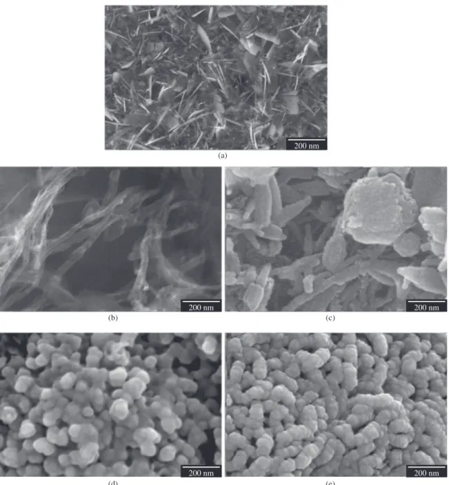
SEM examination revealed morphologic differences in the nHAp deposition layer under different processes. Figure 1a shows details of nHAp crystals formed on the VAMWCNT-O2 surface after electrodeposition process. The plate-like crystal structure covered the entire nanobiomaterial surface, giving the nanobiomaterial a roughness aspect. SEM micrographs, shown in Figure 1b, are VAMWCNT-O2 details of electrodeposition process before biomineralization,
while Figure 1c shows nHAp/VAMWCNT-O2 details
after 7 days of biomineralization. This image shows that there are nHAp not only above, but also between
nHAp/VAMWCNT-O2 nanocomposite, and it also reveals
morphologic differences in formed nHAP, which now has a globular shape. The micrographs indicate a crystallographic structure change from plate-like to a globular morphology of deposited nHAp layer (c, d, e). Furthermore, after 14 days (1d), this layer became denser; however, as this density increases, the roughness decreases as seen after 21 days of biomineralization (1e).
F i g u r e 2 s h o w s H O B c e l l s a d h e s i o n o n
nHAp/VAMWCNT-O2 before biomineralization (2a) and
after 7 days (2b), 14 days (2c), and 21 days (2d) in SBF. SEM examination in Figure 2a shows an HOB adhered
on nHAp/VAMWCNT-O2 surface with short filaments
attached to nHAp crystals. At the same rang (4,000×), Figure 2b shows an increased number of HOB cells adhered with longer cytoplasmatic projections, already proliferated and in a good spreading process, not only on
nHAp/VAMWCNT-O2 surface, but also between nHAp
plate-like crystals clusters. The cell spreading is also evident in Figure 2c, but the presence of HOB is not significant. The same aspect can be found in Figure 2c, but this examination shows another important aspect: an HOB cell well adhered to a nHAp crystal cluster with a long cytoplasmatic projection attached it to another cluster of nHAp crystals between
nHAp/VAMWCNT-O2.
B o t h t h e n H A p a n d VA M W C N T- O2 h a v e physicochemical properties (surface tension, surface free energy, ionic charge, and wettability) suitable for cell adhesion and proliferation6; however, surface topography
and cell protein production activity also interfere with the cell adhesion process13. In relation to surface topography,
geometric structure is related to material roughness and porosity. The aspects found on SEM examination shown in Figure 2 could be proved through the results found in Figure 3.
Figure 3 illustrates the results of ALP activity in percentage for 24 and 48 hours of the control (HOB), nHAp/
VAMWCNT-O2, and biomineralized nHAp/VAMWCNT-O2
nanocomposites for 7, 14, and 21 days. Two culture times (24 and 48 hours) were used to compare calcification
of the ECM, to validate which biomineralized nHAp/
VAMWCNT-O2 nanobiomaterials expressed ECM
calcification through percentage presented in the thymolphthalein micromol/h/mg (normalized by the Equation 1).
When comparing both groups (24 and 48 hours), ALP
showed higher values for nHAp/VAMWCT-O2 and nHAp/
VAMWCNT-O2 nanocomposites biomineralized for 7 days
in SBF (nHAp/VAMWCNT-O27D) compared to HOB
cells. The difference is more significant at 48 hours (nHAp/
VAMWCNT-O27D). The remaining samples showed no
potential for calcification of the ECM as intense as nHAp/
VAMWCNT-O27D.
The ECM released in the first 24 hours, in rougher and less dense surfaces, is more significant, in this case
nHAp/VAMWCNT-O2 and nHAp/VAMWCNT-O27D
(Figure 2a, b), being less significant in the thickest
surfaces-like nHAp/VAMWCNT-O214D and nHAp/
VAMWCNT-O221D nanocomposites.
In the first 6 hours of contact with the biomaterial, cells begin the surface recognition process6. The biomimetic
nHAp/VAMWCNT-O2 nanocomposite surface, before
and after 7 days of biomineralization, characterizes nanocomposite bioactivity. However, after 24 hours the rough surface is replaced by the presence of Ca++, as
shown in Figure 3. Now, the rougher material with a
greater concentration of Ca++ (nHAp/VAMWCNT-O
27D)
is attracting more HOB cells and starting significant ECM calcification. Less significant increase of calcification
can be noticed in nHAp/VAMWCNT- O214D and nHAp/
VAMWCNT-O221D.
These results are complementary to those presented in literature7,14,15, which performed ALP test in CNTs and
observed that HOB cells showed higher proliferation in samples of CNTs than in another biomaterial, which was similar to the result shown in Figures 2 and 3.
4. Conclusion
This study indicates that biomineralized nHAp/
VAMWCNT-O27D nanocomposites showed higher ALP
assay in HOB cell culture, indicating the potential of in vitro
biomineralization. The ALP expression confirms nHAp/
VAMWCNT-O27D nanocomposite bioactivity through
ECM calcification, as shown by SEM analysis of HOB cell adhesion, proliferation, and spreading. These results
also prove that nHAp/VAMWCNT-O2 nanocomposites
biomineralized for 7 days in 5× SBF appear to be privileged candidates as scaffolds for inducing in vivo regenerative bone tissues process.
Acknowledgements
The authors thank the Fundacao de Amparo a Pesquisa do Estado de Sao Paulo (2011/17877-7), (2011/20345-7), (2012/02159-4) and FVE for financial support, and to
everyone form Laboratory of Biomedical Nanotechnology
for all support in the procedures. Special thanks to Priscila Leite for scanning electron microscopy images and Alene Alder-Rangel for reviewing the English.
Figure 3. Comparison of calcification of the ECM as a percentage through ALP assays.
References
1. Souza Filho AG and Fagan SB. Funcionalização de Nanotubos de Carbono. Quimica Nova. 2007; 30(7):1695-1703. http:// dx.doi.org/10.1590/S0100-40422007000700037
2. Herbst MH, Macêdo MIF and Rocco AM. Tecnologias dos nanotubos de carbono: tendências e perspectivas de uma área multidisciplinar. Quimica Nova. 2004; 27(6):986-992. http:// dx.doi.org/10.1590/S0100-40422004000600025
3. Lobo AO, Corat MAF, Antunes EF, Palma MBS, Pacheco-Soares C, Garcia EE et al. An evaluation of cell proliferation and adhesion on vertically-aligned multi-walled carbon nanotube films. Carbon. 2010; 48:245. http://dx.doi. org/10.1016/j.carbon.2009.09.012
4. Oliveira V, Pereira MM, Brandão HM, Brandão MAF, Gattaz WF and Raposo NRB. Nanotubos de carbono aplicados às neurociências: perspectivas e desafios. Revista de Psiquiatria Clínica. 2011; 38(5):201-206. http://dx.doi.org/10.1590/ S0101-60832011000500006
5. Mendes RM, Silva GAB, Caliari MV, Silva EE, Ladeira LO and Ferreira AJ. Effects of single wall carbon nanotubes and its functionalization with sodium hyaluronate on bone repair. Life Sciences. 2010; 87(7-8):215-222. PMid:20600151. http:// dx.doi.org/10.1016/j.lfs.2010.06.010
6. Lobo AO, Corat MAF, Ramos SC, Matsushima JT, Granato AEC, Pacheco-Soares C et al. Fast Preparation of Hydroxyapatite/Superhydrophilic Vertically Aligned Multiwalled Carbon Nanotube Composites for Bioactive
Application. Langmuir. 2010; 26(23):18308-18314.
PMid:20961085. http://dx.doi.org/10.1021/la1034646
7. Marsi TCO, Santos TG, Pacheco-Soares C, Corat EJ, Marciano FR and Lobo AO. Biomineralization of Superhydrophilic Vertically Aligned Carbon Nanotubes. Langmuir. 2012; 28(9):4413-4424. PMid:22320358. http:// dx.doi.org/10.1021/la300111k
8. Irineu JAF, Marsi TCO, Santos TG, Corat EJ, Marciano FR and Lobo AO. Efficient method to produce biomineralizated nanohydroxyapatite/vertically aligned multiwalled carbon nanotube scaffolds. Materials Letters. 2012; 79:166-169. http:// dx.doi.org/10.1016/j.matlet.2012.03.096
9. Oréfice RL, Pereira MM and Mansur HS. Biomateriais: fundamentos e aplicações. Rio de Janeiro: Cultura Médica; 2006.
10. Barrere F, Van Blitterswijk CA, De Groot K and Layrolle P. Nucleation of Biomimetic Ca-P Coatings on Ti6Al4V from a SBF×5 Solution: Influence of Magnesium. Biomaterials. 2002; 23(29):2211-2220. http://dx.doi. org/10.1016/S0142-9612(01)00354-4
11. Barrere F, Van Blitterswijk CA, De Groot K and Layrolle P. Influence of Ionic Strength and Carbonate on the Ca-P Coating Formation from SBF×5 Solution. Biomaterials. 2002; 23(29):1921-1930. http://dx.doi. org/10.1016/S0142-9612(01)00318-0
12. Kokubo T. Apatite formation on surfaces of ceramics, metals and polymers in body environments. Acta Biomaterialia. 1998; 46(7):2519-2527. http://dx.doi. org/10.1016/S1359-6454(98)80036-0
13. Schakenraad JM, Busscher HJ, Wildevuur ChRH and Arends J. Thermodynamic aspects of cell spreading on solid substrata. Cell Biophysics. 1988; 13(1):75-91. PMid:2456154. 14. Li Y, Danmark S, Edlund U, Finne-Wistrand A, He X, Norgard
M et al. Resveratrol-conjugated poly-ε-caprolactone facilitates in vitro mineralization and in vivo bone regeneration. Acta Biomaterialia. 2011; 7(2):751-758. PMid:20849988. http:// dx.doi.org/10.1016/j.actbio.2010.09.008