Functionalization of carbon nanotubes and its application in
nanomedicine: A review
1, 2*H. Sadegh;1,2R. Shahryari-ghoshekandi
1Department of Chemistry, Science and Research Branch, Islamic Azad University, Tehran, Iran
2Young Researchers and Elite Club, Science and Research Branch, Islamic Azad University, Tehran, Iran
AB STRACT:
This review focuses on the latest developments in applications of carbon nanotubes (CNTs) in medicine. A brief history of CNTs and a general introduction to the field are presented.
Then, surface modification of CNTs that makes them ideal for use in medical applications is highlighted. Examples of common applications, including cell penetration, drug delivery, gene delivery and imaging, are given. At the same time, there are concerns about their possible adverse effects on human health, since there is evidence that exposure to CNTs induces toxic effects in experimental models. However, CNTs are not a single substance but a growing family of different materials possibly eliciting different biological responses. As a consequence, the hazards associated with the exposure of humans to the different forms of CNTs may be different. Understanding the structure–toxicity relationships would help towards the assessment of the risk related to these materials. Finally, toxicity of CNTs, are discussed. This review article overviews the most recent applications of CNTs in Nanomedicine, covering the period from 1991 to early 2015.
Keywords:Carbon nanotubes, Drug delivery, Functionalization, Nanomedicine, Surface modification DOI: 10.7508/nmj. 2015.04.001
*Corresponding Author Email:[email protected]
Tel: (+98)21-65933439
Note. This manuscript was submitted on July 13, 2015; approved on August 21, 2015
Received; 13 July 2015 Accepted; 2 August 2015
INTRODUCTION
Since carbon nanotubes (CNTs) were discovered by Iijima in 1991[1], they have become the subject of many studies because of their unique electrical, optical, thermal, and mechanical properties [2-6]. CNTs can be visualized as a sheet of carbon atoms rolled up into a tube with a diameter of around tens of nanometers. There are two main types of CNTs, single-walled (SWCNTs) and multi-walled carbon nanotubes (MWCNTs), the latter being formed by several concentric layers of rolled graphite (Fig 1). In particular, SWCNTs are characterized by a high aspect ratio. In the last decade, CNTs are intensively explored for in vitro and in vivo delivery of therapeutics, which was inspired by an important finding that CNTs can
Nanomed. J., 2(4): 231-248, Autumn 2015H.R. Sadegh et al.
of CNTs such as high absorption in the near-infrared (NIR) range, photoluminescence, and strong Raman shift [10], CNTs are excellent agents for biology detection and imaging. Combined with high surface area of CNTs for attaching molecular recognition molecules, CNT-based, targeted nano-devices have been developed for selective imaging and sensing. There are many areas where CNTs are extremely useful. This review article describes strategies for preparation of CNTs for their use in medicine. Specifically, we focused and highlighted the important nanomedicine applications of CNTs in the field of cell penetration, drug delivery, gene delivery and imaging.
Fig. 1. Schematics of a SWCNTs and MWCNTs
CNTs modifications
Raw CNTs, persisting metallic nature, are highly hydrophobic. Therefore, surfaFce modification of CNTs, or CNTs functionalization so that they could be dispersed in aqueous solutions becomes a key step for their medical applications. The CNTs modification methods involved either non-covalent or covalent strategies. The non-covalent modification utilizes the hydrophobic nature of CNTs, especially, π-π interactionsfor coating of amphiphilic molecules. The covalent modification generates chemical bonds on carbon atoms on CNTs surface via chemical reactions followed by further conjugation of hydrophilic organic molecules or polymers rendering CNTs better solubility. These modifications not only offer CNTs water solubility, but also produce functional moieties that enable linking of therapeutic agents, such as genes, drugs, and recognition molecules for medical applications.
Non-covalent functionalization of CNTs
The methods of functionalization of CNTs based on non-covalent interaction can be performed without
destroying the intrinsic sp2-hybridized structure of the nanotube sidewall, so that the original electronic structure and properties of CNTs can be preserved. Different kinds of non-covalent functionalizations have been explored. The non-covalent functionalization of
CNTs could be achieved byπ–πstacking interactions
between conjugated molecules and the graphitic sidewall of CNTs. Compounds with the pyrene moiety, e.g. N-succinimidyl-1-pyrenebutanoate, could be adsorbed irreversibly onto the surface of SWCNTs throughπ–πinteraction [11, 12]. Furthermore, via the succinimidyl ester group on this bifunctional anchor, various molecules with primary or secondary amines
such as ferritin, streptavidin (SA) or biotin–
parts, while exposing the hydrophilic parts to the aqueous solution to minimize the hydrophobic interface between the nanotubes and their polar environment. Through hydrophobic interactions, water-soluble polymers poly vinyl pyrrolidone (PVP) and poly styrene sulfonate (PSS) wrapped helically around the surface of SWCNTs helped to dissolve the nanotubes in water [55]. The association between the polymer and SWCNTs, which is reversible by changing the solvent system, was very robust, independent of the presence of excess of polymer in solution and uniform along the surface of the nanotubes. Surfactants were also conjugated to CNTs via hydrophobic interactions. So far, various anionic, nonionic and cationic surfactants have been used to suspend CNTs in aqueous medium [56-60]. Among them, bile salt surfactants such as deoxycholic acid (DOC) and taurodeoxycholic acid (TDOC) have been found to be very efficient in solubilizing SWCNTs. This is because the aggregation was further prevented by the coulomb repulsion between surfactant-coated SWCNTs [60]. In addition, if aromatic moieties in the hydrophobic part of the amphiphilic molecule were present, particularly strong interactions between the molecule and the CNTs could be established. This kind of aggregates, forms additionalπ–πstacking between the aromatic part of the molecule and the graphitic sidewalls of CNTs while exposing the hydrophilic groups to the aqueous solution. As an example, sodium dodecyl benzene sulfonate (SDBS) presented an increased ability in suspending CNTs in water [59]. Some of the surfactants including sodium dodecyl sulfate (SDS), SDBS, DOC or TDOC were found to be very effective in dispersing SWCNTs [58-60]. This provided the possibility to study the properties of individual SWCNTs and further manipulate single tubes [61-65]. Hydrophobic interactions were also used to couple biomolecules to CNTs. In particular, an amphiphilic peptide was wrapped around the SWCNT surface. The peptide was able to adoptα-helical structure in aqueous solution, exposing the hydrophobic face of the helix to the nanotube surface [66, 67]. Atomic force microscopy (AFM), transmission electronic microscopy (TEM), and absorption and Raman spectroscopy proved that the SWCNTs were DE bundled by the peptide. The peptide-coated nanotubes were assembled into macromolecular structure through charged peptide-peptide interactions between adjacent peptide-wrapped nanotubes. The size and morphology of the super-molecular structure can be regulated by controlling the factors that affect peptide-peptide interactions. It was also demonstrated
that the ability to disperse individual SWCNTs increased with increasing the amount of aromatic residues inside the peptide sequence [68]. In a similar manner, different proteins were adsorbed onto the surface of SWCNTs and were taken up by the mammalian cells [69].
Another interesting approach involved the conjugation of single-stranded DNA with SWCNTs resulting in helical wrapping around the surface of the tube exposing the hydrophilic sugar-phosphate backbone towards the exterior [70, 71]. Using this strategy, individually dispersed nanotubes in solution were obtained. CNTs wrapping by single-stranded DNA was strongly dependent on the DNA sequence. Moreover, the dependence of the electrostatic properties of the DNA-CNT hybrid on the tube diameter and electronic properties enable nanotube separation by anion exchange chromatography. In addition to a direct interaction with CNTs, biomolecules have also been attached to SWCNTs via amphiphilic molecules as a bifunctional linker. For instance, phospholipid (PL)
molecules with phospholipid–poly(ethylene glycol)
(PEG) chains and terminal amine (PL-PEG-NH2) were
non-covalently conjugated to SWCNTs. PL alkyl chains were adsorbed to the surface of SWCNTs via hydrophobic interactions while the PEG chain extended into the aqueous phase for SWCNTs dispersion. After incorporation of a hetero bifunctional cross-linked sulfosuccinimidyl 6-(30-[2-pyridyldithio] prop-ionamido) hexanoate (sulfo-LC-SPDP), the functionalized SWCNTs (f -SWCNTs) were coupled with DNA and RNA via disulfide linkage. The complexes were subsequently delivered to cells [72].
CNTs covalent functionalization
The CNTs characteristics such as low curvature, low solubility and bundling tendency render carbon nanomaterials a relatively unreactive to chemical treatment. Thus far, a number of ways have been introduced for covalent functionalization of CNTs. With respect to the location of the functional groups, the strategies to covalently functionalize CNTs can be classified into two main types including defect and sidewall functionalizations. The covalent functionalization of nanotubes is more robust and is better controlled compared to functionalization based on non-covalent methods.
CNTs synthesized by any available method are not free of defects. The intrinsic defects include five- or
seven-membered rings called Stone–Wales defects,
-Nanomed. J., 2(4): 231-248, Autumn 2015CNTs in Nanomedicine: A reveiw
hybridized six-membered ring carbon structure of the sidewall [94, 95]. A limited number of defects can be tolerated by CNTs without losing their macroscopic electronic and mechanical properties [3, 75-77]. In addition, the tips of the tubes, forming a hemispherical fullerene, have stronger reactivity than the sidewalls because of their higher curvature [78, 79]. When treated with strong oxidizing agents such as nitric acid, KMnO4/H2SO4, O2, K2Cr2O7/H2SO4or OsO4, CNTs could be opened and cut into short tubes. In the meantime, defects were introduced around the CNT surface [74, 79, 80]. Moreover, oxygenated functional groups such as carboxylic acid, ketone, alcohol and ester groups were generated by the oxidization process to the ends and the defect sites of the nanotubes [81]. These functional groups were very important to further functionalize CNTs. Sonication could also produce defects on the sidewall of CNTs that were eager for further chemical reactions [82-85]. Using the nanotube-bound carboxylic acid groups introduced by oxidization treatment, further functionalization was performed via amidation, esterification or the zwitterion
(COO-NH
3
+) formation. Before covalent modification,
the carboxylic acids are often activated by thionyl chloride, carbodiimide [e.g.
N-(3-dimethylaminopropyl)- N’-ethylcarbodiimide (EDC), N, N’-dicyclohexylcarbodiimide (DCC)] or
N-hydroxysuccinimide (NHS), to get highly reactive intermediate groups. Esterification was achieved by the nucleophilic substitution reaction of nanotube carboxylate salt with alkyl halides [86]. The functionalization of CNTs via nanotube carboxylic acids, located at the ends or pre-existing defects of CNTs, preserved the essential features of the CNT electronic structure as well as the bulk properties of the material [81, 87]. The first shortened soluble SWCNTs were obtained through carboxylic amidation using octadecylamine [81]. Since then, various lipophilic and hydrophilic Dendrons have been attached to CNTs via amide or ester linkages, which significantly improved the solubility of CNTs in organic or aqueous solvents [88]. These groups could be subsequently removed under basic or acidic hydrolysis conditions [89]. In addition, following this functionalization process, CNT bundles were largely exfoliated into individual nanotubes [75]. This enabled further manipulation and investigation of the spectroscopic properties of individual SWCNTs. Via amidation or esterification, polymers like propionyl-ethylenimine-coethylenimine (PPEI-EI),
poly-n-vinylcarbazole (PVK-PS) and PEG were grafted to the surface of CNTs [90]. Another approach called
‘‘graft from’’ was developed to attach polymer to CNTs.
The polymerization initiators were first covalently bound to CNTs and then the nanotube-based macro-initiators were exposed to monomers to accomplish the polymerization. In particular, polymer poly-n-butyl methacry-late (PnBMA), poly-methyl methacrylate (PMMA) and copolymer PMMA-b-polyhydroxyethyl methacrylate (PHEMA) have been coupled to CNTs via atom transfer radical polymerization (ATRP) [91, 92]. Defect functionalization was used to link biological molecules to CNTs via stable covalent bonds. For example, f -SWCNTs and f -MWCNTs with bovine serum albumin (BSA) were synthesized via diimide-activated amidation at ambient condition. It was found that the protein bound to nanotubes remained active [93]. SA (a protein with potential clinical applications in anticancer therapy) was complexed to SWCNTs pre-functionalized with biotin through EDC-activated amidation [94]. Similarly, using the biotin-SA interaction, a biotinylated DNAzyme was covalently attached to MWCNTs pre-functionalized with SA via amide linkage. The nanotube-bound DNAzymes displayed highly active and classical enzymatic behavior [95]. DNA was also bound to CNTs via amide linkage [96, 97]. DNA-CNT conjugates were hybridized with their complementary sequences and this hybridization was apparently reversible upon a denaturation process. The derived single-strand DNA-CNTs could be reused in a second-round of hybridization. In another case, DNA recognition was achieved by peptide nucleic acid (PNA) f -SWCNTs, in which PNA were attached to SWCNTs via NHS activated SWCNTs [98].
The sidewalls of CNTs have an aromatic, six-membered ring structure. So far, various kinds of reactions of the sidewall have been developed such as fluorination, radical addition, nucleophilic addition, electrophilic addition and cycloaddition [99]. There are two principal sources of molecular strain that affect the reactivity in non-planar conjugated molecules: (i) pyramidalization of the conjugated carbon atom and (ii) π-orbital misalignment between adjacent pairs of conjugate carbon atoms. Unlike fullerene, in which the strain is primarily from pyramidalization,π-orbital misalignment is the main source of the strain for the sidewalls of
CNTs. Since π-orbital misalignment, as well as
higher chemical reactivity [78]. Differing from defect reactions, sidewall reactions disrupted the conjugated structure of CNTs, as monitored by Raman or absorption spectroscopy. In the Raman spectrum of pristine SWCNTs,three main characteristic modes were disclosed: (i) the radial breathing (ω
r) below 350 cm -1,
(ii) the tangential(ωt) mode at 1550-1600 cm-1 and (iii) the disorder (sp3) mode at 1250-1450 cm-1. The radial breathing mode was demonstrated to be in strong relationship with the diameter of the nanotube [100]. The relative intensity of the disorder mode was related to the amount of sp3-hybridized carbon atoms in the SWCNTs and thus provided direct information of the extent of sidewall functionalization, in which the carbons linked to functional groups were converted from sp2-hybridized into sp3-hybridized carbon [101]. The absorption spectrum reflected the sidewall covalent modification of SWCNTs. The disruption of the conjugated structure caused by the reaction led to the loss of optical transitions between van Hove singularities in the electronic density of state (DOS) of CNTs, which was detected as the loss of the structure of the absorption spectrum [102].
The fluorination of SWCNTs was accomplished through the reaction of SWCNTs with elemental fluorine at temperatures ranging from 150 to 600 [103]. By controlling the reaction conditions such as temperature and time, the stoichiometry could be regulated. In some cases, a stoichiometry of C2F was obtained. The investigation of absorption and Raman spectra indicated the electronic perturbation of fluorinated SWCNTs. The fluorination allowed exfoliation of SWCNTs bundles into individual nanotubes and remarkably improved the solubility of SWCNTs in tetrahydrofuran (THF), dimethylformamide (DMF) and various alcohols [104]. The C-F bonds were very sensitive to the treatment with hydrazine, which allowed almost complete recovery of the conductivity and the spectroscopic properties of the pristine SWCNTs [103]. It was demonstrated that the Fermi energy level of fluorinated SWCNTs, as well as the conduction band energy, were significantly shifted to lower values compared to those of the pristine SWCNTs. This revealed that fluorinated CNTs were better electron acceptors than pristine nanotubes, and thus displayed higher chemical reactivity to strong nucleophilic reagents like Grignard, alkyllithium reagents and metal alkoxides. The same result was reached by exploiting the eclipsing strain effect that weakened the C-F bonds in fluorinated CNTs relative to the C-F bond in alkyl
fluorides [105, 106]. It was proposed that the substitution of fluorine by nucleophilic reagents proceeds via a concerted allylic displacement [105]. The addition of radicals to SWCNTs was carried out using different radical sources including perfluorinated alkyl radicals photo induced from perfluoroalkyl iodide [107], aryl radicals generated by one-electron reduction of the diazonium salt [108-110], radicals produced by decomposition of benzoyl peroxide in the presence of alkyl iodides [111], or directly produced by decomposition of benzoyl or lauroyl peroxide [112]. The functionalization of SWCNTs with diazonium reagents was achieved by three different methods: electrochemical reaction [98], thermochemical reaction in solvent [109] and using a solvent-free system [110]. The resultant SWCNTs displayed very high degree of functionalization. It was also demonstrated that metallic SWCNTs showed higher reactivity in these types of reactions than semi conductive SWCNTs, which would allow CNT separation [113]. Due to the high electron affinity imparted by the curvature of sidewall [114], CNTs were reacted with strong nucleophile reagents such as nucleophile carbine [107], sec-butyl lithium [115], polymeric carbanions [116] and Birch reduction reagents (lithium/1, 2-diaminoethane [87] or lithium/ ammonia [117]) to form various f-CNTs. In addition, the electrophilic addition was also achieved by the reaction of chloroform with the sidewall of CNTs in the presence of Lewis acid [118].
Various cycloaddition reactions to CNTs have been developed, including [2+1] cycloaddition of dichlorocarbene [87], nitrenes [107] and bromomalonates under Bingel reaction conditions
[119], [4+2]Diels–Alder cycloaddition of
o-quinodimethane performed under microwave irradiation [120], [3+2] cycloaddition of azido groups under ultraviolet (UV) irradiation [121], and azomethine ylides [122]. The 1, 3-dipolar additions of azomethine ylides is particularly versatile for the functionalization of CNTs [122-124]. This strategy is suitable for different types of CNTs (e.g. pristine and purified SWCNTs or MWCNTs), and involves the in situ generation of an azomethine ylide through the decarboxylation of an
immonium salt derived from condensation of α-amino
Nanomed. J., 2(4): 231-248, Autumn 2015H.R. Sadegh et al.
aromatic structure of the sidewalls of the f-SWCNTs was proven by absorption spectroscopy [122]. This functionalization technique was also used to purify CNTs [126]. Protected amino groups were introduced onto the surface of CNTs via 1, 3-dipolar cycloaddition of azomethine ylides [122, 127]. For instance, f-CNTs with N-protected glycine were achieved using N-protected amino acid, which were subsequently used to link biomolecules (e.g. bioactive peptides) by fragment condensation or selective chemical ligation [127,128].The first multi-functionalization of CNTs was achieved using a vertically aligned MWCNT membrane. One method involved the protection of the sidewalls of CNTs with polystyrene in the process of plasma oxidation that produced carboxylate derivatization. Under this process, aligned MWCNT membranes functionalized with carboxylic acid groups at the terminal parts were obtained. Thereafter, bifunctional CNTs with thiol groups on one end and carboxylic groups on the other end were accomplished by floating on top of a 2-aminoethanethiol reaction solution one side of the oxidized CNTs membrane [129]. Another approach was carried out by floating directly one side of an aligned
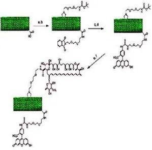
MWCNTs membrane on 3’-azido-3’-deoxythymidine
(AZT) solution in ethanol under UV irradiation and, after removal of absorbed AZT molecules by washing with ethanol, laying the unmodified side on the surface of a perfluorooctyl iodide solution in 1,1,2,2-tetrachloroethane (TEC) under UV irradiation. Thus, bifunctionalized MWCNTs with AZT molecules and perfluorooctyl chains were obtained [130]. These two methods of CNT bifunctionalization are quite complicated and limited to align MWCNTs. Using a different method, Wu and coworkers introduced an orthogonal protecting methodology into the chemical modification of nanotube to achieve the multi-functionalization of CNTs [131]. In particular, diaminotriethylene glycol chains mono-protected by phthalimide were linked to the carboxylic acids of oxidized MWCNTs. In a second step, a suitable N-substituted glycine with a diaminotriethylene glycol chain mono-protected by tert-butyloxycarbonyl (Boc) was introduced via 1, 3-dipolar cycloaddition (Fig 2). Since the phthalimide is stable to harsh acidic conditions and orthogonal to the Boc, the selection and control of attachment of different molecules was possible [131]. In this case, CNTs containing both fluorescein and amphotericin B (AmB) were prepared, and bioactivity of the conjugates was studied [131]. This method of
bifunctionalization is general and suitable for different types of CNTs.
In summary, the functionalization and consequent applications of CNTs have been dramatically increased in the last few years. The chemical modification of CNTs cannot only endow this intriguing nanomaterial with various functions, but also extend their potential applications to different fields of research.
Fig. 2. One of the routes to the bifunctionalization of CNTs. (a) Neat (COCl)2; Pht-N(CH2CH2O)2-CH2CH2-NH2, dry THF, reflux; (b) Boc-NH-(CH2CH2O)2-CH2CH2- NHCH2COOH/ (CH2O)n, DMF, 125 ; (c) hydrated NH2-NH2, EtOH, reflux;
(d) FITC, DMF; (e) HCl 4 M in dioxane; (f) Fmoc-AmB, HOBt/EDC × HCl/DIPEA, DMF; 25% piperidine in DMF.
Boc = tertbutyloxycarbonyl; DIPEA = diisopropylethylamine; FMOC = fluorenyl-methyloxycarbonyl; FITC = fluorescein isothiocyanate;
HOBt = 1-hydroxybenzo-triazole; Pht = phthalimide
Potential Nanomedicine applications of CNTs
Cell penetration
It has already been accepted that CNTs can easily translocate into a wide range of cells [132]. The possible mechanisms of this process are phagocytosis, endocytosis, or membrane piercing, and the differences between the used CNT materials are probably due to the different surface coatings on the CNT backbone. Endocytosis has been reported for DNA-CNT complexes by Kamet al. [133, 134] and Heller et al. (135), while the membrane piercing [132] has been suggested for block copolymer-coated non-covalently functionalized MWCNTs [136] and oxidized, water-soluble CNTs [137].Liu et al. investigated the possibility for carbon nanotubes to penetrate the cell wall and cell membrane of intact plant cells (BY-2 cells), using oxidized SWCNTs non-covalently labeled with fluorescein isothiocyanate (Ox-SWCNT/FITC) or pristine SWCNT complexes with FITC-labeled ssDNA (SWCNT/ssDNA). In both cases, the results were really encouraging; in fact the CNTs enter cells without toxicity. Cells preserved their morphology, cytoplasmic fluidity, and proliferation rate, and, interestingly the ssDNA delivered alone was not founded in the BY-2 cells. The distribution in the cells is different: the Ox-SWCNT/FITC concentrated in the vacuoles while the SWCNT/ssDNA was seen in the cytoplasmic strands. The internalization was time- and temperature dependent, suggesting the involvement of endocytosis that in plant cells is blocked by decrease in the temperature [138].
Drug delivery
As already mentioned, the possibility to fill the internal cavity of CNTs represents a new approach to create drug reservoirs. Hampel et al. explored this field using carboplatin as a filling drug [139]. They used oxidized MWCNTs with different diameters (ranging from 10 to 30 nm) and lengths (10-30 mm). The oxidation process was performed for opening the tubes. The incorporation seems to be dependent on the temperature. With different techniques (such as X-ray photoelectron spectroscopy) the authors confirmed the drug retention into CNTs, although also traces of
external deposition of carboplatin were found. The biological tests performed on bladder cancer cells found that the incubation with carboplatin-MWCNTs inhibits cell growth, while MWCNTs alone do not exert any effects.
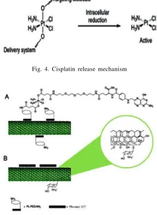
A novel strategy to functionalize carbon nanotubes with two different molecules using the 1,3-dipolar cycloaddition of azomethine ylides was presented by Pastorin et al., who covalently linked methotrexate and FITC to MWCNTs (Fig 3, A). The penetration of this new derivative was studied by epifluorescence and confocal microscopy on human Jurkat T lymphocytes, demonstrating a fast accumulation in the cytoplasm (140). McDevitt et al. prepared CNT derivatives bearing at the same time tumor-specific monoclonal antibodies, radiometal-ion chelates, or fluorescent probes. Water soluble compounds, obtained by dipolar cycloaddition were treated with DOTA-NCS and LC-SMCC, and then I111 in was chelated (Fig 3, B). The conjugates were studied in vitro by flow cytometry and cell-based immunoreactivity assays, while bio-distribution was studied in mice with xenografted lymphoma [141]. SWCNTs coated with modified phospholipids (PL-PEG-NH2) allowed linking a platinum (IV) derivative through the amine residue to one of axial Pt ligands [142]. This molecule itself is almost nontoxic to testicular cancer cells, but becomes significantly cytotoxic when attached to the surface of amine-functionalized soluble SWCNTs. This is due to the fact that the presence of CNTs induces the prodrug cellular uptake, which takes place and confines the complex into endosomes, where the pH is lower than in the cell incubation medium. The acid environment facilitates the reduction from Pt (IV) to Pt (II) and the release of active cisplatin, with loss of axial ligands (Fig 4). More recently, the same authors described a new complex in which one platinum axial ligand is linked to folic acid as targeting moiety (Fig 5, A) [143]. The preparation of high molecular weight complexes should also increase the blood circulation time. They investigated folate receptor-positive [FR (+)] human choriocarcinoma (JAR) and human nasopharyngeal carcinoma (KB) cell lines, using as negative control FR-negative [FR (-)] testicular carcinoma cells (NTera-2), demonstrating that FR (+) cells are more affected. Moreover, the so-prepared prodrug resulted to be much more active than cisplatin itself, forming after intracellular reduction, cisplatin (GpG) intrastrand cross-links with nuclear DNA. It has been recently demonstrated that doxorubicin
adsorbed on MWCNT surface (Fig 5, B) by means of
π-π interaction results to be more efficient than the
Nanomed. J., 2(4): 231-248, Autumn 2015H.R. Sadegh et al.
Fig. 3. Molecular structures of bifunctional CNTs derivatives
Fig. 4. Cisplatin release mechanism
Fig. 5. Non-covalent attachments of anti-tumoral drugs to CNTs
Gene Delivery
One of the first approaches toward genetic material delivery was performed by Lu et al. who complexed RNA polymer poly (rU) to SWCNTs by nonspecific interactions [145]. The translocation across MCF7 breast cancer cells took place and the genetic material was found across the cellular and the nuclear membranes.
Covalent derivative SWCNTs, obtained by amidation of the nanotube carboxylic groups with a chain presenting a terminal ammonium group were studied as telomerase reverse transcriptase small interfering RNA (TERT-siRNA) delivery system into cells [146]. The effect was assessed taking into account the proliferation and the growth of tumor cells (Lewis lung cancer cells and HeLa cells), both in vitro and in vivo. So the silencing of the targeted gene is the evidence of a good intracellular delivery of siRNA, which is locally released from nanotubes and can induce its effect. Dai and coworkers delivered siRNA to human T cells and peripheral blood mononuclear cells. In this case,
SWCNTs were again coated with PL-PEG-NH2.
Differences in efficacy were found to depend on functionalization and degree of hydrophilicity, related to the length of the introduced PEG chain in the PL-PEG construct [147]. Short SWCNTs, non-covalently functionalized with PL-PEG, were used. In this case, a cleavable bond was introduced, linking by disulfide bond the PLPEG unit and the delivering siRNA [148]. The siRNA functionality obtained in this model resulted to be more potent than lipofectamine used as control transfection agent.
Oxidized SWCNTs, bearing positive charges able to interact with fluorescein-labeled dsDNA (dsDNA-FAM), were also coated with folic acid-modified
phospholipids (PL-PEG-NH2), by wrapping the alkyl
chains. The multifunctional system was selectively uptaken by tumor cells, in which folic receptors are overexpressed, driving the oligonucleotide into cells, as demonstrated by fluorescence imaging [149]. SWCNTs were utilized as carriers for ssDNA probe into cells, demonstrating an increased resistance of the oligonucleotides toward nuclease digestion. In fact the ssDNA is protected from enzymatic cleavage and interference from nucleic acid binding proteins, still exerting its function of targeting mRNA [150]. This increases the potentiality of CNTs as oligonucleotide vectors, as already hypothesized by Kateb et al. [136]. They studied the possibility of using CNTs as DNA or
free one, probably because of ameliorate delivery into
RNA delivery systems in brain tumors, thus demonstrating the ability of microglia to efficiently internalize MWCNTs coated with Pluronic F108 as compared to glioma cells.
Anticancer Approaches
The potential of CNTs as a genetic material delivery system is great, with application in gene therapy, considering the general biocompatibility of CNTs themselves. As mentioned already, nanotubes easily bind macromolecules, such as nucleic acids. Li et al. found that oxidized SWCNTs can inhibit DNA duplex association [151, 152]. Moreover, it has been demonstrated that they selectively induce human
telomeric i-motif DNA formation, binding to the
5’-endmajor groove and directly stabilizing the charged CC+base pairs. These findings can be utilized in designing new compounds for cancer therapy, where the modulation of human telomeric DNA can play a fundamental role, although the biological effects of i-motif structure induction is not yet clear.
A polyadenylic acid [poly (rA)] tail is present in eukaryotic cells and human poly (rA) polymerase (PAP) and can be considered a tumor-specific target, and compounds interfering with this structure have interesting potential in the therapy. Zhao et al. studied oxidized SWCNTs and reported that CNTs induce single-stranded poly (rA) to self-structure into duplex but the mechanism is not understood and should involve the characteristics of the oxidized CNTs considering that amino-derivative SWCNTs, surprisingly, do not induce self-structuration [153]. Antitumor immunotherapy is actually not very effective. In the past, Pantarotto et al. [154] have found that viral peptides conjugated to CNTs can improve the anti-peptide immune response. Meng et al. paved the way to improve immunotherapy. Tumor lysate protein, readily obtainable from most solid tumors, has been linked to MWCNTs and administered as a possible tumor-cell vaccine in a mouse model bearing the H22 liver cancer, with positive effects on the cure rate and cellular antitumor immune reaction [155].
Phospholipid–polyethylene glycol chains linked to a
folic acid residue were linked by means of hydrophobic interactions to CNTs. The presence of folate should drive toward a selective uptake of the complex into cells overexpressing folate receptors, as it happens in some cancer cells. After internalization, cell death was induced irradiating with near-infrared (NIR) light. This
action proved to be selective because the presence of CNTs in cells increases the temperature, thereby inducing thermal ablation when irradiated [21]. Analogously, CNTs bearing two specific monoclonal antibodies (insulin-like growth factor 1 receptor, IGF1R and human endothelial receptor 2, HER2), prepared by supra-molecular approach with properly functionalized pyrene units, have been used to kill breast cancer cells with NIR irradiation [156]. The main problem of near infrared irradiation application is due to its poor tissue penetration capacity, which allows the treatment of only superficial cancer lesions. Radiofrequency waves, on the contrary, penetrate more into tissues, and their interaction with internalized CNTs produces an increase in temperature of cells, thereby inducing cell death. Gannon et al. proposed the use of these waves, but in the reported case the limitation is due to the direct injection into tumor of SWCNT coated with poly phenylene-ethynylene polymer [157].
Another atypical approach to cancer treatment is related to angiogenesis. In fact, its inhibition by interfering in the growth factor balance can obstacle the insurgence of diseases, among which tumors can be mentioned. A good model to study angiogenesis is the chick chorio allantoic membrane (CAM). A study performed by Murugesan et al. employed carbon materials (such as CNTs, fullerene, and graphite) to test their capability to inhibit FGF2- or VEGF-induced vessel formation in the CAM model. All the materials showed significant effects in the induced angiogenesis, acting more efficiently in comparison to VEGF, while they did not exert any effect on the basal process in absence of the already mentioned growth factors. The mechanism involved in this process is not yet clarified but it is clear that this anti-antigenic action could be exploited in cancer treatment [158].
CNTs and Neuron Interactions
Nanomed. J., 2(4): 231-248, Autumn 2015CNTs in Nanomedicine: A reveiw
SWCNTs was qualitatively distinguishable from a coupling between SWCNTs and the patch pipette through the patch seal path to ground [160]. Using single-cell electrophysiology techniques, electron microscopy analysis, and theoretical modeling, the same authors demonstrated that CNTs enhance the responsiveness of neurons, due to the formation of tight contacts between CNTs and the cell membranes. These could favor electrical shortcuts between the proximal and distal compartments of the neuron [161]. While in the already mentioned works, the CNTs were deposed on glass coverslips for growing cells, a different approach has been used by Keefer et al. using CNTs to electrochemically coat tungsten and stainless steel wire electrodes. This treatment led to an increase in recording and electrical stimulation of neuronal cultures [162]. Kotov and coworkers also prepared a layer-by-layer composite with SWCNTs and laminin, an essential glycoprotein of human extracellular matrix. The use of this surface for cellular culture induced neural stem cells (NSC) differentiation and the creation of functional neural network. Also in this case, the application of current through CNTs stimulated the action potentials [163]. Carbon nanotubes have also been arranged in vertical alignment to the gate insulator of an ion-sensitive field-effect transistor to act as electrical interfaces to neurons, to study the interactions with them, and an enhance efficiency has been recorded in neuronal electrical activity [164]. Water-soluble SWCNTs grafted to polyethylene glycol have been used in the culturing medium for neural growth. These derivatives demonstrated their ability to block the stimulated membrane endocytosis in neurons and this could justify the evidenced extended neurite length [165], while MWCNTs coated with pluronic F127 (PF127) injected in mouse cerebral cortex do not induce degeneration of the neurons close to the injection site, while PF127 itself can induce apoptosis of mouse primary cortical neurons, implying that the presence of MWCNTs inhibits PF127-induced apoptosis [166].
All these successful works performed on neuronal growth and stimulation led to the hope of using CNTs as material for the reconstruction of neural injured tissues in the near future, paving the way for an application in spinal cord disease resolution and neurodegeneration restoration.
CNTs and antioxidant
CNTs, when instilled in lungs, induced inflammatory and fibrotic response, which was supposed to be due
to oxidative stress derived from free radicals, although no real evidence of ROS generation from CNTs was observed. Moreover Fenoglio et al. reported that MWCNTs do not generate oxygen or carbon-based free radicals in the presence of H2O2 or formatted, respectively, but, on the contrary, when radicals were induced, CNTs act as radical scavengers [167]. Very recently the antioxidant properties of CNTs have been reported by Tour and coworkers. SWCNTs and ultra-short SWCNTs (US-SWCNTs) were derivatized with butyrate hydroxyl toluene (BHT) using two different approaches as covalent attachment of triazene to the sidewalls of pluronic-wrapped SWCNT or amidation of carboxylic residues in the case of US-SWCNT derivatives. The oxygen radical scavenging ability of the different compounds has been evaluated leading to really interesting results, although the different compounds react differently. In the first case, the pluronic-coated SWCNTs were more efficient than the corresponding BHT derivatives, while in the case of oxidized US-SWCNTs, higher the loading of BHT residues, better was the antioxidant activity. This new finding paves the way for SWCNT application as novel medical therapeutics in the antioxidant field [168]. However, it is necessary to remember that a recent report presents contradictory results indicating MWCNT toxicity in A549 cells, which can be reduced by pretreatment with antioxidants to decrease ROS production and interleukin-8 gene expression [169].
CNTs and imaging
SWCNTs are endowed of fluorescent properties that can be exploited in in vitro and in vivo imaging, and that have been used to determine the uptake of nanotubes in macrophages [170], the CNT elimination in rabbits [171], and their toxicity in fruit fly larvae [172]. This surprising use of CNTs for imaging purposes is only possible when some properties of the nanotubes are preserved, that is, at least 100nm of the tubes must be unmodified and the CNTs should not be in bundles, conditions in which the fluorescence is quenched. More recently, Welsher et al. used semiconducting SWCNTs wrapped with polyethylene glycol as near-infrared fluorescent tags. The polyethylene glycol was conjugated to antibodies for the selective recognition for CD20 cell surface receptor on B cells or for HER2/ neu positive breast cancer cells. The intrinsic NIR photoluminescence allowed the detection of the binding to the cells, in spite of the presence of low auto fluorescence in different cells [173]. MRI is another important imaging technique used in medicine. After
Gd@C82 and their derivatives as contrast agents [174-176], the evolution toward the analogous use of CNT has been done by Wilson and coworkers [177]. The tubes, shortened by fluorination down to 20-80 nm,
were loaded with GdCl3. The obtained results
demonstrated that these CNTs have a relaxivity (r1) 40 times greater than any current Gd3+ ion-based clinical agents. The aggregation, that in the case of fullerene endohedrals can vary the relaxivity, does not affect this parameter in the case of CNTs. However, more soluble compounds have been prepared by Ashcroft et al. [178]. The same authors demonstrated the stability of gadonanotubes in buffers, serum, at variation of pH and temperature. Moreover they discovered a great dependence of r1 on the pH, with ultra-sensitivity in a range of pH 7.0-7.4. This suggests the possibility of using the gadonanotubes for the early detection of cancer cells in which the pH can dramatically decrease [179]. A novel study on the bio-distribution and the effect of SWCNTs (raw and super-purified, raw-SWCNT and SP-raw-SWCNT, respectively) after in vivo exposure has been recently reported. The combination
of3He and1Hmagnetic resonance imaging (MRI) was
used in a rat model. Hyperpolarized gases such as3He acting a contrast agent diffuses rapidly in lungs, permitting the determination of ventilated air ways and alveolar spaces. The presence of metal impurity in the raw- SWCNTs was sufficient to induce a drop in magnetic field homogeneity detected in3He MRI, while no significant variation was observed after SP-SWCNT exposition [180]. Proton MRI was used to follow raw-SWCNTs after intravenous injection, finding them in spleen and kidneys. The absence of metal nanoparticles in the SP-SWCNT excluded signal modifications. When this technique was used to determine the fate of CNT after pulmonary instillation, no signal changes in liver, spleen, and kidney were detected, implying the absence of systemic circulation of CNTs after inhalation. This result was also confirmed by histological analysis, establishing the possibility of using noninvasive methodology to detect CNT presence, if associated with a proper iron impurity concentration.
A different approach has been reported by Richard et al. The authors, instead of filling the CNTs with the Gd derivative, used an amphiphilic gadolinium (III) chelate, absorbed on MWCNT external surface, in different concentrations.
The obtained suspension resulted to be stable and r1 was measured in different conditions, finding a dependence on the Gd-chelate concentration. On the contrary, the transversal water proton relaxivity (T2)
was independent from Gd concentration and frequency [181].
Various applications of CNTs
CNTs are incredible sorbent materials for different compounds due to their large surface areas (BET value
is about e”600m2/g, depending on the CNT types taken into account). They have been already used as contaminant remover for water pollution in laboratory experiments [182, 183] and also for dioxin [184]. Chen et al. studied the sorption of americium and thorium on CNT surface, their kinetics and pH dependence [185, 186] but different efficiency probably due to different pretreatment of CNTs, found by other authors [187], and the possibility to use this new form of carbon in nuclear waste management cannot be excluded. CNTs, both single- and multiwalled, were also used to capture bacteria, asStreptococcus mutans. The simple mixture of CNT bundles with bacteria leads to the formation of a precipitate, which shows bacterial adhesion to nanotubes, that depends on the CNT diameters [188].
Toxicity of CNTs
Nanomed. J., 2(4): 231-248, Autumn 2015
indicating that the intrinsic toxicity of CNTs is mainly mediated by the presence of defective sites in their carbon framework [191]. The scavenging activity is related to the presence of defects and seems to go paired with the genotoxic and inflammatory potential of CNT. Tabet et al. used dispersions of MWCNT in dipalmitoyl lecithin, ethanol, and phosphate-buffered saline (PBS), to study the CNT toxicity on human epithelial cell line A549. The presence of PBS induces agglomerate formation on top of the cells, but in all cases MWCNTs decrease the cellular metabolism without permeabilization of the cell membrane or apoptosis, while asbestos fibers penetrate into the cells and increase apoptosis [192]. Wick et al. investigated CNTs with different agglomeration degrees to determine their cytotoxicity on human MSTO-211H cells. Well-dispersed CNTs (obtained by using polyoxyethylene sorbitan monooleate) were less toxic than asbestos and rope-like agglomerates induced higher cytotoxic effects than asbestos fibers [193]. Recently, Guo et al. reported the modification of MWCNTs with glucosamine and decylamine by c-ray irradiation (g-MWCNT and d-MWCNT), and they used these derivatives to perform cytotoxicity test on Tetrahymena pyriformis. The decylamine derivative exhibits a dose-dependent grown inhibition, attributed to the amine toxicity, driven into cells by the nanotubes. The comparison between purified MWCNTs and g-MWCNTs demonstrated growth stimulation by the latter. It seems that the hydrophilic compound is able to complex peptone, present in the medium, and to transfer it into cells. This means that any nonspecific interaction between CNTs and components of the culture medium must be considered in evaluating the cytotoxicity of CNTs (194). Considering that almost nothing is known about CNT impact on natural ecosystems, the ciliated protozoan Tetrahymena thermophile has been also studied because of its role in the regulation of microbial populations through the ingestion and digestion of bacteria. It is, in fact, an important organism in wastewater treatment and, moreover an indicator of sewage effluent quality. SWCNTs have been found in these microorganisms, inducing protozoa aggregations with a consequent inability to interact with bacteria
[195].Salmonella typhimurium andEscherichia coli
strains were used to perform mutagenicity test with MWCNTs. The mutagenic activity did not appear, even in presence of the metabolic activators [196].
Different tests devoted to establish the genotoxic potential of MWCNTs has been performed both in vivo in rats and in vitro on rat lung epithelial cells or human
epithelial cells. Three days after carbon nanotube intratracheal administration, type II pneumocytes present micronuclei with a dose-dependent increase. The same behavior was reported for the in vitro experiments, proving the possibility of MWCNTs to induce clastogenic and aneugenic events (respectively increase the rate of genetic mutation due to DNA breaks and loss or gain of whole chromosomes) [197]. Often the clastogenic is related to ROS generation and the possible presence of metallic nanoparticles (Fe or Co) would justify this action, but the MWCNT antioxidant behavior suggests different pathways and indicates the necessity to explore these systems more in details.
CONCLUSIONS
CNTs have exhibited diverse physical, chemical and mechanical properties suitable for a variety of applications. In last decade, medical applications of CNTs have undergone rapid progress. Their unique properties such as ultrahigh surface area, high aspect ratio and distinct optical properties have been applied to develop innovative, multi-functional CNT-based nanodevices for broad applications. This review has described the chemical and physical methods to prepare CNTs for used in medicine. With these methods, targeting molecules are attached onto CNTs for targeted drug delivery, selective imaging, and other therapies. As a new type of nanomaterial, the toxicity of CNTs has been extensively investigated. To date, tremendous toxicity studies on CNTs have been published. However, the published data are inconsistent. The reason is that CNTs used in these studies vary in dispersion status, size and length of tubes, metal impurities and functionalization methods etc. Moreover, different analysis methods used in the evaluation CNTs toxicity studies also cause disparities. Despite these disparities, there is a broad agreement that well-dispersed CNTs have little or no toxicity both in vitro and in vivo, and therefore are useful for medical applications. Finally, an urgent need has been proposed for long-term studies on the absorption, deposition, metabolism and excretion (ADME) of CNTs. Only after the uncertainty on CNTs toxicity is resolved, the CNT-based therapeutics can be possible applied clinically.
ACKNOWLEDGMENTS
Authors would like to thank Islamic Azad University Science and Research Branch for all supports.
REFERENCES
2. Ouyang M, Huang J L, Lieber CM. One-dimensional energy dispersion of single-walled carbon nanotubes by resonant electron scattering. Phys Rev Lett. 2002; 88(6): 066804. 3. Zare K, Najafi F, Sadegh H. Studies of ab initio and Monte
Carlo simulation on interaction of fluorouracil anticancer drug with carbon nanotube. J Nanostruc Chem. 2013; 3(1): 1-8.
4. Thostenson ET, Ren Z F, Chou TW. Advances in the science and technology of carbon nanotubes and their composites: a review. Compos Sci Technol. 2001; 61(13): 1899-1912. 5. Troiani HE, Miki-Yoshida M, Camacho-Bragado GA, Marques
MAL, Rubio A, Ascencio JA, Jose-Yacaman M. Direct observation of the mechanical properties of single-walled carbon nanotubes and their junctions at the atomic level. Nano Lett. 2003; 3(6): 751-755.
6. Wan XG, Dong JM, Xing DY. Optical properties of carbon nanotubes. Phys Rev B. 1998; 58(11): 6756-6759. 7. Kostarelos K, Lacerda L, Pastorin G, Wu W, Wieckowski S,
Luangsivilay J, Bianco A. Cellular uptake of functionalized carbon nanotubes is independent of functional group and cell type. Nat Nanotechnol. 2007; 2(2): 108-113.
8. Sadegh H, Shahryari-ghoshekandi R, Kazemi M. Study in synthesis and characterization of carbon nanotubes decorated by magnetic iron oxide nanoparticles. Int Nano Lett. 2014; 4(4): 129-135.
9. Sadegh H, Shahryari-ghoshekandi R, Agarwal S, Tyagi I, Asif M, Gupta VK. Microwave-assisted removal of malachite green by carboxylate functionalized multi-walled carbon nanotubes: Kinetics and equilibrium study. J Mol Liq. 2015; 206: 151-158.
10. Ando Y. Carbon nanotube: the inside story. J Nanosci Nanotechnol. 2010; 10(6): 3726-3738.
11. Chen RJ, Zhang Y, Wang D, Dai H. Noncovalent sidewall functionalization of single-walled carbon nanotubes for protein immobilization, J Am Chem Soc. 2001; 123: 3838– 3839.
12. Zare K, Gupta VK, Moradi O, Makhlouf ASH, Sillanpää M, Nadagouda MN, Sadegh H, Shahryari-ghoshekandi R, Pal A, Wang Z, Tyagi I, Kazemi M. A comparative study on the basis of adsorption capacity between CNTs and activated carbon as adsorbents for removal of noxious synthetic dyes: a review. J Nanostruc Chem. 2015; 5(2): 227-236. 13. Besteman K, Lee JO, Wiertz FGM, Heering HA, Dekker C.
Enzyme-coated carbon nanotubes as single-molecule biosensors, Nano Lett. 2003; 3: 727–730.
14. Xin H, Woolley AT. DNAtemplated nanotube localization. J Am Chem Soc. 2003; 125: 8710-8711.
15. Taft BJ, Lazareck AD, Withey GD, Yin A, Xu JM, Kelley SO. Site-specific assembly of DNA and appended cargo on arrayed carbon nanotubes. J Am Chem Soc. 2004; 126: 12750-12751.
16. Gupta VK, Tyagi I, Agarwal S, Moradi O, Sadegh H, Shahryari-ghoshekandi R, Makhlouf ASH, Goodarzi M, Garshasbi A. Study on the removal of heavy metal ions from industry waste by carbon nanotubes: effect of the surface modification-A review. Crit Rev Env Sci Technol (Accepted for publish) DOI:10.1080/10643389.2015.1061874 (2015). 17. Liu L, Wang T, Li J, Guo Z, Dai L, Zhang D, Zhu D. Self-assembly of gold nanoparticles to carbon nanotubes using a thiol-terminated pyrene as interlinker. Chem Phys Lett. 2003; 367: 747-752.
18. Cao L, Chen H, Wang M, Sun J, Zhang X, Kong F. Photoconductivity study of modified carbon nanotube/ oxotitanium phthalocyanine composites. J Phys Chem B. 2002; 106: 8971-8975.
19. Wang X, Liu Y, Qiu W, Zhu D. Immobilization of tetra-tertbutylphthalocyanines on carbon nanotubes: a first step towards the development of new nanomaterials. J Mater Chem. 2002; 12: 1636-1639.
20. Cao L, Chen HZ, Zhou HB, Zhu L, Sun JZ, Zhang XB, Xu JM, Wang M. Carbon nanotube templated assembly of rare-earth phthalocyanine nanowires. Adv Mater. 2003; 15: 909-913.
21. Guldi DM, Rahman GNA, Ramey J, Marcaccio M, Paolucci D, Paolucci F, Qin S, Ford WT, Balbinot D, Jux N, Tagmatarchis N, Prato M. Donor–acceptor nanoensembles of soluble carbon nanotubes. Chem Commun. 2004; 2034-2035.
22. Guldi DM, Rahman GMA, Prato M, Jux N, Qin S, Ford W. Single-wall carbon nanotubes as integrative building blocks for solarenergy conversion. Angew Chem Int Ed. 2005; 44: 2015-2018.
23. Murakami H, Nomura T, Nakashima N. Noncovalent porphyrin-functionalized single-walled carbon nanotubes in solution and the formation of porphyrin–nanotube nanocomposites. Chem Phys Lett. 2003; 378: 481-485. 24. Li H, Zhou B, Lin Y, Gu L, Wang W, Fernando KAS, Kumar
S, Allard LF, Sun YP. Selective interactions of porphyrins with semiconducting single-walled carbon nanotubes. J Am Chem Soc. 2004; 126: 1014-1015.
25. Chen J, Collier CP. Noncovalent functionalization of single-walled carbon nanotubes with water-soluble porphyrins. J Phys Chem B. 2005; 109: 7605-7609.
26. Satake A, Miyajima Y, Kobuke Y. Porphyrin–carbon nanotube composites formed by noncovalent polymer wrapping. Chem Mater. 2005; 17: 716-724.
27. Guldi DM, Rahman GMA, Jux N, Tagmatarchis N, Prato M. Integrating single-wall carbon nanotubes into donor–acceptor nanohybrids. Angew Chem Int Ed. 2004; 43: 5526-5530. 28. Guldi DM, Rahman GMA, Jux N, Balbinot D, Tagmatarchis
N, Prato M. Multiwalled carbon nanotubes in donor–acceptor nanohybrids – towards long-lived electron transfer products. Chem Commun. 2005; 2038-2040.
29. Guldi DM, Taieb H, Rahman GMA, Tagmatarchis N, Prato M. Novel photoactive single-walled carbon nanotube– porphyrin polymer wraps: efficient and long-lived intracomplex charge separation. Adv Mater. 2005; 17: 871-875.
30. Guldi DM, Rahman GMA, Jux N, Balbinot D, Hartnagel U, Tagmatarchis N, Prato M. Functional single-wall carbon nanotube nanohybrids-associating SWCNTs with water-soluble enzyme model systems J Am Chem Soc. 2005; 127: 9830-9838.
31. Chichak KS, Star A, Altoe MVP, Stoddart JF. Single-walled carbon nanotubes under the influence of dynamic coordination and supramolecular chemistry. Small. 2005; 1: 452-461.
32. Tang BZ, Xu H. Preparation, alignment, and optical properties of soluble poly(phenylacetylene)-wrapped carbon nanotubes. Macromol. 1999; 32: 2569-2576.
33. Romero DB, Carrard M, Heer W, Zuppiroli L. A carbon nanotube/organic semiconducting polymer heterojunction. Adv Mater. 1996; 8: 899-902.
Nanomed. J., 2(4): 231-248, Autumn 2015
Suar, M. (eds.) Handbook of Research on Diverse Applications of Nanotechnology in Biomedicine, Chemistry, and Engineering, pp. 90–128. (2015). doi:10.4018/978-1-4666-6363-3.ch006. Accessed 16 Jan 2015
36. Ago H, Shaffer MSP, Ginger DS, Windle AH, Friend RH. Electronic interaction between photoexcited poly( p-phenylene vinylene) and carbon nanotubes. Phys Rev B. 2000; 61: 2286-2290.
37. Wery J, Aarab H, Lefrant S, Faulques E, Mulazzi E, Perego R. Photoexcitations in composites of poly(paraphenylene vinylene) and single-walled carbon nanotubes. Phys Rev B. 2003; 67(11): 115202.
38.Fournet P, Coleman JN, Lahr B, Drury A, Blau WJ, O’Brien DF, Horhold HH. Enhanced brightness in organic light-emitting diodes using a carbon nanotube composite as an electron-transport layer. J Appl Phys. 2001; 90: 969-975. 39.Fournet P, O’Brien DF, Coleman JN, Horhold HH, Blau WJ.
A carbon nanotube composite as an electron transport layer for M3EH-PPV based lightemitting diodes. Synth. Met. 2001; 121: 1683-1684.
40. Moradi O, Gupta VK, Agarwal S, Tyagi I, Asif M, Makhlouf ASH, Sadegh H, Shahryari-ghoshekandi R. Characteristics and electrical conductivity of graphene and graphene oxide for adsorption of cationic dyes from liquids: Kinetic and thermodynamic study. J Ind Eng Chem. 2015; 28: 294-301. 41. Coleman JN, Dalton AB, Curran S, Rubio A, Davey AP, Drury A, McCarthy B, Lahr B, Ajayan PM, Roth S, Barklie RC, Blau WJ. Phase separation of carbon nanotubes and turbostratic graphite using a functional organic polymer. Adv Mater. 2000; 12: 213-216.
42. Star A, Lu Y, Bradley K, Gruner G. Nanotube optoelectronic memory devices. Nano Lett. 2004; 4: 1587-1591. 43. Murphy R, Coleman JN, Cadek M, McCarthy B, Bent M,
Drury A, Barklie RC, Blau WJ. Highyield, nondestructive purification and quantification method for multiwalled carbon nanotubes. J Phys Chem B. 2002; 106: 3087-3091. 44.Coleman JN, O’Brien DF, Dalton AB, McCarthy B, Lahr B,
Barklie RC, Blau WJ. Electron paramagnetic resonance as a quantitative tool for the study of multiwalled carbon nanotubes. J Chem Phys. 2000; 113: 9788-9793. 45. Star A, Stoddart JF, Steuerman D, Diehl M, Boukai A, Wong
EW, Yang X, Chung SW, Choi H, Heath JR. Preparation and properties of polymer-wrapped singlewalled carbon nanotubes. Angew Chem Int Ed. 2001; 40: 1721-1725. 46. Sadegh H, Shahryari-ghoshekandi R, Tyagi I, Agarwal S,
Gupta VK. Kinetic and thermodynamic studies for alizarin removal from liquid phase using poly-2-hydroxyethyl methacrylate (PHEMA). J Mol Liq. 2015; 207: 21-27. 47. Star A, Stoddart JF. Dispersion and solubilization of
single-walled carbon nanotubes with a hyperbranched polymer. Macromol. 2002; 35: 7516-7520.
48. Musa I, Baxendale M, Amaratunga GAJ, Eccleston W. Properties of regioregular poly(3-octylthiophene)/multi-wall carbon nanotube composites. Synth Met. 1999; 102: 1250. 49. Alexandrou I, Kymakis E, Amaratunga GAJ. Polymer-nanotube composites: burying Polymer-nanotubes improves their field emission properties. Appl Phys Lett. 2002; 80: 1435-1437.
50. Valentini L, Armentano I, Biagiotti J, Frulloni E, Kenny JM, Santucci S. Frequency dependent electrical transport between conjugated polymer and single-walled carbon nanotubes. Diam Rel Mater. 2003; 12: 1601-1609.
51. Kymakis E, Amaratunga GAJ. Single-wall carbon nanotube/ conjugated polymer photovoltaic devices. Appl Phys Lett. 2002; 80: 112-114.
52. Kymakis E, Alexandrou I, Amaratunga GAJ. High opencircuit voltage photovoltaic devices from carbon-nanotube-polymer composites. J Appl Phys. 2003; 93: 1764-1768.
53. Bhattacharyya S, Kymakis E, Amaratunga GAJ. Photovoltaic properties of dye functionalized single-wall carbon nanotube/ conjugated polymer devices. Chem Mater. 2004; 16: 4819-4823.
54. Landi BJ, Raffaelle RP, Castro SL, Bailey SG. Single-wall carbon nanotube-polymer solar cells. Prog Photovolt Res Appl. 2005; 13: 165-172.
55.O’Connell MJ, Boul P, Ericson, LM, Huffman C, Wang Y, Haroz E, Kuper C, Tour JM, Ausman KD, Smalley RE. Reversible water-solubilization of single-walled carbon nanotubes by polymer wrapping. Chem Phys Lett. 2001; 342: 265-271.
56. Islam MF, Rojas E, Bergey DM, Johnson AT, Yodh AG. High weight fraction surfactant solubilization of single-wall carbon nanotubes in water. Nano Lett. 2003; 3: 269-273. 57. Richard C, Balavoine F, Schultz P, Ebbesen TW, Mioskowski
C. Supramolecular self-assembly of lipid derivatives on carbon nanotubes. Science. 2003; 300(5620): 775-778.
58.O’Connell MJ, Bachilo SM, Huffman CB, Moore VC, Strano MS, Haroz EH, Rialon KL, Boul PJ, Noon WH, Kittrell C, Ma J, Hauge RH, Weisman RB, Smalley RE. Band gap fluorescence from individual single-walled carbon nanotubes. Science. 2002; 297: 593-596.
59. Moore VC, Strano MS, Haroz EH, Hauge RH, Smalley RE. Individually suspended single-walled carbon nanotubes in various surfactants. Nano Lett. 2003; 3: 1379-1382. 60. Wenseleers W, Vlasov II, Goovaerts E, Obraztsova ED,
Lobach AS, Bouwen A. Efficient isolation and solubilization of pristine single-walled nanotubes in bile salt micelles. Adv Funct Mater. 2004; 14: 1105-1112.
61. Bachilo SM, Strano MS, Kittrell C, Hauge RH, Smalley RE, Weisman RB. Structureassigned optical spectra of singlewalled carbon nanotubes. Science. 2002; 298(5602): 2361–2366. 62. Hagen A, Hertel T. Quantitative analysis of optical spectra
from individual single-wall carbon nanotubes. Nano Lett. 2003; 3: 383-388.
63. Strano MS, Doorn SK, Haroz EH, Kittrell C, Hauge RH, Smalley RE. Assignment of (n, m) Raman and optical features of metallic single-walled carbon nanotubes. Nano Lett. 2003; 3: 1091-1096.
64. Weisman RB, Bachilo SM. Dependence of optical transition energies on structure for singlewalled carbon nanotubes in aqueous suspension: an empirical Kataura plot. Nano Lett. 2003; 3: 1235-1238.
65. Dyke CA, Tour JM. Unbundled and highly functionalized carbon nanotubes from aqueous reactions. Nano Lett. 2003; 3: 1215-1218.
66. Dieckmann GR, Dalton AB, Johnson PA, Razal J, Chen J, Giordano GM, Muňoz E, Musselman IH, Baughman RH, Draper RK. Controlled assembly of carbon nanotubes by designed amphiphilic peptide helices. J Am Chem Soc. 2003; 125: 1770-1777.
individual peptidewrapped single-walled carbon nanotubes. J Am Chem Soc. 2004; 126: 7222-7227.
68. Zorbas V, Smith AL, Xie H, Ortiz-Acevedo A, Dalton AB, Dieckmann GR, Draper RK, Baughman RH, Musselman IH. Importance of aromatic content for peptide/single-walled carbon nanotube interactions. J Am Chem Soc. 2005; 127: 12323-12328.
69. Kam NWS, Dai H. Carbon nanotubes as intracellular protein transporters: generality and biological functionality. J Am Chem Soc. 2005; 127: 6021-6026.
70. Zheng M, Jagota A, Semke ED, Diner BA, Mclean RS, Lustig SR, Richardson RE, Tassi NG. DNA-assisted dispersion and separation of carbon nanotubes. Nat. Mater. 2003; 2: 338-342.
71. Zheng M, Jagota A, Strano MS, Santos AP, Barone P, Chou SG, Diner BA, Dresselhaus MS, Mclean RS, Onoa GB, Samsonidze GG, Semke ED, Usrey M, Walls DJ. Structurebased carbon nanotube sorting by sequence-dependent DNA assembly. Science. 2003; 302(5650): 1545-1548. 72. Kam NWS, Liu Z, Dai H. Functionalization of carbon
nanotubes via cleavable disulfide bonds for efficient intracellular delivery of siRNA and potent gene silencing. J Am Chem Soc. 2005; 127: 12492-12493.
73. Hirsch A. Functionalization of single-walled carbon nanotubes. Angew Chem Int Ed. 2002; 41: 1853-1859. 74. Banerjee S, Hemraj-Benny T, Wong SS. Covalent surface
chemistry of single-walled carbon nanotubes. Adv Mater. 2005; 17: 17-29.
75. Hamon MA, Chen J, Hu H, Chen Y, Itkis ME, Rao AM, Eklund PC, Haddon RC. Dissolution of single-walled carbon nanotubes. Adv Mater. 1999; 11: 834-840.
76. Kukovecz A, Kramberger C, Holzinger M, Kuzmany H, Schalko J, Mannsberger M, Hirsch A. On the stacking behavior of functionalized single-wall carbon nanotubes. J Phys Chem B. 2002; 106: 6374-6380.
77. Chen J, Rao AM, Lyuksyutov S, Itkis ME, Hamon MA, Hu H, Cohn RW, Eklund PC, Colbert DT, Smalley RE, Haddon RC. Dissolution of fulllength single-walled carbon nanotubes J Phys Chem B. 2001; 105: 2525-2528.
78. Niyogi S, Hamon MA, Hu H, Zhao B, Bhowmik P, Sen R, Itkis ME, Haddon RC. Chemistry of single-walled carbon nanotubes. Acc Chem Res. 2002; 35: 1105-1113. 79. Hiura H, Ebbesen TW, Tanigaki K. Opening and purification
of carbon nanotubes in high yields. Adv Mater. 1995; 7: 275-276.
80. Ajayan PM, Ebbesen TW, Ichihashi T, Iijima S, Tanigaki K, Hiura H. Capillarity-induced filling of carbon nanotubes. Nature. 1993; 361: 333-334.
81. Chen J, Hamon MA, Hu H, ChenY, Rao AM, Eklund PC, Haddon RC. Solution properties of single-walled carbon nanotubes. Science. 1998; 282(5386): 95-98.
82. Lago RM, Tsang SC, Lu KL, Chen YK, Green MLH. Filling carbon nanotubes with small palladium metal crystallites: the effect of surface acid groups. Chem Commun. 1995; 1355-1356.
83. Gupta VK, Sadegh H, Yari M, Shahryari Ghoshekandi R, Maazinejad B, Chahardori M. Removal of ammonium ions from wastewater: A short review in development of efficient methods. Global J Environ Sci Manag. 2015; 1(2): 149-158.
84. Monthioux M, Smith BW, Burteaux B, Claye A, Fischer JE, Luzzi DE. Sensitivity of single wall carbon nanotubes to chemical processing: an electron microscopy investigation. Carbon. 2001; 39: 1251-1272.
85. Koshio A, Yudasaka M, Zhang M, Iijima SA. simple way to chemically react single-wall carbon nanotubes with organic materials using ultrasonication. Nano Lett. 2001; 1: 361-363.
86. Qin Y, Shi J, Wu W, Li X, Guo Z, Zhu D. Concise route to functionalized carbon nanotubes. J Phys Chem B. 2003; 107: 12899-12901.
87. Chen Y, Haddon RC, Fang S, Rao AM, Eklund PC, Lee WH, Dickey EC, Grulke EA, Pendergrass JC, Chavan A, Haley BE, Smalley RE. Chemical attachment of organic functional groups to single-walled carbon nanotube material. J Mater Res. 1998; 13: 2423-2431.
88. Sun YP, Huang W, Lin Y, Fu K, Kitaygorodskiy A, Riddle LA, Yu YJ, Carroll DL. Soluble dendron-functionalized carbon nanotubes: preparation, characterization, and properties. Chem Mater. 2001; 13: 2864-2869.
89. Fu K, Huang W, Lin Y, Riddle LA, Carroll DL, Sun YP. Defunctionalization of functionalized carbon nanotubes. Nano Lett. 2001; 1: 439-441.
90. Sun YP, Fu K, Lin Y, Huang W. Functionalized carbon nanotubes: properties and applications. Acc Chem Res. 2002; 35: 1096-1104.
91. Kong H, Gao C, Yan D. Controlled functionalization of multiwalled carbon nanotubes by in situ atom transfer radical polymerization. J Am Chem Soc. 2004; 126: 412-413. 92. Gupta VK, Tyagi I, Agarwal S, Sadegh H,
Shahryari-ghoshekandi R, Yari M, Yousefi-nejat O. Experimental study of surfaces of hydrogel polymers HEMA, HEMA–EEMA– MA, and PVA as adsorbent for removal of azo dyes from liquid phase. J Mol Liq. 2015; 206: 129-136.
93. Huang W, Taylor S, Fu K, Lin Y, Zhang D, Hanks TW, Rao AM, Sun YP. Attaching proteins to carbon nanotubes via diimideactivated amidation. Nano Lett. 2002; 2: 311-314. 94. Kam NWS, Jessop TC, Wender PA, Dai H. Nanotube
molecular transporters: internalization of carbon nanotube– protein conjugates into mammalian cells. J Am Chem Soc. 2004; 126: 6850-6851.
95. Yim T, Liu J, Lu Y, Kane RS, Dordick JS. Highly active and stable DNAzyme-carbon nanotube hybrids. J Am Chem Soc. 2005; 127: 12200-12201.
96. Baker SE, Cai W, Lasseter TL, Weidkamp KP, Hamers RJ. Covalently bonded adducts of deoxyribonucleic acid (DNA) oligonucleotides with single-wall carbon nanotubes: synthesis and hybridization. Nano Lett. 2002; 2: 1413-1417. 97. Hazani M, Naaman R, Hennrich F, Kappes MM. Confocal
fluorescence imaging of DNA-functionalized carbon nanotubes. Nano Lett. 2003; 3: 153-155.
98. Williams KA, Veenhuizen PTM, de la Torre BG, Eritja R, Dekker C. Nanotechnology: carbon nanotubes with DNA recognition. Nature. 2002; 420: 761.
99. Tasis D, Tagmatarchis N, Bianco A, Prato M. Chemistry of carbon nanotubes. Chem Rev. 2006; 106: 1105-1136. 100. Jorio A, Dresselhaus G, Dresselhaus MS, Souza M, Dantas
MSS, Pimenta MA, Rao AM, Saito R, Liu C, Cheng HM. Polarized Raman study of single-wall semiconducting carbon nanotubes. Phys Rev Lett. 2000; 85: 2617-2620. 101. Holden JM, Zhou P, Bi X, Eklund PC, Bandow S, Jishi RA,
Chowdhury KD, Dresselhaus G, Dresselhaus MS. Raman scattering from nanoscale carbons generated in a cobalt-catalyzed carbon plasma. Chem Phys Lett. 1994; 220: 186-191.