1
Original Article
Assessment of the Pulmonary Vascular Blood Supply
in Patients with Pulmonary Atresia, Ventricular Septal
Defect, and Aortopulmonary Collateral Arteries
Ulisses Alexandre Croti, Miguel Lorenzo Barbero Marcial, Carla Tanamati,
Marcelo Biscegli Jatene, Domingo Marcolino Braile, Sergio Almeida de Oliveira
São Paulo, SP - Brazil
Objective
To study the morphometric characteristics of the central pul-monary arteries and aortopulpul-monary collateral arteries by assessing the morphology of the pulmonary vascular blood supply, to determine their significance in surgical treatment.
Method
From January 1990 to June 2001, 40 patients were studied. Those who had the complete cineangiocardiographic study pri-or to the first surgical intervention were included in the study. The morphometric characteristics of the central pulmonary arteries (PPAA) and aortopulmonary collateral arteries (APCA) were analyzed, as was the distribution of blood irrigation to the lungs. The following indices were calculated: pulmonary arteri-al index (PPAAI), aortopulmonary collaterarteri-al arteriarteri-al index (APCAI), and total neopulmonary arterial index (TNPAI = PPAAI + APCAI). The surgical treatment was considered palliative (PT), palliative definitive (PDT), and definitive (DT).
Results
The palliative treatment predominated. No statistically signi-ficant differences were observed in the patients undergoing PT, PDT, and DT, in regard to PPAAI, APCAI, and TNPAI. Comparing PPAAI and APCAI, no difference was observed for DT (P=0.4309); APCAI was greater than PPAAI for PT (P=0.0176); and APCAI was descriptively greater for PDT. The TNPAI of patients undergoing DT was greater than that of patients undergoing PT (P=0.0959). Five morphologically similar subgroups were identified and designa-ted as B1, B2, B3, B4, and B5. Overall mortality was 17.5%.
Conclusion
The morphometric characteristics are important, but the mor-phology of the pulmonary vascular blood supply of the PPAA and APCA proved to be better for guiding the surgical treatment. Independently of the didactical division into subgroups, PT predo-minated. Mortality was not correlated with the morphometric characteristics.
Key words
pulmonary atresia, ventricular septal defect, aortopulmonary collateral arteries
In pulmonary atresia (PA) with ventricular septal defect (VSD) and aortopulmonary collateral arteries (APCA), the pulmonary vas-cular blood supply may be very complex with great anatomical variations, so that some segments of the pulmonary lobes may be supplied by blood flow from the central pulmonary arteries (PPAA) or APCA, or both 1. Due to this extreme heterogeneity, several
anatomical classifications have been proposed, aiming at establi-shing criteria that rationalize the surgical approach. The currently most-used morphological classification 2-5 divides the patients with
PA and VSD into 3 different groups: A, B, and C. In group A, all pulmonary segments are supplied by PPAA. In group B, some pulmonary segments are supplied by PPAA, while others are sup-plied by APCA. In group C, all pulmonary segments are supsup-plied by APCA. PPAA are absent 5.
Based on the analysis of the cineangiocardiographic studies of patients with pulmonary atresia, ventricular septal defect, and aorto-pulmonary collateral arteries (group B), the present study aimed at assessing the morphometric characteristics of the central pulmonary and aortopulmonary collateral arteries, through the assessment of the distribution of the pulmonary vascular blood supply, in an attempt to determine their significance in surgical treatment.
Methods
This study comprised 40 patients with PA, VSD, and APCA operated upon at the Instituto do Coração of the Hospital das Clínicas of the Medical School of the University of São Paulo from January 1990 to June 2001.
This study was approved by the Scientific and Ethical Com-mittee for Analysis of Research Projects of the Instituto do Cora-ção of the Hospital das Clínicas of the Medical School of the Universidade de São Paulo.
The age in the cineangiocardiographic studies ranged from 9 days to 21.6 years (median of 1.1 year). The age in the surgical procedures ranged from 3.7 months to 21.6 years (median of 1.9 years). Twenty-one (52.50%) patients were males and 19 (47.50%) were females.
Patients with morphology of situs solitus and concordant atrio-ventricular and ventriculoarterial connections with complete cineangio-cardiographic studies prior to the first surgical intervention exclusi-vely performed at our institution were included in the study. The medical record analysis focused on the reports of the first cineangio-cardiographic study and the descriptions of all 56 surgeries performed. The following patients were excluded from the study: those in whom the results were not available of the cineangiocardiographic
Instituto do Coração of the Hospital das Clínicas of the Medical School of the Universidade de São Paulo
Mailing address: Ulisses Alexandre Croti - Rua Benjamin Constant, 4035/114 - Cep 15015-600 - São José do Rio Preto, SP, Brazil E-mail: [email protected]
2
study performed before the initial surgical treatment and those in whom not all the possible techniques for demonstrating the pul-monary vascular blood supply were used, resulting in an incomplete examination.
The complete cineangiocardiographic study comprised the fol-lowing: injections of contrast medium into the ascending and des-cending aorta and aortic arch; right and left ventriculographies; selective injections into the ductus arteriosus, PPAA, APCA, and coronary arteries; and retrograde venography. The injections of contrast medium, mainly the selective one into the PPAA and APCA, and retrograde venographies should identify the different anatomical variations and segments supplied.
For quantitative morphometric analysis, CAAS II (cardiovascular angiography analysis system II) software was used. The computer was coupled to an LVA-7000 laser videodisc player (JVC), an SVO-9500 MD videocassette recorder (SONY), and a Tagarno cinevideo camera to allow the analysis of films recorded on digital disk, VHS tape, and 35 mm film.
The system was calibrated based on the diameter of the distal portion of the catheter, which was calculated with CAAS II soft-ware, according to the data previously provided regarding the catheter used during that examination, as for example 5Fr or 6Fr. The images selected for measurement were in the posteroan-terior view, with the vascular segment of interest well contrasted and in ventricular systole.
After this calibration, the PPAA and APCA diameters were mea-sured. The PPAA were measured immediately proximal to the origin of the first lobar branch 6, and the APCA were measured distally to
the point where surgical unifocalization would be ideally possible 7.
With these measurements obtained, the area of the blood vessels was calculated.
Body surface was calculated by applying the Mosteller formu-la 8 to the patients’ weights and heights known on the occasion
of the examination.
Therefore, the following indices could be calculated: PPAAI (pulmonary arterial index), APCAI (aortopulmonary collateral arte-rial index), and TNPAI (total neopulmonary artearte-rial index), as shown in figure 1.
Because some pulmonary segments were supplied by the bran-ches of the PPAA while others were supplied by the APCA, the TNPAI was calculated.
The area was obtained by measuring the PPAA immediately before bifurcation of the lobar branches, as already described 6.
The pulmonary artery was considered hypoplastic when its area had a 25% or greater reduction as compared with the mean areas of PPAA.
When PPAAI was smaller than 100 mm2/m2, the PPAA were
considered hypoplastic 6.
When TNPAI was smaller than 100 mm2/m2, the need for
assessing the ratio between PPAAI and APCAI was considered. The diameter of the APCA was measured distally to the point, in which surgical unifocalization would be ideally possible, as previously described 7.
Stenosis of the APCA was characterized by a luminal reduction greater than 25% 7. The vessel was considered thin when its
area was smaller than 50% of the arithmetic mean of the areas of all APCA; the vessel was considered medium when its area was greater than 50% of the arithmetic mean and smaller than the arithmetic mean of all APCA; and the vessel was considered wide when its area was greater than the arithmetic mean of the areas of all APCA of the patient.
When APCAI was smaller than 100 mm2/m2, the APCA were
considered to have an overall reduced caliber.
Analyzing the areas of pulmonary vascular blood supply, the pulmonary lobes irrigated by the PPAA and those irrigated by the APCA were identified. The relation between the pulmonary lobes and segments was defined as follows: left superior lobe (apicopos-terior, an(apicopos-terior, superior lingular, inferior lingular segments); left inferior lobe (superior, anteromedial basal, lateral basal, posterior basal segments); right superior lobe (apical, anterior, posterior segments); right middle lobe (lateral and medial segments); right inferior lobe (superior, middle basal, anterior basal, lateral basal, posterior basal segments) 9.
When the injection of contrast medium into one of the arteries filled the contralateral pulmonary branch, confluence between the PPAA was considered.
The arteries that supplied at least one intrapulmonary seg-mentary artery and had a trajectory independent of the pulmonary bronchi and intercostal arteries were considered APCA.
The surgical treatments were classified as palliative, “palliative definitive,” or definitive.
Palliative treatment was that in which the patients underwent interventions without closure of the VSD and had future perspectives of conclusion or not of the treatment. They were considered as being under treatment.
“Palliative definitive” treatment was that in which the patients underwent interventions without closure of the VSD and had no other surgical option that enabled the conclusion of the treatment, with RV-PPAA connection and VSD closure. This was the case when the diameter of the PPAA was extremely reduced with no possibility of development, or when the distal pulmonary vascular tree showed signs of pulmonary hypertensive disease with or wi-thout thrombosis, or when unifocalization of at least 10 pulmonary segments for connection to RV was not possible 14.
The definitive treatment consisted of RV-PPAA connection and closure of the VSD, allowing reestablishment of the normal phy-siology. The criteria for closure of the VSD were as follows: pre-sence of PPAA with appropriate calibers, adequate distal pulmonary vascular tree, and presence of at least 10 pulmonary segments that could be connected to the RV.
Results
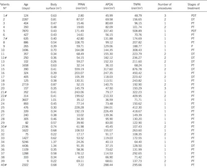
The morphometric analysis of the cineangiocardiographic stu-dies performed before the first surgical procedure and the stages of treatment of all patients are described in table 1.
Fig. 1 - Formulas used for calculating the pulmonary arterial index (PPAAI), aortopulmonary collateral arterial index (APCAI), and total neopulmonary arterial index (TNPAI).
PPAAI= RPA area (mm
2) + LPA area (mm2)
body surface area (m2)
APCAI = APCA area 1 (mm
2) + APCA area 2 (mm2) + ... + APCA area n (mm2)
body surface area (m2)
3
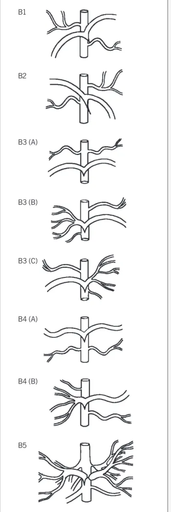
Associating the morphometric data obtained with the dis-tribution of the pulmonary vascular blood supply, 5 subgroups with similar characteristics were identified as follows: B1, B2, B3, B4, and B5 (figure 2). Subgroup B1 comprised 8 (20%) patients, subgroup B2 comprised 4 (10%) patients, subgroup B3 comprised 16 (40%) patients, subgroup B4 comprised 4 (10%) patients, and subgroup B5 comprised 8 (20%) patients.
All subgroup B1 patients had PPAA supplying the segments of the left superior and right inferior lobes. In some patients, the APCA were also present in those segments; the PPAA, however, predominated. All other segments were irrigated by the APCA.
All subgroup B2 patients had PPAA supplying the segments of the right superior and left inferior lobes. In some patients, the APCA were also present in those segments; the PPAA, however, predominated. All other segments were irrigated by the APCA.
All subgroup B3 patients had PPAA supplying the segments of the left and right inferior lobes or supplying the segments of one of the inferior lobes and most of the segments of the lobes of the contralateral lung.
All subgroup B4 patients had PPAA supplying the segments of
the left and right superior lobes or supplying the segments of one of the superior lobes and most of the segments of the lobes of the contralateral lung.
The subgroup B5 patients varied greatly in regard to the dis-tribution of the PPAA and APCA, which resulted in great difficulty in defining their blood supply to the pulmonary segments. There-fore, that was a miscellaneous subgroup. Three patients had age-nesis of the left pulmonary artery, 2 had ageage-nesis of the right pulmonary artery, 2 had fistulas from the left main coronary artery to the pulmonary trunk, and one had hypoplastic PPAA. Adequate identification of which pulmonary segments were supplied by PPAA or APCA could not be performed in all those patients.
An alpha error of 5% was admitted in the statistical analysis. The parametric Student t test and the nonparametric Kruskal-Wallis and Wilcoxon tests were used. The subgroups were corre-lated with the presence of stenoses, TNPAI (PPAAI and APCAI), the number of surgical procedures, and the stage of the treatment. More subgroup B2 patients underwent the definitive treatment than the patients in the other subgroups B; this, however, had no statistical significance.
Table I - Indices obtained in morphometry of the pulmonary arteries and aortopulmonary collateral arteries, surgical procedures, and stages of treatment in regard to age and body surface
Patients Age Body PPAAI APCAI TNPAI Number of Stages of Nº (days) surface (m2) (mm2/m2) (mm2/m2) (mm2/m2) procedures treatment
1# 124 0.63 2.80 45.99 48.79 3 PDT
2 2287 0.81 87.07 69.58 156.65 2 DT
3 404 0.47 15.46 80.69 96.15 1 PT
4 350 0.48 19.65 82.09 101.74 2 PT
5 7870 0.43 171.49 337.40 508.89 1 PDT
6 427 0.43 14.61 56.15 70.76 1 PT
7# 635 0.40 42.80 131.88 174.69 1 PT
8 886 0.54 108.70 99.12 207.82 1 PT
9 265 0.39 59.71 129.06 188.77 1 P
10 1696 0.70 164.08 144.35 308.43 1 PT
11 357 0.34 68.49 155.30 223.79 2 DT
12# 250 0.36 54.15 182.12 236.27 2 DT
13 102 0.26 59.27 152.33 211.60 2 DT
14 1658 0.63 32.14 36.10 68.24 1 PT
15 390 0.41 559.19 317.60 876.78 2 DT
16 324 0.39 203.07 247.35 450.42 1 PT
17 405 0.42 105.39 118.03 223.42 2 DT
18 125 0.38 130.31 113.51 243.82 1 PT
19 2719 0.87 52.15 140.75 192.90 2 PT
20 157 0.35 145.79 47.50 193.29 3 PT
21# 352 0.41 243.06 79.17 322.23 1 PT
22# 314 0.41 199.62 210.33 409.95 2 PT
23 3607 1.01 35.12 36.59 71.71 1 PT
24 893 0.45 77.14 73.48 150.62 1 PT
25 476 0.30 228.28 184.01 412.30 1 DT
26 339 0.34 192.19 226.49 418.67 1 PT
27 240 0.38 10.02 139.36 149.39 1 PT
28 305 0.46 34.30 95.90 130.20 1 PT
29 82 0.57 39.90 83.00 122.90 1 PT
30# 2196 0.74 61.98 45.45 107.43 2 PT
31 1622 0.68 108.53 155.07 263.60 1 PT
32 75 0.22 17.17 91.17 108.35 2 PT
33 1427 0.62 53.52 119.03 172.55 1 PT
34 5246 1.37 24.26 40.13 64.40 1 PT
35 4436 1.34 91.35 37.15 128.50 1 DT
36 1109 0.35 58.36 73.62 131.99 1 PT
37 1288 0.58 178.12 114.53 292.65 1 PT
38 333 0.34 4.53 66.90 71.42 1 PT
39 9 0.22 69.11 68.61 137.73 2 PT
40# 2322 0.68 7.22 288.44 295.66 1 PT
4
The only patients undergoing palliative definitive treatment were in subgroup B1.
No patient undergoing 3 procedures underwent definitive treat-ment. Most patients who had definitive treatment underwent 2 procedures.
All patients with stenosis in at least one PPAA underwent palliative treatment.
The presence of stenosis in the APCA showed no relation to the stage of treatment of the patients.
The number of patients undergoing one, 2, and 3 procedures in the subgroups B1 and B3 is very similar. The same was observed in subgroups B2 and B4. Most patients in subgroup B5 underwent only one procedure, a smaller proportion than that in all other subgroups. No statistically significant difference between the patients undergoing the palliative, palliative definitive, and definitive treatments was observed in regard to the indices PPAAI, APCAI, and TNPAI. Comparing PPAAI and APCAI for each stage of the treatment, the following was observed: no difference occurred between the indices in the definitive stage (P=0.4309); the APCAI was statistically greater than the PPAAI in the palliative stage (P=0.0176); descriptively, the APCAI was greater than the PPAAI in the palliative definitive stage. The TNPAI of patients undergoing definitive treatment was greater than that of patients undergoing palliative treatment (P=0.0959), as shown in table 2.
The greatest proportion of patients who died was found in subgroups B1, B2, and B4; in subgroups B3 and B5, that proportion was smaller. In subgroups B, the number of patients who died was as follows: subgroup B1, 2 (25%) patients; subgroup B2, one (25%) patient; subgroup B3, 2 (12.5%) patients; subgroup B4, one (25%) patient; and subgroup B5, one (12.5%) patient. Considering the entire group, 7 (17.5%) patients died. The greater the number of procedures performed, the greater the proportion of dead patients.
Although lacking statistical significance, the indices of the patients who died were lower than those of the patients who survived. The relation between the mean of the indices and the stage of the treatment for the patients who died and those who survived is shown in table 3.
Discussion
In 1970, Somerville classified pulmonary atresia with VSD as types A, B, C, and D, based on the degree of the development of the PPAA distal to the atresic segment 10. Comparing that
classi-fication with the one adopted in our study, type C (atresia of the pulmonary valve and trunk and one of the pulmonary arteries) in the Somerville classification corresponds to our group B, in which the pulmonary segments are irrigated by the PPAA and APCA 5.
Investigation of the hemodynamic aspects of the pulmonary circulation, using selective catheterization and occlusion with a balloon for visualizing APCA, showed that some pulmonary seg-ments are supplied by blood flow originating from the PPAA, while others are supplied by the APCA 11. Based on this, the important
concept of unifocal or multifocal blood supply to the pulmonary circulation was introduced. This knowledge provided great advan-cement in the treatment of PA with VSD and APCA with the new surgical techniques of unifocalization 12, which were used in
so-me patients in the group.
Fig. 2 - Didactical classification of group B as B1, B2, B3, B4, and B5 subgroups.
B1
B2
B3 (A)
B3 (B)
B3 (C)
B4 (A)
B4 (B)
5
When the blood supply is unifocal, the method proposed by Nakata et al 6 may be used, which allows the calculation of the
areas of the PPAA, and, consequently, the obtainment of PPAAI 6.
Similarly to the way Nakata et al 6 (1984) calculated the
areas of the PPAA (PPAAI), Reddy et al 7 (1997) calculated the
areas of the APCA for patients with multifocal blood supply, who underwent unifocalizations in a single stage, closure of the VSD, and connection between RV and the pulmonary tree. Therefore, the aortopulmonary collateral arterial index (APCAI) was obtained. Addition of the PPAAI and the APCAI provided the total neopulmo-nary arterial index (TNPAI) used in our study.
The one-stage correction was proposed for only one subgroup B2 patient, who died due to low cardiac output syndrome. Reddy et al 7
(1997) also suggested the intraoperative study of blood flow. This is as follows: after complete unifocalization and anastomosis of the extracardiac tube while the patient is still under extracorporeal cir-culation, a cannula is placed in the tube, and a predetermined flow (cardiac output) is injected up to 2.5 L/min/m2, accompanied by a
vigorous aspiration of the left atrium. On that occasion, with a ca-theter in the pulmonary arteries, pressure measurements are taken. Those measurements may or may not suggest closure of the VSD.
The ventricular septal defect may be closed, completing the definitive treatment more safely in patients with TNPAI greater than 200 mm2/m2. When TNPAI is lower than 200 mm2/m2,
other physiological data should be added for one-stage complete correction 7. Others consider it a worse prognosis when TNPAI is
lower than 150 mm2/m213.
Five subgroups with similar characteristics were identified: B1, B2, B3, B4, and B5. The mean TNPAI of patients undergoing definitive treatment was 308.66, and, in subgroups B1, B2, B3, and B5, the mean TNPAI was, respectively, 156.65, 230.03, 431.02, and 128.50. In subgroup B4, no patient underwent definitive treatment.
Of the 8 subgroup B1 patients, only one had all segments of
Table II - Correlation between the pulmonary arterial index, aortopulmonary collateral arterial index, total neopulmonary arterial index, and the stages of treatment in group B
Variable Stages n Minimum Maximum Median Mean SD P
DT 8 54.15 559.19 89.21 156.65 171.82 0.2326
PPAAI PT 30 4.53 243.06 55.94 80.09 69.74 0.0890*
PDT 2 2.80 171.49 87.15 87.15 119.28
DT 8 37.15 317.60 153.81 152.01 84.94 0.3421
APCAI PT 30 36.10 288.44 93.54 110.53 63.58 0.1373*
PDT 2 45.99 337.40 191.69 191.69 206.06
DT 8 128.50 876.78 223.60 308.66 244.42 0.2658 TNPAI PT 30 64.40 450.42 161.59 190.62 110.05 0.0959*
PDT 2 48.79 508.89 278.84 278.84 325.34
n - number; SD - standard deviation; P - descriptive level of the Kruskal-Wallis test for comparing the groups; DT - (definitive treatment); PT - (palliative treatment); PDT - (palliative definitive treatment); P* - descriptive level of the Wilcoxon test for independent samples for comparing the groups DT and PT; PPAAI - pulmonary arterial index; APCAI - aortopulmonary collateral arterial index; TNPAI - total neopulmonary arterial index.
Table III - Correlation between the total neopulmonary arterial index and the occurrence of deaths
Variable Death n Minimum Maximum Median Mean SD P
TNPAI No 33 64.40 876.78 172.55 216.68 165.27 0.5934 Yes 7 48.79 409.95 236.27 227.86 126.57
n - number; SD - standard deviation; P - significance level; PPAAI - pulmonary arterial index; APCAI - aortopulmonary collateral arterial index; TNPAI - total neopulmonary arterial index.
the left superior lobe and right inferior lobe exclusively supplied by the PPAA. The others had one or 2 lobes supplied mainly by the PPAA, but with the presence of APCA. These patients had at least 9 segments with PPAA present, a fact that did not favor definitive surgical correction 14.
Only one patient underwent definitive treatment. That patient had a PPAAI significantly greater than APCAI, showing that, for this type of patient, the presence of developed PPAA is a factor of fundamental importance for the appropriate surgical treatment 15.
The mean TNPAI of the patients undergoing definitive treatment (156.65) was lower than that of patients undergoing palliative definitive treatment (278.84). This occurred due to the high values of APCAI. The 2 patients who underwent palliative definitive treat-ment had extremely hypoplastic PPAA, which did not develop after the first surgical procedure.
Of the 4 patients in subgroup B2, 2 had the right superior lobe and left inferior lobe exclusively supplied by PPAA, and 2 also had APCA, but PPAA predominated. Those patients had only 7 pulmonary segments irrigated by PPAA, justifying the greater mean of APCA supplying the pulmonary segments of those patients.
Two patients underwent definitive treatment and 2 patients underwent palliative treatment. No correlation with TNPAI, which was greater than 200 mm2/m2 in all patients, was observed 7.
Only one patient had PPAAI greater than APCAI. That patient underwent palliative treatment, clearly showing that no relation existed between morphometry and morphology for defining which patient underwent definitive treatment.
Only one patient underwent bilateral unifocalization and one-stage ventricular septal defect closure 15. Although his TNPAI
was 236.27 mm2/m2, and, therefore, concordant with the
re-commendation of total correction when TNPAI is greater than 200 mm2/m2, the patient evolved with low cardiac output and
sup-6
plied by the PPAA, all others had 13 or 14. Four patients achieved the definitive treatment with mean TNPAI of 431.02. One patient had PPAAI lower than APCAI, but TNPAI of 211.60. The others had PPAAI clearly greater than APCAI. This suggests that of the other patients undergoing palliative treatment (mean TNPAI of 233.45), those with PPAAI greater than APCAI have a greater chance of reaching definitive treatment, similarly to that reported by Carotti et al 13.
In subgroup B4, only one patient had 7 segments supplied by PPAA, all others had 14 segments, and yet none of them underwent definitive treatment. The mean TNPAI was 150.57 mm2/m2, with PPAAI smaller than their respective APCAI. The morphological cha-racteristics were more important and significant in the choosing of the treatment. All patients were in the palliative treatment stage, and could evolve to palliative definitive or definitive, but the mor-phometric characteristics did not favor the definitive treatment 13.
In subgroup B5, only one patient underwent definitive treatment with one single procedure and had at least 14 pulmonary segments supplied by PPAA, with TNPAI of 128.50 and PPAAI greater than its APCAI. The other patients underwent palliative treatment, inde-pendently of their indices, showing that the morphological cha-racteristics are more important than the morphometric ones in that subgroup.
Of the 40 patients in group B, only 2 underwent palliative definitive treatment and were coincidentally in subgroup B1, both with hypoplastic PPAA. Their TNPAI were 48.79 and 508.89. The latter had great APCA with signs of hypertension and irrever-sible pulmonary vascular disease, which led him to be considered to have no surgical option for definitive treatment.
It is worth noting that no difference existed between PPAAI and APCAI for the patients undergoing definitive treatment. For those in the palliative and palliative definitive stages, however, APCAI was greater, showing that when APCA predominates, the palliative treatment is more likely to be used.
Thus, independently of the didactical subgroup to which the patient may belong, it is clear that the morphometric measures may help in guiding the treatment. However, the characteristics of irrigation of the pulmonary segments by PPAA or APCA, or both, are mandatory for patient’s management and should be analyzed for each patient individually, as already reported by other authors 14.
Overall mortality was 17.50%, and, in subgroup B1, 2 patients died, one with TNPAI of 48.79 and the other with TNPAI of 174.69. The first patient had PPAAI of 2.80, ie, extremely hypo-plastic PPAA. Thus, after undergoing the Blalock-Taussig (BT) procedure on the right- and left-hand sides, the patient remained hypoxic. As the APCA had unfavorable anatomy, unifocalization was not possible, and the patient evolved with occlusion of the
BT, dying due to hypoxia. In the second patient, a connection between the RV and the PPAA was performed with a PTFE tube; because the VSD was left opened, the patient died on the second postoperative day, due to low cardiac output.
In subgroup B2, one patient with a TNPAI of 236.27 died. The patient underwent bilateral transversal thoracotomy with bila-teral unification 16 (one APCA on the left-hand side with the left
pulmonary artery and 2 APCA on the right-hand side), ligature of one APCA on the right-hand side, and enlargement of the right ventricular outflow tract with bovine pericardium. Left BT and release of one of the ligated APCA were chosen to be performed. The patient died 19 hours after the procedure, due to hypoxemia. In subgroup B3, 2 patients died; their TNPAI was 322.23 and 409.95. The first patient had BT on the right-hand side associated with unification of APCA with the left pulmonary artery and ligature of 2 APCA. The patient developed significant hypoxemia 48 hours after surgery. The cineangiocardiographic study showed a patent BT, which was confirmed during autopsy, which also evidenced a ra-refied pulmonary arterial microcirculation with very thin-walled arte-rioles. The second patient had occlusion of the BT 2 months after surgery and evolved with hypoxemia. Angioplasty was performed at the site of the anastomosis, but without success. A new graft was used, and the PPAA were widened with autologous pericardium, but the patient died after 24 hours due to significant hypoxemia.
In subgroup B4, one patient with a TNPAI of 107.43 died. The patient had BT on the left-hand side and unification of one APCA with the left pulmonary artery, and evolved in the immediate postoperative period with occlusion of the graft, which was re-placed. On the third postoperative day, however, the patient expe-rienced hypoxemia and multiple organ failure.
In subgroup B5, the patient who died had a TNPAI of 295.66. The patient had a BT on the left-hand side and ligature of the APCA. The PPAA were extremely hypoplastic, and the patient developed septicemia (Serratia marcescens) and thrombosis of the graft on the 56th postoperative day.
The mean TNPAI of the patients who died was 227.86 and that of those who survived was 216.68, showing that the mor-phometric characteristics were not important. Morphology of the pulmonary tree is fundamental and should be analyzed for each patient, independently of the patient’s subgroup.
Morphometric characteristics are important, but the morpho-logy of the pulmonary vascular blood supply of the central pulmonary arteries and the aortopulmonary collateral arteries proved to be paramount in guiding surgical treatment, which should be handled in an individualized manner, independently of the didactical division of the patients into subgroups.
The palliative treatment predominated, and mortality showed no correlation with the morphometric characteristics.
1. Haworth SG, Macartney FJ. Growth and development of pulmonary circulation in pulmonary atresia with ventricular septal defect and major aortopulmonary col-lateral arteries. Br Heart J 1980; 44: 14-24.
2. Barbero Marcial M, Jatene AD. Surgical management of the anomalies of the pulmonary arteries in the tetralogy of Fallot with pulmonary atresia. Semin Thorac Cardiovasc Surg 1990; 2: 93-107.
3. Macé L, Dervanian P, Losay J, et al. Défauts d’arborisation pulmonaire des formes
complexes d’atrésie pulmonaire à septum ouvert: unification, unifocalisation et réparation complète. Arch Mal Coeur 1996; 89: 561-8.
4. Barbero Marcial M. Classification of pulmonary atresia with ventricular septal defect. Ann Thorac Surg 2001; 72: 316-7.
5. Croti UA, Barbero Marcial ML, Oliveira SA. Atresia pulmonar com comunicação interventricular. Arq Bras Cardiol 2002; 78: 521-3.
6. Nakata S, Imai Y, Takanashi Y, et al. A new method for the quantitative
7
zation of cross-sectional areas of the pulmonary arteries in congenital heart dis-eases with decreased pulmonary blood flow. J Thorac Cardiovasc Surg 1984; 88: 610-9.
7. Reddy VM, Petrossian E, Mc Elhinney DB, Moore P, Teitel DF, Hanley FL. One-stage complete unifocalization in infants: When should the ventricular septal de-fect be closed? J Thorac Cardiovasc Surg 1997; 113: 858-68.
8. Behrman RE, Kliegman RM, Arvin AM, Nelson WE. Laboratory medicine and ref-erence tables. In: Behrman RE, Kliegman RM, Arvin AM, Nelson WE, eds. Textbook of Pediatrics. 15th Ed. Philadelphia: WB Saunders, 1996: 2079.
9. Tchervenkov CI, Roy N. Congenital Heart Surgery Nomenclature and Database Project: pulmonary atresia – ventricular septal defect. Ann Thorac Surg 2000; 69: S97-105.
10. Somerville J. Management of pulmonary atresia. Br Heart J 1970; 32: 641-51. 11. Macartney FJ, Scott O, Deverall PB. Haemodynamic and anatomical
characteris-tics of pulmonary blood supply in pulmonary atresia with ventricular septal defect - including a case of persistent fifth aortic arch. Br Heart J 1974; 36: 1049-60.
12. Sullivan ID, Wren C, Stark J, De Leval MR, Macartney FJ, Deanfield JE. Surgical unifocalization in pulmonary atresia and ventricular septal defect. A realistic goal? Circulation 1988; 78: lII-5-13.
13. Carotti A, Di Donato RM, Squitieri C, Guccione P, Catena G. Total repair of pulmo-nary atresia with ventricular septal defect and major aortopulmopulmo-nary collaterals: an integrated approach. J Thorac Cardiovasc Surg 1998; 116: 914-23. 14. Shimazaki Y, Tokuan Y, Lio M, et al. Pulmonary artery pressure and resistance late
after repair of tetralogy of Fallot with pulmonary atresia. J Thorac Cardiovasc Surg 1990; 100: 425-40.
15. Puga FJ, Leoni FE, Julsrud PR, Mair DD. Complete repair of pulmonary atresia, ventricular septal defect, and severe peripheral arborization abnormalities of the central pulmonary arteries: experience with preliminary unifocalization procedures in 38 patients. J Thorac Cardiovasc Surg 1989; 98: 1018-29.