Effect of phytoaccumulation of arsenic and
chromium on structural and ultrastructural
changes of brake fern (Pteris vittata)
balaji b. Maruthi sridhar
1*, fengxiang X. han
2, susan V. Diehl
3, David l. Monts
4,5and yi su
41 Department of environmental science and technology, texas southern University, houston, tX 77004
2 Department of chemistry and Biochemistry, Jackson state University, Jackson, Ms 39217
3 Department of Forest Products, Mississippi state University, Mississippi state, Ms 39762
4 institute of clean energy & technology (icet), Mississippi state University, Mississippi state, Ms 39762
5 Department of Physics and astronomy, Mississippi state University, Mississippi state, Ms 39762
* corresponding author: 403Y, New science center; Department of environmental science and technology; texas southern University, houston, tX 77004; tel.: (713) 313-1388, Fax: (713) 313-1853; email: [email protected]
received: 06 september 2010; accepted: 30 October 2011
abstract
structural and ultrastructural changes caused by bioaccumulation of as and cr in brake fern (Pteris vittata), a known arsenic
hyperaccumulator, were investigated. Potted plants of brake fern were exposed to metal treatments of as and cr for three weeks. leaf, stem and root samples were collected periodically and fixed for lM (light Microscopy), seM (scanning electron Microscopy) and teM (transmission electron Microscopy) to evaluate anatomical changes. the fresh weights, dry weights, rWc (relative Water content) and plant heights were obtained before the brake fern plants were harvested for metal accumulation analysis. the as accumulated mainly in the shoots while cr accumulated mainly in the roots of the metal-treated plants. significant changes in the ferns physical characters, including fresh weight, dry weight, rWc, and plant height were observed for only cr-treated plants but not for as-treated plants. Microscopic studies reveal the cr accumulation resulted in dehydration and collapse of internal structure of leaves and cellular breakdown of roots. the as-treated plants showed no significant structural changes in leaves, stems and roots compared to control plants. clotted depositions were observed in roots and stems of plant groups treated with highest concentration of cr and as when compared to control (t0) group. Our study indicates that cr has a profound impact on physiology and structure of fern plants. the accumulation of cr resulted in decrease in growth rate, total biomass and rWc. We believe that brake fern plants can uptake, translocate and sequester as because it caused no significant structural changes in leaves, stems and roots of the plants.
Key words: anatomy, arsenic, chromium, hyperaccumulator, microscopy, phytoremediation, Pteris vittata, translocation, ultrastructure
abbreviations: lM: light Microscopy; seM: scanning electron Microscopy; Faa: Formaldehyde acetic acid; rWc: relative Water content; lsD: least significant Difference
introDUction
chromium and arsenic are serious metal contaminants in soil, sediments and ground water (Bartlett, 1991; Witmer et al., 1991; han et al., 2003). Many remedial measures have
mainly limited by low metal bioavailability in soil and poor metal translocation from roots to shoots (Blaylock and huang, 2000). the bioavailability of metals in soil is strongly influenced by soil ph and complexation with ligands. cr availability for plant uptake depends on the oxidation state of cr, ph, presence of colloidal binding sites and cr-organic complexes in soil (losi et al., 1994).
Decreasing soil ph and the addition of soil amendments can enhance metal solubility, but they are limited by the tolerance of the plant to acid conditions or by excess leaching of the metals to the ground water (Blaylock and huang, 2000). also plant species differ significantly in metal uptake, distribution and tolerance. some of these limitations can be overcome by using metal hyperaccumulators, which can accumulate high concentrations of metals in shoot tissues. these metal accumulators show a remarkable degree of selectivity in accumulating only specific metals from the substrate (salt and Kramer, 2000), as in as hyperaccumulation by brake fern plants (Pteris vittata) (Ma et al., 2001). also the ability of plants to hyperaccumulate one metal may infer some ability to accumulate other metals (reeves and Baker, 1984). hence in this study fern plants are also being evaluated for phytoextraction of cr.
the shoot/root ratios of metal concentrations of hyper-accumulators are always higher than non-hyperhyper-accumulators (salt and Kramer, 2000), as hyperaccumulators possess efficient root-to-shoot translocation. Baker (1981) suggested that metal hyperaccumulators always have shoot/root ratios greater than one while non-hyperaccumulators have ratios less than one. recent studies have evaluated a large number of plant species for uptake and accumulation of cr: Water hyacinth (lytle et al., 1998), smart weed (Qiu et al., 1999), Arabidopsis thaliana (salt et al., 1998), sunflower (shahandeh and hossner, 2000; Mei et al., 2002), indian mustard (salt et al., 1995; shahandeh and hossner, 2000), kale, cauliflower, cabbage (Zayed et al., 1998) and soybean (Mei et al., 2002). salt et al.
(1995) evaluated Thlaspi caerulescens, a hyperaccumulator of
Zn, and found that it did not accumulate cr.
cr concentration in plants from published studies is always higher in roots than shoots. in other words, there is no known hyperaccumulators for cr. this has been attributed to a lack of internal transport of cr (shahandeh and hossner, 2000; han et al., 2004). Plants are known to uptake and reduce cr (Vi) to cr (iii) inside the plant tissues. this conversion of cr is supposed to take place in roots soon after plant uptake (Zayed et al., 1998). however at the cellular level both cr (Vi) and cr (iii) are toxic to plants. cr (Vi) is a strong oxidizing agent and causes severe damage to cell membranes (Barcelo et al., 1986; Vazquez et al., 1987; shahandeh and hossner 2000;
Mei et al., 2002). cr (iii) is toxic to plants because of its ability to form complexes with nucleic acids, proteins and organic
compounds. Barcelo et al. (1988) suggested that increased
metal accumulation resulted in low cellular differentiation and thickenings in vascular bundles of bush bean stems. the foliar changes included breakdown of chloroplasts and decline in chlorophyll synthesis in bush bean plants (Barcelo et al., 1988)
and reduction in the size of mesophyll cells in Arabidopsis
halleri (Zhao et al., 2000). the structural and ultrastructural changes in plant cells caused by bioaccumulaion of metal have resulted in altered plant metabolism and growth.
the objectives of our study were to identify the structural and ultrastructural changes caused by cr and as accumulation in leaves, stems and roots of break fern; and to evaluate brake fern plants for phytoremediation of cr. light micrographs and electron micrographs of leaf, stem and root samples collected during the phytoremediation process are presented here to demonstrate anatomical changes. Biomass, rWc (relative Water content) and metal concentrations of plants are used to evaluate physiological changes caused by metal accumulation. We also monitored the changes in different plant characteristics of fern plants throughout the metal accumulation process using non-destructive and non-invasive plant spectral reflectance techniques. the results of the spectral study were reported in sridhar et al. (2007a).
Material anD MethoDs
plant culture and phytoremediation experimental design: the phytoremediation experiment was conducted in fall 2002. Brake fern plants of 4-5 months age were obtained from edenspace (edenspace inc., Dulles, Va). the plants were transplanted into plastic pots, each containing approximately 1.0-kg of potting mix with one plant in each pot. the transplanting was done two weeks before starting the phytoremediation experiment. the soil used for the pot study was Miracle-Gro Potting Mix from Miracle-Gro lawn Products inc. (Marysville, Oh). Plants were kept outdoors in an enclosed area except during extreme weather conditions. Nutrient solution was supplied to the plants daily, starting one week after transplanting. the composition of the nutrient solution was 0.5 mM ca(NO3)2,3.1 mM Nh4NO3, 0.01 mM Kh2PO4, 50.0 mM
Kcl, 0.2 mM cusO4, 12.0 mM h3BO4, 0.1 mM NisO4•6H2O, 2.0
mM MnsO4•H2O, 0.5 mM ZnsO4•7H2O, and 0.2 mM MgsO4.
the plant treatment groups were supplied with 100 ppm (ast1) and 300 ppm (ast2) arsenic in the form of ca3(asO4)2. ; and
K2cr207. all the treatment groups along with control (untreated,
t0) were arranged in a completely randomized design with five replicates in each group. the metal treatments were applied at the rate of 50 ml pot-1 day-1 starting at the 14th day after
transplanting. the plants were treated with metal solutions for 22 days and then were harvested. the metal treatments were supplemented with nutrient solution and water to avoid any water and nutrient deficiencies.
procedures for microscopic study: leaf samples 5 mm in length were excised from the middle portion of the leaflets of the upper fronds. stem and root samples 5 mm in length were excised from 2 cm above and 2 cm below the stem–root intersection, respectively. the leaf, stem and root samples were prepared for light microscopy (lM), scanning electron microscopy (seM) and transmission electron microscopy (teM).
light microscopy (lM): lM samples were immediately fixed in formaldehyde-acetic acid (Faa). the plant samples were alcohol dehydrated, paraffin embedded and ultramicrotomed. leaf samples were subjected to different stains. stains included copper sulfate for arsenic and chrome azurol s (cas) for cr in order to localize the respective metal in the plant tissues, and 1% toluidine blue and safranin (0.1%) – fastgreen (0.2%) to observe the structural changes (sass, 1958).
For copper sulfate staining, the leaf, stem and root samples were excised and quickly immersed in Faa containing
10% formalin and 2.5% cusO4.5h2O for 5 days. the samples
were prepared for embedding by dehydrating in an ethanol series followed by citrisolve. the samples embedded in paraffin were microtomed to obtain 4µ sections and placed on glass slides. the slides along with sections were cleaned in citrisolve and dehydrated in an ethanol series to remove the paraffin. the sections were further counter stained with 2% safranin and then dehydrated and mounted (Pearse, 1972).
For chrome azurol s (cas) staining, lM sections embedded in paraffin were microtomed to obtain 4µ sections and placed on glass slides which were then cleared in citrisolve and dehydrated in an ethanol series. the sections were treated in 0.2 % cas solution for 24 hours at room temperature. the sections were then washed in a methanol series, dehydrated and mounted (suzuki et al., 1978).
scanning electron microscopy (seM): leaf stem and root samples were also prepared for scanning electron microscopy (seM). stem samples of approximately 5 mm in length were collected from 2cm above the stem-root intersection. the procedure for collection of leaf and root samples was the same
as described for lM. all the samples were immediately fixed in 2.5% glutaraldehyde in 0.05M potassium phosphate buffer (ph 7.1) for 8hrs (sass, 1958; Johansen, 1940) and then dehydrated in an ethanol series. the samples were sealed in parafilm, frozen in liquid nitrogen and fractured transversely using a pre-cooled knife. the cryofractured specimens were critical point dried through carbon dioxide, mounted on stubs and coated with
gold-palladium. all materials were observed with a leO seM.
transmission electron microscopy (teM): leaf and stem segments of approximately 3 mm in length were collected for transmission electron microscopy (teM). the samples were fixed in 2.5% glutaraldehyde in 0.05M potassium phosphate buffer (ph 7.1) for 8hrs and post fixed with OsO4. the samples
were dehydrated in an ethanol series and embedded in spurrs epoxy resin. Ultrathin sections were obtained using an ultramicrotome and stained with uranyl acetate and basic lead citrate for observation using a JeOl teM (Johansen, 1940).
chemical analysis: the plants were cut about 2 cm above the soil at the end of the pot study. the harvested shoots and roots were dried at 80° c in an oven for 48 hours. Dry shoots were then ground and weighed. Plant samples
(approximately 0.5 g) were digested with concentrated hNO3
and h2O2 (Jackson, 1958; han and Banin, 1997; sridhar et al.
2007a, 2007b). the digested solution was filtered and then analyzed for as and cr concentration using inductively coupled plasma-atomic emission spectrometry (icP-aes).
Measurements and statistical analysis: the plant heights from the root-stem intersection to the growing tip of the stem were measured at the end of the experiment. the fresh weights and dry weights of the shoots were obtained using an electronic balance before and after drying in an oven. the rWc (relative Water content) of the plants were obtained using the formula (Fresh weight – Dry weight)/ Fresh weight. the leaf thicknesses were obtained by measuring light micrographs of all the replicates in each of the control (t0), ast2- and crt2- treated groups.
statistical analysis was conducted with sas statistical software (sas institute inc. Nc). the GlM procedure was used for analysis of different metal treatments with means
separated by Duncan’s multiple range test at p<0.05. the
cOrr procedure was used for correlation analysis with means separated at p<0.05.
resUlts
in applied metal solution concentration in both as- (table 1) and cr- (table 2) treated groups. the metal concentrations remained high in roots compared to shoots in cr-treated groups, while metal content was higher in shoots than roots in as-treated groups. the
ratios of shoot/root ratios were calculated to indicate the translocation efficiency of as and cr from root to shoot. the shoot/root ratios were greater than one for as-treated plants (table 1), indicating that plants have a high as uptake efficiency.
table 1. averaged as accumulation in shoot and root (in mg kg-1 dry weight), shoot/root ratio, fresh weight, dry weight, relative water content (rWc), height and
cellular width of leaves of plants treated with as (n=5) at the end of the experiment.
treatment shoot root shoot/root Fresh.wt (g) Dry.wt (g) rWc (%) height (cm) leaf thickness (µm)
t0 18 c* 20 c 0.91 19.30 a 4.15 a 78.34 a 21.6 b 176.3
ast1 4952 b 1229 b 4.02 19.46 a 4.10 a 78.75 a 28.2 ab
-ast2 8206 a 2282 a 3.59 18.16 a 3.82 a 78.75 a 24.0 a 180.5
* Means followed by different letters are significantly different at the 0.05 probability level, grouped into classes a, b and c.
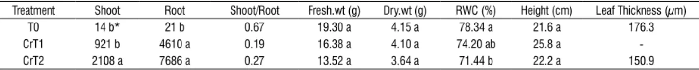
table 2. averaged cr accumulation in shoot and root (in mg kg-1 dry weight), shoot/root ratio, fresh weight, dry weight, relative water content (rWc), height and
cellular width of leaves of plants treated with cr (n=5) at the end of the experiment.
treatment shoot root shoot/root Fresh.wt (g) Dry.wt (g) rWc (%) height (cm) leaf thickness (µm)
t0 14 b* 21 b 0.67 19.30 a 4.15 a 78.34 a 21.6 a 176.3
crt1 921 b 4610 a 0.19 16.38 a 4.10 a 74.20 ab 25.8 a -
crt2 2108 a 7686 a 0.27 13.52 a 3.64 a 71.44 b 22.2 a 150.9
* Means followed by different letters are significantly different at the 0.05 probability level, grouped into classes a, b and c.
the cr concentration is higher in roots than shoots (table 2), hence the shoot/root ratios were less than one for cr-treated plants. even though the cr concentration of shoots and roots of cr treated plants is higher compared to control (t0), the extent of metal accumulation and uptake efficiency of cr was lower than that of as. chlorosis was not visually observed during the treatment process. General effects of cr treatment included stunted growth with an increase in metal concentration. For as-treated plants, the growth rate was not significantly affected.
the averaged fresh weight and dry weight of the plants showed a decrease in ast2 (table 1) and crt2 (table 2) groups compared to control (t0). the rWc of the plants was not significantly different in as-treated plants but showed a significant (p<0.05) decreasing trend with an increase in cr concentration (table 2). the plants heights recorded at the end of the experiment showed no specific trend with accumulation of either metal (table 1 and table 2). the leaf thickness was obtained from the light micrographs decreased for crt2- plants (table 2) while it increased in ast2- plants (table 1).
correlations between the metal accumulation in shoots and roots to the physical and anatomical characters of the plants were analyzed. among the fresh and dry weights of
shoot, only the fresh weight of cr- treated plants showed a significant negative correlation (p<0.05), with an increase in cr accumulation in roots (table 3). the relative Water content (rWc) of the plants showed a significant negative correlation with cr concentration in the roots (p<0.001) and shoots (p<0.05) while no significant correlation was found in as-treated plants (table 3). the leaf thickness was negatively correlated with shoot and root concentration for cr, but only at 0.06 probability level (table 3). No such correlations were observed in as-treated plants.
table 3. correlation coefficients between metal concentrations in shoot and root of plants and physical and anatomical characters: fresh weight, dry weight, relative water content (rWc), height of the plants and cellular width of the leaves.
significant levels: * significant at 0.05 probability level, **significant at 0.001 probability level.
Physical/ anatomical characteristics
as shoot conc.
as root conc.
cr shoot
conc. cr root conc. Fresh. Wt (g) -0.06 -0.17 -0.38 -0.58*
Dry. Wt (g) -0.11 -0.20 -0.17 -0.45
rWc (%) 0.14 0.04 -0.62 * -0.84**
height (cm) 0.31 0.31 -0.07 0.03
structural changes: For as- treated plants, there were no significant changes in the internal structure of leaves compared to the control group (Figure 1a). the ast2-treated leaves appeared healthier with well-developed epidermal and mesophyll cells (Figure 1B) compared to control group in seM micrographs. the cr-treated plants showed significant foliar structural changes compared to the control group (t0). the seM micrographs from the crt2 leaf samples showed reduction in epidermal and palisade cell size and decrease in intercellular spaces and break down of cells (Figure 1c) compared to the control group (Figure 1a).
figure 1. seM micrographs showing transverse section of control (a), ast2- (B) and crt2- (c) treated leaves. No significant changes were observed in ast2 (B) treated leaves compared to control–t0 (a). the crt2- (c) treated leaves showed breakdown of palisade and epidermal cells with an increase in cr concentration.
the teM micrographs of ast2-treated leaves showed well developed chloroplasts with minimum structural changes in both upper and lower palisade cells (Figure 2B) compared to the control group (t0). the only notable structural change observed for leaves of ast2-treated plants, was the loss of the spindle shape in the chloroplasts of the lower mesophyll cells (Figure 2B) compared to the control (Figure 2a).
figure 2. teM micrographs showing the chloroplast of lower palisade parenchyma cells of ast2-(B) treated fern leaves compared to the control-t0 (a). Note the loss of spindle shape (B) in chloroplast with an increase in as concentration compared to control group–t0 (c).
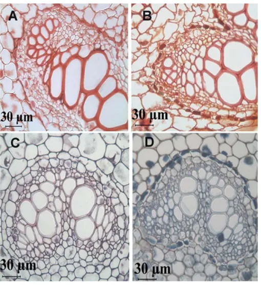
light micrographs of ast2-treated stems (Figure 3B) showed granular as precipitates along the walls of xylem and phloem vessels compared to control (t0) stems (Figure 3a). the transverse sections of the stems were pretreated with cusO4 to precipitate the metal contents. similarly the sections
figure 3. light micrographs showing the transverse section of stem (a, B) and midrib of leaf (c, D). the stems show precipitates of as surrounding the vascular bundles of stems (B) compared to control stems (a). the stems were treated with cusO4 to localize as.the leaf midribs showed areas stained with blue surrounding
the vascular bundles in cr treated leaves (D) compared to control (c). the leaves were treated with cas to localize cr.
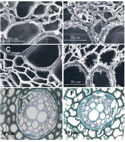
the seM micrographs of stem cross sections of ast2-treated plants (Figure 4B) showed clotted depositions along the walls of xylem and phloem vessels compared to control group (Figure 4a). the depositions were also seen in roots of ast2-treated plants (Figure 4D) but were less intense compared to the stems (Figure 4B). No such depositions were seen in stems and roots of control plants (Figure 5a and Figure 5c). the seM micrographs of stems of crt2- (Figure
figure 4. seM micrographs showing the transverse section of stem (a, B) and root (c, D) of control (t0) and ast2-treated fern plants, respectively. clotted depositions were seen in ast2 treated stems (B) and roots (D) compared to controls (a, c).
significant accumulation of as in shoots and the high shoot/root ratios indicates a high uptake, translocation and accumulation efficiency of brake fern plants for as. in our study, no significant changes in the ferns physical characters, including fresh weight, dry weight, rWc, and plant height were observed for as-accumulating plants. this is consistent with the fact that brake fern is a hyperaccumulator of as, identified by Ma et al. (2001). the only ultrastructural change observed was loss of the spindle shape of chloroplasts in lower palisade cells of leaves for ast2-treated plants. the accumulated as was localized by seM and lM as black depositions along the walls of vascular bundles of stems. similar deposits were also seen in roots, but were less intense. the precipitates suggest a possible mechanism of detoxification of as in stems and roots.
the higher concentration treatment of cr resulted in an increased accumulation in the roots followed by the shoots. the average chromium accumulation in shoots and roots of crt2
treated plants are 2108 and 7686 ppm (mg kg-1 dry weight)
respectively after three weeks of metal treatment (table 2). Our results here indicate that even though brake fern cannot hyperaccumulate cr as it does with as, it can accumulate cr at high concentrations in the stems and roots and hence can be used for phytoremediation of cr-contaminated soils. the decrease in fresh weight and dry weight indicates a decrease in plant growth rate and biomass with cr accumulation. the decrease in rWc (table 2) of the cr accumulating plants and highly significant negative correlation of rWc (table 3) with
both shoot and root (p<0.001) cr concentration, indicates
that cr accumulation resulted in water stress. this is further confirmed by the light micrographs of roots, which show break down of endodermal and cortical cells (Figure 5F). the high cr concentration in roots resulted in cellular break down,
affecting water and metal uptake. Vazquez et al. (1987) and
Barcelo et al. (1986) showed that cr caused severe injury to
bush bean roots resulting in cellular plasmolysis and water stress. the decrease of leaf thickness (table 2) may be due to cr accumulation in leaves or due to cr-induced water stress or the combination of both.
in cr-treated plants, significant amounts of precipitates were seen along the cell walls of the roots. the lM micrographs of roots showed breakdown of endodermal and cortical cells. these changes can be attributed to the intense oxidative action of cr (Vi), which was available to the roots.
the stems showed thickened cell walls and a decrease in the lumen size of xylem and phloem vessels. these effects of cr
(Vi) were similar to as reported by Vazquez et al. (1987) in
bush bean plants and han et al. (2004) in mustard plants.
the decreased vessel size in stems and cellular breakdown of roots may have resulted in the observed low shoot/root ratios and low translocation efficiency of cr. the leaves of cr-treated plants also showed structural changes such as a decrease in intercellular spaces and shrinkage of epidermal, palisade and spongy parenchyma cells.
in our study, the cr-treated plants accumulated significant cr concentrations in both the shoots (upto 2108 mg/kg) and roots (upto 7686 mg/kg) and did not die immediately from phytotoxicity. the cr accumulations reported in this study were higher compared to the cr (Vi) accumulations reported
by su et al. (2005) where brake fern plants accumulated up
to 1,145 mg/kg in shoots and 5,717mg/kg in roots of cr (Vi) treated plants. in the study of su et al. (2005) the brake fern plants were grown on soils added with cr (Vi) but in this study the cr (Vi) was applied in readily available solution form to the plants. this indicates that the brake fern plants have potential to accumulate cr (Vi) at higher concentrations when the cr is made available in the more soluble and readily available form to the plants.
the high translocation efficiency for as shows that brake fern can act as an hyperaccumulator for as. clotted depositions in the vascular bundles of stem and roots in as-treated plants can be suggested as being adaptation and detoxification mechanisms of brake fern. the structural changes in leaves, stems and cellular break down of roots of cr-treated plants indicate that cr has a profound impact and damages the plant physiology. consequently, cr-accumulating brake fern plants exhibit slow growth and decreased biomass. however, as both as and cr occur as co-contaminants in several polluted sites, brake fern plants can be used as potential plant for phytoremediation of both metal contaminants.
for help in sample processing, providing Microtome and other
accessories. this work was supported by funding fromU.s.
Department of energy through cooperative agreement De-Fc26-98Ft-40395.
references
Baker aJM (1981) accumulators and excluders strategies in the response of plants to heavy metals. J. Plant Nutr. 3:643-654.
Barcelo J, Poschenrieder ch, Gunse B (1986) Water relations in chromium Vi treated bush bean plants (Phaseolous vulgaris l. cv. contender) under both normal and water stress conditions. J. exp. Bot. 37:178-187.
Barcelo J, Vazquez MD, Poschenrieder ch (1988) cadmium induced structural and ultrastructural changes in the vascular system of bush bean stems. Bot. acta. 101:254-261.
Bartlett rJ (1991) chromium cycling in soils and water: links and methods. environ. health Perspect. 92:17-29.
Blaylock MJ, huang JW (2000) Phytoextraction of metals. in raskin i, ensley BD, eds. Phytoremediation of toxic metals: using plants to clean the environment. John Wiley & sons. inc., New York. pp. 53-71.
han FX, Banin a, su Y, Monts Dl, Plodinec MJ, Kingery Wl, triplett GB (2002) industrial age anthropogenic inputs of heavy metals into the pedosphere. Naturwissenschaften. 89: 497-504.
han FX, Banin a (1997) long-term transformations and redistribution of potentially toxic heavy metals in arid zone soils. i. under saturated conditions. Water air soil Pollut. 95: 399-423.
han FX, sridhar BBM, Monts Dl, su Y (2004) Phytoavailability and toxicity of trivalent and hexavalent chromium to Brassica juncea l. czern. New Phytol. 169:489-499
han FX, su Y, Monts Dl, Plodinec MJ, Banin a, triplett GB (2003) assessment of global industrial-age anthropogenic arsenic contamination. Naturwissenschaften. 90:395-401.
Jackson Ml (1958) soil chemical analysis. Prentice hall, New Jersey. Johansen Da (1940) Plant Microtechniques. McGraw-hill, New York. losi Me, amrhein c, Frankenberger Wt (1994) Factors affecting chemical and biological reduction of cr (Vi) in soil. environ. toxicol. chem. 13:1727-1735.
lytle cM, lytle FW, Yang N, Qian J, hansen D, Zayed a, terry N (1998) reduction of cr (Vi) to cr (iii) by wetland plants: potential for in situ heavy metal detoxification. environ. sci. technol. 32:3087-3097.
Ma lQ, Komar KM, tu c, Zhang W, cai Y, Kenelley eD (2001) a fern that hyperccumulates arsenic, Nature. 409:579.
Mei B, Puryear JD, Newton rJ (2002) assessment of cr tolerance and accumulation in selected plant species. Plant soil. 247:223-231.
Pearse aGe (1972) histochemsitry theoretical and applied. 3rd edn. Baltimore:
Williams and Wilkins.
Qiu Jh, Zayed a, Zhu Yl, Yu M, terry N (1999) Phytoaccumulation of trace elements by wetland plants:iii uptake and accumulationof ten trace elements by twelve plant species. J. environ. Qual. 28:1448-1455.
raskin i, ensley BD (2000) Phytoremediation of toxic metals: using plants to clean the environment. John Wiley & sons. inc., New York.
reeves rD, Baker aJM (1984) studies on metal uptake by plants from serpentine and non-serpentine populations of thlaspi goesingense halacsy (cruciferae). New Phytol. 98:191-204.
salt De, Blaylock M, Kumar NPBa, Dushenkov V, ensley BD, chet i, raskin i (1995) Phytoremediation: a novel strategy for the removal of toxic metals from the environment using plants. Biotechnology. 13:468-474.
salt De, Kramer U (2000) Mechanisms of metal hyperaccumulation in plants. in raskin i, ensley BD, eds. Phytoremediation of toxic metals: using plants to clean the environment. New York: John Wiley & sons. inc. 231-247. salt De, smith rD, raskin i (1998) Phytoremediation. ann. rev. Plant Physiol. Plant Mol. Biol. 49:643-668.
sass Je (1958) Botanical microtechniques. iowa st. Univ Press, ames. shahandeh h, hossner lr (2000) Plant screening for chromium phytoremediation. int. J. Phytorem. 2:31-51.
sridhar BBM, han FX, Diehl sV, Monts Dl, su Y (2007a) effects of Zn and cd accumulation on structural and physiological characteristics of barley plants. Braz. J. Plant Physiol. 19: 15-22.
sridhar BBM, han FX, Diehl sV, Monts Dl, su Y (2007b) Monitoring the effects of arsenic- and chromium- accumulation in chinese brake fern (Pteris vittata) using microscopy and near infrared spectral reflectance. int. J. of remote sens 28: 1055-1067.
su Y, han FX, sridhar BBM, Monts Dl (2005) Phytotoxicity and phytoaccumulation of trivalent and hexavalent chromium in brake fern . environ toxicol chem 24: 2019-2026.
suzuki t, sumi Y, Miyazaki K, Muraki t, Nokubi K, Kimura M, Kato M (1978) histochemical staining of chromium by azurol s. acta histochemica et cytochemica. 11:46-51.
Vazquez MD, Poschenreider c, Barcelo J (1987) chromium Vi induced structural and ultrastuctural changes in bush bean (Phaseolus vulgaris l.). ann. Bot. 59:427-438
Witmer cM, harris r, shupak si (1991) Oral bioavailability of chromium from a specific site. environ. health Perspect. 92:105-110.
Zayed a, lytle cM, Qian J, terry N (1998) chromium accumulation, translocation and chemical speciation in vegetable crops, Planta. 206:293-299.