268
Comparative Analysis of Intimal Hyperplasia After
Sirolimus-Eluting Stent and Thin-Strut Bare-Metal
Stent Implantation in Small Coronary Arteries
Fernando Stuchi Devito, Amanda G. M. R. Sousa, Fausto Feres, Alexandre Antonio Cunha Abizaid,
Rodolfo Staico, Luis Alberto Piva Mattos, Luis Fernando Leite Tanajura, Andréa C. L. S. Abizaid,
Áurea Jacob Chaves, J. Eduardo M. R. Sousa
Instituto Dante Pazzanese de Cardiologia - São Paulo, SP
M a i l i n g A d d r e s s : F e r n a n d o S t u c h i D e v i t o • P r a ç a 9 d e J u l h o , 1 7 6 / 1 1 1 – 1 5 8 0 0 - 0 0 0 – C a t a n d u v a , S P - B r a z i l E-mail: [email protected] Received on 03/17/05 • Accepted on 05/18/05
O
BJECTIVEThis study aimed at evaluating reduction in intimal hyperplasia volume following angioplasty using sirolimus-eluting stents (Cypher™) compared with thin-strut bare-metal stents (Pixel™) in patients with small vessels.
M
ETHODSEighty patients with coronary artery disease were prospectively included in two consecutive series, the fi rst using sirolimus-eluting stents (50) and the second using bare-metal stents (30).
R
ESULTSThe use of sirolimus-eluting stents reduced: in-stent net volume obstruction [5.0% (SE = 0.77) x 39.0% (SE = 4.72), p < 0.001], in-stent late loss [0.25 mm (SE = 0.03) x 1,11 mm (SE = 0.13), p < 0,001], in-segment late loss [0.30 mm (SE = 0.04) x 0.83 mm (SE = 0.11), p < 0.001], in-stent restenosis (0% x 33.3%, p < 0.001) and in-segment restenosis (4% x 36.7%, p < 0.001). The event-free survival rate was 96% in the sirolimus-eluting stent group versus 86.7% in the bare-metal stent group (BMS) (p = 0.190).
C
ONCLUSIONSirolimus-eluting stents are superior to thin-strut bare-metal stents in reducing intimal hyperplasia (less in-stent obstruction and less late lumen loss) in patients with small vessels. The use of these stents signifi cantly reduced angiographic restenosis at eight months.
K
EY WORDS269
Treatment of patients with coronary artery disease (CAD) in small vessels currently accounts for up to 40%
of all percutaneous revascularizations1,2. Restenosis
may be as high as 50% after bare-metal stenting in small vessels, depending on lesion length and on the
presence of diabetes mellitus3-9. Intimal hyperplasia
is the primary mechanism of in-stent restenosis, and therapies focused on inhibiting neointimal growth should
reduce restenosis10,11.
In the FIM and RAVEL trials, the use of sirolimus-eluting stents in patients with vessels larger than 3.0 mm, compared with bare-metal stents, resulted in marked
reduction in intimal hyperplasia12,14. In the SIRIUS trial,
angiographic restenosis was reduced from 36.3% after bare-metal stenting to 8.9% after sirolimus-eluting stenting (p < 0.001). However, in a subanalysis of this same trial, restenosis after sirolimus-eluting stenting in vessels with mean reference diameter of 2.29 mm was higher (18.4%) when compared to large vessels (1.9%), calling into question the role of these endoprostheses in
patients with small-vessel disease15.
At the same time, stents have been structurally modifi ed to become thinner, and thin-strut stents were assessed in the ISAR-STEREO-2 trial, the results of which
were decreased intimal hyperplasia16,17,18.One study by
Garcia et al evaluating these stents in patients with small vessels reported 19.3% of restenosis and only 4.1% of target-vessel revascularization. Since then, these thin-strut stents have been considered the best option among
uncoated stents for the treatment of small vessels19,20 .
In this study, we compared intimal hyperplasia in small vessels (reference diameter < 2.75 mm) following either sirolimus-eluting or bare-metal stent implantation based on intimal hyperplasia volume measured by intravascular ultrasound.
M
ETHODSFrom December 2002 to December 2003, patients with established diagnosis of coronary artery disease and candidates for elective percutaneous coronary intervention (PCI) were prospectively included in this study according to the following criteria: age equal to or older than 18 years; diagnosis of stable angina defi ned by the Canadian Society Classifi cation (CSC I, II, III or IV), silent ischemia, unstable angina (Braunwald classifi cation IB, IC, IIB, IIC), or myocardial infarction > seven days; reference vessel diameter between 2.20 mm and 2.75 mm (quantitative
coronary angiography); de novo native coronary artery
lesion with target-lesion stenosis between 50% and 99%
and ≤ 30 mm in length. Exclusion criteria were: presence
of cardiogenic shock; serum creatinine > 2.0 mg/dL; peripheral vascular disease; known hypersensitivity or contraindication to heparin, acetylsalicylic acid (ASA), ticlopidine or clopidogrel; end-stage diseases associated with limited life expectancy (less than a year); left
ventricular ejection fraction ≤ 30%; excessive target-vessel
tortuosity, making intracoronary ultrasound diffi cult to
perform; target-lesion ≥ 50% in unprotected left main
coronary artery; ostial lesions; target-lesions located at a
bifurcation involving side branches ≥ 2.0 mm in diameter,
and the presence of thrombus at the target site. The study protocol was approved by the Institutional Research Ethics Committee of the Hospital.
Patients were nonrandomly assigned to the trial and divided into two sequential treatment groups, the fi rst using the Cypher™ stent (coated with sirolimus) and the other using the Multilink Rx Pixel™ stent (uncoated). According to the standard technique, aspirin 200 mg/day was administered at least 24 hours prior to intervention and a thienopyridine (ticlopidine or clopidogrel), during two months. Ticlopidine dosage was 250 mg twice daily (started at least 48 hours prior to intervention). Clopidogrel was given in a loading dose of 300 mg followed by and 75 mg daily (at least 24 hours prior to intervention). Unfractionated heparin was administered by intravenous bolus of 100 IU/kg. The use of both glycoprotein IIb/IIIa inhibitor and predilation was left to the operators’ discretion. Predilation balloons should be undersized at least 0.5 mm to the reference vessel diameter, in addition to being shorter than the chosen stent. Postdilation balloons (pressure > 12 atm) also should be shorter than the implanted stent. When multiple stents were required, they had to overlap by 3 or 4 mm to prevent gaps between the endoprostheses. At the end of the procedure, all patients underwent ultrasound
evaluation. Outpatient visits following discharge were
made one, three, six, and eight months after PCI, when other coronary angiography and ultrasound examinations were scheduled.
As for the endovascular prostheses used, the Cypher™ stent (with sirolimus) manufactured by Cordis, Johnson & Johnson, is made of 316L stainless steel coated with a 50/50 combination of two non-erodible polymers, polyethylene-co-vinyl acetate (PEVA) and poly n-butyl methacrylate (PBMA), mixed with sirolimus. A topcoat of PBMA polymer is applied to the stent surface. All stents
contain 148 µg of sirolimus per cm2 of metal surface area
and, in this formulation, the drug is released gradually over a prolonged period (95% is released up to 28 days after implantation). The Multilink Rx Pixel™, manufactured
by Guidant™, is a stainless-steel, uncoated,
balloon-expandable stent especially designed for small vessels. This thin-strut stent (0.05 mm) is composed of only fi ve rings, allowing complete circumferential coverage with less metal.
270
and in-lesion segments (in-stent plus the 5 mm proximal and distal to the endoprosthesis) at the following time points: preintervention, postintervention, and at follow-up coronary angiography.
Ultrasound assessment was performed using the Clear View™ (CVIS, Boston Scientific Corporation) and a 40-MHz catheter. Using an automated pullback system of the ultrasound catheter at 0.5 mm/s, image acquisition was continuous, starting at least 10 mm distal to the target lesion and advancing up to the aorto-ostial junction. The acquired images were analyzed by the echoPlaque™ 2 software (Indec Systems Inc. U.S). Angiographic and ultrasound images were analyzed at the core lab of the Department of Invasive Cardiology of Institute Dante Pazzanese of Cardiology. Using Simpson’s formula, stent, lumen and intimal hyperplasia
volumes were calculated12.As this study involved stents
of different lengths, these volumes were calculated in
absolute numbers (mm3 per patient) and correlated
to the mean area for each variable (mm2). In-stent
volume obstructionwas calculated as the ratio between
intimal hyperplasia volume and stent volume x 100. Incomplete stent apposition was evaluated and defi ned by the separation of one or more stent strut from the vessel wall with evidence of blood fl ow between the stent and the vessel wall (seen as blood speckles on the ultrasound image). When more than one incomplete stent apposition was detected, the circumferential angle of malapposition was reported. The degree of malapposition was also described and calculated by the sum of distances between
the areas with malapposition14.
In-stent and in-segment restenosis were defi ned by the classic binary criterion (stenosis > 50% at follow-up angiography). Success was defi ned by postprocedural residual stenosis lower than 10%, in the absence of major complications (death, infarction, and emergency surgery).
The primary endpoint was to evaluate, at the term of eight months, the in-stent volume obstruction occupied by intimal hyperplasia. Secondary endpoints included: In-stent and in-segment MLD and late loss, binary restenosis, and incidence of major adverse cardiac events (death, infarction, and target-lesion revascularization).
Calculationof sample size was based on the assumption
of 40% an in-stent volumeobstruction for bare-metal
stents and of only 10% for sirolimus-eluting stents (SD =
30), that is, a relative reduction of 75%21,22. With a
two-tailed hypothesis test, alpha level of 0.05 and statistical power of 90%, 21 patients would be necessary in each group for the primary endpoint to be demonstrated. Considering possible losses and attempting to include the higher number of patients necessary to achieve better distribution of clinical, angiographic, and IVUS variables, we planned to enroll 30 patients in each group. Owing to the availability of fi fty patients who were being treated with sirolimus-eluting stents at that time in the institution,
the fi rst fi fty patients were sequentially treated with these endoprostheses (sirolimus-eluting stents), and the other patients were treated with bare-metal stents.
Categorical variables were expressed as absolute and relative frequencies (percentages), and quantitative variables, as means and standard errors. Pearson’s chi-square test or Fisher’s exact test was used to compare both groups’ categorical variables, when necessary. Student’s t test for independent samples was used to evaluate quantitative variables in a single time point. Quantitative variables measured at three time points (before, immediately after the procedure and at eight months) were determined by Generalized Linear Models with repeat measurements. Two aspects were considered in these models: group (sirolimus-eluting stents and bare-metal stents) and time point (pre-, post-, and late), and comparisons were made between groups (sirolimus-eluting stents vs. bare-metal stents at each time point) and in each group (pre- x post- x late for each group). Statistical analyses were performed using SPSS version 11.0 and SAS version 8.01. Signifi cance level was set at 0.05.
R
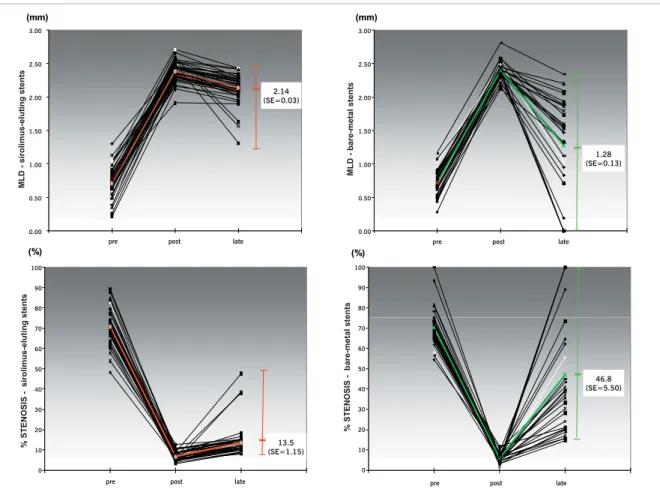
ESULTSEighty patients were included in this study: the fi rst fi fty were sequentially treated with sirolimus-eluting stent (Cypher™) and the last thirty, with Multilink Rx Pixel™ (uncoated). Clinical characteristics of patients in both treatment groups are shown in Table 1, angiographic characteristics in Table 2, as well as the technical variables of procedure in Table 3. During follow-up, 94% of the patients underwent angiographic evaluation. Results of in-stent quantitative coronary angiography (QCA) are shown in Table 4. Mean reference vessel diameter [2.44 mm (SE = 0.02) x 2.37 mm (SE = 0.03), p = 0.075] and mean length of treated lesions [13.75 mm (SE = 0.92) x 12.87 mm (SE = 0.53), p = 0.498] were similar in both groups. Mean minimal lumen diameter did not differ between groups, either in the preprocedural (p = 0.926) or in the postprocedural fi ndings (p = 0.952), but at eight months the vessels treated with sirolimus-eluting stents had increased MLD [2.14 mm (SE = 0.03) x 1.28 mm (SE = 0.13), p < 0.001]. This was due to the late lumen loss, which was signifi cantly lower in the sirolimus group [0.25 mm (SE = 0.03) x 1.11 mm (SE = 0.13), p < 0.001].
271
lumen loss with sirolimus-eluting stents than with bare-metal stents [0.30 mm (SE = 0.04) x 0.83 mm (SE = 0.11), p < 0.001, respectively], in-segment restenosis being signifi cantly lower in the sirolimus arm [4% x 36.7%, (p < 0.001)].
Eighty per cent of the patients underwent intravascular ultrasound examination. Intravascular ultrasound analysis showed that mean intimal hyperplasia volume was 5.0
mm3 (SE = 0.77) in the sirolimus-stent group versus 27.5
mm3 (SE = 3.60) in the BMS-group (p < 0.001). Mean
intimal hyperplasia cross-sectional area was smaller in the sirolimus-stent group compared with the BMS-group
[0.24 mm2 (SE = 0.03) x 1.62 mm2 (SE = 0.19), p <
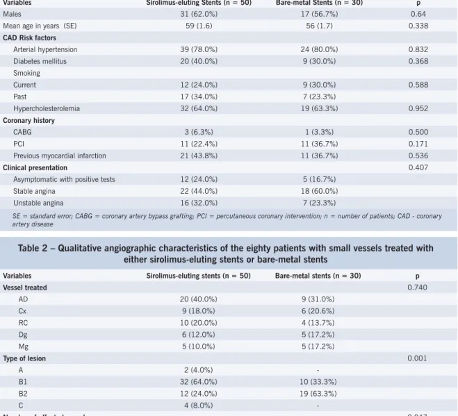
Table 1 – Key clinical characteristics of the eighty patients with small vessels treated with either sirolimus-eluting stents or bare-metal stents
Variables Sirolimus-eluting Stents (n = 50) Bare-metal Stents (n = 30) p
Males 31 (62.0%) 17 (56.7%) 0.64
Mean age in years (SE) 59 (1.6) 56 (1.7) 0.338
CAD Risk factors
Arterial hypertension 39 (78.0%) 24 (80.0%) 0.832
Diabetes mellitus 20 (40.0%) 9 (30.0%) 0.368
Smoking
Current 12 (24.0%) 9 (30.0%) 0.588
Past 17 (34.0%) 7 (23.3%)
Hypercholesterolemia 32 (64.0%) 19 (63.3%) 0.952
Coronary history
CABG 3 (6.3%) 1 (3.3%) 0.500
PCI 11 (22.4%) 11 (36.7%) 0.171
Previous myocardial infarction 21 (43.8%) 11 (36.7%) 0.536
Clinical presentation 0.407
Asymptomatic with positive tests 12 (24.0%) 5 (16.7%)
Stable angina 22 (44.0%) 18 (60.0%)
Unstable angina 16 (32.0%) 7 (23.3%)
SE = standard error; CABG = coronary artery bypass grafting; PCI = percutaneous coronary intervention; n = number of patients; CAD - coronary artery disease
0.001], as was mean in-stent volume obstruction [5% (SE = 0.77) x 39% (SE = 4.72), p < 0.001]. No stent malapposition was found in either group.
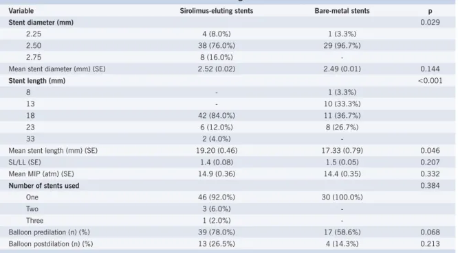
Stent implantation was successful in all the patients, without major in-hospital complications (death, myocardial infarction, or emergency revascularization surgery). No patient in this study was given glycoprotein IIb/IIIa inhibitor. Moreover, there were no in-stent thromboses, nonfatal myocardial infarctions nor deaths, either at 30 days or at eight months. All patients were clinically evaluated during this period, and two patients (4%) treated with sirolimus-eluting stents required repeat target-lesion revascularization. In these cases, additional Table 2 – Qualitative angiographic characteristics of the eighty patients with small vessels treated with
either sirolimus-eluting stents or bare-metal stents
Variables Sirolimus-eluting stents (n = 50) Bare-metal stents (n = 30) p
Vessel treated 0.740
AD 20 (40.0%) 9 (31.0%)
Cx 9 (18.0%) 6 (20.6%)
RC 10 (20.0%) 4 (13.7%)
Dg 6 (12.0%) 5 (17.2%)
Mg 5 (10.0%) 5 (17.2%)
Type of lesion 0.001
A 2 (4.0%)
-B1 32 (64.0%) 10 (33.3%)
B2 12 (24.0%) 19 (63.3%)
C 4 (8.0%)
-Number of affected vessels 0.047
One 38 (76.0%) 15 (51.7%)
Two 11 (22.0%) 11 (37.9%)
Three 1 (2.0%) 3 (0.3%)
272
angioplasty was performed with the implantation of another sirolimus-eluting stent. Six patients in the BMS-group experienced myocardial ischemia symptoms (20.0%), and four (13.3%) required repeat target-lesion revascularization (TLR): two balloon angioplasty and two coronary artery bypass surgery (CABG). Thus, although event-free survival and TLR-free survival were lower in the sirolimus-stent group when compared with the BMS-group, no statistically signifi cant difference was found regarding these clinical outcomes in either group (96% x 86.7%; p = 0.190).
D
ISCUSSIONThis study showed that sirolimus-eluting stenting in patients with small vessels is associated with a lesser degree of intimal hyperplasia compared with the reparative response following bare-metal stenting, and thus results in a lower rate of in-stent and in-segment restenosis. Clinical variables in this study characterized groups of patients with at least moderate complexity for percutaneous coronary intervention. Among all characteristics, it is worth noting the high prevalence Table 3 – Clinical characteristics of procedures performed in the eighty patients with small vessels
treated with either sirolimus-eluting stents or bare-metal stents
Variable Sirolimus-eluting stents Bare-metal stents p
Stent diameter (mm) 0.029
2.25 4 (8.0%) 1 (3.3%)
2.50 38 (76.0%) 29 (96.7%)
2.75 8 (16.0%)
-Mean stent diameter (mm) (SE) 2.52 (0.02) 2.49 (0.01) 0.144
Stent length (mm) <0.001
8 - 1 (3.3%)
13 - 10 (33.3%)
18 42 (84.0%) 11 (36.7%)
23 6 (12.0%) 8 (26.7%)
33 2 (4.0%)
-Mean stent length (mm) (SE) 19.20 (0.46) 17.33 (0.79) 0.046
SL/LL (SE) 1.4 (0.08) 1.5 (0.05) 0.207
Mean MIP (atm) (SE) 14.9 (0.36) 14.4 (0.35) 0.332
Number of stents used 0.384
One 46 (92.0%) 30 (100.0%)
Two 3 (6.0%)
Three 1 (2.0%)
-Balloon predilation (n) (%) 39 (78.0%) 17 (58.6%) 0.068
Balloon postdilation (n) (%) 13 (26.5%) 4 (14.3%) 0.213
SE = standard error; SL/LL = stent length–to–lesion length ratio; MIP = maximal stent implantation pressure; n = number
Table 4 – Variables of in-stent and in-segment quantitative coronary angiography before, immediately after the procedure, and at eight months
Sirolimus-eluting stents Bare-metal stents p
RD (mm) (SE) 2.44 (0.02) 2.37 (0.03) 0.075
Lesion length (mm) (SE) 13.75 (0.92) 12.87 (0.52) 0.498
MLD (mm) (SE)
Preprocedural* 0.71 (0.03) 0.72 (0.04) 0.926
Postprocedural* 2.38 (0.02) 2.39 (0.02) 0.952
At eight months 2.14 (0.03) 1.28 (0.13) <0.001
Stenosis rate (%) (SE)
Preprocedural* 70.64(1.09) 70.57(1.74) 0.980
Postprocedural* 6.78 (0.30) 5.86 (0.30) 0.747
At eight months 13.51(1.15) 46.82 (5.50) <0.001
Acute gain (mm) (SE) 1.67(0.03) 1.69(0.03) 0.661
In-stent late loss (mm) (SE) 0.25 (0.03) 1.11 (0.13) <0.001
In-segment late loss (mm) (SE) 0.30 (0.04) 0.83 (0.11) <0.001
In-stent restenosis (n) (%) 0 10 (33.3) <0.001
In-segment restenosis (n) (%) 2 (4) 11 (36.7) <0.001
273
of diabetes mellitus: 40% in the sirolimus-eluting stent group and 30% in the BMS-group (p = 0.368). In most randomized trials comparing ballooning versus stenting in patients with small vessels, with the exception of the
CHIVAS23 and RAP24 trials, the prevalence of diabetes was
lower than 30%, ranging from 12% and 20%. Even in the
era of sirolimus-eluting stents, the SVELT25 was the trial
that included the highest number of diabetics (26.7%).
In the RAVEL14, SIRIUS, C-SIRIUS26, E-SIRIUS27 and
SES-SMART28 trials, this subgroup accounted for 16%,
25%, 24%, 23%, and 19.4%, respectively.
The most relevant IVUS fi nding was the benefi cial mechanism involved with sirolimus-eluting stent compared with bare-metal stent, that is, inhibition (in 87%) of excessive intimal hyperplasia evaluated by in-stent volume obstruction. This fi nding of marked decrease in in-stent intimal hyperplasia has been consistent in all sirolimus-eluting stent trials that used IVUS as the evaluation tool. Mean intimal hyperplasia
cross-sectional area obtained in this investigation (0.24 mm2)
was similar to that observed in the SVELT25 trial (0.08
mm2), as well as in trials of sirolimus-eluting stents in
larger vessels, such as the RAVEL14 and the SIRIUS15
(0.11 mm2 and 0.50 mm2, respectively), meaning that
intimal hyperplasia inhibition with these stents does not depend on vessel diameter. However, mean intimal hyperplasia associated with the thin-strut, bare-metal
stents used in this study (Pixel™) was 1.62 mm2, lower
than that found in the DANTE29 trial (3.05 mm2) and in
the respective arms of the RAVEL14 and SIRIUS15 trials
(2.05 mm2 and 2.70 mm2, respectively), which used a
thicker strut model.
Moreover, no incomplete apposition of stent struts was found in either group. Unlike in large vessels, stent placement in small vessels is performed with a higher stent-to-vessel diameter ratio, favoring stent strut impaction in the atheromatous plaque. Therefore, incomplete stent apposition is usually less frequent in
small vessels. In the RAVEL14 trial, for example, mean
vessel diameter in ten patients with incomplete stent apposition was 3.16 mm (SD = 0.57), whereas that of 38 patients with well-apposed stents was 2.79 mm (SD = 0.43), (p < 0.05).
In the last decade, recommendations for bare-metal stenting emphasized the need to obtain as much lumen as possible to accommodate hyperplasia and, thus, reduce restenosis. However, in the investigation of drug-eluting stents showing signifi cant decrease in-stent
Fig. 1 - Angiographic measurement variations (MLD above and stenosis rate below) preprocedural, postprocedural, and at eight months in both treatment groups (sirolimus-eluting stents on the left and bare-metal stents on the right). Angiographic measurements are quite similar for both groups, both in preprocedural and postprocedural phases, and are more homogeneous in the sirolimus-stent group compared to the BMS-group, especially at late evolution. MLD = minimal lumen diameter
0 10 20 30 40 50 60 70 80 90 100
pré pós tardio
% DE ESTENOSE stents com sirolimus
0 10 20 30 40 50 60 70 80 90 100
pré pós tardio
% DE ESTENOSE - stents não recobertos
0,00 0,50 1,00 1,50 2,00 2,50 3,00
pré pós tardio
DML stents não recobertos
0,00 0,50 1,00 1,50 2,00 2,50 3,00
pré pós tardio
DML - stents com sirolimus
(%)
(%) (%)(%)
(mm)
(mm) (mm)(mm)
2,14 2,14 (EP=0,03) (EP=0,03) 1,28 1,28 (EP=0,13) (EP=0,13) 13,5 13,5 (EP=1,15) (EP=1,15) 46,8 46,8 (EP=5,50) (EP=5,50)
MLD - sirolimus-eluting stents
3.00 2.50 2.00 1.50 1.00 0.50 0.00
pre post late
2.14 (SE=0.03)
pre post late
pre post late
pre post late 3.00 2.50 2.00 1.50 1.00 0.50 0.00
MLD - bare-metal stents
1.28 (SE=0.13) 100 90 80 70 60 50 40 30 20 10 0
% STENOSIS - sirolimus-eluting stents 13.5
(SE=1.15) 100 90 80 70 60 50 40 30 20 10 0
% STENOSIS - bare-metal stents
274
intimal hyperplasia, as in the SIRIUS15 trial, the fi nding
of border effects attracted special attention, because they accounted for the recurrences in this study. Based on this new perspective, additional technical observations were implemented to guide stent implantation.
One of these aspects is lesion coverage, because an incomplete metallic coverage in sirolimus-eluting stent implantation may affect late results. In this study, stent length–to–lesion length ratio was 1.4, and in the
C-SIRIUS26 and E-SIRIUS27 trials was even higher (1.7
and 1.8, respectively), as well as in the RAVEL14 trial,
in which no follow-up restenosis was found during the fi rst year. Although lesions were more complex in the
C-SIRIUS26 trial than those randomized in the RAVEL14 trial,
requiring more than one stent per lesion in 48% of the cases (67% of which with overlapping stents), restenosis rate was very low (0% in-stent and 2.3% in-segment), demonstrating that longer stents are better, even when more than one stent is required. In this case, this should be done with a 2- or 3 mm overlap, to prevent target lesion ends from being exposed to balloon trauma and, thus, to intimal proliferation.
Among the limitations of this study, the lack of rando-mization should be noted. Logistics regarding limited availability of sirolimus-eluting stents prevented inclusion for a one-year period anticipated by the study design. Nevertheless, it must be emphasized that both treatment groups were quite homogeneous with respect to all variables related to lesion recurrence and thus minimized this aspect. In addition, the way patients were sequentially
included and also the stents’ structural differences prevented a blind analysis of results. However, result analysis by quantitative angiography and intravascular ultrasound, with accurate and objective measurements, performed at a laboratory with large experience in this fi eld, minimizes this aspect.
In view of these fi ndings, we believe that treatment of vessels with reference diameter lower than 2.75 mm changed dramatically with the advent of sirolimus-eluting stents. Moreover, in the near future, as the costs of endoprostheses decrease, the use of sirolimus-eluting stents in patients with small vessels may be even more cost-effective compared with bare-metal stents, owing to a reduction in target-lesion revascularization, hospital admissions, and drug prescriptions.
Therefore, we can conclude that sirolimus-eluting stenting in patients with small vessels leads to reduced intimal hyperplasia, as evaluated by intravascular ultrasound, compared with thin-strut, bare-metal stenting. Angiographic measurements related to late results (degree of stenosis, late lumen loss, and restenosis) are significantly lower after sirolimus-eluting stent implantation. Late minimal lumen diameter is greater following sirolimus-eluting stenting compared with bare-metal stenting. Target-lesion revascularization is about 10% lower in patients treated with sirolimus-eluting stents, even though this difference was not statistically signifi cant in the present study.
No potential confl ict of interest relevant to this article was reported.
R
EFERENCES1. Sociedade Latino-americana de Cardiologia Intervencionista [homepage]. Informe 1988-2002 sobre produtividade em Cardiologia na América Latina. [citado em 8 de fevereiro de 2005]. Disponível em: http://www.solaci.org.
2. Sociedade Brasileira de Hemodinâmica e Cardiologia Intervencionista [homepage]. Central Nacional de Intervenções Cardiovasculares (CENIC): Estatística - Balão vs. stent. [citado em 16 de mar 2004]. Disponível em: http://www.sbhci.org.br.
3. Akiyama T, Moussa I, Reimers B et al. Angiographic and clinical outcome following coronary stenting of small vessels: a comparison with coronary stenting of large vessels. J Am Coll Cardiol 1998; 32: 1610–8.
4. Elezi S, Kastrati A, Neumann FJ et al. Vessel size and long-term outcome after coronary stent placement. Circulation 1998; 98: 1875-80.
5. Al Suwaidi J, Yeh W, Williams DO et al. Comparison of immediate and one-year outcome after coronary angioplasty of narrowing <3 mm with those > or =3 mm (the National Heart, Lung, and Blood Institute Dynamic Registry). Am J Cardiol 2001; 87: 680-6.
6. Briguori C, Nishida T, Adamian M et al. Coronary stenting versus balloon angioplasty in small coronary artery with complex lesions. Catheter Cardiovasc Interv 2000; 50: 390-7.
7. Suselbeck T, Latsch A, Siri H et al. Role of vessel size as a predictor for the occurrence of in-stent restenosis in patients with diabetes mellitus. Am J Cardiol 2001; 88: 243-7.
8. Moreno R, Fernandez C, Alfonso F et al. Coronary stenting versus ballon angioplasty in small vessels. J Am Coll Cardiol 2004; 43: 1964-72.
9. Ho KKL, Sernechia C, Rodriguez O et al. Predictors of angiographic restenosis after stenting: pooled analysis of 1197 patient with protocol-mandated angiographic follow-up from 5 randomized trials. Circulation 1998; 98 (Suppl I): I-362.
10. Mintz GS, Kent KM, Pichard AD et al. Intravascular ultrasound insights into mechanisms of stenosis formation and restenosis. Cardiol Clin 1997; 15: 17-29.
11. Nikol S, Huehns TY, Hofl ing B. Molecular biology and post-angioplasty restenosis. Atherosclerosis1996; 123: 17-31.
12. Sousa JE, Costa MA, Abizaid AC et al. Sustained suppression of neointimal proliferation by sirolimus-eluting stents: one-year angiographic and intravascular ultrasound follow-up. Circulation 2001; 104: 2007-11.
13. Sousa E. FIM 4 years. Presented to TCT 2004. [cited feb 5 2005]. Avaiable from: http://www.tctmd.com/expert-presentations/ slides.html.
14. Morice MC, Serruys PW, Sousa JE et al. RAVEL Study Group. A randomized comparison of a sirolimus-eluting stent with a standard stent for coronary revascularization. N Engl J Med 2002; 346: 1773-80.
15. Moses JW, Leon MB, Popma JJ et al, SIRIUS Investigators. Sirolimus-eluting stents versus standard stents in patients with stenosis in a native coronary artery. N Engl J Med 2003; 349: 1315-23.
275
17. Kastrati A, Mehilli J, Dirschinger J et al. Intracoronary stenting andangiographic results: strut thickness effect on restenosis outcome (ISAR-STEREO) trial. Circulation2001; 103: 2816-21.
18. Pache J, Kastrati A, Mehilli J et al. Intracoronary stenting and angiographic results: strut thickness effect on restenosis outcome (ISAR-STEREO-2) trial. J Am Coll Cardiol 2003; 41: 1283-8.
19. Multi-Link Pixel TM Registry – Six Months Results. 2005 [cited feb 8 2005]. Available from: http://www.tctmd.com/images/Pixel_WP.
20. Garcia E. Randomized Trial of Direct Stenting With the Pixel Stent in Small Vessels. Transcatheter Cardiovascular Therapeutics (TCT) 2003. [cited feb 5, 2005]. Avaiable from: http://www.tctmd.com/expert-presentations/slides.html.
21. Acute platelet inhibition with abciximab does not reduce in-stent restenosis (ERASER study). The ERASER Investigators. Circulation 1999; 100: 799-806.
22. Dussaillant GR, Mintz GS, Pichard AD et al. Small stent size and intimal hiperplasia contribute to restenosis: a volumetric intravascular ultrasound analysis. J Am Coll Cardiol 1995; 26: 720-4.
23. Muramatsu T, Iwasaki K, Inoue Nl. Coronary heart disease stenting in small vessels versus balloon angioplasty study (CHIVAS): a randomized prospective multicenter trial (abstr). J Am Coll Cardiol 2002; 39 (Suppl A): 50A.
24. Garcia E, Gomez-Recio M, Moreno R. Stent reduces restenosis in small vessels: results of the RAP study (abstr). J Am Coll Cardiol 2001; 37 (Suppl. A): 17A.
25. Souza E, for the SVELT investigators. Transcatheter Cardiovascular Therapeutics (TCT) 2004. [cited feb 5, 2005]. Avaiable from: http:// www.tctmd.com/expert-presentations/slides.html.
26. Schampaert E, Cohen EA, Schluter M et al. C-SIRIUS Investigators. The canadian study of the sirolimus-eluting stent in the treatment of patients with long de novo lesions in small native coronary arteries (C-SIRIUS). J Am Coll Cardiol2004; 43: 1110 -5.
27. Schofer J, Schluter M, Gershlick AH et al. E-SIRIUS Investigators. Sirolimus-eluting stents for treatment of patients with long atherosclerotic lesions in small coronary arteries: double-blind, randomised controlled trial (E-SIRIUS). Lancet2003; 362: 1093-99.
28. Ardissino D, Cavallini C, Bramucci E et al. SES-SMART Investigators. Sirolimus-Eluting vs uncoated Stents for Prevention of Restenosis in Small Coronary Arteries. JAMA2004; 292: 2727-34.