Late Outcome of Sirolimus-Eluting Stents: Comparison of
Multidetector Computed Tomography with Quantitative Coronary
Angiography and Intravascular Ultrasound
Ibraim Masciarelli F. Pinto, Amanda G. M. R. Sousa, Walther Ishikama, Luiz Alberto Mattos, Alexandre Abizaid, Fausto
Feres, Luiz Fernando L. Tanajura, Luiz C. B. Sousa, J. Eduardo M. R. Sousa, Adib Jatene
Instituto Dante Pazzanese de Cardiologia e Hospital do Coração (HCor), São Paulo, SP, Brazil
objective: To assess the performance of multidetector computed tomography in determining late clinical outcomes of patients
undergoing sirolimus-eluting stent implantation.
Methods:Thirty patients, successfully submitted to sirolimus-eluting stent implantation for more than six months, were selected to participate in the study. All underwent invasive angiography and intravascular ultrasound following CT angiography using iodinated contrast medium at a dose of 1.5 ml/kg.
Results: Mean proximal reference diameter was 3.01 ± 0.31 mm by tomography and 3.14 ± 0.31 mm by angiography (p
= 0.04). When the left circumflex artery was excluded from the analysis, the difference between both examinations was no longer significant (tomography = 3.01 ± 0.32 mm; angiography = 3.10 ± 0.30 mm, p = 0.65). Mean distal reference diameter was 2.86 ± 0.30 mm by tomography and 2.92 ± 0.32 by angiography (p = 0.25). Mean in-stent minimal lumen diameter was 2.85 ± 0.25 mm by tomography and 2.85 ± 0.29 mm by angiography (p = 0.27). Mean minimal in-stent cross-sectional area was 7.19 ± 1.47 mm2 by tomography and 6.90 ± 1.52 mm2 by intravascular ultrasound (p = 0.36), but there was only a weak correlation between these measurements (r = 0.33).
Conclusion: Computed tomography allows the qualitative assessment of sirolimus-eluting stents, accurate estimate of proximal
and distal reference diameters of the target vessel, and in-stent minimal lumen diameter. Its correlation with measurements performed using intravascular ultrasound, however, is less strong.
key words: Coronary failure, computed tomography, quantitative angiography, intravascular ultrasound.
Mailing Address: ibraim Masciarelli f. pinto • Rua Pedro Doll, 530/51 – 02404-001 – São Paulo, SP, Brazil
Invasive angiography using quantification algorithms was introduced during the 1980s and currently is the method of choice for evaluating patients who underwent coronary stent implantation1-4. This approach allows an accurate determination of coronary artery reference diameters, minimal lumen diameter and percent diameter stenosis1-4.
In addition, this investigation may be complemented by the use of intravascular ultrasound (IVUS), which provides tomographic images of the coronary lumen, so that their diameters and areas can be accurately measured5,6.
In spite of the relevant information provided by these examinations7, their invasive nature causes discomfort, however minimal, involves some risk for the patients and requires at least 8 hours of hospitalization, even when performed as an outpatient procedure. Although postcatheterization incident rates are minimal, minor complications have been reported, usually related to the access route, especially in patients with hypertension, heart failure, and other heart diseases. Major complications have also been reported, albeit rarely7.
This sparked interest in developing noninvasive forms
of evaluation that are effective in determining late clinical outcomes of stent implantation. Among the modalities studied, multidetector computed tomography (MDCT) has come to the fore8,9.
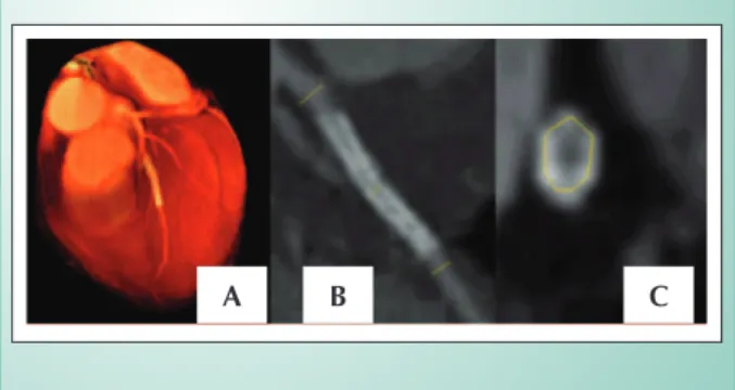
The recorded data were transferred to a workstation (Indigo 02TM, SGITM, Mountain View, CA, USA) for processing and three-dimensional reconstructions, as well as cross-sectional and longitudinal reformatting. The three-dimensional presentations were used for assessing the coronary arteries. The resulting cross-sectional and longitudinal reformatted images allowed a semi-objective measurement of intra-arterial dimensions. For this purpose, electronic calipers were used for manually delimiting the segments to be defined.
Quantitative assessment included measurement of reference diameters, proximal and distal to the stent, as well as minimal lumen diameter. The operator also traced the in-stent minimal area, and the measuring was done on the scanner console (figure 1).
Specific software measured contrast transit time and maximal intensity of opacification in the operator-delimited areas, in addition to projecting contrast media time-density curves. Any hemodynamically significant in-stent obstruction would be indicated by changes in these curves.
This protocol was reviewed and approved by the Institutional Research Ethics Committee of the Hcor, Dante Pazzanese Institute and Medical School of the University of São Paulo.
Statistical analysis -Categorical variables are reported as number and percentage and continuous variables, as mean and standard deviation.
Statistical analysis was conducted using SPSSTM software version 10.0. Sample size was calculated to detect differences of 0.40 mm between means of measurements obtained by angiography and computed tomography, with a 0.36 standard deviation. Alpha level was set at 0.05 and beta level, at 0.10. The 0.40 difference was defined because it reflects the variation inherent to the quantitative angiography system used (CMS). Furthermore, previous studies carried out by our group showed that errors of this magnitude may have clinical implications and also that this has been the mean intraobserver variability of the tomography used 2,8,10. P values < 0.05 were, therefore, considered statistically significant.
Continuous variables were analyzed using the Student’s t-test for paired data. The degree of association among measurements obtained by CT angiography and invasive examinations was determined by means of Pearson’s test.
When subgroups had to be analyzed, reducing the sample to less than 30 patients, non-parametric tests were used. The Wilcoxon test for paired data examined differences between mean continuous variables, and Spearman’s test was used for correlation.
In order to detect whether disagreement between measurements obtained by tomography and angiography could have clinical implications, the Bland-Altman method was applied.
Results
Demographic characteristics of the population - The interval between baseline and invasive reevaluation was 16.80 ± 8.97 months, whereas multidetector CT preceded cardiac catheterization in 28.37 ± 1.33 days. Of the 30 patients The primary purpose of this study was to evaluate the
clinical utility of CT coronary angiography in determining late outcomes in patients treated with drug-eluting stents and comparing their results with those from quantitative coronary angiography and intravascular ultrasound. The secondary purpose was to evaluate the impact of metallic stent struts, without the presence of neointimal proliferation, on the quality of tomographic images.
Patients and methods - In August 2001, we selected thirty patients successfully treated with sirolimus-eluting stent implantation (CypherTM), who were then invasively reevaluated in the late phase (more than six months) and studied through multidector CT 30 days before a control catheterization.
Patients with stent implantation in de novo native coronary lesion for more than six months, neither history of pregnancy, renal failure nor plasma creatinine above 1.5 mg/dl and no known allergy to iodinated contrast material were included. Patients who did not provide written informed consent were excluded from the study.
Methods
This was a prospective study to compare results derived from two different methods of observation of late clinical outcomes after percutaneous interventions. Every examination, namely, quantitative angiography, intravascular ultrasound (IVUS), and CT angiography, was analyzed by independent observers blinded to data from the other two forms of investigation.
Coronary artery angiography was obtained after intracoronary administration of a vasodilator agent (1.20 ml nitroglycerin), and images were transferred to a computer workstation (CMSTM, Medis Medical SystemTM, The Netherlands). This workstation automatically defined artery and stent borders and provided values for reference diameters proximal and distal do the stent, in addition to in-stent minimal lumen diameter.
The invasive investigation ended with intravascular ultrasound examination (Clear ViewTM, Boston ScientificTM, USA), using a 30-MHz mechanical imaging system (Ultra-CrossTM CVISTM, USA). Coronary images were acquired during motorized pullbacks starting 10 mm upstream of the stent and ending 10 mm downstream of the endoprosthesis. In-stent area was measured by IVUS at the end of the study. For this study, only in-stent minimal cross-sectional (CSA) was taken into account, since it could be compared with the results derived from multidetector CT, which does not yet provide volumetric data.
MDCT data were acquired using the Somaton Volume ZoomTM multidetector (Siemens AGTM, Erlhagen, Germany), equipped with a VA 40 protocol15,16. Images were acquired at 500 ms rotation time. As the tomograph is equipped with four rows of detectors, it was possible to obtain eight images per second, each 1.25 mm thick.
included in the study, 23 (76%) were men, mean age 57.67 (SD 8.90); ages, however, ranged from 47 to 77. Vessels treated were the left anterior descending artery in 15 patients (50 %), left circumflex artery in 7 patients (23 %), and right coronary artery in 8 patients (27 %).
Computed tomography: examination characteristics - All the CT examinations were performed successfully and uneventfully, allowing stent localization and blood flow assessment. In six cases (20%) there were motion artifacts, five caused by cardiac arrhythmias and one because the patient was unable to maintain apnea. Nevertheless, even these images could be properly analyzed.
Examination time was 9 ± 4.3 minutes, and contrast volume was 133 ± 22.5 ml. Hospital stay, including patient’s preparation, the examination itself and postprocedural observation was 30.03 ± 5.68 minutes.
Quantitative angiography and intravascular ultrasound
- Mean examination time was 30.90 ± 7 minutes, and mean contrast volume was 129 ± 10.9 ml, which did not differ significantly from those of tomography (p = 0.37).
There were no complications during and after the invasive procedures, and patients were discharged from hospital 12.50 ± 2 hours after the examination.
Qualitative results - Visualization of the entire arterial tree required 2.0 ± 1.0 reconstructions. Only in one case were the coronaries reproduced in their entire extension in a single 3D image with the required diagnostic quality. The visual analysis of contrast dilution showed absence of hemodynamically significant obstructions within all stents.
Quantitative results - Proximal reference diameter by tomography was 3.01± 0.31 mm and by quantitative angiography, 3.14 ± 0.31 mm (p = 0.04). The difference between the two examinations was 3.91% (left anterior descending artery = 2%, left circumflex artery = 5.08% and right coronary artery = 2.97%). The greatest difference was found in the circumflex artery (figure 2). However, when this vessel was excluded from the analysis, differences were no longer significant (tomography = 3.01 ± 0.32 mm, variation: 2.30 to 3.50 mm; angiography = 3.10 ± 0.30 mm, variation: 2.60 to 3.70 mm; p = 0.65).
A significant correlation was found between measurements obtained by computed tomography and invasive angiography (Pearson’s correlation coefficient = 0.72; p = 0.01). This association changed slightly when circumflex arteries were excluded from the analysis (Pearson’s correlation coefficient = 0.74; p = 0.01).
The distal reference diameter by tomography was 2.86 ± 0.30 mm, and by angiography, 2.92 ± 0.32 mm (p = 0.25). The difference between both quantifications was 1.45% (anterior descending artery = 1.92%, circumflex artery = 5.53%, and right coronary artery = 3.91%). Differences never exceeded 5.50%, and the correlation between measurements derived from both examinations was significant (Pearson’s correlation coefficient = 0.66; p = 0.01). When the circumflex artery was excluded from the analysis, no changes were observed (Spearman’s correlation coefficient = 0.63; p = 0.01).
In-stent minimal lumen diameter by tomography was
2.85 ± 0.25 mm and by invasive angiography, 2.85 ± 0.29 mm (p = 0.27). The difference between measurements was 1.14% (anterior descending artery = 1%, circumflex artery = 0.06%, and right coronary artery = 1.14%). No difference exceeded 2.50%, and the significant correlation between these measurements (Pearson’s correlation coefficient = 0.68; p = 0.01) changed slightly when the circumflex artery was excluded from the analysis (Spearman’s correlation coefficient = 0.72; p = 0.01).
Differences between measurements were evaluated using the Bland-Altman method; results are shown in figures 2-4. The analysis of these graphics shows that there was good agreement between both examinations, with few points of disagreement.
In-stent minimal cross-sectional area by computed tomography was 7.19 ± 1.37 mm2 and by intravascular ultrasound, 6.90 ± 1.52 (p = 0.36). Mean difference between both examinations was 7.48 ± 2.82%, and was not affected by the treated artery. The correlation between measurements obtained by both examinations was neither statistically significant (Pearson’s correlation coefficient = 0.33%, p = 0.07) nor affected by the treated artery.
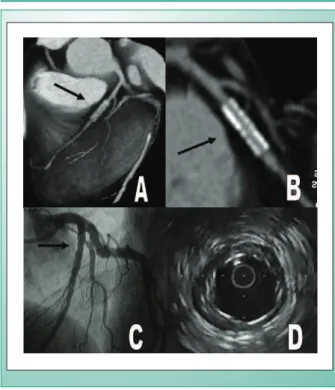
Figure 6 shows an example of a case included in this study. 6,90 +- 1,52 mm2
Discussion
This study was one of the first to evaluate whether multidetector CT allows quantitative and qualitative analysis of coronary stents without neointimal proliferation and to compare its results with those of invasive examinations.
To analyze the degree of coronary stent metallic strut interference in the evaluation performed by multidetector CT, we selected patients who had been treated with sirolimus-eluting stents. All patients included in the study were part of other investigation protocols and were to undergo quantitative coronary angiography and intravascular ultrasound during the late outcome of treatment. Moreover, these patients had already undergone these examinations previously and showed absence of proliferative tissue, representing, therefore, the ideal subgroup for the type of investigation proposed.
Fig. 1 - Three-dimensional reconstructions (A) allowed visual assessment of coronary segments proximal and distal to the stent. Treated vessel lumen diameter and in-stent minimal lumen diameter were measured from reformatted images (B). Cross-sectional area was measured from the cross-sectional reconstruction of the vessel (C).
This was consistent with the study by Sousa et al and Morice and associates, which demonstrated that sirolimus-eluting stents prevent the neointimal proliferation that follows implantation of these prostheses, assuring long-term maintenance of the results obtained immediately after arterial dilation17,18. Sousa also showed that the beneficial effect of this drug is sustained even two years after implantation17. The characteristics described confirm that the analysis of patients treated with this type of stent is the ideal model for this study.
Some procedural aspects related to tomography merit special discussion. As this system obtains eight images per second, the examination takes 25 to 40 seconds; therefore, patients are supposed to hold their breath during this time interval to prevent motion artifacts.
Also important is the patients’ heart rate. Becker et al reported that appropriate reproductions of coronary arteries could be acquired only when this rate was less than 60 beats per minute9. This aspect was facilitated by the introduction of the VA 40 acquisition protocol used in our study. A pilot study carried out in our service demonstrated that examinations of good diagnostic quality can be obtained in patients with heart rate lower than 75 beats per minute19. This may be largely due to the ability of this software to make small adjustments in the cardiac cycle phases19,20. In the present study, artifacts were present in six cases, five of which were related to heart rate and one to breathing, showing the importance of technological advances regarding this point.
A further aspect of practical interest is that, in this type of examination, all contrasted structures are recorded, including heart vessels and, occasionally, even the coronary sinus. Therefore, it is essential that images be analyzed carefully, particularly when it comes to the circumflex artery, because it is in the heart region and the coronary artery and vein may be superimposed. This interference, however, is more pronounced in 3D reconstructions than in longitudinal and cross-sectional reformatted images, which are the projections most frequently used for arterial measurement. 8,19,21,22
Images of good diagnostic quality were obtained in all cases, confirming the effectiveness of the protocol used. Quantitative analysis based on both the visual aspect and estimate of blood flow within the stented vessel was effective
in that it showed absence of hemodynamically significant in-stent obstructions.
Previous studies using electron-beam technology had already suggested the usefulness of non-invasive analysis of coronary stents in the later period by means of time-density curves23,24. Recently, Storto et al reported a case in which they used multidector CT to assess blood flow from time-density curves of contrast media, diagnosing severe in-stent stenosis,
Média dos diâmetros de referência distais (angiografia e tomografia) 2.2 2.4 2.6 2.8 3.0 3.2 3.4 3.6
D if er en ça e n tr e a s m ed id as ( an gi o gr af ia e t o m o gr af ia ) 0.6 0.4 0.2 0.0 -0.2 -0.4 -0.6 -0.8
Fig. 4 - Differences between measurements of the distal reference diameter by angiography and tomography, according to the Bland Altman method.
+1.96 dp 0.45 medium --1.96 dp
-Fig. 5 - Differences between measurements of in-stent minimal lumen diameter by angiography and tomography according to the Bland Altman method.
Média dos diâmetros mínimos intra-prótese (angiografia e tomografia) 2.2 2.4 2.6 2.8 3.0 3.2 3.4 3.6
D if er en ça e n tr e a s m ed id as ( an gi o gr af ia e t o m o gr af ia ) 0.6 0.5 0.4 0.3 0.2 0.1 0.0 -0.1 -0.2 -0.3 -0.4 -0.5 +1.96 dp 0.48 medium 0.05 -1.96 dp -0.39 D if er en ça e n tr e a s m ed id as ( an gi o gr af ia e t o m o gr af ia )
Média dos diâmetros de referência proximais (angiografia e tomografia) 2.4 2.6 2.8 3.0 3.2 3.4 3.6 3.8 0.8 0.6 0.4 0.2 -0.0 -0.2 -0.4 -0.6
Fig. 3 - Differences between measurements of the proximal reference diameter by angiography and tomography, according to the Bland Altman method.
+1.96 dp 0.57 medium 0.12 -1.96 dp
-Mean proximal reference diameters (angiography and tomography)
D if fe re n ce s b e tw e e n me a su re me n ts (a n g io g ra p h y a n d t o mo g ra p h y) D if fe re n ce s b e tw e e n me a su re me n ts (a n g io g ra p h y a n d t o mo g ra p h y)
Mean distal reference diameters (angiography and tomography)
Mean in-stent minimal lumen diameters (angiography and tomography)
D if fe re n ce s b e tw e e n me a su re me n ts (a n g io g ra p h y a n d t o mo g ra p h y) 0.00 0.50 1.00 1.50 2.00 2.50 3.00 3.50 4.00
DADA DADADADADADADADADA DADADADACXCXCXCXCXCXCXCD CDCD CDCD CDCD CD
Fig. 2 - Comparison of proximal reference diameter measured by multidectector tomography (tomography) and invasive angiography (angiography), according to the treated artery.
artéria
m
m
ANGIOGRAFIA TOM OGRAFIA
artery
ANGIOGRAFIA TOM OGRAFIAtomography
which was confirmed by invasive angiography25.
In regard to quantitative results, our study showed that arterial and in-stent lumen diameters obtained by multidetector CT were significantly correlated with those obtained by invasive angiography. A case-by-case analysis revealed that the greatest differences pertained to circumflex artery measurements. The study described by Nieman et al, as well as our own pilot study, had already pointed out difficulties related to this artery8,19. As noted previously, this is due to the difficulty in isolating this vessel for analysis, since it is the only region of the heart where a coronary artery and vein are superimposed8,19. Most often, this happened at the region selected for determining the proximal reference diameter, because the distal end of the stents were usually located in a portion of this artery with no arteriovenous superimposition.
This hypothesis was corroborated by the fact that the correlation between both examinations was strengthened when the circumflex artery was excluded from the analysis, thus suggesting that the tomographic quantitative analysis of this artery is a limitation yet to be circumvented. It should be noted, however, that mean differences between measurements obtained by both examinations, even when taking into account the global sample including the circumflex artery, were lower than the standard deviation of the quantitative angiography system used in this study.
On the other hand, the measurement of distal reference diameter was not affected by the treated vessel, which may be explained, at least partly, by the most distal parts of the circumflex artery not being superimposed by the coronary artery and vein. In-stent lumen diameter was not affected by the treated vessel either. Most probably, this may be accounted
for by the fact that even in the presence of arteriovenous superimposition, stent struts help the operator to identify the boundaries of the regions to be measured. Hence, statistical analysis results were not affected by the exclusion of the circumflex artery from the analysis.
Overall, in our study, differences between all comparisons of measurements obtained by quantitative angiography and computed tomography never exceeded 10%, and the Bland-Altman analysis, most often, showed disagreement between measurements ranging from -0.40 mm and 0.40 mm.
These findings have two major implications. First, although quantitative measurements obtained by tomography did not differ statistically from those obtained by angiography nor from the positive and significant correlation between them, their use in everyday clinical practice should be viewed with caution. Further technological advances are needed for these quantifications to gain widespread acceptance. Additionally, these same data also suggest that this examination may be very promising, because even at an initial stage of development, it yielded results that, for the most part, did not show clinically relevant differences when compared to those considered gold standard for this purpose.
The results obtained when cross-sectional area, measured by ultrasound and invasive tomography, were compared deserve special attention. At first glance, tomography seems to perform excellently in this aspect, because mean values obtained by this examination [6.90 mm2 (SD 1.52)] did not differ statistically from those observed in the invasive study [7.19mm2 (SD 1.47), p = 0.36], and mean disagreement between the results of both examinations was only 7.48 %.
The association between measurements, based on Pearson’s correlation coefficient, was weak and non-significant. This was confirmed by the Bland-Altman analysis, which showed great differences between these measurements. This may be probably due to the interaction between metallic stent struts and radiographic examinations, causing energy gain. This, in turn, leads to the presence of artifacts, which makes it very difficult to determine coronary stent perimeters, inducing error in the definitions of arterial borders. It seems, therefore, that tomographic measurement of in-stent cross-sectional area is not yet ready for clinical application, because the values thus produced do not reflect the actual area of each particular case.
An analysis of the available literature shows that few studies used arterial quantification techniques to analyze images obtained by multidetector computed tomography. Aschenbach et al evaluated 25 asymptomatic volunteers showing no evidence of coronary failure either by multidetector CT or invasive angiography, by measuring the reference diameter of coronary arteries by both techniques. They reported a mean difference of 0.36 mm between both techniques and found excellent correlation between quantitative angiography and tomographic examination (r=0.86)26. The seemingly more favorable results achieved by these authors may be related to the fact that they studied native coronary arteries of normal patients, while we evaluated patients treated with coronary stents.
Very few studies have aimed at using tomography to assess
References
1. Kruger RA. A comparative summary of image processing methods in digital angiography. In: Mancini GBJ, ed. Clinical Applications of Cardiac Digital Angiography. ed. New York: Raven Press: 1988: 19-36.
2. Lefree MT, Simon SB, Mancini GBJ, Bates ER, Vogel RV. A comparison of 35 mm cine film and digital radiographic image recording: implications for quantitative arteriography. Invest Radiol 1988;73: 176-83.
3. Ellis SG, Pinto IMF, Mcgillem M.J, de Boe SF, Lefree M, Mancini GBJ. Accuracy and reproducibility of quantitative coronary arteriography using 6 and 8 French catheters with angiographic acquisition. Cath Cardiovasc Diagn 1991; 22: 52-9.
4. Pinto IMF, Tanajura LFL, Feres F, Mattos LAP, Maldonado GA, Centemero M, et al. Papel da angiografia digital no diagnóstico e avaliação da insuficiência coronária. Rev Soc Cardiol ESP 1992; 6: 75-82.
5. Pinto IMF, Sousa AG, Feres F, Tanajura LF, Mattos LA, Cano MN, et al. Utilidade da angiografia digital quantitativa para a realização da angioplastia coronária: análise de 100 casos. Arq Bras Cardiol 1992; 59: 255-9.
6. Hanet C, Wijns W, Michel X, Schroeder E. Influence of balloon size and stenosis morphology on immediate and delayed elastic recoil after PTCA. J Am Coll Cardiol 1991;18: 506-11.
7. Reiber JHC, Serruys PW. Quantitative coronary angiography. In: Marcus M.L., ed, Cardiac Imaging. Philadelfia: WB Saunders, 1990: 211.
8. F e l d S , G a n i m M , C a r e l l E S , K j e l l g r e n O , K i r k e e i d e R L , Vaughn WK, et al. Comparison of angioscopy, intravascular ultra-s o u n d a n d q u a n t i t a t i v e c o r o n a r y a n g i o g r a p h y i n p r e d i c t i n g o u t c o m e a f t e r c o r o n a r y i n t e r v e n t i o n i n h i g h - r i s k p a t i e n t s . J Am Coll Cardiol 1996: 28: 97-105.
the performance of this examination with the presence of stents. Recently, a case was reported of successful visualization of the lumen inside a sirolimus-eluting stent deployed at the left coronary artery. The authors speculated on the possibility of these results being dependent on the stent’s diameter, but failed to report the threshold value to obtain adequate examinations27.
Limitations - There are some limitations to this study. The technology used was that of four-row detectors, a first generation multidetector system. This examination has been widely used in cardiology practice and is still evolving. Thus, further investigation in the future may yield different results. Nevertheless, our findings are representative of the time in which the study was carried out and prompt similar trials when new equipment capable of rendering 64 images per cycle becomes available. Either way, despite claiming that MDCT scanners that generate 40 images per cycle may be useful for clinical use, the study by Gaspar et al produced only reasonable results using these systems, which had sensitivity of 88.9%, specificity of 80.6%, negative predictive power of 97.4%, and positive predictive power as low as 47.1%13. The difficulties faced by computed tomography regarding non-invasive analysis of stents are well-characterized in the study by Maintz, who analyzed 68 different coronary stents in a CT scanner capable of obtaining 64 images per cycle14. His group demonstrated that, even in optimal conditions, the results of non-invasive analysis are significantly affected by the thickness of the metallic struts, the material of which stents are made, and their design 14. The use of the technology of four detector rows (four images per cycle and eight images per second) with 1.25 mm thick section, as in this study, represents an additional obstacle to viewing inside the implanted stents, especially when there may be neointimal proliferation.
Finally, the study sample followed criteria determined for its calculation, but selected only patients who had undergone sirolimus-stent implantation. Thus, the application of the conclusions drawn from this study to other populations of CAD patients treated with other types of stents should be viewed with reservations.
Clinical and investigational implications and perspectives
- Our findings indicate that multidetector CT is useful for following-up subjects treated with sirolimus-eluting stents, since it enabled the determination of their patency and
luminal diameters.
However, great care must be exercised to reduce heart rate- and respiratory-related motion artifacts. In addition, images should be reconstructed as many times as necessary so that coronary arteries may be reproduced as best as possible. It seems clear that, based on the analysis made by this study, both qualitative and quantitative data should be used to study the clinical course following percutaneous revascularization with coronary stents.
A conclusion that may be inferred from our findings is that CT angiography may be useful for clinical follow-up of patients undergoing drug-eluting stenting and the results of which are similar to those provided by sirolimus. Once some agents currently under investigation or that may be studied in the future show similar performance, these patients’ late evaluation should be conducted using this diagnostic method.
Conversely, major technological advances, such as the introduction of CT scans capable of acquiring 64 images or more per cycle and increase in gantry rotation speed, may greatly benefit non-invasive studies of patients treated with coronary stents.
However, potential limitations to the kind of analysis mentioned in this study and even in investigations using more up-to-date equipment13,14 make it clear that the routine use of multidetector tomography is not recommended for non-invasive follow-up of stent implantation, especially nondrug-eluting stents.
Conclusions
The comparison of multidetector CT results with those of invasive angiography in patients treated with sirolimus-eluting stents has led to the following conclusions: Angiography enables qualitative evaluation of blood flow through the target vessel, allowing measurement of proximal and distal reference diameters of the treated arteries, as well as in-stent minimal lumen diameter.
9. Yock PG, Johnson EL, Linker DT. Intravascular ultrasound: development and clinical potential. Am J Cardiac Imag 1988; 2:185-93.
10. Bom N, Ten Hoff H, Lancee CT, Gussenhoven WJ, Bosch JG. Early and recent intraluminal ultrasound devices. Int J Cardiac Imag 1989; 4: 79-88.
11. Pandian NG, Kreis A, Brockway B, Isner JM, Sacharoff A, Boleza E, et al. Ultrasound angioscopy: real-time, two-dimensional, intraluminal ultrasound imaging: an in vitro study. Am J Cardiol 1988;62: 493-4.
12. Gussenhoven EJ, Essed CE, Lancee CT, Mastick F, Frietman P, Van Egmond FC, et al. Arterial wall characteristics determined by intravascular ultrasound imaging catheter. J Am Coll Cardiol 1989;14: 957-62.
13. Mallery JA, Tobis JM, Griffith J, Gressert J, Mcrae M, Moussabeck O, et al. Assessment of normal and atherosclerotic arterial wall thickness with an intravascular ultrasound imaging catheter. Am Heart J 1990;119: 1392-1400.
14. Abizaid A, Mintz GS, Abizaid AS, Satler LF, Popma JJ, Pichard AD, et al. Aplicações clínicas do ultra-som intracoronário. Arq Bras Cardiol 1997; 69: 263-5.
15. Colombo A, Hall P, Nakamura S, Almagor Y, Maiello L, Martini G, et al. Intracoronary stent without anticoagulation accomplished with intracoronary ultrasound guidance. Circulation 1995;91: 1676-88.
16. De Jaegere P, Mudra H, Figulla H, Almagor Y, Doucet S, Penn I, et al. Intravascular ultrasound-guided optimized stent deployment: immediate and 6 months clinical and angiographic results from the Multicenter Ultrasound Stenting in Coronaries Study (MUSIC Study). Eur Heart J 1998;19:1214-23.
17. Sousa JE, Costa MA, Abizaid AA, Rensing BJ, Abizaid AS, Tanajura LFL, et al. Lack of neointimal proliferation after implantation of sirolimus-coated stent in human coronary arteries: a quantitative coronary angiography and three dimensional intravascular ultrasound study. Circulation 2001; 103: 192-5.
18. AHA/ACC practice guidelines for coronary angiography. A report of the American College of Cardiology / American Heart Association Task Force
on Practice Guidelines (Committee on Coronary Angiography). J Am Coll Cardiol 1999; 33: 1756-824.
19. Gianrossi R, Detrano R, Mulvihill D, Lehmann K, Dubach P, Colombo A, et al. Exercise-induced ST depression in the diagnosis of multivessel coronary artery disease: a meta-analysis. Circulation 1989; 80: 87-98.
20. Wackers FJTH. Exercise myocardial perfusion imaging. J Nucl Med 1994; 35: 726-9.
21. Marangelli V, Iliceto S, Piccinini G, Demartino G, Sorgente L, Rizzon P. Detection of coronary artery disease by digital stress echocardiography: comparison of exercise, transesophageal atrial pacing and dipyridamole echocardiography. J Am Coll Cardiol 1994; 24: 117-24.
22. Nieman K, Oudkerk M, Resing BJ, van Ooijen P, Munne A, van Geuns RJ, et al. Coronary angiography with multi-slice computed tomography. Lancet 2001; 357: 599-603.
23. Kim WY, Danias PG, Stuber M, Flamm SD, Plein S, Nagel E, et al. Coronary magnetic resonance angiography for the detection of coronary stenoses. New Eng J Med 2001; 345: 1863-9.
24. Pinto IMF, Pavanello R, Egito E, Souza LCB, Abibi MH, Bosisio I, et al. Princípios, indicações e limitações da ressonância magnética na avaliação da doença cardiovascular. Rev Soc Cardiol ESP 1992; 6: 63-70.
25. Haberl R, Becker A, Leber A, Becker C, Knez A, Bruning R, et al. Correlation of coronary calcification and angiographically documented stenoses in patients with suspected coronary artery disease: results of 1764 patients. J Am Coll Cardiol 2001: 37: 451-7.
26. Detrano R, Hsiai T, Wang S, Puentes G, Fallavollita J, Shields P, et al. Prognostic value of coronary calcification and angiographic stenoses in patients undergoing coronary angiography. J Am Coll Cardiol 1996: 27: 285-90.