AR
TIGO ORIGINAL / ORIGINAL AR
TICLE
INTRODUCTION
The adenocarcinoma of the ampulla of Vater (AAV) presents low prevalence, with annual estimated incidence - in the United States and France - between three to seven cases per million inhabitants(3, 9). It represents 0.5% to 2% of gastrointestinal cancers(1, 4, 8) and is the second most common periampullary malig-nancy(19). Its occurrence is exponential after the age of 35 (1), with peak of mean incidence between the ages of 60 and 70 years(4). Among the periampullary tumors, it has the best prognosis(5, 17).
Jaundice is one of the most common clinical man-ifestations of AAV (70%-90% of the cases)(4, 20). It is deined by yellowish shades on the skin, sclera and mucous membranes resulting from the deposition of bilirubin in such. It happens when total serum bili-rubin values are evidently higher than 3mg/dL(12, 18).
Some authors state that the classic clinical de-piction of the ampulla of Vater cancer consists of luctuating jaundice (FJ), Courvoisier’s sign, anemia and weight loss(2, 4, 11, 15). Jaundice, in this case, comes from cholestasis caused by the obstruction of the
du-FLUCTUATING JAUNDICE IN THE
ADENOCARCINOMA OF THE AMPULLA OF
VATER: a classic sign or an exception?
José Roberto
ALVES
1, Enio Campos
AMICO
1, Dyego Leandro Bezerra de
SOUZA
2,
Patrick Vanttinny Vieira de
OLIVEIRA
3and
Ícaro Godeiro de Oliveira
MARANHÃO
3ABSTRACT - Background - Some authors consider the luctuating jaundice as a classic sign of the adenocarcinoma of the ampulla of Vater.Objetive - Assessing the frequency of luctuating jaundice in their forms of its depiction in the patients with adenocarcinoma of the ampulla of Vater. Methods - Observational and retrospective study, conducted through analyses of medical records from patients subjected to pancreatic cephalic resections between February 2008 and July 2013. The pathological examination of the surgical specimen was positive to adenocarcinoma of the ampulla of Vater. Concepts and differences on clinical and laboratory luctuating jaundice were standardized. It was subdivided into type A and type B laboratory luctuating jaundice. Results - Twenty patients were selected. One of them always remained anicteric, 11 patients developed progressive jaundice, 2 of them developed clinical and laboratory luctuating jaundice, 5 presented only laboratory luctuating jaundice and one did not present signiicant variations on total serum bilirubin levels. Among the seven patients with luctuating jaundice, two were classiied as type A, one as type B and four were not classiied due to lack information. Finally, progressive jaundice was the prevailing presentation form in these patients (11 cases). Conclusion - This series of cases suggested that clinical luctuating jaundice is a uncommon signal in adenocarcinoma of the ampulla of Vater.
HEADINGS - Adenocarcinoma. Ampulla of Vater. Jaundice. Digestive signs and symptoms.
Declared conflict of interest of all authors: none Disclosure of funding: no funding received
Research performed at: University Hospital Onofre Lopes of the Universidade Federal do Rio Grande do Norte (UFRN), Natal, RN, Brazil.
1 Department of Integrated Medicine, School of Medicine, UFRN; 2 Department of Collective Health, UFRN; 3 School of Medicine, UFRN.
Correspondence: Prof. Dr. José Roberto Alves. Departamento de Medicina Integrada, Hospital Universitário Onofre Lopes, Universidade Federal do Rio Grande do Norte. Av. Nilo Peçanha, 620, Petrópolis - CEP: 59012-300 - Natal, RN, Brasil. E-mail: [email protected]
odenal papilla neoplasm and its luctuating pattern is explained by necrosis and tumor desquamation which allows a decrease in biliary stasis and, consequently, a momentary disappearance of the jaundice(2, 4, 11, 15).
However, there is no real deinition and differ-entiation of clinical and laboratory patterns of jaundice luctuation in AAV in the current medical literature, although some studies describe its physio-pa thology(10, 13, 16).
Moreover, besides the conceptual dificulty re-lated to the clinical featuring of luctuating jaundice related to AAV, there are dificulties related to early pre-operative diagnoses in the absence of histological confirmation, besides the possibility of diagnoses mistakes due to benign diseases(11).
The chosen treatment for AAV is the gastroduode-nopancreatectomy, or Whipple’s surgery, which must be performed in the earliest stages of the disease in order to promote cure and give better life quality to the patients(6, 17).
METHODS
A descriptive, observational and retrospective study per-formed at the University Hospital Onofre Lopes (HUOL) of the Federal University of Rio Grande do Norte, located in Natal, capital city of Rio Grande do Norte, Brazil.
The sample was selected through the analysis of medi-cal records from patients subjected to pancreatic cephalic resections due to AAV, between February 2008 and July 2013, at HUOL.
All patients diagnosed with AAV - conirmed by patho-logic examination of the surgical sample - were excluded from the experiment as well as those who presented lack of information and/or underwent invasive procedures before surgery because it could modify the natural history of the disease. Data collection was performed by researchers and gotten straight from the medical charts.
The design of the current study was approved by the Re-search Ethics Committee of the Norte Rio Grandense League against Cancer, according to decision n. 295676. The menu added the HUOL as an associated Institution according to a second decision, n. 332153. It followed the ethical princi-ples of Resolution 196/96 of the Brazilian National Health Council. In addition, due to the nature of this study, the Reasearch Ethics Committees from the institutions involved in the project have approved the exemption patients signature in the Free and Clariied Term of Consent.
According to the current study, the concept and differ-ence on clinical luctuating jaundice (CFJ) and laboratory luctuating jaundice (LFJ) were standardized. CFJ regarded the presence and subsequent disappearing of a yellowish color shade in the skin and in the mucosa as well as the identiication of sclerae in the patient. Such symptoms can be perceived by him/her or by the physician during the course of the disease. In case of absence of clear information described in the medical records, we considered CFJ patients as those with total serum bilirubin higher than 3mg/dL as well as those with clinical jaundice.
LFJ regarded patients showing variation on the total serum bilirubin throughout the evolution of his/her disease. At least three different dosages of this laboratory test were necessary. The LFJ was subdivided into type A when there was a decrease in the dosage of bilirubin, and the type B, when there was a decrease and a subsequent new increase in bilirubin.
The dosages of total serum bilirubin were performed at the HUOL’s laboratory and it was done by means of the
colorimetric method with the Wiener Lab® diagnostic
re-agent. The method, according to the Wiener Lab protocol, presents amplitude in the coeficient of variation that goes from 1.1% up to 4.7 % when values of total serum bilirubin are measured. Therefore, in order to determine whether luctuations occurred, i.e., we have calculated a statistically signiicant variation in patients’ serum bilirubin values, the mean and standard deviation for each one of them and then the variation in the coeficient. Those who obtained variation coeficient up to 4.7% were not included in the diagnosis for luctuating jaundice, because it was considered as a variation related to the method itself.
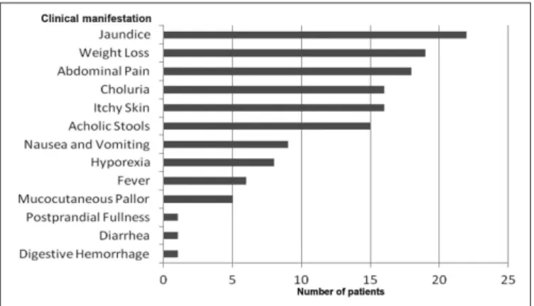
FIGURE 1. The frequency of clinical manifestations identiied in 22 adenocarcinoma of the ampulla of Vater (AAV) patients
RESULTS
The initial sample was collected from 22 patients, most of them male (12 pacients) with mean age of 56.2 years. The most prevaling manifestations in patients were: jaundice (100%), weight loss (86.36%), abdominal pain (81.81%), choluria (72.72%), itchy skin (72.72%) and acholic stools (68.18%) (Figure 1).
Regarding patients from the initial sample (22 patients), 15 of them presented pre-operative suspected AAV diagnosis, by means of an endoscopic exams. Out of them, 10 had their diagnosis conirmed by the anatomopathological outcomes from the endoscopic biopsy.
Seven patients did not make the endoscopic exams, how-ever, due to other image complementary exams (CT and mag-netic resonance of abdomen; cholangiopancreatography), that have presented strong suspected preoperative diagnosis for papillary tumor or other peri-ampullary cancer.
Two patients had to be excluded from the sampling group because one of them (patient 16) underwent a biliodigestive bypass and had no record of total serum bilirubin levels before surgery. The other one (patient 18), underwent an endoscopic retrograde cholangiopancreatography with stent placement and had no levels of serum bilirubin before endos-copy. Thus, the study’s inal sample consisted of 20 patients. Among them, three were up to 40 years old.
Fluctuating jaundice’s identiication and classiication process was done by assessing the clinical history and ana-lyzing the identiied values of total serum bilirubin (Table 1) (Figure 2). The irst measurement of serum bilirubin in the inal sample was performed at a median of 19 days (1-173 days) before surgery, meanwhile the last determination occurred at a median of 2 days (1-20 days) before surgical treatment established.
TABLE 1. Values of total serum bilirubin and statistical analysis from the 20 AAV patients in presence of Wiener Lab reagent, from February 2008 up to July 2013
P TB1 TB2 TB3 TB4 TB5 MEAN SD CV
1 6.3 1.8 --- --- --- 4.07 3.20 78.80
2 28.1 28.2 31.0 21.9 33.0 28.4 4.2 14.8
3 6.3 13.8 --- --- --- 10.0 5.3 52.8
4 31.6 12.5 --- --- --- 22.0 13.5 61.3
5 14.8 18.6 --- --- --- 16.7 2.7 16.2
6 18.2 21.1 24.7 --- --- 21.3 3.2 15.3
7 18.2 22.7 25.4 24.5 --- 22.7 3.2 14.2
8 0.2 --- --- --- --- --- ---
---9 15.0 17.4 --- --- --- 16.2 1.7 10.5
10 6.5 0.8 0.5 0.4 0.3 1.7 2.6 151.6
11 15.1 14.8 15.0 --- --- 14.9 0.1 1.0
12 11.3 6.0 --- --- --- 8.6 3.7 43.3
13 32.5 27.5 --- --- --- 30.0 3.5 11.8
14 11.2 15.8 --- --- --- 13.5 3.2 24.1
15 15.2 16.0 20.1 --- --- 17.1 2.6 15.4
17 13.4 17.0 --- --- --- 15.2 2.5 16.5
19 6.1 14.8 22.2 --- --- 14.3 8.0 56.1
20 23.4 --- --- --- --- --- ---
---21 10.0 10.4 11.5 --- --- 10.6 0.7 7.3
22 15.0 17.4 18.7 19.3 --- 17.6 1.9 10.8
P: patient; TB: value of total serum bilirubin; 1: irst evaluation of TB; 2: second evaluation of TB; 3: third evaluation of TB; 4: fourth evaluation of TB; 5: ifth evaluation of TB; P: patients; SD: standard deviation; CV: coeficient of variation. Note: Patients 16 and 18 were excluded from the sample (For reasons that read the 4th paragraph of Results).
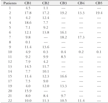
TABLE 2. Values of conjugated serum bilirubin from the 20 AAV patients in presence of Wiener Lab reagent, from February 2008 up to July 2013
Patients CB1 CB2 CB3 CB4 CB5
1 4.5 1.1 --- ---
---2 17.8 17.2 19.2 13.3 19.4
3 4.2 12.4 --- ---
---4 18.6 7.7 --- ---
---5 7.1 9.2 --- ---
---6 12.1 13.8 16.1 --- ---7 9.8 --- 18.2 17.3
---8 0.1 --- --- ---
---9 11.4 13.6 --- ---
---10 4.9 0.3 0.4 0.2 0.1
11 9.2 9.9 8.5 ---
---12 7.9 4.2 --- ---
---13 14.5 11.7 --- --- ---14 7.7 10.1 --- --- ---15 11.4 12.3 16.6 ---
---17 7.5 9.0 --- ---
---19 4.0 12.0 13.3 --- ---20 15.9 --- --- --- ---21 8.0 8.4 9.7 --- ---22 10.0 11.3 10.5 11.4
---CB: value of conjugated bilirubin; 1: irst evaluation of CB; 2: second evaluation of CB; 3: third evaluation of CB; 4: fourth evaluation of CB; 5: ifth evaluation of CB. Note: Patients 16 and 18 were excluded from the sample (For reasons that read the 4th paragraph of Results).
FIGURE 2. Variation of serum total bilirubin of 20 patients with adeno-carcinoma of the ampulla of Vater from February 2008 until July 2013. Note: Patients 16 and 18 were excluded from the sample (For reasons that read the 4th paragraph of Results).
TB: value of total serum bilirubin; 1: irst evaluation of TB; 2: second evaluation of TB; 3: third evaluation of TB; 4: fourth evaluation of TB; 5: ifth evaluation of TB.
Among the seven patients with luctuating jaundice, two were classiied as type A, one as type B, and four were not classiied due to insuficient levels of total serum bilirubin (less than three dosages).
The conjugated bilirubin variation pattern was similar to that of total bilirubin for the 20 patients from the inal sampling (Table 2) (Figure 3).
FIGURE 3. Variation of serum conjugated bilirubin of 20 patients with adenocarcinoma of the ampulla of Vater from February 2008 until July 2013. Note: Patients 16 and 18 were excluded from the sample (For reasons that read the 4th paragraph of Results).
DISCUSSION
Within the initial sample with 22 patients, the most fre-quent identiied symptoms were jaundice, abdominal pain, weight loss, just as presented by other studies(6, 7, 14, 17, 19).
It should be noticed that, to date, the standardization of the jaundice luctuation in AAV had neither been discussed by other studies, nor a classiication on the depictions of clinical and laboratory jaundice had been suggested, as reported in the current study(1-4, 8-13, 15, 16, 18).
Although some authors disclose that luctuating jaundice is a classic clinical manifestation during the natural history of AAV(4, 11, 15, 20), this sign - if we consider the semantic es-sence of jaundice in its clinical depiction - , was found in two patients from the sample.
Jaundice non-luctuation, i.e., the presence of persistent and progressive jaundice was the prevalent depiction in AAV. It was observed in 11 patients. Such fact reinforces the probable exception character of the luctuating jaundice occurrence demonstrated in our study, in its laboratory presentation form, in seven cases and, more rarely, in its clinical form (two cases). Finally, this low luctuating jaundice frequency can also be found in other studies. It happened in two cases within a sample of 35 patients (Hayes et al.)(10); in two cases within a sample of 37 patients (Moody et al.)(13); and in four cases within a sample of 15 patients (Ponka et al.)(16). Furthermore, it is important to emphasize that only one patient in this series of cases, throughout the natural evolution of his/her illness, never presented jaundice signs, i.e., he/she remained anicteric.
CONCLUSIONS
In conclusion, despite the small sampling group presented in the aforementioned series of cases, lack of clinical and laboratory information in medical charts, through a little more than ive years - due to the low incidence of AAV - , it can be suggested that luctuating jaundice, based on concepts standardization proposal, presents itself as low frequency situation during AAV’s natural history. Clinical luctuating jaundice is really an exception condition. Thus, as an indirect consequence, there is the need for conducting studies with a larger sample, possibly multicenter studies - due rarity of the disease - in order to really promote the inal deinition for luctuating jaundice in AAV.
Authors’ contributions
Alves JR mentored medical students in the literature review and data collection, and he was the inal reviewer of the article writing, and responsible for the translation into English, as well as for the text formatting and for the process of submitting the article. Amico EC performed most of the surgeries on patients and contributed to the revision of the article writing. Souza DLB performed the statistical analysis and contributed to the inal revision. Oliveira PVV and Ma-ranhãoIGO conducted a literature review and participated in the writing of the manuscript. All authors read and approved the inal version of the article.
Alves JR, Amico EC, Sousa DLB, Oliveira PVV, Maranhão IGO. Icterícia lutuante em adenocarcinoma de ampola de Vater: um sinal clássico ou uma exceção? Arq Gastroenterol. 2015,52(2):147-51.
REFERENCES
1. Albores-Saavedra J, Schwartz AM, Batich K, Henson DE. Cancers of the ampulla of vater: Demographics, morphology, and survival based on 5,625 cases from the SEER program. J Surg Oncol. 2009;100(7):598-605.
2. Beale GL, Trulock AS. Carcinoma of the ampulla of Vater. Am J Surg. 1952;83(1):20-5.
3. Benhamiche AM, Jouve JL, Manfredi S, Prost P, Isambert N, Faivre J. Cancer of the ampulla of Vater: Results of a 20-year population-based study. Eur J Gastroenterol Hepatol. 2000;12(1):75-9.
4. Chan M, Adler DG. Ampullary cancer: review and clinical uptodate. Community Oncology. 2010;7:61-6.
5. Chen SC, Shyr YM, Wang SE. Longterm survival after pancreaticoduodenectomy for periampullary adenocarcinomas. HPB (Oxford). 2013;15(12):951-7. 6. Di Giorgio A, Alieri S, Rotondi F, Prete F, Di Miceli D, Ridolini MP, et al.
Pancreatoduodenectomy for tumors of vater's ampulla: Report on 94 consecutive patients. World J Surg. 2005;29(4):513-8.
7. Gaspar B, Beuran M, Paun S, Ganescu R, Hostiuc S, Negoi I. Current strategies in the therapeutic approach for adenocarcinoma of the ampulla of Vater. J Med Life. 2013;6(3):260-5.
8. Gassler N, Knüchel R. Tumors of vater's ampulla. Pathologe. 2012; 33(1):17-23. 9. Goodman MT, Yamamoto J. Descriptive study of gallbladder, extrahepatic bile
duct, and ampullary cancers in the United States, 1997-2002. Cancer Causes and Control. 2007;18(4):415-22.
10. Hayes DH, Bolton JS, Willis GW, Bowen JC. Carcinoma of the Ampulla of Vater. Ann Surg. 1987;206(5): 572-7.
11. Kim MH, Lee SK, Seo DW, Won SY, Lee SS, Min YI. Tumors of the major duodenal papilla. Gastrointestinal Endosc. 2001;54(5):609-20.
12. Martinelli ALC. Icterícia. Medicina (Ribeirão Preto). 2004;37:246-52. 13. Moody F, Thorbjarnarson B. Carcinoma of the ampulla of Vater. Am J Surg.
1964;107:572-9.
14. Nikfarjam M, Muralidharan V, McLean C, Christophi C. Local resection of ampullary adenocarcinomas of the duodenum. ANZ J Surg. 2001;71(9):529-33. 15. Polli CA, Gonçalves ACA, Defácio A, Bechaalani P, Moraes CM, Paula RA.
A importância da semiologia no diagnóstico diferencial das icterícias aliada a exames complementares. Arq Med Hosp Fac Cienc Med Santa Casa São Paulo. 2008;53:113-7.
16. Ponka JL, Uthappa NS. Carcinoma of the ampulla of Vater. Am J Surg. 1971;121(3):263-70.
17. Qiao QL, Zhao YG, Ye ML, Yang YM, Zhao JX, Huang YT, Wan YL. Carcinoma of the ampulla of Vater: Factors inluencing long-term survival of 127 patients with resection. World J Surg. 2007;31(1):137-43.
18. Roche SP, Kobos R. Jaundice in Adult Patient. Am Fam Physician. 2004;69(2): 299-304.
19. Talamini MA, Moesinger RC, Pitt HA, Sohn TA, Hruban RH, Lillemoe KD, et al. Adenocarcinoma of the ampulla of Vater: A 28-year experience. Ann Surg. 1997;225(5):590-600.
20. Tsukada K, Takada T, Miyazaki M, Miyakawa S, Nagino M, Kondo S, et al. Diagnosis of biliary tract and ampullary carcinomas. J Hepatobiliary Pancreat Surg 2008;15(1):15-40.