INTRODUCTION
Article/Artigo
1. Departamento de Doenças Tropicais e Diagnóstico por Imagem, Faculdade de Medicina de Botucatu, Universidade Estadual Paulista, Botucatu, SP. 2. Departamento de Dermatologia e Radioterapia, Faculdade de Medicina de Botucatu, Universidade Estadual Paulista, Botucatu, SP. 3. Departamento de Pediatria, Faculdade de Medicina de Botucatu, Universidade Estadual Paulista, Botucatu, SP. 4. Departamento de Patologia, Faculdade de Medicina de Botucatu, Universidade Estadual Paulista, Botucatu, SP. 5. Centro de Estudos de Venenos e Animais Peçonhentos, Universidade Estadual Paulista, Botucatu, SP.
Address to: Dr. Benedito Barraviera. Depto Doenças Tropicais e Diagnóstico Imagem/FMB/UNESP.
Distrito de Rubião Junior s/n, 18618-970 Botucatu, SP, Brasil. Phone: 55 14 3811-6212; Fax: 55 14 3815-9898.
e-mail: [email protected]
Received in 03/12/2010
Accepted in 16/06/2011
Africanized honeybee stings: how to treat them
Picadas de abelhas africanizadas: como tratá-las?
Ricardo Augusto Monteiro de Barros Almeida
1, Taylor Endrigo Toscano Olivo
1, Rinaldo Poncio Mendes
1,
Silvia Regina Catharino Sartori Barraviera
2, Lenice do Rosário Souza
1, Joelma Gonçalves Martins
3,
Miriam Hashimoto
3, Viciany Erique Fabris
4, Rui Seabra Ferreira Junior
5and Benedito Barraviera
1,5ABSTACT
Introduction: In 1956, Africanized honeybees (AHB) migrated from Brazil to other regions of the Western Hemisphere, including South, Central, and North America, except for Canada. Despite being productive, they are highly aggressive and cause fatal accidents. his study aimed to evaluate patients at the Clinical Hospital of Botucatu Medical School (HC-FMB) and to propose treatment guidelines. Methods: From 2005 to 2006, the clinical and laboratorial aspects of 11 patients (7 male and 4 female) and the anatomopathological aspects of one patient who had died in 2003 were analyzed. Results: he age of the surviving patients varied from 5 to 87 years, with a mean of 42.5 years. he majority of accidents occurred in the aternoon, and the number of stings ranged from 20 to 500. he principal signs and symptoms were pain and local inlammatory signs, nausea, tachycardia, and vomiting. Biochemical indings presented increased levels of creatine phosphokinase, lactate dehydrogenase, and aspartate/alanine aminotransferase. An 11-year-old male patient died upon entering the atic of a two-storey building where he was atacked by a swarm, receiving more than 1,000 stings. He was sent to HC-FMB where he was treated, but he died 24h later. Observed at the autopsy were erythematous-purpuric skin lesions besides necrosis at the sting locations, rhabdomyolysis, focal myocardial necrosis, tubular hydropic degeneration and focal tubular acute necrosis of the kidneys, myoglobinuria, and centrolobular necrosis in the liver. Conclusions: Accidents caused by multiple AHB stings always constitute a medical emergency. As there is no speciic antivenom, we have developed guidelines, including irst aid, drugs, and the proper removal of stingers.
Keywords: Africanized honeybee stings. Treatment guidelines. Apis mellifera.
RESUMO
Introdução: As abelhas africanizadas (AHBs) migraram do Brasil em 1956 para todo o continente Americano. Apesar de produtivas, são agressivas causando acidentes fatais. O objetivo foi avaliar pacientes atendidos no Hospital das Clínicas da Faculdade de Medicina de Botucatu (HC-FMB) e propor um roteiro de tratamento. Métodos: Entre 2005 e 2006, foram analisados os aspectos clínicos e laboratoriais de 11 pacientes e anatomopatológicos de um que foi a óbito em 2003. Resultados: A idade dos pacientes variou entre 5 e 87 com média de 42,5 anos. Sete eram do sexo masculino e quatro do feminino. O número de picadas variou entre 20 e 500. Nove deles receberam mais de 50 picadas. Os principais sinais e sintomas foram dor local, náuseas, taquicardia e vômitos. Os exames hematológicos mostraram leucocitose, neutroilia, anemia e desvio à esquerda escalonado. Os exames bioquímicos revelaram níveis elevados de creatinofosfoquinase, desidrogenase lática e aspartato/alanina aminotransferase. O paciente que foi a óbito 24h após o atendimento tinha 11 anos, era do sexo masculino e foi atacado ao adentrar um edifício de dois andares recebendo mais de 1.000 picadas. O exame anatomopatológico mostrou lesões eritemato-purpúricas, além de necrose nos locais das picadas. Apresentou também rabdomiólise, necroses focais do miocárdio, degeneração hidrópica acompanhada de necrose tubular renal aguda,mioglobinúria e necrose centrolobular no fígado. Conclusões: Os pacientes acometidos por múltiplas picadas necessitam de tratamento imediato e por não dispormos de um soro especíico desenvolvemos um roteiro que inclui os primeiros socorros, as drogas a serem empregadas e a retirada dos ferrões corretamente. Palavras-chaves: Picadas de abelhas africanizadas. Roteiro de tratamento. Apis mellifera.
At the outset of the 19th century, setlers from
Europe brought the Apis mellifera mellifera bees to southern Brazil1. hese bees were adapted to the
European temperate climate, and upon arriving in Brazil, they presented low production of honey and its derivatives. his led the Brazilian government to implement a program to develop bees that would be more productive and beter adapted to the tropical climate. hus, in 1956, Apis mellifera scutellata queen bees were brought from Africa to Rio Claro, State of São Paulo, Brazil2. It was known at the time that the
African bees were more productive and had greater swarming capacity but were much more aggressive. he objective of the project was to crossbreed the European bees already existent in Brazil with the African bees in an atempt to obtain a hybrid with the tameness of the European bees and the productivity of the African. By accident, some African queens escaped and initiated the natural Africanization of the bee group, initially in Brazil and subsequently throughout the Americas3-6. In 1990, the Africanized
honeybees could be found in Texas and, today, are also distributed in Florida in the United States7-15.
It is probable that in the near future, they will reach the vicinity of Canada, where they will face a harsh winter as their only natural barrier. On the one hand, pollination and honey productivity have increased substantially, making Brazil a major exporter of honey products today; on the other hand, the swarming capacity, aggressiveness, and atacks
en masse have caused serious and fatal accidents in humans and other animals. Despite the small seasonal variations, the great diference between European and Africanized venoms is not in the quality but rather in the inoculated quantity resulting from massive attacks16-18. This causes serious
METHODS
RESULTS
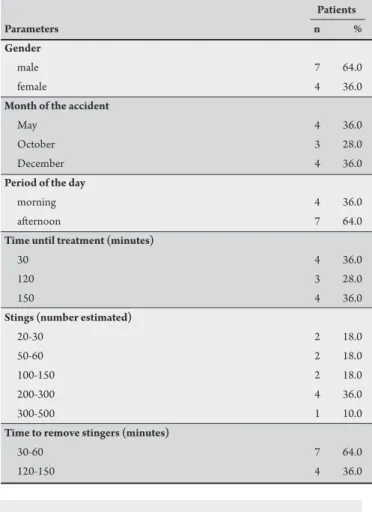
TABLE 1 - Identiication of patients atacked by Africanized honeybees. Patients
Parameters n %
Gender
male 7 64.0
female 4 36.0
Month of the accident
May 4 36.0
October 3 28.0
December 4 36.0
Period of the day
morning 4 36.0
aternoon 7 64.0
Time until treatment (minutes)
30 4 36.0
120 3 28.0
150 4 36.0
Stings (number estimated)
20-30 2 18.0
50-60 2 18.0
100-150 2 18.0
200-300 4 36.0
300-500 1 10.0
Time to remove stingers (minutes)
30-60 7 64.0
120-150 4 36.0
TABLE 2 - Clinical aspects of patients atacked by Africanized honeybees.
Present Absent
Clinical aspects n % n %
Local pain, edema, erythema, and itching 11 100.0 0 0.0
Nausea 8 73.0 3 27.0
Anemia 7 64.0 4 36.0
Tachycardia and vomiting 6 55.0 5 45.0
Hypertension and diarrhea 5 45.0 6 55.0
Generalized edema 4 36.0 7 64.0
Dizziness, hypotension, fever, urticaria, and weakness 3 27.0 8 73.0
Palpebral edema and wheezing lungs 2 18.0 9 82.0
Dry mouth, syncope, agitation, abdominal pain, laryngeal stridor, arthralgia, myalgia, anaphylaxis,and acute renal failure 1 9.0 10 91.0 Shock, arrhythmia, headache, convulsion, respiratory failure, oliguria, anuria, and central nervous system depression 0 0.0 11 100.0
a safe, eicacious antivenom, we have developed guidelines for the most appropriate care aimed at reducing the risk of death among these patients32. It must be emphasized that the Clinical Hospital of Botucatu
Medical School (HC-FMB/UNESP) has been renowned in its region for over 40 years for the treatment of accidents by venomous animals. he objectives of this study were to evaluate the patients treated from 2005 to 2006 survived a massive attack of Africanized honeybees (AHB), to discuss the anatomopathological indings of one case that resulted to death in 2003, and to propose treatment guidelines that comprise emergency care, drugs, and the proper removal of stingers.
A retrospective analysis of 11 accidents caused by multiple stings from AHB that occurred in the region of Botucatu, State of São Paulo, Brazil (48º 21' W, 22º 48' S) was carried out. hese included eight adults and three children treated at the Tropical Diseases Unit of HC-FMB from 2005 to 2006. he epidemiological, clinical, and therapeutic data were collected by conducting a review of the respective patient charts, whereas the laboratory data were obtained by means of an informational system of the network of HC-FMB laboratories. he case of an 11-year-old boy from Botucatu, São Paulo, Brazil, who died in 2003 was presented. He was atacked by a swarm of AHB ater he had entered the atic of a two-storey building located downtown to remove pigeon nests, unwitingly stimulating the beehive. Desperate, he jumped to the street from a height of approximately 10m. He was sent in critical condition to the HC-FMB but died ater 24h.
Ethical considerations
his project was submited to the Research Ethics Commitee of Botucatu Medical School-UNESP and received approval on November 5, 2007 (Of.455/2007-CEP).
he 11 surviving patients ranged in age from 5 to 87 years, averaging 42.5 years. Seven (64%) were male; four were female. Four patients were atacked in May, three in October, and four in December. As to the period of the day, most (64%) of them were atacked in the aternoon and received medical care within 2h ater the accident. he number of stings varied from 20 to 500, with 9 (82%) of the patients receiving more than 50 stings. None of the patients reported a previous atack by bees or said that the beehives were mechanically stimulated. he method adopted to remove the
stingers was blade for three patients, tweezers for six, and unknown for two patients. Table 1 summarizes these data.
Table 2 shows the clinical signs manifested by the patients. All patients reported local pain and local inlammatory signs (edema, erythema, itching). Nausea occurred in 73% of the cases, anemia in 64%, and tachycardia followed by vomiting in 55%. Hypertension and diarrhea, and generalized edema were detected in 45% and 36%, respectively. Dizziness, hypotension, fever, urticaria, weakness, palpebral edema, and wheezing lungs were also observed. Among these patients, arterial shock was not detectable.
DISCUSSION TABLE 3 - Laboratorial tests.
Increased Normal Not informed
Laboratorial tests n % n % n %
Leukocytes and neutrophils 11 100.0 0 0.0 -Creatine phosphokinase (CPK) 10 91.0 - 1 9.0 Lactate dehydrogenase (LDH) 5 46.0 3 27.0 3 27.0 Aspartate aminotransferase (AST) 5 46.0 4 36.0 2 18.0 Erythrocyte sedimentation rate 2 18.0 2 18.0 7 64.0 Urea and creatinine 1 9.0 10 91.0 -Alanine aminotransferase (ALT) 1 9.0 8 73.0 2 18.0
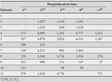
TABLE 4 - Creatine phosphokinase* levels during hospitalization. Hospitalization time
Patients 1st 2nd 3rd 4th >5th
1 - - - -
-2 - 1,297 1,128 1,185
-3 - 1,216 630 1,658
-4 175 4,090 1,101 5,777 1,511 5 167 4,670 5,024 4,251 1,367
6 288 225 - -
-7 139 2,255 982 1,465
-8 239 2,584 3,724 1,016 276
9 353 996 711 147 37
10 155 - 98 59
-11 279 1,554 4,738 - 721
*CPK (U/L).
Table 4 reveals the serum level of CPK observed over five days of hospitalization. On the irst day CPK level did not exceed 353U/L, but it increased from the second day onward, reaching levels of 5.777U/L on the fourth day then decreasing from the ith day onward.
Treatment employed and clinical outcome
All the patients received stimulation to promote diuresis, principally by intravenous administrations of electrolytic solutions (100%), mannitol (91%), and/or loop diuretic (18%). Sixty-four percent received sodium bicarbonate by intravenous route for alkalinization of urine. Among the analgesics utilized, opioids (64%) were prominent. Finally, all received antihistamines, initially intramuscularly and subsequently orally. Only one patient did not receive intravenous corticotherapy. Non-opioid and diuretic analgesics were employed for ive (45%) and two (18%) patients, respectively. he average time of hospitalization was 3.5 days, varying between 2 and 9 days, and all evolved to a complete cure without sequelae. he patient who received more than 1,000 bee stings arrived at the hospital unconscious, developed respiratory arrest, and subsequently died following a cardiac arrest.
Anatomopathological examination
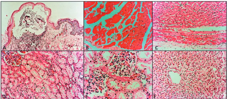
he anatomopathological examination of the patient who died revealed the following: skin showing subepidermal vesicle with epidermal necrosis and mononuclear inlammatory iniltrate at the base associated with vasculitis, congestion, and edema in the adjacent dermis; skeletal muscle including rhabdomyolysis characterized by iber eosinophilia, vacuolization, and absence of nuclei; heart showing necrotic cardiocytes characterized by iber anucleation, eosinophilia, and lumpy cytoplasmic material, associated with discrete interstitial
mononuclear iniltrate; kidney with tubular hydropic degeneration and myoglobinuria with eosinophilic amorphous material in Bowman’s and intratubular space; and liver with centrilobular areas with hepatocytic necrosis and discrete steatosis.
Accidents by Apis bee stings present distinct clinical manifestations, depending on the sensitivity of the individual to venom and on the number of stings. he most frequent accident is that in which an individual who is not sensitized to the venom is afected by a few stings. In these cases, the clinical picture is limited to local inlammatory reaction with erythematous papules, pain, and heat. In most instances, this situation is resolved without the participation of a physician.
Another clinical presentation is that in which an individual previously sensitized to one or more venom components manifests immediate hypersensitivity. This can be triggered by only one sting and requires immediate medical intervention. In general, the clinical picture is manifested by laryngeal edema and bronchospasm accompanied by anaphylactic shock.
The third presentation is that of multiple stings. Generally, the accident occurs in the ield and includes inoculation of a large quantity of venom caused by hundreds or thousands of stings33-36.
he clinical picture of accidents always initiates on the skin, the location of a sting with venom inoculation. his event can evolve to necrosis, as shown in Figure 1A. his image shows a subepidermal vesicle with epidermal necrosis and mononuclear inlammatory iniltrate at the base associated with vasculitis. In addition, there was congestion and edema in the adjacent dermis. he presence of diferent molecules in the venom, including phospholipase A2, hyaluronidase, melitin, apamine37-40, and, most recently, a serine
protease-like protein studied by Lima et al.41, may contribute to skin
necrosis at the sting site. Such necrosis has frequently occurred and has been reported by most studies in Brazil33-45.
The presence of rhabdomyolysis characterized by fiber eosinophilia, vacuolization, and absence of nuclei can be seen in Figure 1B. Rhabdomyolysis is a well-known cause of acute renal failure (ARF). Myoglobin toxicity has been related to renal vasoconstriction, intraluminal cast formation, and direct heme-protein cytotoxicity46. In a study conducted by Vanholder et al.47,
bee venom caused an early and significant increase in CPK, a reliable marker of the presence and intensity of muscle injury, and a massive tubular deposition of myoglobin. Other intramuscular cell enzymes like LDH and AST increased as well. Experimental bee venom-induced rhabdomyolysis with enzyme elevations was previously described by Azevedo-Marques and colleagues44. he
main factor responsible for rhabdomyolysis in bee venom is most likely melitin48. his substance inserts itself into the phospholipid
bilayer of cell membranes, causing hydrolysis and cell disruption49.
Phospholipase A2 may also damage the cell membrane and may play a role in rhabdomyolysis48. Clinically, a number of cases of ARF
induced by bee venom presented rhabdomyolysis that was evidenced by increased enzymes or myoglobinemia and myoglobinuria33,36,50.
In the present study, the maximum levels of CPK were observed on the fourth day of hospitalization, in agreement with the literature33-36.
FIGURE 1 - a Skin showing subepidermal vesicle with epidermal necrosis and mononuclear inlammatory iniltrate at the base associated with vasculitis. Congestion and edema in the adjacent dermis HE (40X). b Skeletal muscle including rhabdomyolysis characterized by iber eosinophilia, vacuolization, and absence of nuclei HE (400X). c Heart showing necrotic cardiocytes characterized by iber anucleation, eosinophilia, and lumpy cytoplasmic material, associated with discrete interstitial mononuclear iniltrate HE (200X). d Kidney with tubular hydropic degeneration: cytoplasmic vacuolization with regular nuclei. Spots of acute tubular necrosis: nuclear pyknosis and cytoplasmic eosinophilia HE (200X). e Myoglobinuria: eosinophilic amorphous material in Bowman’s space and intratubular space HE (400X). f Liver: Detail of centrilobular areas with hepatocytic necrosis: hepatocyte rarefaction, nuclear pyknosis, cytoplasmic eosinophilia, sinusoidal congestion, and edema of Disse’s space. Discrete steatosis HE (400X).
anucleation, eosinophilia, and lumpy cytoplasmic material, associated with mild interstitial mononuclear iniltrate. he venom provokes rhabdomyolitic activity in both experimental animals and humans44. In severe cases, there are areas of myocardial necrosis
similar to microinfarctions. here might be direct toxicity of the venom or only microinfarctions resulting from thrombosis of the microcirculation associated with the propagation of intravascular cloting36,44-45. Ferreira et al.45,51 demonstrated enzymatic changes
and morphological lesion of the acute myocardial infarction type, showing a possible direct toxic action of the venom on cardiac muscle.
On the other hand, the cardiac changes previously demonstrated in Wistar rats suggest that the components of the venom themselves or even substances released in the organism play some role in peripheral arteriolar resistance and may contribute to the changes in mean arterial pressure. In addition to presenting several vasoactive components, AHB venom contains melitin, a polypeptide toxin known to mobilize arachidonic acid from the cell membrane. Melitin evokes endothelium-derived hyperpolarizing factor-type relaxation by activating endothelial Ca2+-dependent phospholipase A2 followed by the transmission of a chemical and/or electrical signal via myoendothelial gap junctions. his vasorelaxation mechanism may be negatively regulated by nitric oxide44. Recent studies conducted by Guimarães et al.43 concluded that
the fall in mean arterial pressure is probably due to several factors, in addition to the cardiac changes already demonstrated in Wistar rats; it is possible that the venom components themselves or even substances released in the organism play some role in peripheral arteriolar resistance and may contribute to the changes in mean arterial pressure.
he kidneys require special atention in these accidents because hemolysis, rhabdomyolysis, and other direct efects of the venom contribute to acute tubular necrosis. Figure 1D shows a kidney with tubular hydropic degeneration: cytoplasmic vacuolization with regular nuclei and spots of acute tubular necrosis. Acute renal failure, which occurs after massive attacks by AHB, results
from toxic and ischemic mechanisms accompanied by hypovolemic and/or anaphylactic shock associated with tubular lesion caused by pigments released by muscular lesion (myoglobinuria), by hemolysis (hemoglobinuria), and by the direct toxic efect of the venom itself33-35.
his type of accident is followed by complications such as hypotension, hemolysis, rhabdomyolysis, clotting disturbances, and hepatic involvement34,52. According to Gabriel et al.53, four hypotheses must be
considered to explain the pathogenesis of acute tubular necrosis: I) a direct toxic efect of AHB venom on renal tubules, especially proximal tubules; II) a toxic efect of myoglobin and hemoglobin on the tubules (Figure 1E); III) an ischemic efect mediated by AHB or by substances released in the organism, or by a fall in cardiac output due to acute myocardial infarction-like lesions with a consequent fall in mean arterial pressure, reducing renal plasma low; and iv) a stress-potentiating efect acting, for example, on nicotinamide adenine dinucleotide release in the myocardium, aggravating the cardiac ischemic component and consequently renal perfusion.
The hepatic changes shown in Figure 1F are similar to those described by Barraviera et al.52,54 and reported in ophidian accidents.
These alterations were recently confirmed by other authors55-56.
hese are unspeciic lesions that may occur in the acute envenoming syndrome19-20,32 previously described and in situations of shock. Initially,
there is mitochondrial edema followed by Na+/K+ pump failure, hydropic degeneration, and hepatic necrosis19-20,52,54. In addition, lesions
due to the venom itself may worsen necroses observed in severe cases57.
Recently, Fighera et al.57 studied six fatal cases of dogs atacked by the
Finally, the accidents caused by multiple AHB stings always constitute a medical emergency. Early treatment, the removal of stingers, and precautions in relation to the status of hydration and renal function, in addition to the use of antihistamines, corticosteroids, non-opioid analgesics and diuretics, when indicated at appropriate doses, can be lifesavers in this type of accident.
Concluding remarks and recommendations
he treatment of patients stung by AHB should be approached as described below. First, we classify the case as an allergic or a toxic reaction. Next, we observe whether there is a local or a systemic aliction. he treatment must be conducted on a case-by-case basis, such as:
Allergic reactions: a) local (one or more stings): edema >10cm, progression up to 48h, duration in days, serous blister; b) systemic (one or more stings): reactions of hypersensitivity from degrees I to IV, including anaphylactic shock.
Toxic reactions: a)local (few stings): pain, erythema, low-intensity local edema, duration in hours; b) systemic (from 50 to 100 stings): pruritus, lushing, urticaria, sweating, fever, hypotension, headache, nausea or vomiting, abdominal cramps, bronchospasm, shock, respiratory failure, rhabdomyolysis, hemolysis, and ARF.
Allergic reactions
Accidents involving bee stings have diferent clinical manifestations
depending on the individual’s sensitivity to the venom and the number of stings. he most frequent accident is that in which an individual not sensitized to the venom is afected by a few stings. he clinical signs in these cases are limited to local inlammatory reaction, including erythematous papules, pain, and heat, and this situation is usually solved without medical assistance. When medical intervention is required, however, systemic antihistamines and topical corticosteroids are recommended. Dextrochlorpheniramine maleate at a dose of 2 to 6mg, administered orally every 6h for adults, and 0.15-0.30mg/kg weight up to a maximum of 5mg for children has been the drug of choice. his dosage must be maintained for at least 3 to 5 days depending on the case. In addition, we can use topical corticosteroids either alone or associated with menthol at 0.5%.
Another clinical presentation involves an individual previously sensitized to one or more venom components who manifests an immediate hypersensitivity reaction. his is a severe case that may be triggered by only one sting, and it requires immediate medical intervention. he general clinical signs include laryngeal edema and bronchospasm followed by anaphylactic shock. his patient requires hospitalization and immediate medical care. A thick vein from the patient’s forearm should be catheterized for liquid and drug infusion. he later should be maintained with glucose solution at 5% until the patient presents total hemodynamic stabilization. To promote hemodynamic stabilization in case of shock, it is necessary to ofer 20ml/kg of saline solution (0.9%) or lactate ringer intravenously as fast as possible (less than 5min). his volume must be repeated until the signals of shock have disappeared. In case of anaphylactic shock, the drugs should be injected in the following order. First is liquid adrenaline at 1:1,000 dilution. It is the only effective and immediate measure and should be used subcutaneously or intramuscularly at the dose of 0.3 to 1ml (0.01ml/kg weight). In cases of cardiac arrest, intravenous and/or endotracheal routes should be used at 1:10,000 dilution. Second is promethazine: one to two ampoules by intramuscular or intravenous route. For children, use 0.5 to 1.0mg/kg weight. his drug has antihistaminic efect.
We can also use diphenidramine in an intravenous route at a dose of 5mg/kg every 6h. In case there is no response, we must use anti-H2 antihistaminics. hird is aminophylline: his is a bronchodilator drug indicated for bronchospasm. Use 5mg/kg weight (0.3ml/kg weight). For children, we prefer to use inhalatory short-acting beta-2 agonists. Concomitantly, oxygen must be ofered in a maximum concentration to maintain pulse oximetry above 92%. Fourth drug is hydrocortisone: his is a corticosteroid-type drug that inhibits the action of inlammation mediators. Use 5-10mg/kg body weight diluted in 100ml glucose solution at 5%. Administer repeated intravenous doses every 6h.
Toxic reactions
Multiple stings are the third presentation of this type of accident. In this case, treatment is always a medical emergency. Unfortunately, a speciic antivenom is not yet available, although many researches have been conducted. Based on the experience of other Brazilian authors33-36 and on the protocol used at the Department of Tropical
Diseases, Botucatu Medical School, UNESP, the following measures should be taken as soon as the patient arrives at the hospital. A) Inject, by intramuscular route, an ampoule of promethazine; for children, use 0.5-1.0 mg/kg body weight. B) Inject, by intramuscular route, an ampoule of meperidine-type hypnoanalgesic; for children, administer 1.5mg/kg weight/day. C) If the patient is in shock, inject subcutaneously or intramuscularly from 0.5 to 1 ampoule of liquid adrenaline 1:1,000; for children, use 0.01mg/kg body weight. D) In case of bronchospasm inject, by intramuscular route, 1 ampoule of aminophylline; for children, preferentially, use short-acting beta-2 agonist. E) Catheterize a central vein, and then install central venous pressure; in case of shock, use intraosseous route if necessary. F) Administer intravenously 1g hydrocortisone; for children, use 5-10mg/kg body weight. his scheme should be maintained for at least three to ive days, according to the clinical evolution. G) he patient must be well hydrated with colloids and crystalloids; then, osmotic diuresis should be induced intravenously with 20% mannitol, at a dose of 100ml, every 6h for adults and from 10 to 12.5ml/kg body weight for children. Mannitol administration should continue for at least ive days. However, special atention should be paid to avoid iatrogenic dehydration. For patients presenting anuria, mannitol is contraindicated. H) Alkalinize the urine with sodium bicarbonate solution at a dose of 1 to 2mEq/kg weight/dose every 6h to prevent renal lesions caused by Hemoglobinuria and myoglobinuria.
Finally, regarding such injuries, treatment choice is always challenging and must be assessed case by case, especially when the patient is a child or an adult over 60 years. In these situations age, weight, and possible associated morbid conditions should be considered when choosing the best therapy to avoid unfavorable clinical outcomes.
he authors declare that there is no conlict of interest.
CONFLICT OF INTEREST
FINANCIAL SUPPORT
REFERENCES
Fundação de Amparo à Pesquisa do Estado de São Paulo (FAPESP);
Process number 2006/55545-8 (RSFJr), 2007/05159-7 (BB).
1. Rutner F. Isolated populations of honeybees in Australia. J Apic Res 1986; 15:97-104.
2. Kerr WE. he history of the introduction of African bees in Brazil. S Afr Bee J 1967; 39:33-35.
3. Clarke KE, Rinderer TE, Franck P, Quezada-Euán JG, Oldroyd BP. The Africanization of honeybees (Apis mellifera L.) of the Yucatan: a study of a massive hybridization event across time. Evolution 2002; 56:1462-1474.
4. Cornuet JM. Population genetics. In: Rinderer TE, editor. Bee genetics and breeding. Orlando, FL: Academic Press; 1986. p. 235-254.
5. Rutner F, Tassencourt L, Louveaux J. Biometrical-statistical analysis of the geographic variability of Apis mellifera L. Apidologie 1978; 9:363-381. 6. Rothenbuhler WC. Semidomesticated insects: honeybee breeding.
In: Moy MA, McKelvey JJ, editors. Genetics and beneicial organisms. Bellagio, Italy: Rockefeller Foundation; 1979. p. 84-92.
7. Taylor OR. he past and possible future spread of Africanized honeybees in the Americas. Bee World 1977; 58:19-30.
8. Alaniz J. he buzz: health care workers step up training to prepare for killer bee atacks [Internet]. Nurseweek; 2000 - [cited 2010 Apr 26]. Available from: htp:// www.nurseweek.com/news/features/00-08/bees.html.
9. Kaplan J. What’s buzzing with Africanized honeybees? [Internet]. Agri Res; 2004 Mar - [cited 2010 Apr 26]. Available from: htp://www.ars.usda.gov/is/ AR/archive/mar04/bees0304.html.
10. Oklahoma State University. Africanized honey bees in Oklahoma [Internet]. Oklahoma: Department of Entomology, Oklahoma State University. [cited 2010 Apr 26]. Available from: htp://www.entoplp.okstate.edu/ahb.
11. University Of California. Africanized honey bee information in brief [Internet]. California: Department of Entomology. [cited 2010 Mar 10]. Available from: htp://bees.ucr.edu/ahb-facts.html.
12. United States Department of Agriculture. Africanized honey bees [Internet]. [place unknown]: United States Department of Agriculture. [cited 2010 Apr 26]. Available from: http://www.ars.usda.gov/Research/docs. htm?docid=11059&pf=1&cg_id=0.
13. Agricultural Research Service. United States Department of Agriculture. Africanized honey bees [Internet]. [place unknown]: United States Department of Agriculture. [cited 2010 Jun 05]. Available from: htp://www.ars.usda.gov/ Research/docs.htm?docid=11059&page=6.
14. Taylor OR. African bees: potential impact in the United States. Bull Entomol Soc Am 1985; 31:15-24.
15. De La Rua P, Jaffé R , Dall’Olio R , Muñoz I, Serrano J. Biodiversity, conservation and current threats to European honeybees. Apidologie 2009; 40:263-284.
16. Ferreira Jr RS, Sciani JM, Marques-Porto R, Lourenço Jr A, Orsi RO, Barraviera B, et al. Africanized honeybee (Apis mellifera) venom profiling: seasonal variation of melitin and phospholipase A2 levels. Toxicon 2010; 56:355-362.
17. Schumacher MJ, Schmidt JO, Egen NB, Dillon A. Biochemical variability of venoms from individual European and Africanized honeybees (Apis mellifera). J Allergy Clin Immunol 1992; 90:59-65.
18. Schumacher MJ, Schmidt JO, Egen NB, Lowry JE. Quantity, analysis and lethality of European and Africanized honeybee venoms. Am Trop Med Hyg 1990; 43:79-86.
19. Barraviera B, Lomonte B, Tarkkowski A, Hanson LA, Meira DA. Acute-phase reactions, including cytokines, in patients biten by Bothrops and Crotalus snakes in Brazil. J Venom Anim Toxins1995; 1:11-22.
20. Barraviera B. Acute-phase response in snake bite. Toxicon 1994; 32:861-862. 21. Betten DP, Richardson WH, Tong TC, Clark RF. Massive honey bee
envenomation-induced rhabdomyolysis in an adolescent. Pediatrics 2006; 117:231-235.
22. Petricevich VL. Scorpion venom and the inlammatory response. Mediators Inlamm 2010; 2010:903295.
23. Voronov E, Apte RN, Sofer S. he systemic inlammatory response syndrome related to the release of cytokines following severe envenomation. J Venom Anim Toxins 1999; 5:5-33.
24. Murthy KRK, Murthy RK. On scorpion envenoming syndrome. Problems of medical ethics and accountability in medical research in India. J Venom Anim Toxins 2002; 8:3-17.
25. D’Suze G, Moncada S, González C, Sevcik C, Aguilar V, Alagón A. Relationship between plasmatic levels of various cytokines, tumour necrosis factor, enzymes, glucose and venom concentration following Tityus scorpion sting. Toxicon 2003; 41:367-375.
26. Amaral CF, Rezende NA. Both cardiogenic and non-cardiogenic factors are involved in the pathogenesis of pulmonary oedema ater scorpion envenoming. Toxicon 1997; 35:997-998.
27. Magalhães MM, Pereira ME, Amaral CF, Rezende NA, Campolina D, Bucaretchi F, et al. Serum levels of cytokines in patients envenomed by Tityus serrulatus
scorpion sting. Toxicon 1999; 37:1155-1164.
28. Bertazzi DT, Assis-Pandochi AI, Azzolini AE, Talhaferro VL, Lazzarini M, Arantes EC. Efect of Tityus serrulatus scorpion venom and its major toxin, TsTX-I, on the complement system in vivo. Toxicon 2003; 41:501-508.
29. Petricevich VL. Cytokine and nitric oxide production following severe envenomation. Curr Drug Targets Inlamm Allergy 2004; 3:325-332. 30. Moura-da-Silva AM, Laing GD, Paine MJ, Dennison JM, Politi V, Crampton JM,
et al. Processing of pro-tumor necrosis factor-alpha by venom metalloproteinases: a hypothesis explaining local tissue damage following snake bite. Eur J Immunol 1996; 26:2000-2005.
31. Hernández Cruz A, Garcia-Jimenez S, Zucatelli Mendonça R, Petricevich VL. Pro- and anti-inlammatory cytokines release in mice injected with Crotalus
durissus terriicus venom. Mediators Inlamm 2008; 2008:874-962.
32. Ferreira Jr RS, Almeida RAMB, Barraviera SRCS, Barraviera B. Historical perspective and human consequences of Africanized bee stings in the Americas. J Toxicol Environm Health 2011; Forthcoming.
33. Bresolin NL, Carvalho FC, Goes JC, Fernandes V, Baroto AM. Acute renal failure following massive atack by Africanized bee stings. Pediatr Nephrol 2002; 17:625-627.
34. Daher EF, Silva Jr GB, Bezerra GP, Pontes LB, Martins AMC, Guimarães JA. Acute renal failure ater massive honeybee stings. Rev Inst Med Trop São Paulo 2003; 45:45-50.
35. Daher EF, Oliveira A, Silva LSV, Silva EMB, Morais TP. Insuiciência renal aguda por picada de abelhas: relato de casos. Rev Soc Bras Med Trop 2009; 42:209-212. 36. França FO, Benvenuti LA, Fan HW, Dos Santos DR, Hain SH, Picchi-Martins FR, et al. Severe and fatal mass atacks by “killer” bees (Africanized honey bees -
Apis mellifera scutellata) in Brazil: clinicopathological studies with measurement of serum venom concentrations. Q J Med 1994; 87:269-282.
37. Koçer U, Ozer Titikcioglu Y, Mete Aksoy H, Karaaslan Ö. Skin and sot tissue necrosis following hymenoptera sting. J Cutan Med Surg 2003; 7:133-135. 38. Lima PR, Brocheto-Braga MR. Hymenoptera venom review focusing on Apis
mellifera. J Venom Anim Toxins Incl Trop Dis 2003; 9:149-162.
40. Funari SRC, Zeidler PR, Rocha HC, Sforcin JM. Venom production by Africanized honey bees (Apis mellifera) and Africanized-European hybrids. J Venom Anim Toxins 2001; 7:190-198.
41. Lima PRM, Brochetto-Braga MR, Chaud-Netto J. Proteolytic activity of Africanized honeybee (Apis mellifera: hymenoptera, apidae) venom. J Venom Anim Toxins 2000; 6:64-76.
42. Daher EF, Silva Junior GB, Bruneta DM, Pontes LB, Bezerra GP. Rhabdomyolysis and acute renal failure ater strenuous exercise and alcohol abuse: case report and literature review. São Paulo Med J 2005; 123:33-37.
43. Guimarães JV, Costa RS, Machado BH, Reis MA. Cardiovascular proile ater intravenous injection of Africanized bee venom in awake rats. Rev Inst Med Trop São Paulo 2004; 46:55-58.
44. Azevedo-Marques MM, Ferreira DB, Costa RS. Rhabdomyonecrosis experimentally induced by Wistar rats by Africanized bee venom. Toxicon 1992; 30:344-348.
45. Ferreira DB, Costa RS, De Olivera JA, Muccillo G. An infarct-like myocardial lesion experimentally induced in Wistar rats with Africanized bee venom. J Pathol 1995;177:95-102.
46. Zager RA. Rhabdomyolysis and myohemoglobinuric acute renal failure. Kidney Int 1996; 49:314-326.
47. Vanholder R, Sever MS, Erek E, Lameire N. Rhabdomyolysis. J Am Soc Nephrol 2000; 11:1553-1561.
48. Ownby CL, Powell JR, Jiang MS, Fletcher JE. Melitin and phospholipase A2 from bee (Apis mellifera) venom cause necrosis of murine skeletal muscle in vivo. Toxicon 1997; 35:67-80.
49. Schumacher MJ, Egen NB. Signiicance of Africanized bees for public health. A review. Arch Intern Med1995; 155:2038-2043.
50. Kolecki P. Delayed toxic reaction following massive bee envenomation. Ann Emerg Med 1999; 33:114-116.
51. Ferreira DB, Costa RS, Oliveira JS, Muccillo G. Cardiac noradrenaline in experimental rat envenomation with Africanized bee venom. Exp Toxicol Pathol 1994; 45:507-511.
52. Barraviera B, Bonjorno Júnior JC, Arkaki D, Domingues MA, Pereira PC, Mendes RP, et al. A retrospective study of 40 victims of Crotalus snake bites. Analysis of the hepatic necrosis observed in one patient. Rev Soc Bras Med Trop 1989; 22:5-12.
53. Gabriel DP, Rodrigues Junior AG, Barsante RC, Santos-Silva V, Caramori JT, Martim LC, et al. Severe acute renal failure ater massive atack of Africanized bees. Nephrol Dial Transplant 2004;19:2680.
54. Barraviera B, Coelho KYR, Curi PR, Meira DA. Liver dysfunction in patients bitten by Crotalus durissus terrificus (Laurenti, 1768) snakes in Botucatu (State of São Paulo - Brazil). Rev Inst Med Trop São Paulo 1995; 37:63-70. 55. França RF, Vieira RP, Ferrari EF, Souza A, Osorio AL, Prianti Jr ACG, et al.
Acute hepatotoxicity of Crotalus durissus terriicus (South American ratlesnake) venom in rats. J Venom Anim Toxins incl Trop Dis 2009;15:61-78.
56. Teixeira GN, Vilela-Goulart MG, Iakimof A, Bastos-Ramos WP, Matos-Filho TR, Lopes-Martins A, et al. Preliminary characterization of hepatotoxicity of crotalic venom. J Venom Anim Toxins incl Trop Dis 2003; 9:431.