Myocardial Dysfunction in Chagasic Patients with no Apparent Heart
Disease
Airandes de Sousa Pinto, Braulio Muzzi Ribeiro de Oliveira, Fernando Antonio Botoni, Antonio Luiz Pinho Ribeiro,
Manoel Otavio da Costa Rocha
Department of Internal Medicine and Postgraduate Program in Tropical Medicine, Faculdade de Medicina da Universidade Federal de Minas Gerais, Belo Horizonte, MG - Brazil
Summary
Background: The Tei index simultaneously evaluates the ventricular systolic and diastolic functions. It has a good correlation with hemodynamic data and has enabled the detection of initial ventricular function alterations of different etiologies.
Objective: To study initial biventricular function alterations in chagasic patients with no apparent heart disease, using the Tei index.
Methods: Forty eight individuals were evaluated. Group 1 was comprised of 25 patients diagnosed with Chagas disease with no apparent heart disease and normal echocardiogram; group 2 was comprised of 23 healthy individuals with negative serological tests for Chagas disease.
Results: The Tei index was significantly higher in the group of chagasic patients with no apparent heart disease when compared to that of the control group: left ventricular Tei index (0.48 ± 0.11 vs. 0.36 ± 0.06, p < 0.001) , right ventricular Tei index (0.34 ± 0.10 vs. 0.26 ± 0.07, p = 0.001). The left and right ventricular Tei indexes were considered abnormal in 48% and 28% of the chagasic patients, respectively.
Conclusion: Chagasic patients with no apparent heart disease present early alterations of the right and left ventricles. (Arq Bras Cardiol 2007;89(6):348-353)
Key words: Ventricular dysfunction; Chagas disease; Chagas cardiomyopathy.
Mailing address: Airandes de Sousa Pinto •
Rua Aristides Novis, 139/301 - Kalilândia - 44025-300 Feira de Santana, BA - Brazil
E-mail: [email protected]
Manuscript received August 13, 2006; revised manuscript received May 18, 2007; accepted June 18, 2007
Introduction
The expression Chagas disease with no apparent heart disease refers to asymptomatic patients with positive serological test for Trypanosoma cruzi infection, and normal electrocardiogram and chest radiograph. Since radiological studies of their digestive system have not been performed, they cannot be classified in the indeterminate form 1. Fifty
percent of the chagasic patients are estimated to have this clinical form in endemic areas.
The assessment of the ventricular function is important for the treatment and prognostic evaluation in the different chronic heart diseases. The assessment of early myocardial impairment in patients with Chagas disease may be useful to establish subgroups of patients with a worse prognosis. The Tei index, which simultaneously assesses the ventricular systolic and diastolic functions, has proven useful to assess the ventricular function in several heart diseases 2-5.
The objective of this study is to evaluate the presence and
magnitude of ventricular function alterations in chagasic patients with no apparent heart disease, by using the Tei index.
Methods
Patients - Patients consecutively recruited in the Chagas Disease Referral Outpatient Service of Hospital das Clínicas da Universidade Federal de Minas Gerais, referred by blood banks or primary care units for serological confirmation and medical care were studied. For this investigation, asymptomatic chagasic patients with normal cardiac silhouette on chest radiograph, ejection fraction equal to or higher than 55% and no alterations of segmental motion on echocardiogram, as well as normal esophagogram and electrocardiogram were selected (group 1). T. cruzi infection was confirmed by at least two positive serological tests, among those routinely used in the hospital (indirect immunofluorescence, ELISA and indirect hemagglutination).
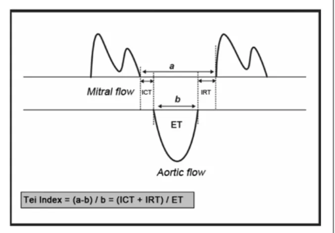
Interval “a”, which corresponds to the time extending from the end of the “A” wave to the beginning of the tricuspid “E” wave, incorporates the sum of the right ventricular isovolumic contraction time, ejection time, and isovolumic relaxation time. Interval “b” corresponds to the right ventricular ejection time. The sum of the isovolumic contraction time and isovolumic relaxation time was obtained by subtracting “b” from “a”. The right ventricular Tei index was calculated by dividing the sum of the isovolumic contraction time and isovolumic relaxation time by the ejection time8.
Statistical analysis - Continuous data were expressed as mean ± standard deviation.
To compare the groups in relation to discrete variables, the chi-square test was used and also the Fisher’s exact test, when necessary. Parametric continuous variables were analyzed using the Student’s t test. When necessary, logarithmic transformation was performed to allow the analysis by parametric methods.
All results were considered significant for a significance probability lower than 5%.
Results
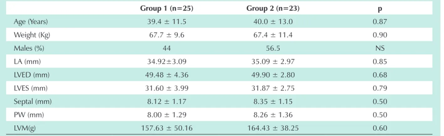
A total of 48 individuals participated in this study, 25 (52.08%) in group 1 (with no apparent heart disease) and 23 (47.92%) in group 2 (control group). General and echocardiographic characteristics of the study sample are shown in Tables 1 and 2.
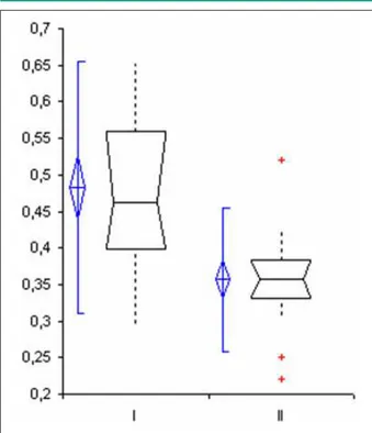
When analyzed as regards conventional variables, group 1 did not show any differences in relation to the control group. There was a trend of greater deceleration time in group 1 (190.38 ± 35.17 vs. 169.29 ± 35.50, p = 0.06). However, the left ventricular and right ventricular Tei indexes were significantly higher in chagasic patients when compared to the control group: left ventricular Tei index (0.48 + 0.11 vs. 0.36 + 0.06, p < 0.001), right ventricular Tei index (0.34 + 0.10 vs. 0.26 + 0.07, p = 0.001) (Figures 2 and 3).
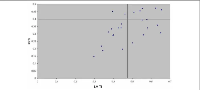
The cut-off point of Tei indexes was obtained considering the control group mean + 2 standard deviations. Tei indexes ≤ 0.48 and ≤ 0.40 for left and right ventricles, respectively, were considered normal. Twelve (48%) patients had an abnormal left ventricular Tei index, and seven (28%) patients had an abnormal right ventricular Tei index. Five (20%) patients had concomitant right and left ventricular alterations, and 11 (44%) patients had normal left and right ventricular indexes. Two (8%) patients had right ventricular alteration alone, and seven (28%) patients had abnormal left ventricular Tei index alone (Figure 4). In the control group, two (8.7%) individuals had Tei index alterations. One (4.4%) patient had left ventricular alteration alone, and one (4.4%) patient had right ventricular alteration alone.
Discussion
Approximately half of the chagasic patients in endemic areas do not have any cardiovascular complaint and have normal electrocardiogram and chest radiograph. Longitudinal studies demonstrate that some of them may remain like this indefinitely9-11, whereas others develop
Patients with the following characteristics were excluded: any evidence of cardiovascular disease; diabetes mellitus; chronic obstructive pulmonary disease; alcohol use; renal failure; anemia; pregnancy; thyroid dysfunction; and any other disease that could interfere with the ventricular function. Written informed consent to participate in the study was obtained from all patients, and the study protocol was approved by the University’s Research and Ethics Committee.
Echocardiographic assessment - Echocardiographic study of all individuals was performed by a single examiner using the HDI 5000-ATL equipment, in the Service of Cardiology and Cardiovascular Surgery of Hospital das Clínicas da UFMG. It was comprised of standard views from the parasternal, apical, subcostal and suprasternal positions.
The left ventricular systolic function was assessed using the ejection fraction. Final diastolic and systolic volumes were estimated using the Teichholz et al’s method 6.
The left ventricular Tei index was measured using the mean of three consecutive intervals. Time intervals obtained with pulsed Doppler were measured in the mitral flow with the sample positioned at the tip of the leaflets, and in the left ventricular outflow tract (sample positioned directly anterior to the aortic valve). Interval “a”, which corresponds to the time between the end of the “A” wave and the beginning of the mitral “E” wave, incorporates the sum of the isovolumic contraction time, ejection time, and isovolumic relaxation time. Interval “b” corresponds to the left ventricular ejection time. The sum of the isovolumic contraction time and isovolumic relaxation time was obtained by subtracting “b” from “a” (Figure 1). The left ventricular Tei index was calculated by dividing the sum of the isovolumic contraction time and isovolumic relaxation time by the ejection time7.
Quantitative analysis of the right ventricle was performed using the Tei index. The right ventricular Tei index was evaluated using the mean of three measurements in each time interval. The time intervals, obtained with pulsed Doppler, were measured in the tricuspid valve flow (sample positioned close to the ring) and in the right ventricular outflow tract (sample positioned directly anterior to the pulmonary valve).
some clinical form of the disease within a period of 10 to 20 years, at a rate of 2% to 5% a year12. Survival analysis
of chagasic patients with no apparent heart disease demonstrates that it is similar to that of normal individuals13.
However, sudden death, although uncommonly, may be the first manifestation of the disease14.
Autopsy findings are not frequently available because of the low mortality rate of these patients. Complete pathological studies are practically limited to examinations of patients who died of violent deaths15 and to experimental models in dogs16.
The analysis of endomyocardial biopsy of these individuals shows focal areas of myocarditis17. Little cell destruction was
demonstrated on ultrastructural studies. Inflammatory cells accumulate in focal myocardial areas, with destruction of the intercellular matrix in some areas. Unlike in the acute phase, few cardiomyocytes are invaded or destroyed18.
These alterations have neither a clinical expression nor do they promote changes in the genesis and conduction of the cardiac impulse, but may be responsible for the presence of alterations in more sensitive tests for cardiovascular assessment such as exercise test19, cardiopulmonary exercise
test20, Holter monitoring21, radionuclide ventriculography22,
autonomic function tests23, and echocardiography24-26.
However, the prognostic implications of these findings are still not fully known27.
The analysis of the diastolic function in chagasic patients with no evidence of heart disease has demonstrated alterations in the mitral “E” wave deceleration time and in the isovolumic relaxation time25. In the present study, the trend of an increase
in deceleration time was observed in group 1.
The ventricular function depends on the systole and diastole, both energy-dependent. However, this dichotomization is artificial because the first phase of diastole – the relaxation, starts in the second phase of systole28.
The Tei index, calculated by the formula TI = (IRT + ICT) / ET, incorporates the systolic and diastolic functions in a single value. Systolic dysfunction results in the prolongation of the isovolumic contraction time and shortening of the ejection time. The isovolumic relaxation time is prolonged by the alteration in relaxation that results from the diastolic dysfunction29. The Tei index, therefore, increases in the
presence of myocardial dysfunction.
Tei et al2 found a good correlation between the Tei index
and invasive contractility and relaxation measurements, Table 1 - General and echocardiographic characteristics of patients with Chagas disease and of control group individuals
Group 1 (n=25) Group 2 (n=23) p
Age (Years) 39.4 ± 11.5 40.0 ± 13.0 0.87
Weight (Kg) 67.7 ± 9.6 67.4 ± 11.4 0.90
Males (%) 44 56.5 NS
LA (mm) 34.92±3.09 35.09 ± 2.97 0.85
LVED (mm) 49.48 ± 4.36 49.90 ± 2.80 0.68 LVES (mm) 31.60 ± 3.99 31.87 ± 2.75 0.79 Septal (mm) 8.12 ± 1.17 8.35 ± 1.15 0.50
PW (mm) 8.00 ± 1.29 8.26 ± 1.36 0.50
LVM(g) 157.63 ± 50.16 164.43 ± 38.25 0.60
LA - Left atrial diameter; LVED - Left ventricular end-diastolic diameter; LVES - Left ventricular end-systolic diameter; Septal - Interventricular septal thickness; PW - Posterior wall thickness; LVM - left ventricular mass
Table 2 - Echocardiographic characteristics of patients with Chagas disease and of control group individuals
Group 1 (n=25) Group 2 (n=23) P
EF (%) 65.34±6.46 65.34±5.37 0.32
LV TI 0.48±0.11 0.36±0.06 < 0.001
RV TI 0.34±0.10 0.26±0.07 0.001
E (cm/s) 75.19±20.30 75.43±11.32 0.96
A (cm/s) 56.83±13.37 56.46±12.87 0.55
E/A 1.32±0.41 1.39±0.29 0.50
DT (ms) 190.38±35.17 169.29±35.50 0.06
Fig. 2 - Box-whisker plots of left ventricular Tei index in 25 chagasic patients with no apparent heart disease, and in 23 patients of the control group.
Fig. 3 - Box-whisker plots of right ventricular Tei index in 25 chagasic patients with no apparent heart disease, and in 23 patients of the control group.
DP/DT and Tau, respectively, regardless of pre and afterload, thus demonstrating that the Tei index is a measurement of the global ventricular function2.
Ejection fraction measurement using echocardiography
is based on the similarity of the ventricle with geometric models30. However, the right ventricle has a complex
structure and, in practice, it has been subjectively assessed. Because the Tei index uses values obtained with Doppler, which are not dependent on the right ventricular geometry, it is an alternative for the quantitative measurement of the right ventricular function. Tei et al8 used the right ventricular
Tei index in patients with primary pulmonary hypertension. The Tei index was the best variable to discriminate patients with pulmonary hypertension, in addition to being a predictor of survival8. Moller et al31 studied the left and right
ventricular Tei indexes after the first myocardial infarction. They observed that the right ventricular Tei index had a high sensitivity and low specificity for the diagnosis of right ventricular infarction. In the multivariate analysis, the left and right ventricular Tei indexes were independent predictors of mortality. Chockalingam et al32 demonstrated that a Tei
index ≥ 0.30 has a high sensitivity and specificity for the diagnosis of right ventricular infarction in the presence of inferior infarction32.
In this study, the Tei index demonstrated an early alteration of the left and right ventricular myocardial function. The ability to detect early myocardial alterations was also demonstrated by Sato et al33 when evaluating
anthracycline cardiotoxicity in children. On the other hand, the Tei index also represents an important tool in the verification of initial improvement of the left ventricular function after therapeutic interventions34-35.
Early right ventricular impairment has already been determined in Chagas disease, although right ventricular dysfunction alone, as assessed by echocardiography, is not a predominant event in chronic chagasic cardiopathy36.
Marin-Neto et al37 demonstrated early right ventricular
impairment in the absence of left ventricular impairment in patients with no heart disease37. Other authors, however,
observed that despite the fact that few alterations of pulmonary congestion are seen in patients with chronic chagasic cardiopathy, the finding of right ventricular dilatation is usually concomitant with left ventricular functional impairment.
This study had the following limitations: 1) other diastolic function measurements were not taken; 2) there is no consensus as regards normal Tei index values; 3) lack of intra and interobserver variability measurements; 4) there was a considerable overlapping of results between the control and chagasic groups; 5) the clinical importance and prognostic implications of these findings are still not fully known.
However, our objective was merely to draw comparisons between the two groups, without studying the diastolic function alone, or determining absolute values of the Tei index in patients with no apparent heart disease.
In conclusion, the use of the Tei index demonstrated the capability to detect early alterations in the global biventricular function in chagasic patients with no apparent heart disease.
Potential Conflict of Interest
References
1. I Reunião de Pesquisa Aplicada em Doença de Chagas: validade do conceito de forma indeterminada de doença de Chagas. Rev Soc Bras Med Trop. 1985; 18: 46.
2. Tei C, Nishimura RA, Seward JB, Tajik AJ. Noninvasive Doppler-derived myocardial performance index: correlation with simultaneous measurements of cardiac catheterization measurements. J Am Soc Echocardiogr. 1997; 10: 169-78.
3. Moyssakis I, Tzanetea R, Tsaftaridis P, Rombos I, Papadopoulos DP, Kalotychou V, et al. Systolic and diastolic function in middle aged patients with sickle ß thalassaemia: an echocardiographic study. Postgrad Med J. 2005; 81: 711-4.
4. Veselka J, Procházková S, Bolomová-Homolová I , Duchoňová R, Tesař D. Effects of alcohol septal ablation for hypertrophic obstructive cardiomyopathy on Doppler Tei Index: a midterm follow-up. Echocardiography. 2005; 22: 105-9.
5. Seyfarth HJ, Pankau H, Hammerschmidt S, Schauer J, Wirtz H, Winkler J. Bosentan improves exercise tolerance and Tei Index in patients with pulmonary hypertension and prostanoid therapy. Chest. 2005; 128: 709-13.
6. Teichholz LE, Kreulen T, Herman MV, Gorlin R. Problems in echocardiographic volume determinations: echocardiographic-angiographic correlations in the presence of absence of asynergy. Am J Cardiol. 1976; 37: 7-11.
7. Tei C, Ling LH, Hodge DO, Bailey KR, Oh JK, Rodeheffer RJ, et al. New index of combined systolic and diastolic myocardial performance: a simple and reproducible measure of cardiac function - a study in normals and dilated cardiomyopathy. J Cardiol. 1995; 26: 357-66.
8. Tei C, Dujardin KS, Hodge DO, Bailey KR, McGoon MD, Tajik AJ, et al. Doppler echocardiographic index for assessment of global right ventricular function. J Am Soc Echocardiogr. 1996; 9: 838-47.
9. Coura JR, Anunziato N, Willcox, HP. Morbidade da doenca de Chagas: I - estudo de casos procedentes de vários estados do Brasil, observados no Rio de Janeiro. Mem Inst Oswaldo Cruz. 1983; 78: 363-72.
10. Pereira JB, Willcox HP, Coura JR. Morbidade da doença de Chagas: III. estudo longitudinal de seis anos em Virgem da Lapa, Minas Gerais, Brasil. Mem Inst Oswaldo Cruz. 1985; 80: 63-71.
11. Ianni BM, Arteaga E, Frim CC, Pereira Barretto AC, Mady C. Chagas’ heart disease: evolutive evaluation of electrocardiographic and echocardiographic parameters in patients with the indeterminate form. Arq Bras Cardiol. 2001; 77: 59-62.
12. Dias JC. The indeterminate form of human chronic Chagas’ disease: a clinical epidemiological review. Rev Soc Bras Med Trop. 1989; 22: 147-56.
13. Kloetzel K, Dias JC. Mortality in Chagas’ disease: life table for the period 1949-1967 in an unselected population. Rev Inst Med Trop São Paulo. 1968; 10: 5-8.
14. Prata A. Clinical and epidemiological aspects of Chagas disease. Lancet Infect Dis. 2001; 1: 92-100.
15. Lopes ER, Chapadeiro E, Andrade ZA, Almeida HO, Rocha A. Anatomia patológica dos corações de chagásicos assintomáticos falecidos de modo violento. Mem Inst Oswaldo Cruz. 1981; 76: 189-97.
16. Scalabrini A, Cardoso A, Andrade SG, Andrade ZA. Correlação clínico-patológica na forma indeterminada da doença de Chagas experimental do cão. Arq Bras Cardiol. 1996; 67: 385-8.
17. Mady C, Pereira-Barretto AC, Ianni BM, Lopes EA, Pileggi F. Right ventricular endomyocardial biopsy in undetermined form of Chagas’ disease. Angiology. 1984; 35: 755-9.
Fig. 4 - Characterization of 25 chagasic patients with no apparent heart disease in relation to the left and right ventricular Tei indexes. Sources of Funding
This study was funded by Conselho Nacional de Desenvolvimento Científico e Tecnológico (CNPq).
Study Association
This article is part of the thesis of master submitted by Airandes
de Souza Pinto, from Programa de Pós-graduação em Medicina
18. Andrade, Z. Immunopathology of Chagas disease. Mem Inst Oswaldo Cruz. 1999; 94: 71-80.
19. Bellini AJ, Santos RC, Bilac A. Prova de esforço na forma sub-clínica da doença de Chagas. Arq Bras Cardiol. 1977; 30: 261.
20. Mady C, Yasbek P Jr , Pereira Barretto AC , Vianna CB, Serro-Azul LG, Bellotti G, et al. Estudo da capacidade funcional máxima pela ergoespirometria em pacientes portadores da doença de Chagas. Arq Bras Cardiol. 1986; 47: 201-5.
21. Marins N, Flores AP, Seixas TN, da Costa Fagundes J, Ostrowsky M, De Marco Martins A, et al. Estudo da forma indeterminada da doença de Chagas através da eletrocardiografia dinâmica. Arq Bras Cardiol. 1982; 39: 303-7.
22. Giorgi MC, Meneguetti JC, Hironaka FH, Barretto AC, Arteaga-Fernandez E, Bellotti, et al. Quantificação de captação miocárdica de Gálio 67 em pacientes portadores de doença de Chagas. Arq Bras Cardiol. 1985; 45: 132.
23. Ribeiro AL, Moraes RS, Ribeiro JP, Ferlin EL, Torres RM, Oliveira E, et al. Parasympathetic dysautonomia precedes left ventricular systolic dysfunction in Chagas disease. Am Heart J. 2001;141: 260-5.
24. Combellas I, Puigbo JJ, Acquatella H, Tortoledo F, Gomez JR. Echocardiographic features of impaired left ventricular diastolic function in Chagas’s heart disease. Br Heart J. 1985; 53: 298-309.
25. Barros MV, Rocha MO, Ribeiro AL, Machado FS. Doppler tissue imaging to evaluate early myocardium damage in patients with undetermined form of Chagas’ disease and normal echocardiogram. Echocardiography. 2001; 18: 131-6.
26. de Almeida-Filho OC, Maciel BC, Schmidt A, Pazin-Filho A, Marin-Neto JA. Minor segmental dyssynergy reflects extensive myocardial damage and global left ventricle dysfunction in chronic Chagas disease. J Am Soc Echocardiogr. 2002; 15: 610-6.
27. Ribeiro ALP, Rocha MOC. Forma indeterminada da doença de Chagas: considerações acerca do diagnóstico e do prognóstico. Rev Soc Bras Med Trop. 1998; 31: 301-14.
28. Nishimura RA, Housmans PR, Hatle LK, Tajik AJ. Assessment of diastolic function of the heart: background and current applications of Doppler
echocardiography. Part I. Physiologic and pathophysiologic features. Mayo Clin Proc. 1989; 64: 71-81.
29. Lax JA, Bermann AM, Cianciulli TF, Morita LA, Masoli O, Prezioso HA. Estimation of the ejection fraction in patients with myocardial infarction obtained from the combined index of systolic and diastolic left ventricular function: a new method. J Am Soc Echocardiogr. 2000; 13: 116-23.
30. Schiller NB, Shah PM, Crawford M, DeMaria A, Devereux R, Feigenbaum H, et al. Recommendations for quantitation of the left ventricle by two-dimensional echocardiography. American Society of Echocardiography Committee on Standards, Subcommittee on Quantitation of Two-Dimensional Echocardiograms. J Am Soc Echocardiogr. 1989; 2: 358-67.
31. Moller JE, Sondergaard E, Poulsen SH, Appleton CP, Egstrup K. Serial Doppler echocardiographic assessment of left and right ventricular performance after a first myocardial infarction. J Am Soc Echocardiogr. 2001; 14: 249-55.
32. Chockalingam A, Gnanavelu G, Alagesan R, Subramaniam T. Myocardial performance index in evaluation of acute right ventricular myocardial infarction. Echocardiography. 2004; 21: 487-94.
33. Sato T, Harada K, Tamura M, Watanabe A, Ishii M, Takada G. Cardiorespiratory exercise capacity and its relation to a new Doppler index in children previously treated with anthracycline. J Am Soc Echocardiogr. 2001; 14: 256-63.
34. Palloshi A, Fragasso G, Silipigni C, Locatelli M, Cristell N, Pala MG. Early detection by the Tei index of carvedilol-induced improved left ventricular function in patients with heart failure. Am J Cardiol. 2004; 94: 1456-9.
35. Nearchou NS, Tsakiris AK, Lolaka MD, Karatzis EN, Tsiafoutis IN, Flessa CD, et al. Influence of Angiotensin II receptors blocking on overall left ventricle’s performance of patients with acute myocardial infarction of limited extent. Echocardiographic assessment, Int J Cardiovasc Imaging. 2005; 191-8.
36. Nunes MC, Barbosa MM, Brum V.A, Rocha MOC. Análise morfofuncional do ventrículo direito na miocardiopatia dilatada chagásica. Rev Bras Ecocardiogr. 2002; 15: 59-63.