DOI: 10.1590/0004-282X20150133
ARTICLE
Cognitive deficits in post-stroke aphasia
Déficits cognitivos na afasia pós-AVE
Milena V. Bonini1,2, Márcia Radanovic1
Approximately one third of patients who survive the irst
weeks after stroke are aphasic. Aphasia due to stroke is as-sociated with increased mortality, worse functional recovery, and lower chances of returning to work activities1.
Language processing depends on other cognitive func-tions such as attention, memory, executive funcfunc-tions and visuospatial abilities, which act as supportive systems. On the other hand, much of human`s thinking pattern rely on language itself, which renders it to be a critical function in reasoning, abstract thinking, and problem solving. It is well established that language and other cognitive functions are
interrelated, and researchers’ eforts are currently focused on searching for evidence on how and to what degree the difer -ent cognitive domains are recruited to interact with each oth-er, as well as on the impact of a particular cognitive function impairment in other functions2. One factor that permeates
this interrelation between cognitive functions is the overlap
of anatomical sites afected in vascular lesions, because the
same brain region can be simultaneously required to
partici-pate in diferent cognitive functions3.
Most neuropsychological tests used to assess aphasics’ performance in non-linguistic tasks depend on verbal expres-sion, and therefore, have proven to be unsuitable, especially in severe aphasia4.
In recent years, the concept of vascular cognitive impair-ment has gained much attention in the literature, as it pro-poses a new framework for establishing the relationship be-tween cerebrovascular disease and cognitive decline, which encompasses a spectrum varying from various forms of vascular mild cognitive impairment (VaMCI) to vascular de-mentia (VD). Aphasia may hamper an accurate appraisal of
general cognitive abilities due to the diiculties in performing
1Universidade de São Paulo, Faculdade de Medicina, Departamento de Neurologia, Sao Paulo SP, Brazil; 2Universidade de São Paulo, Hospital Universitário, Sao Paulo SP, Brazil.
Correspondence: Marcia Radanovic; Rua Cristiano Viana, 163/ap. 92; 05411-000 São Paulo SP, Brasil; E-mail: [email protected]
Conflict of interest: There is no conlict of interest to declare.
Received 04 February 2015; Received in inal form 20 May 2015; Accepted 08 June 2015.
ABSTRACT
The assessment of aphasics’ cognitive performance is challenging and such patients are generally excluded from studies that describe cognitive deicits after stroke. We evaluated aphasics’ performance in cognitive tasks compared to non-aphasic subjects. A sample of 47 patients (21 aphasics, 17 non-aphasics with left hemisphere lesions and 9 non-aphasics with right hemisphere lesions) performed cognitive tasks (attention, verbal and visual memory, executive functions, visuospatial skills and praxis). Aphasic patients performed poorer than all non-aphasics in Digit Span (p < 0.001), Clock-Drawing Test (p = 0.006), Verbal memory (p = 0.002), Visual Memory (p < 0.01), Verbal Fluency (p < 0.001), and Gesture Praxis (p < 0.001). Aphasia severity correlated with performance in Trail Making test part B (p = 0.004), Digit Span forward (p < 0.001) and backwards (p = 0.011), and Gesture Praxis (p = 0.002). Aphasia is accompanied by deicits not always easy to be evaluated by cognitive tests due to speech production and motor impairments. Assessment of cognitive functions in aphasics might contribute to optimize therapeutic intervention.
Keywords: aphasia, stroke, memory, executive function, attention, cognition.
RESUMO
A avaliação cognitiva de afásicos é difícil e tais pacientes são frequentemente excluídos dos estudos que descrevem déicits cognitivos pós-AVC. Avaliamos o desempenho de afásicos em tarefas cognitivas comparados a não-afásicos. Um grupo de 47 indivíduos (21 afásicos, 17 não-afásicos com lesão à E e 9 não-afásicos com lesão à D) realizou testes cognitivos (atenção, memória verbal e visual, funções executivas, habilidades visoespaciais e praxias). Afásicos apresentaram pior desempenho do que não-afásicos em Extensão de Dígitos (p < 0,001), Desenho do Relógio (p = 0,006), Memória Verbal (p = 0,002), Memória Visual (p < 0,01), Fluência Verbal (p < 0,001) e Praxias Gestuais (p < 0,001). A gravidade da afasia correlacionou-se com o desempenho no Teste de Trilhas parte B (p = 0,004), Extensão de Dígitos direta (p < 0,001) e inversa (p = 0,01), e Praxias Gestuais (p = 0,002). Afasia é acompanhada por déicits difíceis de ser avaliados devido às deiciências de expressão e motoras. A avaliação das funções cognitivas em afásicos pode otimizar a intervenção terapêutica.
a reliable neuropsychological evaluation in this population.
his, in turn, may lead to misclassiication of patients as pos -sible VaMCI / VD instead of probable VaMCI / VD if there is not documented evidence of normal cognitive function prior to the onset of aphasia5. In fact, there is a tendency toward
excluding aphasic patients in studies addressing cognitive outcome in stroke patients6.
he heterogeneity of response in patients with the same
degree of aphasia under similar therapeutic interventions has raised the question about the role of other cognitive func-tions in this variability. Nicholas7 have found poorer
perfor-mance when training alternative communication in aphasics
who presented executive dysfunction, and Fillingham et al.8
reported that episodic and working memory, as well as
rea-soning impairments afected aphasics’ performance during anomia therapy. hese studies point to the importance of the
assessment of cognitive functions in aphasic patients in
or-der to optimize the therapeutic eforts.
Although the notion that aphasia constitutes a major obstacle to an adequate neuropsychological evaluation is considered “common sense”, very few studies addressed this issue objectively in order to investigate which abilities are more compromised and to quantify this impairment. A survey on PuBMed database using the terms: “aphasia” and “neuropsychological assessment” returned 61 papers (back to 1977), only one addressing cognitive evaluation of apha-sic patients; “aphasia” and “neuropsychological tests” re-turned 1844 papers (back to 1975), only 21 addressing the subject; “aphasia” and “cognition” or “cognitive assessment” returned 1617 papers (back to 1949), only 17 addressing the
subject; inally, “aphasia” and “cognitive tests” returned 752
papers (back to 1967), only 12 addressing the subject. After excluding papers in duplicate, we reached the total num-ber of 33 studies addressing cognitive functions in aphasia due to stroke in a period of over 60 years. Also, to the best of our knowledge, there are not similar studies conducted in Brazil. Data driven from these studies display a very
het-erogeneous proile in performance for aphasics; this is due
mainly to methodological bias, as the criteria for patients’ enrollment have dramatically changed over decades, as a result of advances in neuroimaging diagnosis. But, in gen-eral, aphasic patients tend to perform poorly in attention, executive functions, working memory and verbal memory tasks, with a great deal of dispersion that can be attributed to type and severity of aphasia9.
Considering the diiculties in the cognitive evaluation of
aphasic patients and the clinical implications for diagnosis and rehabilitation, a better understanding of the cognitive
proile of aphasic patients is warranted.
he present study aimed to: a) evaluate the performance of aphasic patients in cognitive tasks (attention, verbal
memory, non-verbal memory, executive functions, visuo-spatial skills); b) compare the performance of aphasic and non-aphasic stroke patients in the mentioned tasks; and c)
correlate the performance of aphasic patients in cognitive tasks with aphasia severity and time elapsed from stroke.
METHOD
Participants
Forty-seven individuals over 18 years old, with diagnosis
of irst stroke episode conirmed by CT brain scan were en -rolled in the study. Patients were recruited from an outpa-tient service at a university hospital, as part of the “Stroke Morbidity and Mortality Study”1 (EMMA – Estudo da Morbidade e Mortalidade do Acidente Vascular Encefálico), a epidemiological surveillance study of cerebrovascular
dis-ease in progress at the institution. he minimum time inter -val between stroke occurrence and enrollment in the study was two months.
Individuals who presented previous strokes, previous or current history of drug abuse (including alcohol), current use
of drugs afecting the Central Nervous System in doses that
could impair cognitive performance, previous history of neu-rological and / or psychiatric disorders that could hamper cognitive performance or communication (such as epilep-sy, schizophrenia, depression, severe brain trauma,
demen-tia), and non-correctible visual / auditory deicits that could
interfere with the evaluation were excluded from the study.
From an original cohort of 466 patients, only 47 fully satisied all the inclusion criteria. he main causes for non-enrollment were: previous stroke (130 patients), stroke not conirmed by
neuroimaging (85), death (83), and schooling below 2 years (45). All patients signed a consent form prior to the
enroll-ment in the study. he study was approved by the ethics com -mittee of the institution where it was performed.
Materials and procedures
Participants underwent the following battery of tests and questionnaires: Boston Diagnostic Aphasia Examination (BDAE)10, Gesture Praxis Protocol (BDAE)10, Trail Making
Test (TMT A and B)11, Visual Cancellation Test3, Word List
Memory, Word List Recall, Word List Recognition, Praxis and Constructional Praxis Recall (CERAD)12, Digit Span (
for-ward and backfor-wards)13, Visual Memory (BCB-Edu)14, the
Clock-Drawing Test (CDT)15, and FAS-COWA. Depressive
symptoms were evaluated through the Hamilton Rating Depression Scale 21-item version (HRDS-21)16, with a cut of
score of 7. Handedness was evaluated through the Edinburgh Inventory17. Quality of life of aphasic subjects was assessed by
the Stroke and Aphasia Quality of Life Scale-39 (SAQOL-39), a scale that measures the perception of quality of life in four do-mains: Physical, Psychosocial, Communication, and Energy18.
Our selection of tests was based on the Cognitive Linguistic
Quick Test (CLQT), a battery developed by Helm-Estabrooks
communication disorders19. We sought for tests that are well
known in the literature, as well as those for which normative data in the Brazilian population is available.
According to the results obtained in the BDAE and BNT (see Appendix 1), participants were classiied as aphasics (n = 21) and non-aphasics (n = 26). he distribution of pa -tients according to aphasia type was as follows: global sia (n = 5); Broca`s aphasia (n = 3); transcortical motor apha-sia (n = 1); Wernicke’s aphaapha-sia (n = 3); transcortical sensory aphasia (n = 3); conduction aphasia (n = 1); anomic aphasia (n = 2); mixed transcortical aphasia (n = 3).
Patients were examined by a speech therapist with exper-tise in Neurolinguistics. Evaluation sessions lasted two hours, in average. More than one session was scheduled when the participant demonstrated or reported fatigue, so we were able to obtain the subject’s best performance.
To compare the performance on cognitive tests, our sample
was divided into three groups: aphasics (n = 21), non-aphasics with left hemisphere (LH) lesion (NAph L, n = 17) and non-aphasic with right hemisphere (RH) lesion (NAph R, n = 9).
his comparison aimed to estimate the efect of the lesion side
and of the presence of aphasia in the participants’ performance. Continuous variables (which presented a non-gaussian distribution) were compared among the three groups by the non-parametric Kruskall-Wallis test, and multiple compari-sons were carried out when appropriate, using the Dunn’s
post-hoc test. To compare the frequency of distribution of categorical variables, we used the Fisher´s Exact Test and
Chi-square test for independent samples.
Spearman’s correlation was employed to verify the rela-tion between severity of aphasia and time elapsed from stroke onset and the performance of aphasics in all non-linguistic cognitive tasks. Aphasia severity was measured using the Aphasia Severity Scale, from the BDAE, with a score vary-ing from 0 to 5 (the higher the score, the milder the aphasia).
he statistical analysis was performed using the software MedCalc® for Windows version 10.0. A signiicance level (p) of
0.05 was adopted for all tests.
RESULTS
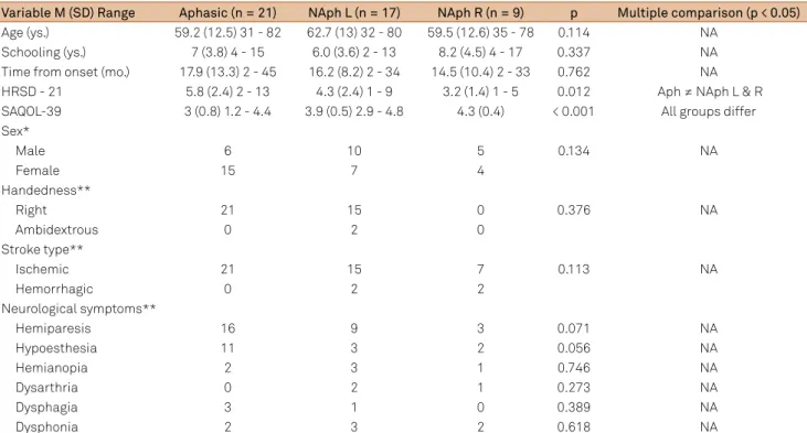
he groups were matched by age, education and time
between the stroke and evaluation, and the homogeneity of
the sample is presented in Table 1. here was a statistically signiicant diference between groups in the HRSD-21, with
a higher frequency of depressive symptoms among aphasics.
he most common neurological impairment was hemipare -sis (observed in 76% of aphasics, 53% of non-aphasics with LH lesion, and 33% of non-aphasics with RH lesions) fol-lowed by hypoesthesia (observed in 52% of aphasics, 18% of non-aphasics with LH lesion, and 33% of non-aphasics with RH lesions). A description of lesion sites for aphasics and non-aphasics is provided in Appendix 2.
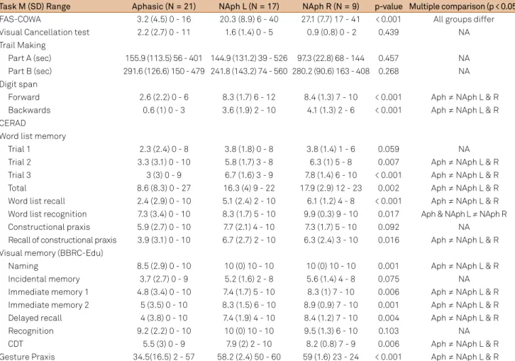
Aphasics had poorer performance than non-aphasics in the Digit Span ( forward and backwards), Word List Memory, Visual Memory, Constructional Praxis Recall,
CDT, FAS-COWA), and Gesture Praxis. There were not
Table 1. Sample characterization according to demographic and clinical data.
Variable M (SD) Range Aphasic (n = 21) NAph L (n = 17) NAph R (n = 9) p Multiple comparison (p < 0.05)
Age (ys.) 59.2 (12.5) 31 - 82 62.7 (13) 32 - 80 59.5 (12.6) 35 - 78 0.114 NA
Schooling (ys.) 7 (3.8) 4 - 15 6.0 (3.6) 2 - 13 8.2 (4.5) 4 - 17 0.337 NA
Time from onset (mo.) 17.9 (13.3) 2 - 45 16.2 (8.2) 2 - 34 14.5 (10.4) 2 - 33 0.762 NA
HRSD - 21 5.8 (2.4) 2 - 13 4.3 (2.4) 1 - 9 3.2 (1.4) 1 - 5 0.012 Aph ≠ NAph L & R
SAQOL-39 3 (0.8) 1.2 - 4.4 3.9 (0.5) 2.9 - 4.8 4.3 (0.4) < 0.001 All groups differ
Sex*
Male 6 10 5 0.134 NA
Female 15 7 4
Handedness**
Right 21 15 0 0.376 NA
Ambidextrous 0 2 0
Stroke type**
Ischemic 21 15 7 0.113 NA
Hemorrhagic 0 2 2
Neurological symptoms**
Hemiparesis 16 9 3 0.071 NA
Hypoesthesia 11 3 2 0.056 NA
Hemianopia 2 3 1 0.746 NA
Dysarthria 0 2 1 0.273 NA
Dysphagia 3 1 0 0.389 NA
Dysphonia 2 3 2 0.618 NA
significant differences in the performance of aphasic and
non-aphasic patients in the Visual Cancellation Test and in the TMT (A and B), although there was a trend for a
worse performance of LH damaged and aphasic patients. In the Word Recognition task, LH damaged (both apha-sic and non-aphaapha-sic) performed poorer than RH damaged
patients (Table 2).
Aphasia severity correlated with scores in the TMT part B
(r = -0.917, p = 0.004), Digit Span forward (r = 0.860, p < 0.001) and backwards (r = 0.543, p = 0.01), and Gesture Praxis
(r = 0.668, p = 0.002). Time elapsed from stroke onset did not
correlate with any measures of cognitive function.
DISCUSSION
Impairment in cognitive tasks in aphasic patients has been systematically described in the literature for decades20.
In 2002, Helm-Estabrooks9 systematized the approach to
cognitive deicits in aphasic patients, proposing a new instru
-ment to assess ive main domains in this population. Ever
since, most papers have put in evidence the interrelation be-tween language and attention, executive functions, memory, and visuospatial skills as a two-way street where language is
afected by impairment in any other function and vice-versa.
Such approach aims to provide optimization of rehabilitation
eforts, which must consider the global cognitive status of the patient. Seniów et al.21 described visuospatial memory and
abstract thinking deicits in aphasics, with great heterogene -ity in performance.
In the TMT, assessing selective attention, speed of per
-ceptual processing and mental lexibility, performance was
found to be below normal for all groups, especially in Part B. Only seven aphasics were able to perform the test;
four-teen patients had impeding right hemiparesis. herefore, our
results are not to be considered representative of cognitive impairment per se in the aphasic group, and this test may be considered as a non-suitable method for evaluating LH dam-aged and aphasic patients.
Aphasic patients performed poorer than non-aphasic in the Digit Span test (both forward and backwards), which assesses attention, working memory, and mental control. Although this task requires verbal output, there was no time limit to perform it; even in these conditions, aphasic pa-tients displayed a much worse performance. De Renzi and Nichelle22 described the same inding, which is now thought
to relect a deicit in the phonological loop (the verbal com -ponent of working memory23.
Table 2. Performance of Aphasics and non-Aphasics in cognitive tasks.
TaskM (SD) Range Aphasic (N = 21) NAph L (N = 17) NAph R (N = 9) p-value Multiple comparison (p < 0.05)
FAS-COWA 3.2 (4.5) 0 - 16 20.3 (8.9) 6 - 40 27.1 (7.7) 17 - 41 < 0.001 All groups differ
Visual Cancellation test 2.2 (2.7) 0 - 11 1.6 (1.4) 0 - 5 0.9 (0.8) 0 - 2 0.439 NA
Trail Making
Part A (sec) 155.9 (113.5) 56 - 401 144.9 (131.2) 39 - 526 97.3 (22.8) 68 - 144 0.457 NA
Part B (sec) 291.6 (126.6) 150 - 479 241.8 (143.2) 74 - 560 280.2 (90.6) 163 - 408 0.268 NA Digit span
Forward 2.6 (2.2) 0 - 6 8.3 (1.7) 6 - 12 8.4 (1.3) 7 - 10 < 0.001 Aph ≠ NAph L & R
Backwards 0.6 (1) 0 - 3 3.6 (1.9) 2 - 10 4.1 (1.3) 2 - 6 < 0.001 Aph ≠ NAph L & R
CERAD
Word list memory
Trial 1 2.3 (2.4) 0 - 8 3.8 (1.8) 0 - 8 3.8 (1.4) 1 - 6 0.059 NA
Trial 2 3.3 (3.1) 0 - 10 5.8 (1.7) 3 - 8 6.3 (1) 5 - 8 0.007 Aph ≠ NAph L & R
Trial 3 3 (3) 0 - 9 6.7 (1.6) 3 - 9 7.8 (1.4) 6 - 10 < 0.001 Aph ≠ NAph L & R
Total 8.6 (8.3) 0 - 27 16.3 (4) 9 - 22 17.9 (2.9) 12 - 23 0.002 Aph ≠ NAph L & R
Word list recall 2.4 (2.9) 0 - 10 5.1 (2.4) 2 - 10 6.1 (1.2) 4 - 8 < 0.001 Aph ≠ NAph L & R Word list recognition 7.3 (3.4) 0 - 10 8.3 (1.7) 5 - 10 9.9 (0.3) 9 - 10 0.017 Aph & NAph L ≠ NAph R
Constructional praxis 5.9 (2.7) 0 - 10 7.7 (2.1) 4 - 10 7.3 (1.7) 5 - 10 0.092 NA
Recall of constructional praxis 3.9 (3.1) 0 - 10 6.7 (2.7) 2 - 10 6.3 (2.4) 3 - 10 0.016 Aph ≠ NAph L & R Visual memory (BBRC-Edu)
Naming 8.5 (2.9) 0 - 10 10 (0) 10 - 10 10 (0) 10 - 10 0.001 Aph ≠ NAph L & R
Incidental memory 3.7 (2.7) 0 - 9 5.2 (1.6) 2 - 8 5.6 (1.4) 4 - 8 0.075 NA
Immediate memory 1 4.8 (3.4) 0 - 10 7.4 (1.7) 5 - 10 8.3 (1) 7 - 10 0.006 Aph ≠ NAph L & R Immediate memory 2 5 (3.5) 0 - 10 8.3 (1.5) 6 - 10 8.9 (0.9) 7 - 10 0.001 Aph ≠ NAph L & R
Delayed recall 4 (3.8) 0 - 10 7.4 (1.9) 4 - 10 8.4 (1.2) 7 - 10 0.004 Aph ≠ NAph L & R
Recognition 9.2 (2.2) 0 - 10 10 (0) 10 - 10 9.5 (1.3) 6 - 10 0.103 NA
CDT 5.5 (3) 0 - 9 7.9 (2) 2 - 10 8.2 (0.8) 7 - 9 0.006 Aph ≠ NAph L & R
Attention has been extensively studied in aphasic patients
in the recent years, as attentional deicits may impair audi -tory comprehension (i.e., comprehension of single words) as well as sentence comprehension and production4,24.
Most previous studies tended to focus on a speciic atten -tion ability (i.e., sustained, selective, divided atten-tion, and so on) or modality (auditory, visual), and therefore results tend to be quite inconsistent. Korda and Douglas25 compared
aphasics’ and normal subjects’ performance in a sustained at-tention task reporting only an increasing in reaction time for both groups. Helm-Estabrooks9 found preserved visual
dis-crimination and attention in a group of aphasics. Kalbe et al.4
reported that aphasic patients performed poorly in at least one of the three cognitive functions assessed in their study (memory, attention and reasoning). Murray2 also reported
attentional deicits in aphasic patients, and correlated those deicits with language and other cognitive functions, with implications for rehabilitation strategies. Hinckley et al.26
de-scribed impairment of attention in aphasic patients,
includ-ing poor performance in the Visual Cancellation Test. We
found a trend to a poorer performance of patients with LH damage in this task, more pronounced in the aphasic group. However, attention is a complex function encompassing sev-eral abilities, and the relations between distinct attention functions and language are yet to be understood.
Verbal luency tasks demand intact executive functioning
as well as semantic storage. As would be expected, aphasic
patients performed poorer in phonological luency as a re
-sult of diiculties in speech initiation and production, and se -mantic impoverishment. Impairment in the lexical-se-mantic processing in the LH lesions is well described, but there is evidence of impairment in this process also in RH lesions27.
In the Constructional Praxis task, all groups presented poor performance, but there was no disadvantage for the
aphasic group. Only the CDT (which poses greater execu
-tive demand) diferentiated aphasics from non-aphasics.
Helm-Estabrooks9 found greater diiculty of aphasic sub
-jects in performing the tasks proposed in the CLQT19, which
increased proportionally to executive functioning demands.
he author concludes that executive functions are the second most afected cognitive abilities in brain damage associated with aphasia (aside from language itself). Hinckley et al.26,
seeking for evidence about executive functions interference
in aphasia therapy, described a delay of aphasics to fulill the
criteria to initiate formal language therapy.
In the Word List memory task, which assesses verbal mem-ory, aphasics had a performance below normal in immediate and delayed recall tasks, while NAph L and NAph R groups showed normal performance. De Renzi and Nichelli22 reported
LH damaged patients to perform poorer in short-term memo-ry tasks (verbal and non-verbal), with aphasic patients exhib-iting greater impairment than non-aphasics. Verbal memory impairment in aphasics is currently considered to be due to
phonological loop deicit, which hampers the ability to retrieve
verbal encoded material23. However, aphasic patients showed
improvement of performance in the word recognition task when compared to spontaneous recall, which shows a rela-tively preserved capacity of encoding information, suggesting also the interference of organizational spontaneous searching
strategies diiculties.
In the Visual Memory task, aphasics performed poorer than non-aphasics in all subtasks, except for recognition, a
inding previously published28. he improved performance in
the Recognition task suggests a relative preservation of
non-verbal encoding with deicit in retrieval strategies, similar to
that observed for verbal stimuli. Verbal encoding is used for storing visual as well as verbal information, and subvocal re-hearsal is one of the mechanisms by which material stored in the visuospatial sketch gains access to the phonological
output bufer23.
Regarding Gesture Praxis, ideomotor apraxia, speech apraxia, and orofacial apraxia are well known to frequently oc-cur in association with aphasia. In our sample, there was im-provement in aphasics’ performance on imitation for natural gestures, conventional gestures, use of functional objects, and
orofacial praxis (raw data not shown). his improvement oc -curred in 78%, 68%, 65%, and 50% of aphasic patients, respec-tively for each of the aforementioned task, and it might be related to the presence of dissociative apraxia, in which the conceptual system for action (the stored knowledge of actions) and the action production system (sensorimotor programmes) are preserved, but cannot be accessed by verbal command29.
Aphasics show great heterogeneity in performance on cognitive tasks9. he interference of hemiparesis and the dif
-iculties of verbal production are factors that pose an addi -tional burden in this group of patients. In tasks with
great-er motor requirements, such as the TMT, which depends on motor speed is an additional obstacle to these patients. he
same holds true for tasks relying mostly on verbal output.
he linguistic-cognitive interrelation is evidenced by the
correlations found between severity of aphasia (which is re-lated to lesion size30) and the subjects’ performance in the
TMT (Part B), Digit Span, and Gesture Praxis Test, where
the performance was inversely proportional to the severity of aphasia; there is a large overlapping of fronto-parietal net-works for language, attentional-executive, praxis, and mo-tor functions) rather than for memory or visuospatial tasks (in which there is participation of mesial temporal and right
hemisphere structures). his methodological diiculty is un -likely to be overcome in clinical studies as strokes are distrib-uted according to the vascular anatomy and not according to the underlying cognitive circuits. Studies enrolling a great-er numbgreat-er of cases, which allow the comparison of groups
according to more speciic and isolated cerebral lesions (e.g.
understanding of the impact that speciic changes in verbal
production and comprehension exert on the performance of aphasic subjects.
Limitations of our study are the small number of subjects in the sample, the heterogeneity of vascular lesions and clini-cal types of aphasia, as well as the need for verbal response in most non-linguistic cognitive tests. Right hemiparesis may also account for the poorer performance of aphasic patients, although it is true only for those tasks that require drawing
(e.g., Visual Cancellation Test, TMT A and B, Constructional Praxis, and CDT).
One of the challenges faced by clinicians and rehabili-tation professionals working with aphasics is how to per-form a reliable cognitive assessment to identify which func-tions are preserved and which are impaired. Such knowledge
can directly inluence the choice of the most appropriate
therapeutic intervention for each patient. For this reason,
speciic batteries have been developed in order to assess cog -nitive functions in aphasic patients, such as the CLQT18, and
the Aphasia Check List (ACL)2, the latter designed for
pa-tients with severe aphasia. However, these batteries are not yet regularly used in the assessment of Brazilian Portuguese
speakers. he lack of appropriate instruments to evaluate
aphasics can lead them to be regarded as VCI or even VD, and vice–versa, as many clinicians tend to be overly lenient and never submit these patients to a thorough cognitive eval-uation due to their language impairment.
In conclusion, our study contributes to the understand-ing of Brazilian aphasics’ pattern of performance in cognitive functions. Moreover, this study reinforces the need to
devel-op and/or validate speciic instruments for the assessment of
cognitive abilities in aphasic subjects in Brazil.
Appendix 1. Performance of Aphasics and Non-aphasics in the BDAE, BNT and verbal luency tests.
Task M (SD) Range Aphasic (N = 21) N Aph L (N = 17) N Aph R (N = 9) p-value Multiple comparison (p < 0.05) Conversation and Narrative
Simple social questions 3 (2.6) 0 - 7 6.9 (0.24) 6 - 7 7 (0) 7 - 7 < 0.001 Aph ≠ NAph L & R
Spontaneous speech 1.5 (1.3) 0 - 3 5 (0) 5 - 5 5 (0) 5 - 5 < 0.001 Aph ≠ NAph L & R
Cookie Theft Picture 1.5 (1.4) 0 - 4 4.6 (0.5) 4 - 5 4.9 (0.3) 4 - 5 < 0.001 Aph ≠ NAph L & R Oral Comprehension
Word discrimination 11.4 (4.3) 1 - 16 14.9 (1) 13 - 16 15.1 (0.7) 14 - 16 0.002 Aph ≠ NAph L & R
Commands 5 (2.9) 0 - 9 9.6 (0.6) 8 - 10 9.5 (0.5) 9 - 10 < 0.001 Aph ≠ NAph L & R
Complex ideational material 2.2 (1.9) 0 - 5 4.8 (0.6) 4 - 6 5.1 (0.6) 4 - 6 < 0.001 Aph ≠ NAph L & R Oral Expression
Automatized sequences 2 (1.5) 0 - 4 4 (0) 4 - 4 4 (0) 4 - 4 < 0.001 Aph ≠ NAph L & R
Word repetition 2.7 (2.2) 0 - 5 5 (0) 5 - 5 5 (0) 5 - 5 0.001 Aph ≠ NAph L & R
Phrase repetition 0.7 (0.9) 0 - 2 2 (0) 2 - 2 2 (0) 2 - 2 < 0.001 Aph ≠ NAph L & R
Responsive naming 3.7 (4.1) 0 - 10 9.8 (0.3) 9 - 10 9.8 (0,4) 9 - 10 < 0.001 Aph ≠ NAph L & R Category speciic naming 5.7 (5.3) 0 - 12 11.9 (0.2) 11 - 12 11.9 (0.3) 11 - 12 < 0.001 Aph ≠ NAph L & R Reading
Letter-word matching 2.9 (1.5) 0 - 4 4 (0) 4 - 4 4 (0) 4 - 4 < 0.001 Aph ≠ NAph L & R
Number matching 3.2 (1.2) 0 - 4 3.8 (0.3) 3 - 4 4 (0) 4 - 4 0.027 Aph ≠ NAph L & R
Word-picture matching 2.7 (1.1) 0 - 4 3.8 (0.3) 3 - 4 3.9 (0.3) 3 - 4 < 0.001 Aph ≠ NAph L & R Word reading 6.8 (6.4) 0 - 15 14.6 (1) 11 - 15 15 (0) 15 - 15 < 0.001 Aph ≠ NAph L & R
Sentence reading 1.2 (1.8) 0 - 5 4.2 (1.5) 1 - 5 5 (0) 5 - 5 < 0.001 Aph ≠ NAph L & R
Sentence comprehension 0.9 (0.9) 0 - 3 2.3 (0.9) 0 - 3 2.9 (0.3) 2 - 3 < 0.001 Aph ≠ NAph L & R Reading comprehension 1.5 (1.1) 0 - 3 3.2 (0.7) 1 - 4 3.2 (0.4) 3 - 4 < 0.001 Aph ≠ NAph L & R Writing
Letter form 6.5 (5.3) 0 - 14 11.7 (2.7) 7 - 14 13.7 (0.4) 13 - 14 0.001 Aph ≠ NAph L & R
Letter choice 9.3 (7.4) 0 - 21 19.3 (2.6) 12 - 21 20.6 (0.7) 19 - 21 < 0.001 Aph ≠ NAph L & R Motor ability 5.6 (4.7) 0 - 14 11.2 (3.1) 7 - 14 13.2 (1.6) 9 - 14 0.001 Aph ≠ NAph L & R Coding skills 2.8 (3.2) 0 - 9 7.9 (1.9) 2 - 9 8.5 (0.5) 8 - 9 < 0.001 Aph ≠ NAph L & R Written confrontation naming 1 (1.3) 0 - 4 3.1 (1.3) 0 - 4 4 (0) 4 - 4 < 0.001 Aph ≠ NAph L & R Narrative writing
Writing mechanics 0.6 (0.8) 0 - 2 1.3 (0.4) 1 - 2 1.8 (0.5) 1 - 2 0.001 Aph ≠ NAph L & R
Vocabulary access 0.4 (0.6) 0 - 2 2 (0.9) 0 - 3 2.3 (0.8) 1 - 3 < 0.001 Aph ≠ NAph L & R
Syntax 0.4 (0.6) 0 - 2 1.9 (0.8) 0 - 3 2.3 (0.5) 2 - 3 < 0.001 Aph ≠ NAph L & R
Content adequacy 0.3 (0.5) 0 - 2 2 (0.8) 0 - 3 2.5 (1) 0 - 3 < 0.001 Aph ≠ NAph L & R
Total Writing 1.7 (2.3) 0 - 8 7.2 (2.7) 1 - 11 8.9 (2.3) 4 - 11 < 0.001 Aph ≠ NAph L & R
BNT 17.5 (16.8) 0 - 45 46.2 (8.5) 33 - 58 48.9 (5.9) 40 - 59 < 0.001 Aph ≠ NAph L & R
References
1. Laska AC, Hellblom A, Murray V, Kahan T, Von Arbin M. Aphasia in acute stroke and relation to outcome. J Intern Med. 2001;249(5):413–22. doi:10.1046/j.1365-2796.2001.00812.x
2. Murray, LL. Attention and other cognitive deicits in aphasia: presence and relation to language and communication measures Am J Speech Lang Pathol, 2012;21:S51-64. doi:10.1044/1058-0360(2012/11-0067)
3. Mesulam, MM, editor. Principles of behavioral and cognitive neurology. 2nd ed. New York: Oxford University Press; 2000. Chapter nº 1, Behavioral neuroanatomy, p. 1-120.
4. Kalbe E, Reinhold N, Brand M, Markowitsch HJ, Kessler J. A new test battery to assess aphasic disturbances and associated cognitive dysfunctions - German normative data on the aphasia check list. J Clin Exp Neuropsychol. 2005;27(7):779-94. doi:10.1080/13803390490918273
5. Gorelick PB, Scuteri A, Black SE, Decarli C, Greenberg SM, Iadecola C et al. Vascular contributions to cognitive impairment and dementia: a statement for healthcare professionals from the American Heart Association/American Stroke Association. Stroke. 2011;42(9):2672-713. doi:10.1161/STR.0b013e3182299496
6. Sachdev PS, Brodaty H, Valenzuela MJ, Lorentz L, Looi JC, Wen W et al. The neuropsychological proile of vascular cognitive impairment in stroke and TIA patients. Neurology. 2004;62(6):912-9. doi:10.1212/01.WNL.0000115108.65264.4B
7. Nicholas M, Sinotte MP, Helm-Estabrooks N. Using a computer to communicate: Effect of executive function impairments in people with severe aphasia. Aphasiology, 2005;19 (10/11), 1052:65.
8. Fillingham JK, Sage K, Lambon Ralph MA. The treatment of anomia using errorless learning. Neuropsychol Rehabil. 2006;16(2):129-54. doi:10.1080/09602010443000254
9. Helm-Estabrooks N. Cognition and aphasia: a discussion and a study. J Commun Disord. 2002;35(2):171-86. doi:10.1016/S0021-9924(02)00063-1
10. Goodglass H, Kaplan E, Barresi B. The Boston diagnostic aphasia examination. 2nd ed. Philadelphia: Lippincot Williams Wilkins; 2001.
11. Reitan RM. Validity of the Trail Making Test as an indicator of organic brain damage. Percept Mot Skills.1958;8(3):271-6. doi:10.2466/pms.1958.8.3.271
12. Morris JC, Heyman A, Mohs RC, Hughes JP, Belle G, Fillenbaum G et al. The Consortium to Establish a Registry for Alzheimer’s Disease (CERAD). Part I. Clinical and neuropsychological
assessment of Alzheimer’s disease. Neurology. 1989;39(9):1159-65. doi:10.1212/WNL.39.9.1159
13. Wechsler D. Wechsler memory scale-revised manual. San Antonio: The Psychological Corporation; 1987.
14. Nitrini R, Caramelli P, Porto CS, Charchat-Fichman H, Formigoni AP, Carthery-Goulart MT et al. Brief cognitive battery in the diagnosis of mild Alzheimer’s disease in subjects with medium and high levels of education. Dement Neuropsychol. 2007;1(1):32-6.
15. Sunderland T, Hill JL, Mellow AM, Lawlor BA, Gundersheimer J, Newhouse PA et al. Clock drawing in Alzheimer´s disease: a novel measure of dementia severity. J Am Geriatr Soc. 1989;37(8):725-29. doi:10.1111/j.1532-5415.1989.tb02233.x
16. Hamilton M. Rating depressive patients. J Clin Psychiatry. 1980;41(12 Pt 2):21-4.
17. Oldield RC. The assessment and analysis of handedness: the Edinburgh inventory. Neuropsychology. 1971;9(1):97-113. doi:10.1016/0028-3932(71)90067-4
18. Hilari K, Byng S, Lamping DL, Smith SC. Stroke and Aphasia Quality of Life Scale-39 (SAQOL-39): evaluation of acceptability, reliability, and validity. Stroke. 2003;3498):1944-50. doi:10.1161/01.STR.0000081987.46660.ED
19. Helm-Estabrooks N. Cognitive linguistic quick test. San Antonio: The Psychological Corporation; 2001.
20. Beeson PM, Bayles KA, Rubens AB, Kaszniak AW. Memory
impairment and executive control in individuals with stroke-induced aphasia. Brain Lang. 1993;45(2):253-75. doi:10.1006/brln.1993.1045
21. Seniów J, Litwin M, Leśniak M. The relationship between non-linguistic cognitive deicits and language recovery in patients with aphasia. J Neurol Sci. 2009;283(1-2):91-4. doi:10.1016/j.jns.2009.02.315
Appendix 2. Cerebral lesion sites for Aphasic and Non-aphasic groups.
Lesion site Aphasic (n = 21) Non-aphasic (n = 26)
Left 21 16
Frontal 1 1
Parietal 5 2
Temporal 1
-Occipital - 1
Fronto-parietal 3 3
Temporo-parietal 4
-Temporo-occipital - 1
Parieto-occipital - 1
Fronto-temporo-parietal 5
-PVWM - 1
Basal ganglia 2 1
Thalamus - 5
Right 0 9
Frontal - 1
Occipital - 1
Fronto-temporal - 1
Fronto-parietal - 2
Fronto-temporo-parietal - 1
22. De Renzi E, Nichelli P. Verbal and non-verbal short-term memory impairment following hemispheric damage. Cortex. 1975;11(4):341-54. doi:10.1016/S0010-9452(75)80026-8
23. Baddeley A. Working memory and language: an overview. J Commun Disord. 2003;36(3):189-208. doi:10.1016/S0021-9924(03)00019-4
24. Kemper S, Schmalzried R, Herman R, Mohankumar D. The effects of varying task priorities on language production by young and older adults. Exp Aging Res. 2011;37(2):198-219. doi:10.1080/0361073X.2011.554513
25. Korda RJ, Douglas JM. Attention deicits in stroke patients with aphasia. J Clin Exp Neuropsychol. 1997;19(4):525-42. doi:10.1080/01688639708403742
26. Hincley JJ, Carr TH, Patterson JP. Relationship between cognitive abilities, treatment type, and treatment time in aphasia. Paper
presented at the 31st Annual Clinical Aphasiology Conference; 2001 May 29-June 2; Santa Fe, NM. (Aphasiology. 2002;16(4-6).
27. Joanette Y, Ansaldo AI, Kahlaoui K, Côté H, Abusamra V, Ferreres A et al. [The impact of lesions in the right hemisphere on linguistic skills: theoretical and clinical perspectives]. Rev Neurol. 2008;46(8):481-8. Spanish.
28. Gainotti G, Cappa A, Perri R, Silveri MC. Disorders of verbal and pictorial memory in right and left brain-damaged patients. Int J Neurosci. 1994;78(1-2):9-20. doi:10.3109/00207459408986041
29. Greene JD. Apraxia, agnosias, and higher visual function abnormalities. J Neurol Neurosurg Psychiatry. 2005;76(Suppl 5):v25-34. doi:10.1136/jnnp.2005.081885