Arq Neuropsiquiatr 2009;67(2-A):311-313
311 Clinical / Scientiic note
ChroniC inflammatory demyelinating
polyneuropathy in an adoleSCent
with type 1 diabeteS mellituS
Crésio Alves, Zilda Braid, Nadja Públio
polineuropatia inflamatória deSmielinizante CrôniCa em um adoleSCente Com diabeteS melito tipo 1
Pediatric Endocrinology Service, Hospital Universitário Professor Edgard Santos, Faculty of Medicine, Universidade Federal da Bahia, Salvador BA, Brazil. Received 5 September 2008, received in inal form 5 December 2008. Accepted 19 February 2009.
Dr. Crésio Alves – Rua Plínio Moscoso 222 / 601 - 40157-190 Salvador BA - Brasil. E-mail: cresio.alves@uol.com.br
Chronic inlammatory demyelinating
polyradiculopa-thy (CIDP) is an immune-mediated disorder for which
spe-ciic clinical cerebrospinal luid (CSF), electrophysiologic
and histological criteria have been proposed
1. CIPD is an
uncommon cause of childhood polyneuropathy.
Descrip-tions of children with CIPD are scarce. Therefore, this
arti-cle intends to report a case about an adolescent with type
1 diabetes mellitus (T1DM) with a satisfactory therapeutic
response to immunoglobulin and methylprednisolone.
This presentation highlights the importance of
investi-gating diabetic patients, including children, with
polyneu-ropathy in an attempt to identify the ones with
demyeli-nating polyneuropathy, because of the likelihood of these
patients to beneit from immumodulatory and/or
immu-nossupressive treatment.
CaSe
Male, 14 year-old, with a seven year history of T1DM and one year history of diabetic nephropathy. Under insulinotherapy and captopril with unsatisfactory glycemic control. Nine months be-fore the initial evaluation he presented pain, paresthesia and a progressive, symmetric muscular weakness, in both lower limbs. After ive months of therapeutic failure under carbamazepine and amytryptiline, he was referred to our service. The initial evaluation showed a mild and symmetric, proximal and distal lower limb weakness (the upper limbs were not affected). The proximal compromise was worse than distal. There were patellar and Achilles tendon hyporelexia, with absence of Babinski sign. Pain, vibratory and position stimulus modalities were normal, al-though tactile sensory tests evidenced paresthesias. Anal and vesical sphincter function and muscular tone were preserved.
Complete blood count, erythrocyte sedimentation rate, urea, creatinine, electrolytes, AST, ALT, GGT, alkaline phos-phatase, prothrombine time, total proteins, albumin, creatine-kynase (CK), CK-MB, thyroid function tests, lipid proile and ce-rebrospinal luid evaluation were normal.
The irst electromyography (EMG), done ive months after the beginning of the neurologic symptoms (Nihon Khoden
Elec-tromyography, 4 channels), evidenced decreased conduction ve-locities with normal amplitude in the median, ulnar, left ibular and posterior tibial nerves, except in the deep right ibular nerve, where although the amplitude was 75% of the lower limit of the normal range (LLNR), the conduction study was 80% lower than LLNR. The distal motor latencies of the median, left ulnar, su-pericial ibular and sural nerves were prolonged (above 120% of the normal range). The F waves presented prolonged latencies in the ulnar and posterior tibial nerves. The examination with mo-nopolar needle showed decrease in the recruitment of action potentials of the motor unit.
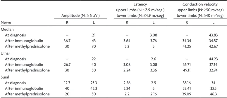
The patient was initially treated with human immunoglobu-lin (400 mg/Kg/day, IV, 5 days), improving the sensory symptoms for about a month, with subsequent relapse, in smaller intensity. There was not improvement of the lower limb weakness. A sec-ond EMG, performed 2 months after therapy with immunoglob-ulin, evidenced increment in the amplitude of the sensory and motor action potential, and decrease of the distal motor laten-cies, with persistence of the conduction velocities though. The examination showed F waves with prolonged latency in the left ulnar and posterior tibial nerves, with normal latency in the left ulnar nerve. The examination with monopolar needle showed normal recruitment.
Due to partial response to immunoglobulin, it was decide on instituting treatment with methylprednisolone (900 mg/ day, IV, 5 days), without any side effect. At reassessment, three months after starting corticotherapy, the patient presented im-provement of neurologic symptoms. A third EMG conducted 2 months after methylprednisolone displayed normal ampli-tude, with slight decrease in the sensory (median, ulnar, right sural and left supericial nerves) and motor (median and left fibular nerves) conduction velocity, and prolonged distal tency in the median nerves. The F waves presented normal la-tencies. Examination with monopolar needle showed normal recruitment.
Arq Neuropsiquiatr 2009;67(2-A)
312
Chronic inlammatory demyelinating polyneuropathy Alves et al.
Table 1. Eletroneuromyographic indings (motor potentials) before and after treatment with immunoglobulin and methylprednisolone.
Nerve Amplitude (N: ≥4 mV)
Latency
upper limbs (N: ≤3.9 m/seg) lower limbs (N: ≤4.9 m/seg)
Conduction velocity upper limbs (N: ≥50 m/seg) lower limbs (N: ≥40 m/seg)
R L R L R L
Median At diagnosis After immunoglobulin After methylprednisolone – 9.3 9 7 6.6 7.27 – 4.3 3.6 4.2 3.9 3.6 – 40.65 47.17 40.65 38.89 44.55 Ulnar At diagnosis After immunoglobulin After methylprednisolone – 10 8.8 8.2 12.2 7.6 – 3 2.2 3.25 3 2.6 – 41.23 49.58 42.86 44.23 50 Fibular At diagnosis After immunoglobulin After methylprednisolone 3.0 (75% LLNR) 4.3 4.3 4.3 6.3 4.67 6.05 6 4.5 8.3 (169% ULNR) 6.8 (138% ULNR) 4.9 31.79 (79% LLNR) 32.29 40.12 36.67 30.91 39.16 Tibial At diagnosis After immunoglobulin After methylprednisolone 7.7 11.7 11.2 7.9 13.5 8.6 6.7 (136% ULNR) 4.8 3.6 7.15 (145% ULNR) 5.9 3.6 – – – – – – N: normal reference values; ULNR: upper limit of the normal range; LLNR: lower limit of the normal range; R: right; L: left.
Table 2. Eletroneuromyographic indings (sensitive potentials) before and after treatment with immunoglobulin and methylprednisolone.
Nerve
Amplitude (N: ≥ 5 µV )
Latency
upper limbs (N: ≤3.9 m/seg ) lower limbs (N: ≤4.9 m/seg)
Conduction velocity upper limbs (N: ≥50 m/seg) lower limbs (N: ≥40 m/seg)
R L R L R L
Median At diagnosis After immunoglobulin After methylprednisolone – 36.7 30 21 43 70 – 3.64 3.2 3.08 3.76 3 – 34.34 41.25 43.83 34.57 42.67 Ulnar At diagnosis After immunoglobulin After methylprednisolone – 26.7 30 22 40 30 – 3.08 2.24 2.6 3.08 3.36 – 35.71 49.11 44.23 37.34 32.74 Sural At diagnosis After immunoglobulin After methylprednisolone 12.7 40 20 23.3 43.3 30 2.56 3.24 2.2 2.5 3 2.16 35.16 32.41 39.09 34 33.3 46.3 N: normal reference values; R: right; L: left.
diSCuSSion
Chronic inlammatory demyelinating
polyradiculopa-thy is an acquired neuropapolyradiculopa-thy characterized by a chronic,
rapidly progressive, proximal and distal symmetric
weak-ness, associated with hyporelexia and sensory symptoms
1.
It has an immunologic pathophysiology involving both the
humoral and the cellular immunologic response, which
supports the rationale to the immunotherapy
1,2.
Arq Neuropsiquiatr 2009;67(2-A)
313
Chronic inlammatory demyelinating polyneuropathy Alves et al.
diabetic somatic polyneuropathy from CIDP is not a
sim-ple task. While CIDP progresses in months, with
relaps-es in a third of the casrelaps-es, in the diabetic neuropathy the
course is slower, progressing in years, and usually related
to the length of diabetes
1-4,5. In addition, the
electromyo-graphy and nerve conduction studies reveal
demyelina-tion with nervous conducdemyelina-tion abnormalities, contrary to
the axonal pattern of diabetic neuropathy.
The diagnosis of the classic diabetic CIDP
6, is achieved
when the patient presents: (1) diabetes; (2) progressive
chronic or relapsing, sensory or sensory-motor
polyneu-ropathy, with length of at least 2 months, associated with
hyporelexia; (3) eletrophysiologic criteria for
demyeli-nating neuropathy; (4) increase of CSF proteins without
increase of leukocytes; (5) clinical improvement with
im-munotherapy; and (6) in selected cases, evidence of loss
of myelinated ibers in the nerve biopsy. However, besides
the classical presentation, CIDP encompasses a large set
of phenotypes, including one with a normal CSF study.
Therefore there is not a single diagnostic pattern which
can cover every expression of the disease
1,2,4,7.
In this patient, the diagnosis of CIDP was based on:
di-agnosis of T1DM, progressive pattern of the neuropathy,
and electromyography studies compatible with
demyeli-nating polyneuropathy, without evidence of axonal lesion.
The axonal degeneration and the chronic evolution
are predictors of a worse prognosis
2. Simmons et al.
8and
Hattori et al.
9reported subacute evolution and a
predom-inantly motor neuropathy in the juvenile age group, with
a better recovery after relapse than adults. On the
oth-er hand, Nevo et al.
10did not ind signiicant
differenc-es in the clinical characteristics of children and adults
with CIDP.
The therapeutic scheme of choice is elusive. Some
au-thors start with human immunoglobulin, while others
pre-fer costicosteroids
2,7,11. The immunosuppressive drugs are
reserved for failure of the initial therapy. The comparison
among studies about therapeutic response is hindered by
the diversity of presentations of CIDP, the type and the
dose of the drugs employed
7.
The mechanism of action of immunoglobulin remains
uncertain. Two double-blind randomized clinical trial
demonstrated beneits of the use of immunoglobulin
12.
Side effects include renal dysfunction, thromboembolic
events and high cost
4.
The exact mechanism of action of steroids in CIDP
is unknown. In mild disease, one can start prednisone (1
mg/Kg/day) and when improvement of symptoms
fol-lows, restart a gradual decrease. If relapse or worsening
of symptoms occurs, the dose can be increased or an
im-munosuppressive drug can be added up. When the
op-tion is methylprednisolone, the start dose, in adults, was
1.000 mg/day for 3 to 5 consecutive days, afterwards
ta-pering off the dose
11. Among the side effects were: poor
glycemic control. weight gain, hypertension and Cushing
syndrome features.
This article advises about the importance of thinking
of CIDP when evaluating a sensory-motor diabetic
periph-eral neuropathy.
referenCeS
1. Kalita J, Mistra UK, Yadav RK. A comparative study of inlam -matory demyelinating polyradiculoneuropathy with and with-out diabetes mellitus. Eur J Neurol 2007;14:638-643.
2. Said G. Chronic inlammatory demyelinating polyneuropathy. Neuromuscul Disord 2006;16:293-303.
3. Said G. Focal and multifocal diabetic neuropathies. Arq Neu-ropsiquiatr 2007;65:1272-1278.
4. Sharma KR, Cross J, Farronay O, Ayyar DR, Shebert RT, Brad-ley WG. Demyelinating neuropathy in diabetes mellitus. Arch Neurol 2002b;59:758-765.
5. Stewart JD, McKelvey R, Duncan L, Carpenter S, Karpati G. Chronic inlammatory demyelinating polyneuropathy (CIPD) in diabetics. J Neuro Sci 1996;142:59-64.
6. Cornblath DR, Asbury AK, Albers JW, et al. Report from an Ad Hoc Sucommittee of the American Academy of Neurology AIDS Task Force: research criteria for the diagnosis of chron -ic inlammatory demyelinating polineuropathy (CIPD). Neu -rology 1991;41:617-618.
7. Saperstein D, Katz J, Amato A, Barohn R. Clinical spectrum of chronic acquired demyelinating polyneuropathies. Muscle Nerve 2001;24:311-324.
8. Simmons Z, Wald JJ, Albers JW. Chronic inflammatory de-myelinating polyradiculoneuropathy in children: II. Long-term follow-up, with comparison to adults. Muscle Nerve 1997;20:1569-1575.
9. Hattori N, Misu K, Koike H, et al. Age of onset inluences clin -ical features of chronic inlammatory demyelinating polyneu -ropathy. J Neurol Sci 2001;184:57-63.
10. Nevo Y, Pestronk A, Kornberg AJ, et al. Childhood chronic in -lammatory demyelinating neuropathies: clinical course and long-term follow-up. Neurology 1996:47:98-102.
11. Lopate G, Pestronk A, Al-Lozi M. Treatment of chronic in -lammatory demyelinating polyneuropathy with high-dose intermittent intravenous methylprednisolone. Arch Neurol 2005;62:249-254.
