Arq Neuropsiquiatr 2011;69(2-A):262-263
262
Letter
Arachnoid cysts and absence epilepsy
An evidence or a coincidence?
Nina Ventura1, Isabella D’Andrea2, Maria Fátima Bento de Souza Cardoso1,
Soniza V. Alves-Leon2, Emerson L. Gasparetto2
Correspondence
Nina Ventura
Rua São Clemente 398 / 103 22260-000 Rio de Janeiro RJ - Brasil E-mail: niventura@hotmail.com
Received 30 July 2010
Received in final form 4 October 2010 Accepted 11 October 2010
EPILEPSIA COM CRISES DE AUSÊNCIA E CISTO ARACNÓIDE: UMA EVIDÊNCIA OU COINCIDÊNCIA?
Hospital Universitário Clementino Fraga Filho, Epilepsy Program, Federal University of Rio de Janeiro, Rio de Janeiro RJ, Brazil: 1MD; 2MD, PhD.
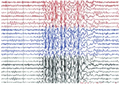
While evaluating a cohort of 22 pa-tients with absence epilepsy, we found two cases of 46- and 41-year-old females (Pa-tient 1 and 2), who presented arachnoid cysts in the choroid issure. he Patient 1 presented eyelid myoclonic epilepsy, early onset (11 years old) and no family history. Patient 2 presented juvenile absence epi-lepsy, characterized by sudden and brief impairment of consciousness, also early onset (14 years old) and positive family history. During video-electroencepha-lography (EEG) monitoring, Patient 1 presented lickering after eye-closure in bright light room and consciousness im-pairment during some of the flickering (Fig 1). Photic stimulation increased the frequency of eyelid myoclonic absence seizures. he ictal register showed spike-wave discharges of 4 HZ with bilateral projection (3 seconds interval) and ante-rior prevalence. he video-EEG of the Pa-tient 2 showed 2.5- to 3 Hz generalized spike and wave discharges. Both patients underwent brain magnetic resonance
(MR) imaging that showed well deined lesions compressing the hippocampus, presenting high signal on T2-weighted images and low signal on T1-weighted im-ages, with no contrast enhancement, both located in the right choroid issure, sizing 1.9 cm (AP) × 1.6 cm (L) × 0.9 cm (T) in Patient 1 and 1.8 cm (AP) × 1.8 cm (L) × 1.2 cm (T) in Patient 2 (Fig 2).
he relation between arachnoid cysts and absence seizures was not previous discussed in literature. In addition, there are some theories supporting that a cor-tical focus could play a leading role in the origin of generalized spike wave dis-charges1. Meeren et al.1 suggested that
primary generalized epilepsies, including absence epilepsy, are the expression of a cortical abnormality. They argued that the initial leading spike appears first in a circumscribed area of the perioral re-gion of the somatosensory cortex, which has a low threshold for spike generation. he spike rapidly spreads over the cortex, thus giving generalized appearance to the
Arq Neuropsiquiatr 2011;69(2-A)
263
Arachnoid cysts: absence epilepsy Ventura et al.
discharges. However, to the best of our knowledge, the only report of a focal lesion associated with absence sei-zures was presented by Nakanishi et al.2. hey reported a
case of typical absence epilepsy in a 34-year-old woman without history of seizures, who had hypoparathyroidism and hyperostosis frontalis interna causing mild compres-sion over the frontal lobes. he authors suggested that the compression of the superior medial frontal lobes by the hyperostosis could be deeply involved in the devel-opment of spike-wave stupor in the patient.
Corroborating the cortical focus theory and previous
reports of a focal lesion associated to absence epilepsy, these cases suggest that arachnoids cysts in the hippo-campus region might be associated with absence epi-lepsy, but further studies should be conducted in order to deine whether that association is causal or casual.
REFERENCES
1. Meeren H, van Luijtelaar G, Lopes da Silva F, Coenen A. Evolving concepts on the pathophysiology of absence seizures: the cortical focus theory. Arch Neurol 2005;62:371-376.
2. Nakanishi M S, Neshige R, Kuroda Y, Inoue Y. A case of adult-onset spike-wave stupor associated with hypoparathyroidism and hyperostosis fron-talis interna (HFI). Rinsho Shinkeigaku 1990;30:1114-1117.
![Fig 2. [A] Axial T2-weighted MR image of case one shows a hiperintense cystic appearing lesion in the right choroid issure, which is com- com-pressing the head and body of the hippocampus](https://thumb-eu.123doks.com/thumbv2/123dok_br/15433484.595388/2.955.109.896.94.379/axial-weighted-hiperintense-cystic-appearing-choroid-pressing-hippocampus.webp)
