Key words:
Carcinoma, Renal Cell; Tissue Array Analysis; Immunohistochemistry; Carbonic Anhydrases
Int Braz J Urol. 2013; 39: 484-92
__________________
Submitted for publication: March 27, 2012
__________________
Accepted after revision: August 30, 2012 Introduction: The knowledge about the molecular biology of clear cell renal cell
carci-noma (ccRCC) is evolving, and Carbonic Anhydrase type IX (CA-IX) has emerged as a potential prognostic marker in this challenging disease. However, most of the literature about CA-IX on ccRCC comes from series on metastatic cancer, with a lack of series on non-metastatic cancer. The objective is to evaluate the expression of CA-IX in a cohort of non-metastatic ccRCC, correlating with 1) overall survival, and 2) with established prognostic parameters (T stage, tumor size, Fuhrman nuclear grade, microvascular in-vasion and peri-renal fat inin-vasion).
Materials and Methods: This is a retrospective cohort study. We evaluated 95 patients with non-metastatic clear cell renal cell carcinoma, as to the expression of CA-IX. The analyzed parameters where: overall survival (OS), TNM stage, tumor size (TS), Fuhr-man nuclear grade (FNG), microvascular invasion (MVI), peri-renal fat invasion (PFI). We utilized a custom built tissue microarray, and the immunoexpression was digitally quantified using the Photoshop® software.
Results: The mean follow-up time was 7.9 years (range 1.9 to 19.5 years).
The analysis of CA-IX expression against the selected prognostic parameters showed no correlation. The results are as follows: Overall survival (p = 0.790); T stage (p = 0.179); tumor size (p = 0.143); grouped Fuhrman nuclear grade (p = 0.598); micro-vascular invasion (p = 0.685), and peri-renal fat invasion (p = 0.104).
Conclusion: Carbonic anhydrase type IX expression does not correlate with overall survival and conventional prognostic parameters in non-metastatic clear cell renal cell carcinoma.
INTRODUCTION
Renal cell carcinoma corresponds to 3% of all cancers (1), and its incidence is rising (2). The clear cell type (ccRCC) is the most common and one of the most aggressive forms of renal cancer (3,4).
The knowledge about the molecular biolo-gy of ccRCC is evolving, and Carbonic Anhydrase type IX (CA-IX) has emerged as a potential prog-nostic marker in this challenging disease (5,6).
In response to either hypoxia or VHL mu-tation, the HIF-1α accumulates and stimulates a
Carbonic Anhydrase IX is Not a Predictor of Outcomes
in Non-Metastatic Clear Cell Renal Cell Carcinoma – A
Digital Analysis of Tissue Microarray
_______________________________________________
Marcelo Zerati, Kátia R. M. Leite, José Pontes-Junior, Cesar Camara Segre, Sabrina Thalita Reis,
Miguel Srougi, Marcos Francisco Dall’Oglio
Laboratory of Medical Investigation (LIM55), Urology Department, University of Sao Paulo Medical School and Uro-Oncology Group, Urology Department, University of Sao Paulo Medical School, Sao Paulo, Brazil
ABSTRACT
ARTICLE
INFO
range of downstream effectors, including CA-IX expression (7).
CA-IX is an enzyme responsible for the cellular pH control, and in tumors with high CA-IX expression there is a better prognosis, and also a better response to therapy, probably because such tumors express a less aggressive phenotype (7-9). Low CA-IX expressing tumors demonstrate a more aggressive phenotype, probably because such tu-mors thrive in an acidic and hypoxic milieu, whi-ch is traditionally known to render tumors more aggressive and less responsive to therapy (7-9).
However, most of the literature about the utility of CA-IX as a prognostic marker on ccRCC comes from series of patients with metastatic can-cer (10-12), with a lack of series on non-metastatic cancer.
Patients with localized ccRCC are curable with surgery, but approximately one third of the patients operated with curative intent will even-tually develop metastatic disease in the course of follow-up (13). Therefore it would be very inte-resting if CA-IX could help predict which patients would require closer follow-up or even more ag-gressive adjuvant therapy (14).
The aim of this paper is to evaluate the expression of CA-IX in a cohort of non-metasta-tic ccRCC, correlating it with overall survival and conventional prognostic factors.
MATERIALS AND METHODS
Patient selection
We identified 227 patients with renal can-cer operated between 1988 and 2006 at the Sírio--Libanês Hospital and Beneficência Portuguesa Hospital in São Paulo, Brazil. Eighty-three patients were excluded for having non-clear cell cancers. Among the 144 ccRCC patients, 49 were excluded for various reasons: specimen blocks irretrievable, incomplete charts, metastatic disease. The remai-ning 95 patients were included in the cohort. Table 1 shows the baseline characteristics of the cohort.
Follow-up time ranged from 1.9 to 19.5 ye-ars, median follow-up was 7.9 years.
Demographic and clinical data were retrie-ved from hospital medical charts, anatomopatho-logical data was provided by the final pathoanatomopatho-logical
report. The final clinical condition of the patients was obtained by either office chart review or tele-phone contact with patients or relatives.
The study was submitted to and approved by the Institutional Ethics Committee, and an informed consent was obtained from patients or relatives.
Tissue microarray
A custom built tissue microarray was constructed with the technique adapted from Ko-nonen et al. (15). Using a Beecher system (Beecher Instruments, Sun Prairie, WI, USA), which collects 0.6 mm cylinders, two samples from each patient were arrayed. Representative 4µm sections of the tissue microarray were transferred to glass slides.
Immunohistochemistry
Samples underwent antigenic recovery by heat using a citrate buffer (1µM, pH 6.0) and he-ated for 30 minutes in an electrical heater. The slides were incubated overnight with CA-IX mo-noclonal antibody (Abcam, Cambridge, USA; 1:1,000). For immunostaining, the LSAB system (Dako, USA) was used.
Digital Image Capture
Each histospot was photographed with a Olympus BX60 microscope (Olympus Corpo-ration, Tokyo, Japan), coupled with a Olympus DP71 camera, controlled by the DP Controller software (version 3.2.1.276). Images were mana-ged with Olympus DP Manager software (version 3.1.1.208).
The microarray was initially inspected under the optical microscope to assure optimal quality of image and illumination. The histospots were analyzed with the camera photometer, and once a good image quality was achieved, all the camera controls were shifted to manual, in order to obtain standardized images throughout the ar-ray. The settings were ISO 200, shutter speed of 1/2,500 seconds. All the microscope settings (light intensity, condenser distance and aperture) were kept unchanged for the entire digital acquisition session. Focus of each histospot was adjusted as necessary.
Digital quantification of immunoexpression
The immunoexpression was analyzed using the Photoshop CS4 software, Portuguese version 11 (Adobe Systems, CA, EUA).
The technique consists in counting the pi-xels of the brown color of interest, in an area mea-surement, adapted from Lehr et al. (16,17).
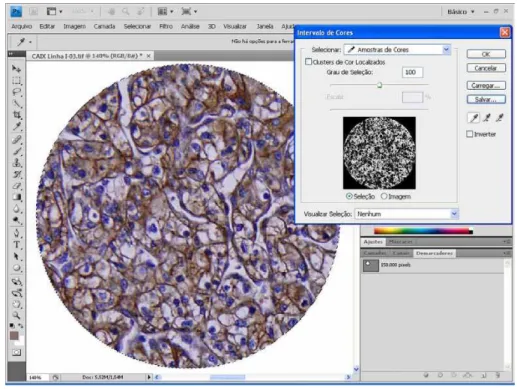
The picture of each histospot was inspected looking for an area with the following characteris-tics: 1) contains only neoplasic tissue, and 2) has compact and homogeneous histology. A circular marker was used to delimitate a region of interest (ROI) with a fixed area of 150,000 pixels, and was applied over the picture. The remaining tissue was digitally excluded. The next step was to zoom in the area of interest until some slight pixelization of the image was obtained. The color selection tool was used to pinpoint the brown color in a region of citoplasmic membrane representing the immu-noexpression of interest, and all the corresponding pixels were selected, as shown on Figure-1. All the remaining pixels of any other color were digitally excluded, with the remaining pixels corresponding only to the brown color of interest. The histogram tool was then used to count the pixels of the brown color of interest, as shown on Figure-2. The result was transferred to a spreadsheet, and the mean va-lue of the pixel counting of both the samples of each patient was calculated.
Statistical analysis
The statistical analyses were performed with the SPSS 16 Software for Windows. Survival cur-ves were calculated by the Kaplan-Meier method and the difference between the curves was demons-trated with the log-rank test.
The expression of CA-IX for each prog-nostic parameter was analyzed with Kruskal-Wallis and Mann-Whitney tests.
RESULTS
CA-IX expression was evaluated in 95 va-lid cases.
The area of CA-IX expression ranged from zero pixels to 68,780 pixels, with a median ex-pression of 20,924 pixels and standard deviation
Table 1 - Patient and tumor characteristics
n = 95 %
Sex
Male 70 73.7
Female 25 26.3
Age (years)
Range 9 / 81
Median (CI, 95%) 59.2 (56.5 / 61.8)
Follow-up (years)
Range 1.9 / 19.5
Median (CI, 95%) 7.9 (6.9 / 8.8)
Tumor size (cm)
Range 1.2 / 19.5
Median (CI, 95%) 5.0 (4.38 / 5.60)
Size
Right 39 41.0
Left 53 55.8
Bilateral 3 3.2
T Stage
T1 69 72.6
T2 8 8.4
T3 18 19.0
N+ Stage 0 0
M+ Stage 0 0
Tumor size (categorized)
Up to 4.0 cm 50 52.6
Between 4.1 and 7.0 cm 27 28.4
Larger than 7.1 cm 18 19.0
Fuhrman nuclear grade
G1 25 26.3
G2 37 38.9
G3 26 27.3
G4 7 7.5
Microvascular invasion
Absent 70 73.7
Present 25 26.3
Peri-renal fat invasion
Absent 76 80.0
Figure 1 - Region of Interest (ROI), and selection of brown color of interest using the color selection tool.
15,902 pixels. The mean expression area was 23,700 pixels, with 95% confidence interval be-tween 20,461 and 26,939 pixels.
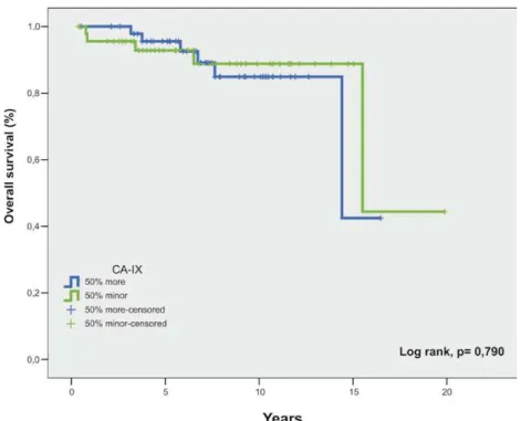
When comparing the survival curves of the patients with the 50% higher expression versus the 50% lower expression, there was no statistical sig-nificance between the curves (p= 0.790), (Figure-3). When we analyzed the expression of CA--IX against the selected prognostic parameters, we found no correlation of its expression with T
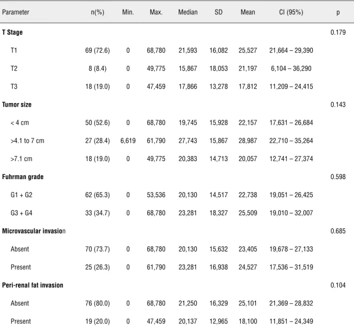
stage (p = 0.179); tumor size (p = 0.143); grouped Fuhrman nuclear grade (p = 0.598); microvascular invasion (p = 0.685), and peri-renal fat invasion (p = 0.104) (Table-2).
DISCUSSION
This is a cohort of non metastatic ccRCC evaluated for CA-IX expression using a tissue micro-array and digital immunoexpression quantification.
Figure 3: Overall survival curve by the expression of CA-IX.
Tissue microarray technology has many advantages over conventional samples processing. With all the samples being immunostained simul-taneously in the same batch, there is a very high level of standardization, virtually eliminating te-chnical bias seen with the individual processing of samples. Furthermore, TMA technology provides great economy of research resources, personnel, and tissue specimens. It is currently the technique of choice for high throughput research (15,18-20).
Digital quantification of immunoexpres-sion eliminates the bias of one of the most subjec-tive steps of the research:specimen interpretation. Even specialized pathologists differ in the inter-pretation of the specimens (21). Immunohistoche-mistry is a qualitative method, however, it has re-cently being quantitatively evaluated with the use of digital analysis software (22-25).
Table 2: Expression of CA-IX against the study parameters
Parameter n(%) Min. Max. Median SD Mean CI (95%) p
T Stage 0.179
T1 69 (72.6) 0 68,780 21,593 16,082 25,527 21,664 – 29,390
T2 8 (8.4) 0 49,775 15,867 18,053 21,197 6,104 – 36,290 T3 18 (19.0) 0 47,459 17,866 13,278 17,812 11,209 – 24,415
Tumor size 0.143
< 4 cm 50 (52.6) 0 68,780 19,745 15,928 22,157 17,631 – 26,684
>4.1 to 7 cm 27 (28.4) 6,619 61,790 27,743 15,867 28,987 22,710 – 35,264 >7.1 cm 18 (19.0) 0 49,775 20,383 14,713 20,057 12,741 – 27,374
Fuhrman grade 0.598
G1 + G2 62 (65.3) 0 53,536 20,130 14,517 22,738 19,051 – 26,425 G3 + G4 33 (34.7) 0 68,780 23,281 18,327 25,509 19,010 – 32,007
Microvascular invasion 0.685
Absent 70 (73.7) 0 68,780 20,130 15,632 23,405 19,678 – 27,133
Present 25 (26.3) 0 61,790 23,281 16,938 24,527 17,536 – 31,519
Peri-renal fat invasion 0.104
Absent 76 (80.0) 0 68,780 21,250 16,329 25,101 21,369 – 28,832
Present 19 (20.0) 0 47,459 20,137 12,965 18,100 11,851 – 24,349
using the histogram (16,17). Adobe Photoshop® is a widely available, low cost software, and its use makes this technology available and reproducible to other centers (16,17,25-27).
The gene that codifies CA-IX is regulated by HIF-1α expression, and HIF-1α is controlled by the VHL protein. CA-IX expression is common in ccRCC and its role is to regulate pH in the hypoxic neoplasic environment. Either hypoxia or the VHL mutation causes HIF-1α accumulation, therefore activating a
range of proangiogenic factors, including CA-IX ex-pression (28-34).
a predictor of survival, prognosis, and therapeutic response (11,38-41).
In the setting of non-metastatic ccRCC, ho-wever, CA-IX has not shown the same performance as in metastatic ccRCC.
Leibovich et al. (42) reported a cohort of 933 RCC patients, in which 730 where ccRCC, with a median follow-up of 10 years. The results did not demonstrate CA-IX to be a prognostic marker when compared to conventional prognostic factors. The paper from Leibovich et al. show some differences when compared to ours. Although most patients had ccRCC, they analyzed mixed histological types. And also, patients with metastatic disease where included. In this regard, it’s worth mention that expression of CA-IX was not statistically significant among the Nx/N0 versus N1/N2 groups, and also among M0 versus M1 groups. One could expect to see some differences in CA-IX expression in metastatic ver-sus non-metastatic patients, but the authors failed to confirm this rationale, therefore, further suppor-ting the limited role of CA-IX in the non-metastatic scenario. They also reported high levels of CA-IX in many other organs (gastric mucosa, pancreatic and biliary epithelia, and base of epithelial crypts of small intestine) and therefore question that CA-IX would have limited utility as both independent prog-nostic factor and therapeutic target in RCC.
We found one published paper that is very similar to ours. Klatte et al. (43), in the search for a molecular signature of ccRCC, evaluated exclusively non-metastatic ccRCC as to the expression of twen-ty molecular markers, including CA-IX. Although they used a qualitative immunohistochemical analy-sis, the result showed CA-IX did not correlate with the studied parameters on univariate analysis (p = 0.651), and therefore was not considered for multi-variate analysis.
In our cohort of non-metastatic ccRCC pa-tients, with long term follow-up, evaluated with tissue microarray and digital quantification of im-munoexpression, we found no correlation of CA-IX expression with either overall survival, or with con-ventional prognostic parameters: T stage, tumor size, Fuhrman nuclear grade, microvascular invasion and peri-renal fat invasion.
Our results further support an emerging con-cept in the literature about the limited usefulness of
CA-IX as a prognostic marker in non-metastatic cle-ar cell renal cell ccle-arcinoma.
Carbonic Anhydrase IX expression did not correlate with overall survival, T stage, tumor size, Fuhrman nuclear grade, microvascular invasion and peri-renal fat invasion in non-metastatic clear cell renal cell carcinoma.
ABREVIATIONS
CA-IX: Carbonic anhydrase type 9
ccRCC: Clear cell renal cell carcinoma
HE: Hematoxilin-eosin
HIF-1α: Hypoxia inducible factor one-alpha
KPS: Karnofsky performance status
RCC: Renal cell carcinoma
RGB: Red, Green, Blue
ROI: Region of interest
TIFF: Tagged Image File Format
TMA: Tissue microarray
TNM: Tumor, Nodule, Metastasis
VHL: von Hippel-Lindau
ACKNOWLEDGEMENT
This project received financial support from FAPESP (State of Sao Paulo Agency for Re-search Support), Grant nº: 2008/52908-8
CONFLICT OF INTEREST
None declared.
REFERENCES
1. Jemal A, Siegel R, Xu J, Ward E: Cancer statistics, 2010. CA Cancer J Clin. 2010; 60: 277-300. Erratum in: CA Cancer J Clin. 2011; 61: 133-4.
2. Chow WH, Devesa SS, Warren JL, Fraumeni JF Jr: Rising in-cidence of renal cell cancer in the United States. JAMA. 1999 5; 281: 1628-31.
3. Novick A, Campbell SC: Renal Tumors. In: Walsh PC, Retik AB, Darracott Vaughan Jr. E, Wein AJ, (ed.), Campbell’s Urology. 8 ed. Philadelphia: Saunders; 2002; pp. 2672-731.
5. Linehan WM, Walther MM, Zbar B: The genetic basis of cancer of the kidney. J Urol. 2003; 170: 2163-72.
6. Linehan WM, Grubb RL, Coleman JA, Zbar B, Walther MM: The genetic basis of cancer of kidney cancer: implications for gene-specific clinical management. BJU Int. 2005; 95(Suppl 2): 2-7. 7. Pantuck AJ, Zeng G, Belldegrun AS, Figlin RA: Pathobiology,
prognosis, and targeted therapy for renal cell carcinoma: ex-ploiting the hypoxia-induced pathway. Clin Cancer Res. 2003 15; 9: 4641-52.
8. Pastorekova S, Parkkila S, Zavada J: Tumor-associated car-bonic anhydrases and their clinical significance. Adv Clin Chem. 2006; 42: 167-216.
9. Wykoff CC, Beasley NJ, Watson PH, Turner KJ, Pastorek J, Sibtain A, et al.: Hypoxia-inducible expression of tumor-asso-ciated carbonic anhydrases. Cancer Res. 2000; 60: 7075-83. 10. Atkins MB, Choueiri TK, Cho D, Regan M, Signoretti S:
Treat-ment selection for patients with metastatic renal cell carcino-ma. Cancer. 2009; 115: 2327-33.
11. Bui MH, Seligson D, Han KR, Pantuck AJ, Dorey FJ, Huang Y, et al.: Carbonic anhydrase IX is an independent predictor of survival in advanced renal clear cell carcinoma: implications for prognosis and therapy. Clin Cancer Res. 2003; 9: 802-11. 12. Kim HL, Seligson D, Liu X, Janzen N, Bui MH, Yu H, et al.:
Using tumor markers to predict the survival of patients with metastatic renal cell carcinoma. J Urol. 2005; 173: 1496-501. 13. Linehan WM, Bates SE, Yang JC: Cancers of the genitouri-nary system: cancers of the kidney. In: De Vita JVT, Hell-man S, Rosenberg SA, (ed.), Cancer: Principles & Prac-tice of Oncology. 7 ed. Philadelphia: Lippincott Williams & Wilkins; 2005; pp. 1139-68.
14. Leppert JT, Lam JS, Pantuck AJ, Figlin RA, Belldegrun AS: Carbonic anhydrase IX and the future of molecular markers in renal cell carcinoma. BJU Int. 2005; 96: 281-5.
15. Kononen J, Bubendorf L, Kallioniemi A, Bärlund M, Sch-raml P, Leighton S, et al.: Tissue microarrays for high-throughput molecular profiling of tumor specimens. Nat Med. 1998; 4: 844-7.
16. Lehr HA, Mankoff DA, Corwin D, Santeusanio G, Gown AM: Application of photoshop-based image analysis to quantifi-cation of hormone receptor expression in breast cancer. J Histochem Cytochem. 1997; 45: 1559-65.
17. Lehr HA, van der Loos CM, Teeling P, Gown AM: Complete chromogen separation and analysis in double immunohis-tochemical stains using Photoshop-based image analysis. J Histochem Cytochem. 1999; 47: 119-26.
18. Hoos A, Cordon-Cardo C: Tissue microarray profiling of cancer specimens and cell lines: opportunities and limita-tions. Lab Invest. 2001; 81: 1331-8.
19. Karlsson C, Bodin L, Piehl-Aulin K, Karlsson MG: Tissue microarray validation: a methodologic study with special reference to lung cancer. Cancer Epidemiol Biomarkers Prev. 2009; 18: 2014-21.
20. Torhorst J, Bucher C, Kononen J, Haas P, Zuber M, Köchli OR, et al.: Tissue microarrays for rapid linking of molecu-lar changes to clinical endpoints. Am J Pathol. 2001; 159: 2249-56.
21. Gavrielides MA, Gallas BD, Lenz P, Badano A, Hewitt SM: Observer variability in the interpretation of HER2/neu im-munohistochemical expression with unaided and comput-er-aided digital microscopy. Arch Pathol Lab Med. 2011; 135: 233-42.
22. Kohlberger PD, Obermair A, Sliutz G, Heinzl H, Koelbl H, Bre-itenecker G, et al.: Quantitative immunohistochemistry of fac-tor VIII-related antigen in breast carcinoma: a comparison of computer-assisted image analysis with established counting methods. Am J Clin Pathol. 1996; 105: 705-10.
23. Goto M, Nagatomo Y, Hasui K, Yamanaka H, Murashima S, Sato E: Chromaticity analysis of immunostained tumor specimens. Pathol Res Pract. 1992; 188: 433-7.
24. Masmoudi H, Hewitt SM, Petrick N, Myers KJ, Gavrielides MA: Automated quantitative assessment of HER-2/neu immunohistochemical expression in breast cancer. IEEE Trans Med Imaging. 2009; 28: 916-25.
25. Mofidi R, Walsh R, Ridgway PF, Crotty T, McDermott EW, Keaveny TV, et al.: Objective measurement of breast cancer oestrogen receptor status through digital image analysis. Eur J Surg Oncol. 2003; 29: 20-4.
26. Brunner J, Krummenauer F, Lehr HÁ: Quantification of video-taped images in microcirculation research using in-expensive imaging software (Adobe Photoshop). Microcir-culation. 2000; 7: 103-7.
27. Saad HA, Terry MA, Shamie N, Chen ES, Friend DF, Holiman JD, et al.: An easy and inexpensive method for quantitative analysis of endothelial damage by using vital dye staining and Adobe Photoshop software. Cornea. 2008; 27: 818-24. 28. Gnarra JR, Tory K, Weng Y, Schmidt L, Wei MH, Li H, et
al.: Mutations of the VHL tumour suppressor gene in renal carcinoma. Nat Genet. 1994; 7: 85-90.
29. Harris AL: Hypoxia--a key regulatory factor in tumour growth. Nat Rev Cancer. 2002; 2: 38-47.
30. Maranchie JK, Vasselli JR, Riss J, Bonifacino JS, Linehan WM, Klausner RD: The contribution of VHL substrate bind-ing and HIF1-alpha to the phenotype of VHL loss in renal cell carcinoma. Cancer Cell. 2002; 1: 247-55.
31. Maxwell PH, Wiesener MS, Chang GW, Clifford SC, Vaux EC, Cockman ME, et al.: The tumour suppressor protein VHL targets hypoxia-inducible factors for oxygen-depen-dent proteolysis. Nature. 1999 20; 399: 271-5.
32. Semenza GL: HIF-1 and tumor progression: pathophysiol-ogy and therapeutics. Trends Mol Med. 2002; 8: S62-7. 33. Zhong H, De Marzo AM, Laughner E, Lim M, Hilton DA,
34. Zhong H, Chiles K, Feldser D, Laughner E, Hanrahan C, Georgescu MM, et al.: Modulation of hypoxia-inducible factor 1alpha expression by the epidermal growth factor/ phosphatidylinositol 3-kinase/PTEN/AKT/FRAP pathway in human prostate cancer cells: implications for tumor angio-genesis and therapeutics. Cancer Res. 2000; 60: 1541-5. 35. Atkins M, Regan M, McDermott D, Mier J, Stanbridge E,
Youmans A, et al.: Carbonic anhydrase IX expression pre-dicts outcome of interleukin 2 therapy for renal cancer. Clin Cancer Res. 2005; 11: 3714-21.
36. Tunuguntla HS, Jorda M: Diagnostic and prognostic mo-lecular markers in renal cell carcinoma. J Urol. 2008; 179: 2096-102.
37. Hong YS, Cho HJ, Kim SY, Jung KH, Park JW, Choi HS, et al.: Carbonic anhydrase 9 is a predictive marker of survival benefit from lower dose of bevacizumab in patients with previously treated metastatic colorectal cancer. BMC Can-cer. 2009; 9: 246.
38. Bui MH, Visapaa H, Seligson D, Kim H, Han KR, Huang Y, et al.: Prognostic value of carbonic anhydrase IX and KI67 as predictors of survival for renal clear cell carcinoma. J Urol. 2004; 171: 2461-6.
39. Pastorekova S, Zavada J: Carbonic anhydrase IX (CA IX) as a potential target for cancer therapy. Cancer Therapy. 2004; 2: 245-62.
40. Sandlund J, Oosterwijk E, Grankvist K, Oosterwijk-Wakka J, Ljungberg B, Rasmuson T: Prognostic impact of carbon-ic anhydrase IX expression in human renal cell carcinoma. BJU Int. 2007; 100: 556-60.
41. Soyupak B, Erdoğan S, Ergin M, Seydaoğlu G, Kuzgunbay B, Tansuğ Z: CA9 expression as a prognostic factor in renal clear cell carcinoma. Urol Int. 2005; 74: 68-73.
42. Leibovich BC, Sheinin Y, Lohse CM, Thompson RH, Chev-ille JC, Zavada J, et al.: Carbonic anhydrase IX is not an independent predictor of outcome for patients with clear cell renal cell carcinoma. J Clin Oncol. 2007; 25: 4757-64. 43. Klatte T, Seligson DB, LaRochelle J, Shuch B, Said JW,
Riggs SB, et al.: Molecular signatures of localized clear cell renal cell carcinoma to predict disease-free survival after nephrectomy. Cancer Epidemiol Biomarkers Prev. 2009; 18: 894-900.