Introduction
Piperine (PIP) is an alkaloid amide isolated from several species of the Piper genus, including Piper tuberculatum
which occurs in Northeast Brazil. The drug was shown to protect against oxidative stress, as evaluated by in vitro
studies and also to lower peroxidation and oxidative stress [1]. Furthermore, piperine has been found to have immunomodulatory, oxidant, asthmatic, carcinogenic, inlammatory, nociceptive, anti-arthritic, anti-ulcer, anti-amoebic and anti-pyretic activities [2-5]. Recently [6] PIP was shown to inhibit key signaling pathways involved in T lymphocyte activation and acquisition of efector function, pointing out to the drug usefulness in the management of T lymphocyte-mediated autoimmune and chronic inlammatory disorders.
Earlier [7], PIP was reported to signiicantly block convulsions induced by kainate, but showed only slight efects on convulsions induced by glutamate, NMDA and guanidine succinate. Lately [8], it was shown to have protective efects on glutamate-induced decreases of cell viability and apoptosis of hippocampal neurons. PIP inhibited phytohemagglutinin-stimulated human peripheral blood mononuclear cells and also inhibited the production of IL-2 and IFN-gamma [2].
and Therapeutics
Original research
Correia AO et al., J Neurol Ther 2015, 1(1):1-8 http://dx.doi.org/10.14312/2397-1304.2015-1
Neuroprotective efects of piperine, an alkaloid from the Piper
genus, on the Parkinson's disease model in rats
Alyne Oliveira Correia1, Abílio Augusto Pimentel Cruz1, Arôdo Tenório Ribeiro de Aquino1, Joanisson Rubens Gomes Diniz1, Karizia Bianca
Ferreira Santana1, Pedro Ivo Martins Cidade1, Jaine Dantas Peixoto1, Daniel Luna Lucetti1, Maria Elizabeth Pereira Nobre1, Giovany Michely Pinto da Cruz1, Kelly Rose Tavares Neves2 and Glauce Socorro de Barros Viana1,2,
1 Faculty of Medicine Estácio of Juazeiro do Norte, Ceará, Brazil 2 Faculty of Medicine of the Federal University of Ceará, Fortaleza, Brazil
Abstract
Piperine (PIP), an alkaloid from the Piper genus plants, presents biological properties, including potent anti-inlammatory actions. Since neuroinlammation plays a key role in Parkinson's disease (PD), the objectives were to evaluate the neuroprotective activity of PIP in a model of PD. Male Wistar rats were divided as: sham-operated (SO), untreated 6-OHDA (lesioned in the right striatum) and 6-OHDA lesioned and treated orally with PIP (5 and 10 mg/kg, 2 weeks). The SO group was injected with saline into the right striatum. The SO and untreated 6-OHDA groups were administered with water, for 2 weeks. All animals were subjected to behavioral (open ield, rotarod, and apomorphine-induced rotations tests), neurochemical (DA and DOPAC determinations), histological (luoro jade staining) and immunohistochemical analyses (TH, DAT, TNF-alpha and iNOS). The results showed that PIP reversed behavioral alterations observed in the untreated 6-OHDA group. DA and DOPAC contents decreased in the striatal lesioned side of the untreated 6-OHDA group, but this change was in part reversed by PIP, at the higher dose. The luoro jade luorescence, observed in the untreated 6-OHDA group was attenuated after PIP treatments. Furthermore, increased immunoreactivities for TH and DAT were completely reversed in the lesioned group after PIP treatments. In addition, a partial recovery of increased striatal immunoreactivies for TNF-alpha and iNOS was observed
in the lesioned group after PIP treatments. In conclusion, PIP presented a neuroprotective action, probably a consequence of its
anti-inlammatory and antioxidant properties, making the drug a potential candidate for the treatment of neurodegenerative diseases as PD.
Keywords: piperine; parkinson's disease; neuroinlammation; oxidative stress; neurodegeneration; neuroprotection
Corresponding author: Dr. Glauce Socorro de Barros Viana, Rua Barbosa
de Freitas, 130/1100, Fortaleza 60170-020, Brazil. E-mail: gbviana@live.com
Received 3 September 2015 Revised 15 October 2015 Accepted 21 October
2015 Published 28 October 2015
Citation: Correia AO, Cruz AAP, Aquino ATRD, Diniz JRG, Santana KBF,
Cidade PIM, Peixoto JD, Lucetti DL, Nobre MEP, Cruz GMPD, Neves KRT, Glauce Socorro de Barros Viana. Neuroprotective efects of piperine, an alkaloid from the Piper genus, on the Parkinson's disease model in rats. J Neurol Ther. 2015; 1(1):1-8. DOI:10.14312/2397-1304.2015-1
Copyright: 2015 Correia AO, et al. Published by NobleResearch
Publishers. This is an open-access article distributed under the terms of
the Creative Commons Attribution License, which permits unrestricted use,
distribution and reproduction in any medium, provided the original author and source are credited.
An important antidepressant-like activity and a cognitive enhancement efect has been also demonstrated by PIP (5-20 mg/kg) administered for 4 weeks [9] and these properties were also observed by others [10]. Quantitative analyses of brain homogenates by HPLC indicated PIP to be distributed in the hippocampus at a higher extent than at the cortex. Pal et al. [11], observed that the antidepressant activity of PIP on post-status epilepticus, in the model of pilocarpine-induced convulsions in rats, may be attributed to its MAO inhibitory and neuroprotective activities.
Open Access
A potent anticonvulsant efect of PIP was shown by us, in the pilocarpine-induced convulsions in mice [12]. The drug increased latencies to the 1st convulsion and to death, as well as percentage of survivals. These parameters were further increased by atropine, but not by memantine (a NMDA receptor blocker) or nimodipine (a calcium channel blocker) after their association with PIP. Moreover, the diazepam plus PIP group showed an increased latency to the 1st convulsion, suggesting the GABAergic involvement. Moreover, the PIP efect was blocked by lumazenil (a benzodiazepine antagonist). In addition, it increased the striatal levels of inhibitory amino acids and reversed pilocarpine-induced increases in sera and brain nitrite. Hippocampi from the pilocarpine-untreated group showed an increased TNF-alpha immunoreactivity, as opposed to that presented after PIP. The anticonvulsant efect is probably the result of PIP anti-inlammatory and antioxidant activities as well as its efect on inhibitory amino acids.
In order to support PIP neuroprotective properties and to clarify its action mechanism, the objective of the present study was to explore further our previous indings [12], by evaluating the neuroprotective efect of PIP in the model of Parkinson’s disease in rats, focusing on behavioral, neurochemical, histological and immunohistochemical efects of the drug.
Material and methods
Drugs and reagents
Piperine was purchased from Sigma (St. Louis, MO, USA), as well as 6-hydroxydopamine, apomorphine and HPLC standards. Ketamine and xylazine were from Konig (Santana de Parnaíba, São Paulo, Brazil). Antibodies for immunohistochemistry assays were from Santa Cruz Biotechnology (Dallas, TX, USA) or Merck-Millipore (Darmstadt, Germany). All other reagents were of analytical grade.
Animals
Male Wistar rats (200-250 g) were maintained at a 242oC
temperature, in a 12 h dark/12 h light cycle, with standard food and water ad libitum. The study was submitted to the Ethical Committee for Animal Experimentation of the Faculty of Medicine Estácio of Juazeiro do Norte-FMJ, Ceará (Brazil). All experiments followed the ethical principles established in the Guide for the Care and Use of Laboratory Animals, USA, 2011.
The 6-OHDA model of PD and the experimental protocol
The animals were anesthetized with an association of xylazine (10 mg/kg, i.p.) and ketamine (80 mg/kg, i.p.), and had their head superior region shaved. Then, each animal was ixed to the stereotaxic frame by its ear canals and a longitudinal midline incision was made and the tissues were separated for bregma visualization. The following coordinates (at two diferent points) were used: 1st point
(AP, 0.5; ML, -2.5; DV, 5.0) and 2nd point (AP, -0.9; ML,
-3.7; DV, 6.5). A thin hole was drilled in the skull, over the
target area, and a 1 L solution containing 6 g 6-OHDA was injected into each point. The syringe stayed in place for 5 min, to assure the solution difusion, and then the incision was sutured. The sham-operated (SO) animals
were subjected to all procedures, except for the injection of saline, instead of 6-OHDA. Afterwards, the animals returned to their cages for recovering, and divided into the following groups: SO and 6-OHDA-lesioned (both groups treated by gavage with distilled water), 6-OHDA-lesionedPIP5 and 6-OHDA-lesionedPIP10 (these two groups were orally treated with PIP, at the doses of 5 and 10 mg/kg). All treatments started 24 h after the surgical procedure and continued for 15 days, with drug volumes of 0.2 mL/100 g body weight. Then, the animals were behaviorally evaluated and at the next day, they were euthanized and their brain tissues removed for neurochemical, histological and immunohistochemical studies.
Behavioral testing
Open ield test: This test evaluates a stimulant or depressant drug activity and may also indicate an anxiolytic action. The arena was made of wood, whose dimensions were 50 cm x 50 cm x 30 cm (length, width, height). The loor was divided into 4 quadrants of equal size. At the time of the experiment (always performed in the morning), the apparatus was illuminated by a red light. The following parameters were observed for 5 min: number of crossings with the four paws from one quadrant to another (this measures the locomotor spontaneous activity) and the number of rearings (stereotyped vertical exploratory movements not shown).
Rotarod test: This test is widely used to evaluate the deicit in motor coordination of rodents. An animal with dopamine depletion presents a motor deicit, depending upon the degree of the 6-OHDA striatal lesion [13]. The animal was placed on a horizontal rotating bar (12 rpm/min), for 2 min, and the number of falls/min was measured.
Apomorphine-induced rotations: The contralateral rotation (opposite to the lesioned right side) induced by apomorphine (1 mg/kg, i.p.) was monitored for 1 h. The cause of this apomorphine-induced rotational behavior is related to the unbalance, in the nigrostriatal dopaminergic pathways, between the right (lesioned) and left (unlesioned) brain hemispheres.
Neurochemical studies
Determinations of DA and DOPAC by HPLC: The striatal contents of DA and DOPAC were determined by HPLC. Homogenates were prepared in 10% HClO4 and centrifuged at 4C (15,000 rpm, 15 min). The supernatants were iltered and 20 L injected into the HPLC column. For that, an electrochemical detector (model L-ECD-6A from Shimadzu, Japan), coupled to a column (Shim-Pak CLC- ODS, 25 cm) with a low rate of 0.6 mL/min, was employed. A mobile phase was prepared with monohydrated citric acid (150 mM), sodium octyl sulfate (67 mM), 2% tetrahydrofuran and 4% acetonitrile in deionized water. The mobile phase pH was adjusted to 3.0, with NAOH (10 mM). Monoamines were quantiied by comparison with standards, processed the same manner as the samples. The results are expressed as ng/g tissue.
Histological and immunohistochemical assays
neurons undergoing degeneration. After parain removal (by immersion in xylol), sections were mounted on slides surrounded by gelatin. The tissue was rehydrated by immersions in decreasing concentrations of alcohol and inally in distilled water. The slices were transferred into a 0.06% potassium permanganate solution, for 15 min, washed in distilled water and transferred to a luoro jade solution where they stayed for 30 min. After staining, the slices were washed in distilled water (3 times, 2 min each time). The excess water was discarded and the dry slices mounted in Fluoromount media and examined with a
luorescence microscope.
Immunohistochemical assays in rat striata: Brain striatal sections were ixed in 10% bufered formaldehyde for 24 h, followed by a 70% alcohol solution. The sections were embedded into parain wax, for slices processing on appropriate glass slides. These were placed in the oven at 58oC, for 10 min, followed by deparainization in
xylol, rehydration in alcohol at decreasing concentrations, washing in distilled water and PBS (0.1 M sodium phosphate bufer, pH 7.2), for 10 min. The endogenous peroxidase was blocked with a 3% hydrogen peroxide solution, followed by incubation with the appropriate primary anti-antibody for TH, DAT, TNF-alpha and iNOS,and diluted according to the manufacturers’ instructions (Santa Cruz or Millipore, USA), for 2 h, at room temperature in a moist chamber. The glass slides were washed with PBS (3 times, 5 min each) and incubated with the biotinylated secondary antibody, for 1 h, at room temperature in a moist chamber. Then, they were washed again in PBS and incubated with streptavidin-peroxidase, for 30 min, at room temperature in a moist chamber. After another wash in PBS, they were incubated in 0.1% DAB solution (in 3% hydrogen peroxide). Finally, the glass slides were washed in distilled water and counterstained with Mayers hematoxilyn, washed in tap water, dehydrated in alcohol (at increasing concentrations), diaphanized in xylol and mounted on Entelan for optic microscopy examination.
Statistical analyses
The results are presented as meansSEM and the data were analyzed by one-way ANOVA, followed by Newman-Keuls as the post hoc test. Whenever needed (measurements of DA and DOPAC), the data were analyzed by the paired Student’s t-test, comparing diferences between the right and left striata from the same animal. Alternatively, the data were analyzed by the Student’s t-test for comparing right striata from diferent groups.
Results
Our working hypothesis was based on a previous work [12], showing that PIP exerted a neuroprotective action. In the present study, we wanted to support those indings by evaluating behavioral, neurochemical, histological and immunohistochemical efects of PIP on a PD model in rats.
Behavioral testing
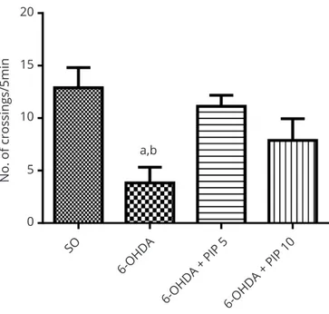
Open ield test: Parkinsonian rats usually present a decreased locomotor activity, in the open ield test. In the present work, the untreated 6-OHDA-lesioned group showed around 70% decreases in the number of
crossings/5 min, and this was almost completely reversed after the PIP treatment (Figure 1).
Figure 2 Piperine (PIP) treatments completely reversed the motor deicit
of the 6-OHDA group, as evaluated by the rota rod test. The values are means±SEM of 5-9 animals. a. vs. SO, q=7.095, p<0.01; b. vs. 6-OHDA+PIP5, q=7.621, p<0.01; c. vs. 6-OHDA+PIP10, q=5.150, p<0.05 (One-way ANOVA
and Newman-Keuls as the post hoc test).
Rotarod test: While a signiicant increase in the number of falls, indicative of a deicit in motor coordination, was demonstrated in the untreated 6-OHDA group, as related to the SO group, this motor deicit was totally reversed after PIP treatments with both doses, and the results were similar to those of the SO group (Figure 2).
SO
6-OHDA
6-OHDA + PIP 56-OHDA + PIP 10
No. of falls/min
4
3
2
1
0
a,b,c
Figure 1 Piperine (PIP) treatments reversed partly the decrease in locomotor
activity of the 6-OHDA group, as evaluated by the open ield test. The values are means±SEM of 6-9 animals. a. vs. SO, q=5.016 and b. vs. 6-OHDA+PIP5, q=3.942. Both at p<0.05 (One-way ANOVA and Newman-Keuls as the post hoc test).
SO
6-OHDA
6-OHDA + PIP 56-OHDA + PIP 10
No. of crossings/5min
20
a,b 15
10
5
Apomorphine-induced rotations: We demonstrated that the untreated 6-OHDA lesioned group presented around 168 contralateral rotations/h, which is an indication of the degree of striatal lesion. On the other hand, 53% and almost 90% reductions were observed in the lesioned groups, after PIP treatments with 5 and 10 mg/kg, respectively, suggesting a neuroprotective efect (Figure 3).
Neurochemical measurements
Striatal DA and DOPAC contents: While no alteration was observed in both striatal sides of the SO group, the untreated 6-OHDA group presented a 73% decrease in DA levels in the lesioned right side, as related to its unlesioned left side. The decreases were of only 49 and 35%, after PIP treatments with the doses of 5 and 10 mg/ kg, respectively, in the right sides of the 6-OHDA groups, relatively to their unlesioned sides. Decreases of 55% were seen in DOPAC contents, in the lesioned right side of the untreated 6-OHDA group, in relation to its left side. After PIP treatments, the lower dose presented a greater efect (only a 28% reduction in DOPAC contents), as compared to the higher dose which showed a 48% reduction in its lesioned right side. This unexpected result was due to the fact that higher DOPAC contents were detected in the left side of this group (Figure 4).
Histological assays
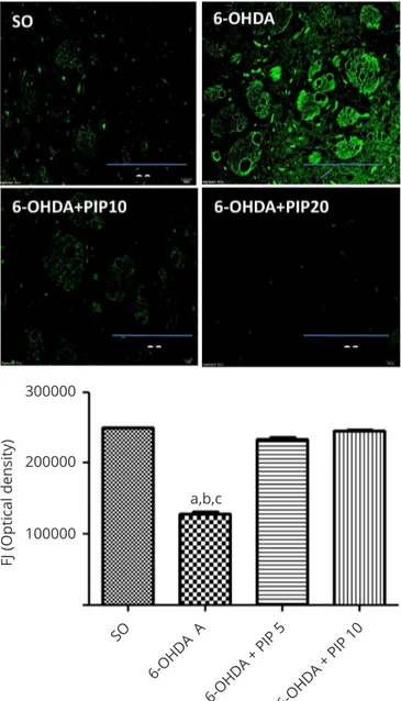
Fluoro jade staining: An intense luorescence, indicative of neurodegeneration, was observed in the ipsilateral striatum (lesioned right side) of the untreated 6-OHDA group (expressed as a 40% decrease in the optical density), as related to the SO group. A complete reversal of this efect was demonstrated in the 6-OHDA group after PIP treatments with both doses (Figure 5).
Figure 4 Piperine (PIP) treatments reverse partly the decreased DA and
DOPAC contents observed in the striatum right side (lesioned) of the 6-OHDA group. The values are means±SEM of 8-13 animals. DA a. vs. SO R, p<0.0001; b. vs. 6-OHDA L, p<0.0001; c. vs. 6-OHDA R, p<0.0001; d. vs. SO R, p<0.024; e. vs. SO R, p<0.0063; f. vs. 6-OHDA R, p<0.0076. DOPAC: a. vs. SO R, p>0.0068; b. vs. 6-OHDA L, p<0.0212; c. vs. 6-OHDA R, p<0.0168; d. vs. 6-OHDA R, p<0.0077; e. vs. 6-OHDA+PIP5 L, p<0.0381; f. vs. 6-OHDA+PIP10 L, p<0.0135 (Two-tailed paired or unpaired Student t-test).
Figure 5 Piperine (PIP) treatments of the 6-OHDA group decreased cell
degeneration as detected by less luorescence after the luoro jade staining in the lesioned right striatum. a. vs. SO, q=18.29; b. vs. 6-OHDA+PIP5, q-15.39; c. vs. 6-OHDA+PIP10, q=17.39. All at p<0.01 (One-way ANOVA and
Newman-Keuls as the post hoc test). Representative photomicrographs
(x400), analyzed by the Image J software (NIH, USA). Scale bar=200 µm.`
SO
6-OHDA A
6-OHDA + PIP 10 6-OHDA + PIP 5
FJ (Optical density)
SO L
4000
a,b a,b
c,d
c,d
e,f e,f
4000
3000 3000
2000 2000
1000 1000
0 0
SO L
SO R SO R
6-OHDA +PIP5 L6-OHDA +PIP5 R6-OHDA +PIP10 L6-OHDA +PIP10 R 6-OHDA +PIP5 L6-OHDA +PIP5 R6-OHDA +PIP10 E6-OHDA +PIP10 R
6-OHDA R 6-OHDA R
6-OHDA L 6-OHDA L
DA (ng/g tecido)
DOPAC (ng/g tecido)
300000
200000
100000
a,b,c
SO R SO L 6-OHDA R
6-OHDA + PIP 10 L 6-OHDA + PIP 10 R 6-OHDA + PIP 5 R6-OHDA + PIP 5 L 6-OHDA L
No. of rotations/h
200
150
100
50
0
a,b,c
Figure 3 Piperine (PIP) treatments reversed the increased number of
Immunohistochemical assays
Immunohistochemistry for tyrosine hydroxylase (TH): A reduction of 54% in TH immunoreactivity was demonstrated in the lesioned right striata of the untreated 6-OHDA group, relatively to the SO group. Values of optical density for TH immunoreactivity, in the 6-OHDA group after treatments with both PIP doses, were not signiicantly diferent from those of the SO group (Figure 6).
Immunohistochemistry for dopamine transporter (DAT): A drastic decrease (around 95%) in DAT immunoreactivity was observed in the lesioned right striatum of the untreated 6-OHDA rats, as related to the SO group; and
Figure 6 Piperine (PIP) treatments almost completely reversed the drastic
decrease in TH immunoreactivity of the 6-OHDA group in the lesioned right striatum. a. vs. SO, q=8.316, p<0.01; b. vs. 6-OHDA+PIP5, q=5.079, p<0.05; c. vs. 6-OHDA+PIP10, q=7.957, p<0.01 (One-way ANOVA and Newman-Keuls
as the post hoc test). Representative photomicrographs (x400) analyzed by
the Image J software (NIH, USA). Scale bar=200 µm.
this efect was completely reversed after PIP treatments with both doses (Figure 7).
Figure 7 Piperine (PIP) treatments reversed almost completely the drastic
decrease in DAT immunoreactivity of the 6-OHDA group in the lesioned right striatum. a. vs. SO, q=28.40; b. vs. 6-OHDA+PIP5, q=27.18; c. vs. 6-OHDA+PIP10, q=26.63. All at p<0.001 (One-way ANOVA and
Newman-Keuls as the post hoc test). Representative photomicrographs (x400),
analyzed by the Image J software (NIH, USA). Scale bar=200 µm.
Immunohistochemistry for TNF-alpha: A drastic increase for TNF-alpha immunoreactivity of 13.2-fold was observed in the lesioned right striatum of the untreated 6-OHDA group, as related to SO controls. PIP treatments of the 6-OHDA group were not able to completely reverse the increase in these cytokine levels. However, the increase was much lower after the treatment with the higher PIP dose (6.7-fold) (Figure 8).
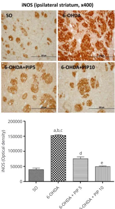
Immunohistochemistry for iNOS: A 3.8-fold increase was seen in iNOS immunoreactivity in the lesioned right striatum of the untreated 6-OHDA group, as related to the SO group. This efect was almost completed reversed in the 6-OHDA group after treatments with the high PIP dose and the result was not signiicantly diferent from those of the SO group (Figure 9).
SO
SO
6-OHDA A
6-OHDA
6-OHDA + PIP 10
6-OHDA + PIP 10
6-OHDA + PIP 5
6-OHDA + PIP 5
TH (Optical density)
TH (Optical density)
20000
20000
150000
150000
100000
100000
50000
50000
a,b,c
Discussion
Parkinson’s disease (PD) is a chronic, progressive neurologic pathology, presenting four cardinal motor manifestations, such as tremor at rest, rigidity, bradykinesia and postural instability. However, PD patients have dysfunctions, extending beyond the classical motor disabilities associated with the disease. Indeed, PD patients appear to be at increased risk for cognitive and psychiatric dysfunctions, mainly dementia and depression [14]. In addition, they often have disturbing sensory symptoms and pain in afected limbs that are signs of autonomic failure.
Neuroinlammation and oxidative stress can damage DA neurons in PD patients [15], and in vivo and in vitro
studies demonstrate these factors have a role in the disease. It has been shown that TNF-alpha, IL-1beta and IL-6-positive neurons increased in the substantia nigra and
putamen, during the progress of PD [16, 17]. Besides, the levels of pro-inlammatory cytokines in peripheral blood tend to be higher in PD patients [18]. Furthermore, the concept that free radical-mediated injury may underlie the neurodegeneration occurring in PD, continues to be the leading hypothesis for its pathogenesis. The possibility that DA neurons may undergo free radical-mediated injury is supported by animal studies using neurotoxins, as 6-OHDA [14]. Interestingly, DA itself is a selective neurotoxin for neurons, what is largely due to its ability to form ROS, making DA a source of oxidative stress.
Presently, the available treatments for PD ofer only symptomatic relief and there is no disease-modifying treatment yet. Furthermore, the protective therapy for PD is based on the concept that SNpc neurons can
Figure 9 Piperine (PIP) treatments reversed partly the increase in iNOS
immunoreactivity of the 6-OHDA group in the lesioned right striatum. a. vs. SO, q=26.80, p<0.001; b. vs. 6-OHDA+PIP5, q=18.16, p<0.001; c. vs. 6-OHDA+PIP10, q=24.51, p<0.001; d. vs. SO, q=8.639, p<0.01; e. vs. 6-OHDA+PIP5, q=6.344, p<0.01 (One-way ANOVA and Newman-Keuls as
the post hoc test). Representative photomicrographs (x400), analyzed by
the Image J software (NIH, USA). Scale bar=200 µm.
Figure 8 Piperine (PIP) treatments reversed partly the increase in
TNF-alpha immunoreactivity of the 6-OHDA group in the lesioned right striatum. a. vs. SO, q=22.05, p<0.001; b. vs. 6-OHDA+PIP10, q=11.15, p<0.01; c. vs. SO, q=17.12, p<0.001; d. vs. SO, q=9.640, p<0.01; e. vs. 6-OHDA+PIP5, q=7.098, p<0.01 (One-way ANOVA and Newman-Keuls as
the post hoc test). Representative photomicrographs (x400) analyzed by
the Image J software (NIH, USA). Scale bar=200 µm.
Figure 8 Piperine (PIP) treatments reversed partly the increase in
TNF-alpha immunoreactivity of the 6-OHDA group in the lesioned right striatum. a. vs. SO, q=22.05, p<0.001; b. vs. 6-OHDA+PIP10, q=11.15, p<0.01; c. vs. SO, q=17.12, p<0.001; d. vs. SO, q=9.640, p<0.01; e. vs. 6-OHDA+PIP5, q=7.098, p<0.01 (One-way ANOVA and Newman-Keuls as the post hoc test).
Representative photomicrographs (x400) analyzed by the Image J software (NIH, USA). Scale bar=200 µm.
SO SO
6-OHDA 6-OHDA
6-OHDA + PIP 10 6-OHDA + PIP 10
6-OHDA + PIP 5 6-OHDA + PIP 5
iNOS (Optical density)
TNF - alpha (Optical density)
200000 200000
150000 150000
100000 100000
50000
50000
0
0 a,b
c,d
a,b,c
d
somehow be spared from degenerative processes leading to cell death and DA depletion. Thus, neuroprotective therapy supported by animal studies, using neurotoxins such as 6-OHDA and agents targeting oxidative stress, mitochondrial dysfunction and inlammation, are prime candidates for neuroprotection [19].
Previously, PIP was shown to act signiicantly on early acute changes in inlammatory processes and on chronic granulative changes [20]. In the arthritis model in rats, PIP was efective in decreasing several parameters, as MPO, LPO, GSH, catalase, SOD and NO, known to be involved in rheumatoid arthritis. It also reduced the levels of pro-inlammatory mediators, as IL-1B, TNF-alpha and PGE2. Surprisingly, although PIP is a potent anti-inlammatory drug, it seems to have no analgesic property [21].
Recently [22], PIP was shown to display signiicant protective efect in an experimental model of periodontitis, what may be ascribed to its inhibitory activity on the expression of IL-1B, MMP-8, MMP-9 and MMP-13. Besides, PIP showed an anti-inlammatory efect at colorectal sites, in acetic acid-induced colitis in mice, by downregulating the production and expression of inlammatory mediators [23].
Although the literature presents several studies on the peripheral efects of PIP, only a decade ago [24], its antidepressant-like properties, mediated at least in part by MAO inhibition, were demonstrated [25, 26]. Others [9] observed that PIP (5-20 mg/kg, administered for 1-4 weeks) possessed an antidepressant-like activity and cognitive enhancing efect. At this same dose range, PIP signiicantly
improved memory impairment and hippocampal
neurodegeneration, in a model of Alzheimer's disease in rats [27]. According to these authors, the possible underlying mechanisms might be partly associated with the decrease in lipoperoxidation and AChE activity. Another in vitro study [28] also revealed the PIP neuroprotective action.
In the model of 6-OHDA-lesioned rats, we showed that PIP reversed in part or almost completely the increased apomorphine-induced rotational behavior and also the decreased locomotor activity, both observed in the untreated lesioned animals. Others [29] also demonstrated that PIP reduced contralateral rotations induced by apomorphine, in the same experimental model. A similar efect was observed with alkaloids from Piper longum, including piperine, in a MPTP model of PD in mice, where these compounds increased their movements, as related to the untreated MPTP animals [30].
A hallmark of PD is the degeneration of dopaminergic neurons in the SNpc, coupled with a depletion of DA and its metabolites in the nigrostriatal projections [31], and many of the motor features of PD result primarily from the loss of those dopamine neurons [32]. In the present study, PIP treatments partly reversed the decreases in striatal DA contents, observed in the untreated 6-OHDA group. A similar, although lower, efect was observed in DOPAC contents in the lesioned side of the striatum, emphasizing the potential neuroprotective properties of PIP.
We showed a drastic decrease in TH immunoreactivity in the striatum lesioned side of the untreated 6-OHDA
animals, as related to the SO group, what was partly reversed after PIP treatments. TH catalyzes the formation of L-DOPA, the rate-limiting step in the biosynthesis of DA, and thus PD can be considered a TH-deiciency syndrome of the striatum [33]. Therefore, an eicient strategy for PD treatment is based on correcting or bypassing the enzyme deiciency, what could be achieved by a neuroprotective treatment. Although we did not observe a complete recovery of TH activity, in the 6-OHDA group after PIP treatments, its decrease was highly attenuated, particularly after the higher dose. We feel that a longer treatment (e.g., 3 weeks) would result in a more intense efect.
The dopamine transporter (DAT) controls the spatial and temporal dynamics of DA neurotransmission, by driving the reuptake of this extracellular transmitter into presynaptic neurons [34]. Besides regulating synaptic DA in the striatum, DAT modulation can afect locomotor activity and, in PD, the DAT loss could afect DA clearance and locomotor activity [35]. DAT is considered to be the single most important determinant of extracellular DA concentrations and it is reduced up to 70% in PD [36]. We showed a drastic decrease in DAT immunoreactivity in striata from untreated 6-OHDA animals, what was completely reversed after PIP treatments. PIP results on both DAT and TH immunoreactivities point out to its great potential for PD treatment.
Despite a large number of studies, the cause of neuronal loss in PD is not fully understood. Neuroinlammatory mechanisms might contribute to the cascade of events leading to neuronal degeneration, and thus multiple neuroinlammatory processes are exacerbated in PD, including glial-mediated reactions and increased expression of pro-inlammatory cytokines in the substantia nigra [37, 38]. We showed that PIP treatments signiicantly reversed the increased immunoreactivities for TNF-alpha and iNOS, observed in the untreated 6-OHDA-lesioned animals.
Evidences indicate an upregulation of iNOS and COX-1 and -2 in the substantia nigra of PD patients [39], and the loss of dopaminergic neurons may result from inlammation-induced proliferation of microglia and reactive macrophages expressing iNOS [40]. Furthermore, pro-inlammatory cytokines as TNF-alpha have been implicated as main efectors for functional consequences of neuroinlammation on neurodegeneration in PD models [41]. Under pathological conditions, microglia release large amounts of TNF-alpha which is an important component of the neuroinlammatory response associated to neurodegenerative diseases [42].
Furthermore, the progress in PD treatments will rely on understanding genetic mutation or susceptibility factors that lead to the disease, better translation between preclinical animal models and clinical research, and improvements in the design of clinical trials [43].
Conclusion
Our indings give further support for PIP oxidant, anti-inlammatory and anti-apoptotic properties. Moreover, these efects are probably related to PIP neuroprotective actions, making this drug a promising candidate to translational studies for the prevention or treatment of degenerative diseases as PD.
Acknowledgments
The authors are grateful for the inancial support from the Brazilian National Research Council (CNPq). The authors also thank the technical assistance of Ms. V. R. Bastos, Ms. A.C. de Araújo and Ms. Janice P. Lopes and to Prof. M.O.L. Viana for his skilled manuscript orthographic revision.
Conlict of Interests
The authors declare no conlict of interest.
References
[1] Srinivasan K. Black pepper and its pungent principle-piperine: a review
of diverse physiological efects. Crit Rev Food Sci Nutr. 2007; 47(8):735– 748.
[2] Chuchawankul S, Khorana N, Poovorawan Y. Piperine inhibits cytokine
production by human peripheral blood mononuclear cells. Genet Mol Res. 2012; 11:617–627.
[3] Meghwall M, Gowami TK. Piper nigrum and piperine: an update. Phytother Res. 2013; 27(8):1121–1130.
[4] Bang JS, Oh DH, Choi HM, Sur B-J, Lim SJ, et al. Anti-inlammatory and antiarthritic efects of piperine in human interleukin 1beta-stimulated ibroblast-like synoviocytes and in rat arthritis models. Arthritis Res Ther. 2009: 11(2):R49.
[5] Sabina EP, Nasreen A, Vedi M, Rasool M. Analgesic, antipyretic and ulcerogenic efects of piperine: an active ingredient of pepper. J Pharm Sci & Res. 2013; 5(10):203–206.
[6] Doucette CD, Rodgers G, Liwski RS, Hoskin DW. Piperine from black pepper inhibits activation-induced proliferation and efector function of T lymphocytes. J Cell Biochem. 2015; 116(11):2577–2588.
[7] D’Hooge R, Pei YQ, Raes A, Lebrun P, Van Bogaert PP, et al. Anticonvulsant
activity of piperine on seizures induced by excitatory amino acids
receptor agonists. Arzneimittelforschung. 1996; 46(6):557–560. [8] Fu M, Sun ZH, Zuo HK. Neuroprotective efect of piperine on primarily
cultured hippocampal neurons. Biol Pharm Bull. 2010; 33(4):598–603. [9] Wattanathorn J, Chonpathompikunlert P, Muchimapura S, Priprem A,
Tankamnerdthai O. Piperine, the potential functional food for mood and cognitive disorders. Food Chem Toxicol. 2008; 46(9):3106–3110. [10] Priprem A, Chanpathompikunlert P, Sutthiparinyamont S,
Wattanathorn J. Antidepressant and cognitive activities of intranasal piperine-encapsulated liposomes. Adv Biosci Biotechnol 2011, 2:108–
116.
[11] Pal A, Nayak S, Sahu PK, Swain T. Piperine protects epilepsy associated depression: a study on role of monoamines. Eur Rev Med Pharmacol Sci. 2011; 15(11):1288–1295.
[12] da Cruz GM, Felipe CF, Scorza FA, da Costa MA, Tavares AF, et al. Piperine decreases pilocarpine-induced convulsions by GABAergic mechanisms. Pharmacol Biochem Behav. 2013; 104:144–153. [13] Przedborski S, Levivier M, Jiang H, Ferreira M, Jackson-Lewis V, et al.
Dose-dependent lesions of the dopaminergic nigrostriatal pathway induced by intrastriatal injection of 6-hydroxydopamine. Neuroscience. 1995; 67(3):631–647.
[14] Zigmond MJ, Burke RE. Pathophysiology of Parkinson’s disease, in: Neuropsychopharmacology: The Fifth Generation of Progress. Edited by Kenneth L Davis, Dennis Charney, Joseph T Coyle, Charles Nemerof. American College of Neuropsychopharmacology. 2002.
[15] Kouti L, Noroozi M, Akhondzadeh S, Abdollahi M, Javadi MR, et al. Nitric oxide and peroxinitrite serum levels in Parkinson’s disease:
correlation of oxidative stress and the severity of the disease. Eur Rev
Med Pharmacol Sci. 2013; 17(7):964–970.
[16] Nagatsu T, Sawada M. Inlammatory process in Parkinson's disease:
role for cytokines. Curr Pharm Des. 2005; 11(8):999–1016.
[17] Sawada M, Imamura K, Nagatsu T. Role of cytokines in inlammatory process in Parkinson's disease. J Neural Transm. 2006; Suppl 70:373– 381.
[18] Sadek HL, Almohari SF, Renno WM. The inlammatory cytokines in the pathogenesis of Parkinson's disease. Journal of Alzheimers Disease & Parkinsonism. 2014; 4:148.
[19] Seidi SE, Potashkin JA. The promise of neuroprotective agents in Parkinson’s disease. Front Neurol. 2011; 2:68.
[20] Mujumdar AM, Dhuley JN, Deshmukh VK, Raman PH, Naik SR. Anti-inlammatory activity of piperine. Jpn J Med Sci Biol. 1990; 43(3):95– 100.
[21] Sudjarwo SA. The potency of piperine as anti-inlammatory and analgesic in rats and mice. Folia Medica Indonesiana. 2005; 41:190– 194.
[22] Dong Y, Huihui Z, Li C. Piperine inhibits inlammation, alveolar bone loss and collagem ibers breakdown in a rat periodontitis model. J Periodontal Res. 2015; 50(6):758–765.
[23] Gupta RA, Motiwala MN, Dumore NG, Danao KR, Ganjare AB. Efect of piperine on inhibition of FFA induced TLR4 mediated inlammation and amelioration of acetic acid induced ulcerative colitis in mice. J Ethnopharmacol. 2015; 164:239–246.
[24] Lee SA, Hong SS, Han XH, Hwang JS, Oh GJ, et al. Piperine from the
fruits of Piper longum with inhibitory efect on monoamine oxidase
and antidepressant-like activity. Chem Pharm Bull (Tokyo). 2005; 53(7):832–835.
[25] Lee CS, Han ES, Kim YK. Piperine inhibition of
1-methyl-4-phenylpyridinium-induced mitochondrial dysfunction and cell death
in PC12 cells. Eur J Pharmacol. 2006; 537(1–3):37–44.
[26] Li S, Wang C, Wang M, Li W, Matsumoto K, et al. Antidepressant like efects of piperine in chronic mild stress treated mice and its possible mechanisms. Life Sci. 2007; 80(15):1273–1381.
[27] Chonpathompikunlert P, Wattanathorn J, Muchimapura S. Piperine the
main alkaloid of Thai black pepper, protects against neurodegeneration
and cognitive impairment in animal model of cognitive deicit like condition of Alzheimer’s disease. Food Chem Toxicol. 2010; 48:798– 802.
[28] Mao QQ, Huang Z, Ip SP, Xian YF, Che CT. Protective efects of piperine against corticosterone-induced neurotoxicity in PC12 cells. Cell Mol Neurobiol. 2012; 32(4):531–537.
[29] Shrivastava P, Vaibhav K, Tabassum R, Khan A, Ishrat T, et al. Anti-apoptotic and anti-inlammatory efect of piperine on 6-OHDA induced Parkinson's rat model. J Nutr Biochem. 2013; 24(4):680–687.
[30] Bi Ying, Qu PC, Wang QS, Zheng Li, Liu HL, et al. Neuroprotective efects
of alkaloids from Piper longum in a MPTP-induced mouse model of
Parkinson's disease. Pharm Biol. 2015; 53(10):1516–1524.
[31] Bernheimer H, Birkamyer W, Hornykiewicz O, Jellinger K, Seitelberger F. Brain dopamine and the syndromes of Parkinson and Huntington. J
Neurol. 1973; 20(4):415.
[32] Fahn S, Sulzer D. Neurodegeneration and Neuroprotection in Parkinson's disease. NeuroRx. 2004; 1(1):139–154.
[33] Tabrez S, Jabir NR, Shakil S, Greig NH, Alam Q, et al. A synopsis on the role of tyrosine hydroxylase in Parkinson’s disease. CNS Neurol Disord Drug Targets. 2012; 11(4):395–409.
[34] Vaughan RA, Foster JD. Mechanisms of dopamine transporter regulation in normal and disease states. Trends Pharmacol Sci. 2013; 34(9):489–496.
[35] Chotibut T, Apple DM, Jeferis R, Salvatore MF. Dopamine transporter loss in 6-OHDA Parkinson’s model is unmet by parallel reduction in dopamine uptake. PLos One. 2012; 7(12):e52322.
[36] Nutt JG, Carter JH, Sexton GJ. The dopamine transporter: importance in Parkinson's disease. Ann Neurol. 2004; 55(6):766–773.
[37] Cebrián C, Loike JD, Sulzer D. Neuroinlammation in Parkinson's disease animal models: a cell stress response or a step in neurodegeneration
Curr Top Behav Neurosci. 2015; 22:237–270.
[38] Hirsch E, Hunot S. Neuroinlammation in Parkinson's disease: a target
for neuroprotection Lancet Neurol. 2009; 8(4):382–397.
[39] Knott C, Stern G, Wilkin GP. Inlammatory regulators in Parkinson's disease: iNOS, lipocortin-1, and cyclooxygenases-1 and -2. Mol Cell Neurosci. 2000; 16:724–739.
[40] Iravani MM, Kashei K, Mander P, Rose S, Jenner P. Involvement of inducible nitric oxide synthase in inlammation-induced dopaminergic neurodegeneration. Neuroscience. 2002, 110(1):49–58.
[41] Leal MC, Casabona JC, Puntel M, Pitossi FJ. Interleukin-1beta and tumor necrosis factor-alpha: reliable targets for protective therapies in Parkinson's disease Front Cell Neurosci. 2013; 7:53.
[42] Olmos G, Lladó J. Tumor necrosis factor alpha: a link between neuroinlammation and excitotoxicity. Mediators of Inlammation. 2014; 2014:861231.


