Research Article
Neuroprotective Properties of the Standardized
Extract from
Camellia sinensis
(Green Tea) and Its Main
Bioactive Components, Epicatechin and Epigallocatechin
Gallate, in the 6-OHDA Model of Parkinson’s Disease
Natália Bitu Pinto,
1,2Bruno da Silva Alexandre,
2Kelly Rose Tavares Neves,
1Aline Holanda Silva,
1Luzia Kalyne A. M. Leal,
1and Glauce S. B. Viana
1,21Faculty of Medicine of the Federal University of Cear´a, Rua Nunes de Melo 1127 (Rodolfo Te´oilo), 60430-270 Fortaleza, CE, Brazil
2Faculty of Medicine Est´acio of Juazeiro do Norte, Avenida Tenente Raimundo Rocha 515 (Cidade Universit´aria),
63048-080 Juazeiro do Norte, CE, Brazil
Correspondence should be addressed to Glauce S. B. Viana; [email protected]
Received 7 February 2015; Revised 25 April 2015; Accepted 25 May 2015
Academic Editor: Cheorl-Ho Kim
Copyright © 2015 Nat´alia Bitu Pinto et al. his is an open access article distributed under the Creative Commons Attribution License, which permits unrestricted use, distribution, and reproduction in any medium, provided the original work is properly cited.
Camellia sinensis(green tea) is largely consumed, mainly in Asia. It possesses several biological efects such as antioxidant and
anti-inlammatory properties. he objectives were to investigate the neuroprotective actions of the standardized extract (CS), epicatechin (EC) and epigallocatechin gallate (EGCG), on a model of Parkinson’s disease. Male Wistar rats were divided into SO (sham-operated controls), untreated 6-OHDA-lesioned and 6-OHDA-lesioned treated for 2 weeks with CS (25, 50, or 100 mg/kg), EC (10 mg/kg), or EGCG (10 mg/kg) groups. One hour ater the last administration, animals were submitted to behavioral tests and euthanized and their striata and hippocampi were dissected for neurochemical (DA, DOPAC, and HVA) and antioxidant activity determinations, as well as immunohistochemistry evaluations (TH, COX-2, and iNOS). he results showed that CS and catechins reverted behavioral changes, indicating neuroprotection manifested as decreased rotational behavior, increased locomotor activity, antidepressive efects, and improvement of cognitive dysfunction, as compared to the untreated 6-OHDA-lesioned group. Besides, CS, EP, and EGCG reversed the striatal oxidative stress and immunohistochemistry alterations. hese results show that the neuroprotective efects of CS and its catechins are probably and in great part due to its powerful antioxidant and anti-inlammatory properties, pointing out their potential for the prevention and treatment of PD.
1. Introduction
Green tea has attracted signiicant attention worldwide for its beneits for a varied number of disorders, ranging from
cancer to weight loss [1]. Although there are several
polyphe-nolic catechins in green tea, epigallocatechin-3-gallate is the most abundant one, accounting for 65% of its total catechin
content [2], being probably responsible for most of green tea
medicinal properties. Green tea and its bioactive constituents are best known for their antioxidant properties, leading to clinical studies in diseases associated with reactive oxygen species, such as cancer or cardiovascular and
neurodegener-ative diseases [3–9].
Evidences [10–12], including those from our laboratory
[13], have indicated the anti-inlammatory properties of green
tea and epigallocatechin gallate. Furthermore, inlammation has been implicated in neurodegenerative pathologies, as
Parkinson’s disease [14–16]. Parkinson’s disease is the second
most common neurodegenerative disorder ater Alzheimer’s disease. It is characterized by a slow and progressive
degen-eration of dopaminergic neurons in thesubstantia nigra pars
compacta. Although the cause of this neuronal degeneration
may be poorly understood, it is largely accepted that
neuroin-lammatory mechanisms are certainly involved [14–22].
In addition, oxidative stress is shown to be associated with
neuronal degeneration, as shown in previous [23, 24] and
more recent works [25–30]. he oxidative stress contributes to the cascade leading to dopamine cell degeneration in PD and is closely linked to other components of neurodegenera-tion, as mitochondrial dysfuncneurodegenera-tion, excitotoxicity, increased oxygen free radical production, and inlammation, as well
[31]. An important consequence of oxidative stress is an
increased lipid peroxidation found in thesubstantia nigraof
PD patients [23].
Polyphenols, such as epigallocatechin gallate, due to their anti-inlammatory and antioxidant efects, are known to present neuroprotective properties, what could explain their
beneits in neurodegenerative diseases [32–36]. Although
there are some data in the literature on the efects of green tea and its polyphenols on Parkinson’s disease, the great majority
of them deal with in vitro models [37–39] or studied the
beneicial efects of green tea consumption by Parkinson’s
disease patients [39].
hus, the objectives of the present work were to evaluate the neuroprotective properties of the standardized extract of green tea and its catechins on the 6-OHDA model of Parkinson’s disease in rats. For that, behavioral evaluations (apomorphine-induced rotation, open ield, water maze, rota rod, and forced swimming tests) and neurochemical deter-minations (striatal DA, DOPAC and HVA measurements, and antioxidant activity) were performed. Besides, immuno-histochemistry assays for TH (dopaminergic degeneration marker) and for COX-2 and iNOS (inlammation-related enzyme) were also carried out in 6-OHDA-lesioned animals, without and with drugs treatments.
2. Material and Methods
2.1. Drugs and Reagents. 6-hydroxydopamine, apomorphine,
and HPLC standards were from Sigma-Aldrich (St. Louis, MO, USA); ketamine and xylazine were from K¨onig (Santana de Parna´ıba, S˜ao Paulo, Brazil). Antibodies for immunohisto-chemistry assays were from Santa Cruz Biotechnology (Dal-las, TX, USA) or Merck-Millipore (Darmstadt, Germany). All other reagents were of analytical grade.
2.2. Animals. Male Wistar rats (200–250 g) from the Animal
House of the Faculty of Medicine Est´acio of Juazeiro do
Norte were maintained at a 24 ± 2∘C temperature, in a
12 h dark/12 h light cycle, with standard food and waterad
libitum. he study was submitted to the Ethical Committee
for Animal Experimentation of the Faculty of Medicine of the Federal University of Cear´a (Brazil) and was approved under the number 104/2011. All experiments followed the ethical principles established in the Guide for the Care and Use of Laboratory Animals, USA, 1986.
2.3. Determination of the Total Phenol Content in the Dried
Extract from C. sinensis. Samples (10 mg) of the extract
were transferred to a 10 mL volumetric lask, containing 0.25 mL 1 N Folin-Ciocalteau reagent and 4 mL Milli-Q water.
Ater alkalinization of the medium (1.25 mL 20% Na2CO3
solution), the volume was completed to 10 mL with Milli-Q
water. Ater 40 min, at room temperature (25∘C) and at dark,
the mixture was submitted to spectrophotometric reading
at 715 nm [40]. he calibration curve was prepared with
standard gallic acid (dissolved in Milli-Q water), at
con-centrations ranging from 2 to 16�g/mL. he results showed
32.6% total phenols in theC. sinensisextract, determined as
gallic acid equivalents.
2.4. Experimental Protocol Used for the 6-OHDA Model
of PD. he animals were anesthetized with an association
of xylazine (10 mg/kg, i.p.) and ketamine (80 mg/kg, i.p.), submitted to trichotomy of the head superior region, and ixed to the stereotaxic frame by their ear canals. hen, a longitudinal midline incision was done and tissues were separated for bregma visualization. he following coordinates (at two diferent points from bregma) were used: 1st point
(AP, +0.5; ML,−2.5; and DV,−5.0) and 2nd point (AP,−0.9;
ML,−3.7; and DV, +6.5) according to Paxinos and Watson,
1997 [41]. hen, a thin hole was performed in the skull,
over the target area, and a 1�L solution containing 6�g
6-OHDA was injected into each point. he syringe stayed in place for 5 min to assure the solution difusion, and then the incision was sutured. he sham-operated (SO) animals were subjected to all procedures, except that saline was injected into the two points. Aterwards, the animals returned to their cages for recovering. hey were divided into the following groups: SO (treated by gavage with distilled water); 6-OHDA-lesioned (also treated by gavage with distilled water); 6-OHDA-lesioned and treated with 25 or 50 mg/kg
of the standardized extract ofC. sinensis(CS25 and CS50);
epicatechin, 10 mg/kg (EC10); or epigallocatechin gallate, 10 mg/kg (EGCG10). All treatments started 1 h before the stereotaxic surgery and continued daily for 15 days, with drug volumes of 0.2 mL/100 g body weight. hen, ater treatments and 1 h ater the last drug administration, the animals were submitted to behavioral tests. At the next day, they were euthanized (decapitation) and brain tissues were removed for neurochemical, histological, and immunohistochemical studies.
2.5. Behavioral Testing
2.5.1. Apomorphine-Induced Rotations. Contralateral
rota-tions (opposite to the lesioned right-side) induced by apo-morphine (1 mg/kg, i.p.) were monitored for 1 h. he cause of this apomorphine-induced rotational behavior is related to the imbalance, in the nigrostriatal dopaminergic pathways, between the right (lesioned) and let (unlesioned) brain hemispheres. All groups were treated for two weeks. he SO and untreated-6-OHDA groups were administered with distilled water, for the same period of time.
2.5.2. Open Field Test. his test evaluates a stimulant or
depressant drug activity and may also indicate an anxiolytic action. he arena was made of wood, whose dimensions were
50 cm×50 cm×30 cm (length, width, and height). he loor
2.5.3. Forced Swimming Test. he test is based on the obser-vation that when the animals are subjected to a stressful situation with no possibility for escaping, they adopt a posture of immobility ater an initial period of agitation. he reduction of this immobility time is suggestive of an antidepressant action. he animals were placed individually in a cylinder (40 cm height and 23 cm diameter), containing water up to 25 cm below the top. he immobility time was monitored for 5 min.
2.5.4. Water Maze Test. he water maze consisted of a black
circular pool (1.7 m diameter and 1 m height) with water
(0.59 m deep) at 22∘C, and visual cues on the walls at one
of four equally spaced locations: north (N), east (E), south (S), and west (W). he pool was divided into 4 quadrants: NW, NE, SE, and SW. Ater the treatment, each animal was submitted to two trials in the pool, for the next three days. In these trials, the animals were placed into the pool, facing the wall, and had a maximum time of 54 s to ind a submersed platform. he animal stayed on the platform for 10 s. Animals that did not ind the platform at the end of 54 s were placed on it for 10 s. Ater this time, the animal was removed from the pool for 30 s, and the procedure was repeated (6 times for each animal). he procedure was repeated again 24 h later (pretest). Ater 48 h of the pretest, the procedure was repeated once more for evaluating the time needed by the animal to ind the platform; this correlates with the animal’s spatial memory learning ability.
2.6. Oxidative Stress Evaluation
2.6.1. MDA Determination. he degree of lipoperoxidation
was measured by means of the thiobarbituric acid reactive
substances (TBARS) method [42]. For that, 10% striatal
homogenates from all groups studied were prepared in 1.15% KCl. hen, 0.25 mL of each homogenate was added to 1 mL 10% trichloroacetic acid and 1 mL 0.6% thiobarbituric acid. Ater agitation, the mixture was maintained in water
bath (95–100∘C) for 15 min, followed by the addition of n
-butanol (2 : 1, v/v). he mixture was cooled in an ice bath
and centrifuged (800×g, 5 min). he TBARS content in
the supernatants was spectrophotometrically determined at
535 nm. he results are expressed in�mol malondialdehyde
(MDA) per g tissue.
2.6.2. Nitrite Determination. For nitrite determination, 10%
striatal homogenates from all groups studied were
pre-pared in 1.15% KCl. Ater centrifugation (800×g, 10 min),
100�L of supernatant samples was incubated with 100�L
Griess reagent (1% sulfanilamide in 1% H3PO4, 0.1%
N-naphthylethylenediamide dihydrochloride in 1% H3PO4)
diluted in distilled water (1 : 1 : 1 : 1), at room temperature for 10 min. he absorbance was measured at 550 nm and nitrite concentrations were determined with a standard curve of
NaNO2 and the results are expressed as�mol nitrite per g
tissue [43].
2.6.3. Antioxidant Potential Evaluation by Chemiluminescence
in Human Neutrophils. his method is commonly employed
for a direct measurement of reactive oxygen species (ROS) generation. It is capable of quantifying both intracellular
and extracellular ROS. Briely, human neutrophils (5×10⁶
cells/mL) were incubated, for 20 min at 37∘C, with the
standardized extract of C. sinensis (0.1 to 100�g/mL) and
the luminol chemiluminescent probe (280�M). Ater that,
the tubes were transferred to the luminometer, and phorbol
myristate acetate (PMA, 107M), as the stimulant reagent, was
added. hen, the production of chemiluminescence (CL),
in cpm (photons/min), was followed for 20 min at 37∘C.
he spontaneous production of CL by neutrophils was also performed in the absence of the stimulus, and quercetin
(50�g/mL) was used as standard [44].
2.7. Neurochemical Determinations of DA and DOPAC by
HPLC. he striatal contents of DA and DOPAC were
deter-mined by HPLC. Homogenates were prepared in 10% HClO4
and centrifuged at 4∘C (15,000 rpm, 15 min). he supernatants
were iltered and 20�L injected into the HPLC column. For
that, an electrochemical detector (model L-ECD-6A from Shimadzu, Japan) coupled to a column (Shim-Pak CLC-ODS, 25 cm) with a lux of 0.6 mL/min was employed. A mobile phase was prepared with monohydrated citric acid (150 mM), sodium octyl sulfate (67 mM), 2% tetrahydrofuran, and 4% acetonitrile in deionized water. he mobile phase pH was adjusted to 3.0 with NAOH (10 mM). Monoamines were quantiied by comparison with standards, processing the same manner as the samples. he results are expressed as ng/g tissue.
2.8. Immunohistochemical Analyses in Rat Brain Areas
2.8.1. Immunohistochemistry Assays for TH, iNOS, and
COX-2. Brain striatal or hippocampal sections were ixed in
10% bufered formol, for 24 h, followed by a 70% alcohol solu-tion. he sections were embedded into parain wax for slices processing on appropriate glass slides. hese were placed in
the oven at 58∘C, for 10 min, followed by deparainization in
0 100 200
300 a, b, c, d
e, f g h i SO 6 -O HD A 6 -O HD A + CS 25 6 -O HD A + CS 50 6 -O HD A + EC 10 6 -O HD A + EGC G 10 N u m b er o f r o ta tio n s/ 60 min
Figure 1: Efects presented by the 6-OHDA-lesioned animals, before and ater treatments with CS25, CS50, EC25, or EGCG10, on the apomorphine-induced rotational behavior. a: versus SO,� = 11.44; b: versus 6-OHDA + CS25,� = 3.19; c: versus 6-OHDA + CS50,
� = 6.65; d: versus 6-OHDA + EC10,� = 7.53; e: versus 6-OHDA +
EGCG10,� = 6.82; f: versus SO,� = 8.97; g: versus SO,� = 4.54; h: versus SO,� = 3.63; i: versus SO,� = 4.36(one-way ANOVA and Newman-Keuls as thepost hoctest).
2.9. Statistical Analyses. For statistical analyses, one-way
ANOVA, followed by the Newman-Keuls as thepost hoctest,
was used for multiple comparisons. Whenever needed, the
paired or unpaired Student’s�-test was used, for comparisons
between two means. Diferences were considered signiicant at� < 0.05.
3. Results
3.1. Behavioral Evaluation
3.1.1. Apomorphine-Induced Rotations. Two weeks ater the
intrastriatal 6-OHDA lesion, all animals were administered with apomorphine (1 mg/kg, i.p.) and assessed for rotational behavior, for 1 h. A signiicant increase in the number of contralateral rotations/h was observed in the untreated
6-OHDA group (248.8 ± 38.40), as related to the SO group
(3.0 ± 1.43). A partial recovery was exhibited in the 6-OHDA
groups ater treatments with CS25 (179.4 ± 21.62), CS50
(98.8±21.8), EC10 (79.7±10.40), and EGCG10 (95.2±15.52) (Figure 1).
3.1.2. Open Field Test. he results showed a signiicant
decrease in locomotor activity (65%) in the untreated 6-OHDA group, as related to the SO group. his efect was smaller in the 6-OHDA groups ater treatments with the
stan-dardizedC. sinensisextract (CS) that exhibited decreases of
50 and 34%, with the doses of 25 and 50 mg/kg, respectively. Lower decreases were observed in the 6-OHDA group ater
treatments with EC10 (13%) and EGCG10 (30%) (Figure 2).
3.1.3. Forced Swimming Test. he untreated 6-OHDA animals
presented a 3.9-fold increase in the immobility time, as
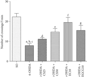
0 10 20 30
a, b, c d e f g SO 6 -O HD A 6 -O HD A + CS 25 6 -O HD A + CS 50 6 -O HD A + EC 10 6 -O HD A + EGC G 10 N u m b er o f cr ossin gs/ 5 min
Figure 2: Efects presented by the 6-OHDA-lesioned animals, before and ater treatments with CS25, CS50, EC10, or EGCG10, on the locomotor activity as assessed by the open ield test. a: versus SO,
� = 8.75; b: versus 6-OHDA + CS50,� = 4.15; c: versus 6-OHDA +
EC10,� = 6.76; d: versus EGCG10,� = 4.44; e: versus SO,� = 6.77; f: versus SO,� = 4.45; g: versus SO, 3.83 (one-way ANOVA and Newman-Keuls as thepost hoctest).
0 50 100
150 a, b, c
d e f g SO 6 -O HD A 6 -O HD A + CS 25 6 -O HD A + CS 50 6 -O HD A + EC 10 6 -O HD A + EGC G 10 T ime o f immob ili ty/ 5 min
Figure 3: Efects presented by the 6-OHDA-lesioned animals, before and ater treatments with CS25, CS50, EC10, or EGCG10, on the immobility time as assessed by the forced swimming test. a: versus SO,� = 8.86; b: versus 6-OHDA + EC10,� = 4.66; c: versus 6-OHDA + EGCG10,� = 5.89; d: versus SO,� = 5.07; e: versus SO,
� = 5.17; f: versus SO,� = 3.93(one-way ANOVA and
Newman-Keuls as thepost hoctest).
related to the SO group, indicating a depressant activity. his alteration was partly reversed ater treatments with CS25 and CS50 (around 2.8-fold increase). Similarly, only 2.4- and 2.0-fold increases in the immobility time were observed ater treatments of the 6-OHDA lesioned animals with EC10 and
0 5 10 15 20
a, b, c, d
e f g T ime t o find p la tf o rm (s) SO 6 -O HD A 6 -O HD A + CS 25 6 -O HD A + CS 50 6 -O HD A + EC 10 6 -O HD A + EGC G 10
Figure 4: Efects presented by the 6-OHDA-lesioned animals, before and ater treatments with CS25, CS50, EC10, or EGCG10, on the time (s) to ind the platform as assessed by the water maze test. a: versus SO,� = 5.92; b: versus 6-OHDA + CS50,� = 4.54; c: versus 6-OHDA + EC10,� = 3.71(one-way ANOVA and Newman-Keuls as thepost hoctest).
3.1.4. Water Maze Test. We demonstrated (Figure 4) a
3.2-fold increase in the time to ind the platform by the untreated 6-OHDA group, as related to the SO, indicating an alteration of the spacial memory and hippocampal dysfunction. he increase was lower in the 6-OHDA groups ater treatments with CS25 and CS50 (2.4- and 1.6-fold, resp.). In the 6-OHDA groups treated with EC10 and EGCG10 the values were close to those of the SO group.
3.2. Evaluation of the Oxidative Stress
3.2.1. TBARS Determination. he untreated 6-OHDA group
presented a 2.3-fold increase in lipid peroxidation, as related to the SO group. Lower changes (2.0- and 1.4-fold increases) were observed in the 6-OHDA groups ater treatments with CS25 and CS50, respectively. Similarly, decreases around 1.2-fold were seen ater treatments with EC10 or EGCG10 (Figure 5).
3.2.2. Nitrite Determination. An increase of 2.6-fold in nitrite
contents was observed in the untreated 6-OHDA group, as related to the SO group. his increase was of 1.8- and 1.4-fold in the 6-OHDA group, ater treatments with CS25 and CS50, and around 1.4-fold ater treatments with EC10 or EGCG10 (Figure 6).
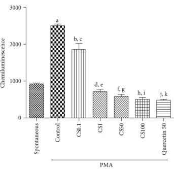
3.2.3. Antioxidant Capacity in Human Neutrophils by
Chemi-luminescence. An increase of 2.7-fold in oxidative capacity
was observed in the controls, ater PMA addition, as related
to the spontaneous oxidation capacity (Figure 7). Although
an increase was also detected with the lower concentration
of CS (0.1�g/mL), it was smaller than that in the presence
of PMA. However, the oxidation capacity in the presence of CS decreased by 23, 37, and 45%, as related to controls (spontaneous oxidation), at concentrations of 1, 50, and
0 100 200 300 400 500
a, b, c, d
e SO 6 -O HD A 6 -O HD A + CS 25 6 -O HD A + CS 50 6 -O HD A + EC 10 6 -O HD A + EGC G 10 TB ARS ( 𝜇 mo l MD A/g tissue)
Figure 5: Efects presented by striatal tissues from 6-OHDA-lesioned animals, before and ater treatments with CS25, CS50, EC10, or EGCG10, on the lipid peroxidation as determined by the TBARS assay. a: versus SO,� = 7.75; b: versus 6-OHDA + CS50,
� = 5.24; c: versus 6-OHDA + EC10,� = 6.89; d: versus 6-OHDA
+ EGCG10,� = 6.77(one-way ANOVA and Newman-Keuls as the
post hoctest).
0 1 2 3 4 5 6
a, b, c, d
SO 6 -O HD A 6 -O HD A + CS 25 6 -O HD A + CS 50 6 -O HD A + EC 10 6 -O HD A + EGC G 10 N itr it e/ni tra te ( 𝜇 mo l/g tissue)
Figure 6: Efects shown by striatal tissues from 6-OHDA-lesioned animals, before and ater treatments with CS25, CS50, EC10, or EGCG10, on the nitrite/nitrate contents as determined by the Griess assay. a: versus SO,� = 7.09; b: versus 6-OHDA + CS25,� = 3.48; c: versus 6-OHDA + CS50,� = 4.83; d: versus 6-OHDA + EC10,
� = 4.89; e: versus 6-OHDA + EGCG10,� = 5.36(one-way ANOVA
and Newman-Keuls as thepost hoctest).
100�g/mL, respectively. his last value was similar to that
observed in the presence of quercetin, used as standard (a 48% decrease).
3.3. Neurochemical Evaluation
3.3.1. DA, DOPAC, and HVA Determinations in the Rat
Striata. DA striatal depletion in untreated 6-OHDA animals
0 1000 2000 3000
PMA a
b, c
d, e f, g
h, i j, k
Chemil
umines
cence
Sp
ont
an
eou
s
Co
n
tr
o
l
CS
0.1 CS1
CS
50
CS
100
Quer
cetin
50
Figure 7: he standardized extract fromCamellia sinensis(CS), at concentrations ranging from 0.1 to 100�g/mL, signiicantly decreased chemiluminescence in human neutrophils, as related to the PMA-stimulated cells, indicative of an antioxidant potential. a: versus spontaneous (spont.),� = 25.67; b: versus spont.,� = 14.09; c: versus spont.,� = 3.24; d: versus spont.,� = 5.16; e: versus spont.,� = 7.25; f: versus control (cont.),� = 9, 68; g: versus cont.,
� = 27.01; h: versus cont.,� = 28.93; i: versus cont.,� = 30.05; j:
versus cont.,� = 32.93(one-way ANOVA and Newman-Keuls as
thepost hoctest).
(91 and 87% decreases, resp.). However, a signiicant reversion of these efects was demonstrated in the 6-OHDA groups, ater treatments with CS25 and CS50, where dose-dependent decreases (45 and 23%) were observed in DA contents. Furthermore, smaller decreases were also seen in DA levels of the 6-OHDA + EC10 (52%) and 6-OHDA + EGCG10 (50%) groups. As far as DOPAC and HVA contents are concerned, signiicant decreases (62 and 44%) were demonstrated only in the 6-OHDA group, ater treatment with the higher CS dose (6-OHDA + CS50), as related to the untreated 6-OHDA group. Similar results were observed with the 6-OHDA + EC10 and 6-OHDA + EGCG10 that presented decreases of 66 and 24% for DOPAC and 52 and 87% decreases for HVA, respectively, as related to the untreated 6-OHDA group
(Figures8(a),8(b), and8(c)).
3.4. Immunohistochemistry Assays
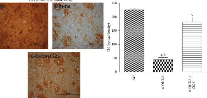
3.4.1. Efects of Green Tea and Its Catechins on TH, COX-2,
and iNOS Immunostainings. he untreated 6-OHDA group
showed around 80% decreases in striatal immunostaining
for TH (Figure 9), as related to the SO group. However, the
results observed in the 6-OHDA group ater treatment with CS (6-OHDA + CS25) presented immunostaining similar to that observed in the SO group. A 4-fold increase in
COX-2 immunostaining (Figure 10) was observed in striatal
tissue from untreated 6-OHDA animals, as related to the
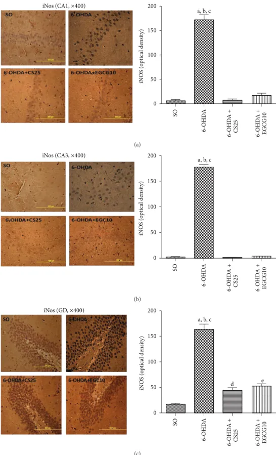
SO group. In addition, increases of only 2.7- and 2.5-fold were observed in the 6-OHDA groups ater treatments with CS25 or EGCG10. Immunohistochemistry assays were also performed for hippocampal tissues in these same groups. he results showed around 24-, 129-, and 10-fold increases in immunostainings for iNOS, in CA1, CA3, and DG subields in the untreated 6-OHDA animals, as related to the SO group. he values decreased towards those of the SO group in the 6-OHDA group ater treatments with CS25 and EGCG10,
mainly at CA1 and CA3 areas (Figure 11).
4. Discussion
Parkinson’s disease (PD) is a chronic, neurodegenerative dis-order, involving the degeneration of dopaminergic neurons in
thesubstantia nigra pars compactaand leading to the loss of
dopaminergic terminals in the striatum. Gait disturbances are among the most important motor problems associated with PD; they occur in all stages of the disorder and are one of the
hallmarks for PD progression [45].
C. sinensisand its polyphenolic catechins have attracted
signiicant scientiic attention for their health beneits in a variety of disorders, including neurodegenerative ones, such
as PD [1]. In the present study, we showed that the C.
sinensisstandardized extract and its catechins, epicatechin
and epigallocatechin gallate, present a neuroprotective efect on the striatal 6-OHDA model of PD, in rats.
We showed that these drugs reversed, at least partly, the behavioral changes observed in the untreated 6-OHDA-lesioned animals, at very low doses. hus, in the apomor-phine-induced rotational test, signiicant decreases occurred in the 6-OHDA group ater treatments with CS50, as well as with the two catechins. he drugs also reversed the decreased locomotor activity observed in the untreated lesioned group; and similar results were seen with the forced swimming test and water maze test, indicative of antidepressant and spatial
learning efects, respectively. Previously [13], we showed that
catechin, at small doses (10 and 20 mg/kg), attenuated the increased rotational behavior induced by apomorphine and also the decrease in locomotor activity and working memory deicits, what support our present indings.
Evidences demonstrated that green tea polyphenols
pos-sess an antidepressant activity in mice [46], while others
[47] showed that several green tea catechins, including EC
and EGCG, ater a long-term oral administration to rats, improved reference and working memory related to learning ability. All these data agree with ours and point out that these green tea efects could be of beneit in neurodegenerative disorders, as Parkinson’s disease.
CS50, EC10, and EGCG10 signiicantly reversed the decreases in striatal DA, DOPAC, and HVA contents, observed in the untreated lesioned animals. Interestingly, similar efects were seen with all three drugs. Although there are several studies on the neuroprotective efects of green tea and its catechins, most of them concentrate their objective
on cellular orin vitromodels of PD [37,38,48–56]. hus,
there are relatively few works dealing with in vivo animal
0 1000 2000 3000 4000
a, b, c, d, e
f g
h
D
o
pa
mine (n
g/g t
ecido)
SO
6
-O
HD
A
6
-O
HD
A
+
CS
25
6
-O
HD
A
+
CS
50
6
-O
HD
A
+
EC
10
6
-O
HD
A
+
EGC
G
10
(a)
0 1000 2000 3000 4000
a, b, c, d, e
f g
h
D
O
PA
C (n
g/g t
ecido)
SO
6
-O
HD
A
6
-O
HD
A
+
CS
25
6
-O
HD
A
+
CS
50
6
-O
HD
A
+
EC
10
6
-O
HD
A
+
EGC
G
10
(b)
0 500 1000 1500 2000 2500
a, b, c d
e f
g
HV
A (n
g/g t
ecido)
SO
6
-O
HD
A
6
-O
HD
A
+
CS
25
6
-O
HD
A
+
CS
50
6
-O
HD
A
+
EC
10
6
-O
HD
A
+
EGC
G
10
(c)
Figure 8: he decreases in striatal DA contents (a), DOPAC (b), and HVA (c) were signiicantly reversed in the 6-OHDA-lesioned group, ater treatments with CS (25 or 50 mg/kg), EC (10 mg/kg), or EGCG (10 mg/kg). (a) a: versus SO,� = 8.72; b: versus 6-OHDA + CS25,� = 4.99; c: versus 6-OHDA + CS50,� = 6.91; d: versus 6-OHDA + EC10,� = 3.52; e: versus SO,� = 4.38; f: versus SO,� = 4.46; g: versus SO,� = 4.78. (b) a: versus SO,� = 8.69; b: versus 6-OHDA + CS25,� = 6.26; c: versus SO,� = 5.26; d: versus SO,� = 5.27. (c) a: versus SO,� = 9.57; b: versus 6-OHDA + CS50,� = 5.06; c: versus SO,� = 7.71; d: versus SO,� = 4.24; e: versus SO,� = 4.82; f: versus SO,� = 7.10(one-way ANOVA and Newman-Keuls as thepost hoctest).
et al., 2001 [57], investigated the efects of
epigallocatechin-3-gallate on the MPTP-induced dopaminergic neuron loss in
thesubstantia nigraand striatal dopamine depletion, showing
that the pretreatment of mice with the green tea extract or EGCG prevented these efects.
However, in our study, we evaluated not only DA but also DOPAC and HVA contents in the 6-OHDA-lesioned group, ater treatments with green tea and its catechins, epicatechin and epigallocatechin gallate. Growing evidences have indicated that oxidative stress and inlammation play a key role in neurodegenerative disorders, including PD, and probably the antioxidant efects of green tea are associated
with its neuroprotective action [33]. Supporting this, we
demonstrated that green tea and its catechins presented
antioxidant efects, as evaluated by the decrease of lipid peroxidation and nitrite contents, observed in striatal tissue from 6-OHDA lesioned animals, ater treatments with those drugs. Besides, they present an antioxidant potential in
human neutrophilsin vitro. Others [59] also observed that
green tea polyphenols protected dopaminergic neurons, by preventing 6-OHDA-induced increase in ROS and NO levels, lipid peroxidation, and nitrite content, among other efects.
Also, growing evidences from previous and more recent
works [11, 50, 62–66] have indicated the antioxidant
prop-erties of green tea and its major catechins. Furthermore, it is largely accepted that, in PD, oxidative stress is a common underlying mechanism that leads to cellular dysfunction and
a, b
SO
6
-O
HD
A
6
-O
HD
A
+
CS
25
TH (o
p
tical den
si
ty)
200 250
150
100
50
0
c
TH (ipsilateral striatum,×400)
Figure 9: he treatment with the standardized extract ofC. sinensis(CS) reverses almost completely the depletion of the striatal tyrosine hydroxylase (TH) activity in 6-OHDA-lesioned animals, as evaluated by immunohistochemistry assays. he data were quantiied by the Image J sotware. a: versus SO,� = 17.38; b: versus 6-OHDA + CS25,� = 12.24; c: versus SO,� = 3.85(one-way ANOVA and Newman-Keuls as thepost hoctest).
a, b, c
d e
SO
6
-O
HD
A
6
-O
HD
A
+
CS
25
6
-O
HD
A
+
EGC
G
10
200 250
150
100
50
0
CO
X
-2
(o
p
tical den
si
ty)
COX-2(ipsilateral striatum,×400)
Figure 10: Treatments with CS25 or EGCG10 of 6-OHDA-lesioned animals partly reversed the drastic increase in immunostaining for COX-2 in the striata. he data were quantiied by the Image J sotware. a: versus SO,� = 12.27; b: versus 6-OHDA + CS25,� = 6.07; c: versus 6-OHDA + EGCG10,� = 6.68; d: versus SO,� = 7.12; e: versus SO,� = 6.47(one-way ANOVA and Newman-Keuls as thepost hoctest).
respiration and other cellular processes, and some, as super-oxide anion, nitric super-oxide and hydrogen persuper-oxide are essential
for redox signaling and cellular function [26].
In the present work, green tea and catechins treatments of 6-OHDA-lesioned animals partly reversed the striatal
a, b, c
SO
6
-O
HD
A
6
-O
HD
A
+
CS
25
6
-O
HD
A
+
EGC
G
10
iNos (CA1,×400)
iN
OS (o
p
tical den
si
ty)
200
150
100
50
0
(a)
a, b, c
SO
6
-O
HD
A
6
-O
HD
A
+
CS
25
6
-O
HD
A
+
EGC
G
10
iN
OS (o
p
tical den
si
ty)
200
150
100
50
0
iNos (CA3,×400)
(b)
a, b, c
SO
6
-O
HD
A
6
-O
HD
A
+
CS
25
6
-O
HD
A
+
EGC
G
10
d e
200
150
100
50
0
iN
OS (o
p
tical den
si
ty)
iNos (GD,×400)
(c)
Figure 11: Treatments with CS25 or EGCG10 of 6-OHDA-lesioned animals reversed at least partially the increased immunostainings for iNOS, mainly in CA1, CA3 areas, but also in DG hippocampal subields. he data were quantiied by the Image J sotware. CA1: a: versus SO,
� = 23.22; b: versus 6-OHDA + CS25,� = 23.26; c: versus 6-OHDA + EGCG10,� = 21.82. CA3: a: versus SO,� = 74.33; b: versus 6-OHDA +
the regulation of TH activity. PD is characterized by a severe loss of dopaminergic neurons and depletion of dopamine in
thesubstantia nigra pars compacta. Furthermore, reduction of
TH expression results in diminished DA synthesis, making this enzyme essential in the pathogenesis of PD, which is
considered as a TH-deiciency syndrome of the striatum [67–
69].
In addition, we demonstrated that CS and its catechins decrease immunostaining for COX-2, in striatal tissues from 6-OHDA lesioned animals. Unlike COX-1, COX-2 expression is usually minimally expressed in normal tissue, but when activated COX-2 regulates prostaglandin produc-tion, primarily within inlammatory cells. his inlammatory response is an important part in healing and repairing. Although some studies also showed that green tea and its catechins decrease COX-2 expression, most of them were
performed in human cancer cells [70–73]. Evidences link
COX-2 to the progression of PD. hus, it was demonstrated that the ablation of COX-2 markedly reduced the deleterious
efects of MPTP on the nigrostriatal pathway [74]. Besides,
COX-2 inhibition prevented the formation of the oxidant species dopamine-quinone, implicated in the pathogenesis of
PD [74–76].
Inlammation and oxidative stress are involved in the onset and progression of PD. hus, evidences have conirmed elevated oxidative stress and inlammatory response occur-ring early in the disease, contributing to and/or exacerbating
the nigrostriatal degeneration [77]. We showed that
treat-ments of 6-OHDA animals with CS25 or EGCG signiicantly decreased immunostainings for iNOS in rats’ hippocampi. Besides, epigallocatechin and epigallocatechin gallate were shown to greatly reduce NO production from the three
NOS isoforms, including iNOS [78]. Furthermore, green tea
anti-inlammatory and antioxidant properties are certainly justiied by its inhibition of proinlammatory enzymes as COX-2 and iNOS.
It is important to notice that in our study we used very low doses of the standardized green tea extract and of its cat-echins, epicatechin and epicatechin-3-gallate. Furthermore, considering the high rate of green tea consumption world-wide and its potential beneits in several health problems, including neurodegenerative diseases as PD, translational studies should be stimulated, in order to better understand the possible mechanism for the drug neuroprotective action. In this sense, we believe that the drug anti-inlammatory and antioxidant efects are certainly pivotal for its inclusion as a putative candidate in clinics.
Conflict of Interests
he authors declare that there is no conlict of interests regarding the publication of this paper.
References
[1] N. T. Zaveri, “Green tea and its polyphenolic catechins: medic-inal uses in cancer and noncancer applications,”Life Sciences, vol. 78, no. 18, pp. 2073–2080, 2006.
[2] D. C. Chu and L. R. Juneja, “General chemical composition of green tea and its infusion,” inChemistry and Applications of
Green Tea, T. Yamamoto, L. R. Juneja, D. C. Chu, and M. Kim,
Eds., pp. 13–22, CRC Press, New York, NY, USA, 1997. [3] Z. Y. Wang, S. J. Cheng, Z. C. Zhou et al., “Antimutagenic activity
of green tea polyphenols,”Mutation Research, vol. 223, no. 3, pp. 273–285, 1989.
[4] C. Han, “Screening of anticarcinogenic ingredients in tea polyphenols,”Cancer Letters, vol. 114, no. 1-2, pp. 153–158, 1997. [5] Y. H. Cao and R. H. Cao, “Angiogenesis inhibited by drinking
tea,”Nature, vol. 398, no. 6726, p. 381, 1999.
[6] H. Mukhtar and N. Ahmad, “Green tea in chemoprevention of cancer,”Toxicological Sciences, vol. 52, no. 2, pp. 111–117, 1999. [7] C. S. Yang, P. Maliakal, and X. Meng, “Inhibition of
carcinogen-esis by tea,”Annual Review of Pharmacology and Toxicology, vol. 42, pp. 25–54, 2002.
[8] U. Pfefer, N. Ferrari, M. Morini, R. Benelli, D. M. Noonan, and A. Albini, “Antiangiogenic activity of chemopreventive drugs,”
International Journal of Biological Markers, vol. 18, no. 1, pp. 70–
74, 2003.
[9] S. Mandel and M. B. H. Youdim, “Catechin polyphenols: neurodegeneration and neuroprotection in neurodegenerative diseases,”Free Radical Biology and Medicine, vol. 37, no. 3, pp. 304–317, 2004.
[10] G. L. Tipoe, T.-M. Leung, M.-W. Hung, and M.-L. Fung, “Green tea polyphenols as an anti-oxidant and anti-inlammatory agent for cardiovascular protection,”Cardiovascular and
Hematologi-cal Disorders—Drug Targets, vol. 7, no. 2, pp. 135–144, 2007.
[11] M. E. Cavet, K. L. Harrington, T. R. Vollmer, K. W. Ward, and J.-Z. Zhang, “Anti-inlammatory and anti-oxidative efects of the green tea polyphenol epigallocatechin gallate in human corneal epithelial cells,”Molecular Vision, vol. 17, pp. 533–542, 2011. [12] C. Pryanka, S. Chandra, P. Dey, and S. Bhattacharya,
“Evalu-ation of anti-inlammatory efects of green tea and black tea: a comparativein vitrostudy,”Journal of Advanced Pharmaceutical
Technology & Research, vol. 3, no. 2, pp. 136–138, 2012.
[13] M. D. A. Teixeira, C. M. Souza, A. P. F. Menezes et al., “Catechin attenuates behavioral neurotoxicity induced by 6-OHDA in rats,”Pharmacology Biochemistry and Behavior, vol. 110, pp. 1– 7, 2013.
[14] A. Hald and J. Lotharius, “Oxidative stress and inlammation in Parkinson’s disease: is there a causal link?” Experimental
Neurology, vol. 193, no. 2, pp. 279–290, 2005.
[15] H. Wilms, L. Zecca, P. Rosenstiel, J. Sievers, G. Deuschl, and R. Lucius, “Inlammation in Parkinson’s diseases and other neu-rodegenerative diseases: cause and therapeutic implications,”
Current Pharmaceutical Design, vol. 13, no. 18, pp. 1925–1928,
2007.
[16] A. Hald, J. van Beek, and J. Lotharius, “Inlammation in Parkinson’s disease: causative or epiphenomenal?”Sub-Cellular
Biochemistry, vol. 42, pp. 249–279, 2007.
[17] M. F. Beal, “Mitochondria, oxidative damage, and inlammation in Parkinson’s disease,” Annals of the New York Academy of
Sciences, vol. 991, pp. 120–131, 2003.
[18] R. Lee Mosley, E. J. Benner, I. Kadiu et al., “Neuroinlammation, oxidative stress, and the pathogenesis of Parkinson’s disease,”
Clinical Neuroscience Research, vol. 6, no. 5, pp. 261–281, 2006.
[20] S. Phani, J. D. Loike, and S. Przedborskia, “Neurodegeneration and inlammation in Parkinson’s disease,”Parkinsonism and
Related Disorders, vol. 18, supplement 1, pp. S207–S209, 2012.
[21] K. U. Tufekci, R. Meuwissen, S. Genc, and K. Genc, “Inlamma-tion in parkinson’s disease,”Advances in Protein Chemistry and
Structural Biology, vol. 88, pp. 69–132, 2012.
[22] E. C. Hirsch, S. Vyas, and S. Hunot, “Neuroinlammation in Parkinson’s disease,”Parkinsonism and Related Disorders, vol. 18, supplement 1, pp. S210–S212, 2012.
[23] E. C. Hirsch, “Does oxidative stress participate in nerve cell death in Parkinson’s disease?” European Neurology, vol. 33, supplement 1, pp. 52–59, 1993.
[24] P. Jenner and C. W. Olanow, “Oxidative stress and the pathogen-esis of Parkinson’s disease,”Neurology, vol. 47, no. 6, pp. S161– S170, 1996.
[25] S. Nikam, P. Nikam, S. K. Ahaley, and A. V. Sontakke, “Oxidative stress in Parkinson’s disease,”Indian Journal of Clinical
Biochem-istry, vol. 24, no. 1, pp. 98–101, 2009.
[26] A. L. Friedlich, M. A. Smith, X. Zhu et al., “Oxidative stresss in Parkinson's disease,”he Open Pathology Journal, vol. 3, pp. 38–42, 2009.
[27] A. Federico, E. Cardaioli, P. Da Pozzo, P. Formichi, G. N. Gallus, and E. Radi, “Mitochondria, oxidative stress and neurodegener-ation,”Journal of the Neurological Sciences, vol. 322, no. 1-2, pp. 254–262, 2012.
[28] M. Varc¸in, E. Bentea, Y. Michotte, and S. Sarre, “Oxidative stress in genetic mouse models of Parkinson’s disease,” Oxidative
Medicine and Cellular Longevity, vol. 2012, Article ID 624925,
25 pages, 2012.
[29] S. Chakraborty, J. Bornhorst, T. T. Nguyen, and M. Aschner, “Oxidative stress mechanisms underlying Parkinson’s disease-associated neurodegeneration inC. elegans,”International
Jour-nal of Molecular Sciences, vol. 14, no. 11, pp. 23103–23128, 2013.
[30] O. Hwang, “Role of oxidative stress in Parkinson’s disease,”
Experimental Neurobiology, vol. 22, no. 1, pp. 11–17, 2013.
[31] P. Jenner, “Oxidative stress in Parkinson’s disease,”Annals of
Neurology, vol. 53, supplement 3, pp. S26–S38, 2003.
[32] T. Pan, J. Jankovic, and W. Le, “Potential therapeutic properties of green tea polyphenols in Parkinson’s disease,”Drugs & Aging, vol. 20, no. 10, pp. 711–721, 2003.
[33] S. Mandel, O. Weinreb, T. Amit, and M. B. H. Youdim, “Cell signaling pathways in the neuroprotective actions of the green tea polyphenol (-)-epigallocatechin-3-gallate: implications for neurodegenerative diseases,”Journal of Neurochemistry, vol. 88, no. 6, pp. 1555–1569, 2004.
[34] O. Weinreb, S. Mandel, T. Amit, and M. B. H. Youdim, “Neu-rological mechanisms of green tea polyphenols in Alzheimer’s and Parkinson’s diseases,”Journal of Nutritional Biochemistry, vol. 15, no. 9, pp. 506–516, 2004.
[35] M. Roth, B. N. Timmermann, and B. Hagenbuch, “Interactions of green tea catechins with organic anion-transporting polypep-tides,”Drug Metabolism and Disposition, vol. 39, no. 5, pp. 920– 926, 2011.
[36] S. A. Mandel, O. Weinreb, T. Amit, and M. B. H. Youdim, “Molecular mechanisms of the neuroprotective/neurorescue action of multi-target green tea polyphenols,” Frontiers in
Bioscience, vol. 4, no. 2, pp. 581–598, 2012.
[37] G. Nie, Y. Cao, and B. Zhao, “Protective efects of green tea polyphenols and their major component, (− )-epigallocatechin-3-gallate (EGCG), on 6-hydroxydopamine-induced apoptosis in PC12 cells,”Redox Report, vol. 7, no. 3, pp. 171–177, 2002.
[38] R. R. Hou, J. Z. Chen, H. Chen, X. G. Kang, M. G. Li, and B. R. Wang, “Neuroprotective efects of (− )-epigallocatechin-3-gallate (EGCG) on paraquat-induced apoptosis in PC12 cells,”
Cell Biology International, vol. 32, no. 1, pp. 22–30, 2008.
[39] H. Chen, Y. Zhang, X. Lu, and Z. Qu, “Comparative studies on the physicochemical and antioxidant properties of diferent tea extracts,”Journal of Food Science and Technology, vol. 49, no. 3, pp. 356–361, 2012.
[40] H. P. S. Makkar,Quantiication of Tannins in Tree and Shrub
Foliage: A Laboratory Manual, FAO/IAEA Working Document,
IAEA, Vienna, Austria, 2000.
[41] G. Paxinos and C. Watson,he Rat Brain in Stereotaxic
Coordi-nates, Academic Press, San Diego, Calif, USA, 1997.
[42] H. H. Draper and M. Hadley, “[43] Malondialdehyde determi-nation as index of lipid peroxidation,” inOxygen Radicals in
Biological Systems Part B: Oxygen Radicals and Antioxidants,
vol. 186 ofMethods in Enzymology, pp. 421–431, Elsevier, New York, NY, USA, 1990.
[43] L. C. Green, D. A. Wagner, J. Glogowski, P. L. Skipper, J. S. Wishnok, and S. R. Tannenbaum, “Analysis of nitrate, nitrite, and [15N]nitrate in biological luids,”Analytical Biochemistry, vol. 126, no. 1, pp. 131–138, 1982.
[44] Y. Li, H. Zhu, and M. A. Trush, “Detection of mitochondria-derived reactive oxygen species production by the chemilumi-genic probes lucigenin and luminol,”Biochimica et Biophysica
Acta—General Subjects, vol. 1428, no. 1, pp. 1–12, 1999.
[45] N. Giladi and Y. Balash, “he clinical approach to gaite disturbances in Parkinson’s disease: maintaining independent mobility,”Journal of Neural Transmission, vol. 70, supplement, pp. 327–332, 2006.
[46] W.-L. Zhu, H.-S. Shi, Y.-M. Wei et al., “Green tea polyphenols produce antidepressant-like efects in adult mice,”
Pharmaco-logical Research, vol. 65, no. 1, pp. 74–80, 2012.
[47] A. M. Haque, M. Hashimoto, M. Katakura, Y. Tanabe, Y. Hara, and O. Shido, “Long-term administration of green tea catechins improves spatial cognition learning ability in rats,”Journal of
Nutrition, vol. 136, no. 4, pp. 1043–1047, 2006.
[48] Y. Levites, M. B. H. Youdim, G. Maor, and S. Mandel, “Atten-uation of 6-hydroxydopamine (6-OHDA)-induced nuclear factor-kappaB (NF-�B) activation and cell death by tea extracts in neuronal cultures,”Biochemical Pharmacology, vol. 63, no. 1, pp. 21–29, 2002.
[49] G. Nie, C. Jin, Y. Cao, S. Shen, and B. Zhao, “Distinct efects of tea catechins on 6-hydroxydopamine-induced apoptosis in PC12 cells,”Archives of Biochemistry and Biophysics, vol. 397, no. 1, pp. 84–90, 2002.
[50] H. Raza and A. John, “In vitroefects of tea polyphenols on redox metabolism, oxidative stress, and apoptosis in PC12 cells,”
Annals of the New York Academy of Sciences, vol. 1138, pp. 358–
365, 2008.
[51] R. Moldzio, K. Radad, C. Krewenka et al., “Efects of epigal-locatechin gallate on rotenone-injured murine brain cultures,”
Journal of Neural Transmission, vol. 117, no. 1, pp. 5–12, 2010.
[52] K.-K. Tai and D. D. Truong, “(-)-Epigallocatechin-3-gallate (EGCG), a green tea polyphenol, reduces dichlorodiphenyl-trichloroethane (DDT)-induced cell death in dopaminergic SHSY-5Y cells,”Neuroscience Letters, vol. 482, no. 3, pp. 183–187, 2010.
6-hydroxydopamine,”Neuroscience Letters, vol. 469, no. 3, pp. 360–364, 2010.
[54] V. L´opez and M. I. Calvo, “White Tea (Camellia sinensisKuntze) exerts neuroprotection against hydrogen peroxide-induced tox-icity in PC12 cells,”Plant Foods for Human Nutrition, vol. 66, no. 1, pp. 22–26, 2011.
[55] M.-Y. Lee, E. J. Choi, M.-K. Lee, and J.-J. Lee, “Epigallocatechin gallate attenuates L-DOPA-induced apoptosis in rat PC12 cells,”
Nutrition Research and Practice, vol. 7, no. 4, pp. 249–255, 2013.
[56] N. Lorenzen, S. B. Nielsen, Y. Yoshimura et al., “How epigal-locatechin gallate can inhibit�-synuclein oligomer toxicity in vitro,”he Journal of Biological Chemistry, vol. 289, no. 31, pp. 21299–21310, 2014.
[57] Y. Levites, O. Weinreb, G. Maor, M. B. H. Youdim, and S. Mandel, “Green tea polyphenol (-)-epigallocatechin-3-gallate preventsN -methyl-4-phenyl-1,2,3,6-tetrahydropyridine-induced dopaminergic neurodegeneration,”Journal of
Neuro-chemistry, vol. 78, no. 5, pp. 1073–1082, 2001.
[58] J.-Y. Choi, C.-S. Park, D.-J. Kim et al., “Prevention of nitric oxide-mediated 1-methyl-4-phenyl-1,2,3,6-tetrahydropyridine-induced Parkinson’s disease in mice by tea phenolic epigallo-catechin 3-gallate,”NeuroToxicology, vol. 23, no. 3, pp. 367–374, 2002.
[59] S. Guo, J. Yan, T. Yang, X. Yang, E. Bezard, and B. Zhao, “Protective efects of green tea polyphenols in the 6-OHDA rat model of Parkinson’s disease through inhibition of ROS-NO pathway,”Biological Psychiatry, vol. 62, no. 12, pp. 1353–1362, 2007.
[60] J. S. Kim, J.-M. Kim, O. Jeong-Ja, and B. S. Jeon, “Inhibition of inducible nitric oxide synthase expression and cell death by (−)-epigallocatechin-3-gallate, a green tea catechin, in the 1-methyl-4-phenyl-1,2,3,6-tetrahydropyridine mouse model of Parkinson’s disease,”Journal of Clinical Neuroscience, vol. 17, no. 9, pp. 1165–1168, 2010.
[61] J. S. AL-amri, M. M. Hagras, and M. I. Mujallid, “Efect of epigallocatechin-3-gallate on inlammatory mediators release in LPS-induced Parkinson’s disease in rats,”Indian Journal of
Experimental Biology, vol. 51, no. 5, pp. 357–362, 2013.
[62] N. Salah, N. J. Miller, G. Paganga, L. Tijburg, G. P. Bolwell, and C. Rice-Evans, “Polyphenolic lavanols as scavengers of aqueous phase radicals and as chain-breaking antioxidants,”Archives of
Biochemistry and Biophysics, vol. 322, no. 2, pp. 339–346, 1995.
[63] F. Nanjo, K. Goto, R. Seto, M. Suzuki, M. Sakai, and Y. Hara, “Scavenging efects of tea catechins and their derivatives on 1,1- diphenyl-2-picrylhydrazyl radical,”Free Radical Biology and
Medicine, vol. 21, no. 6, pp. 895–902, 1996.
[64] I. Morel, G. Lescoat, P. Cogrel et al., “Antioxidant and iron-chelating activities of the lavonoids catechin, quercetin and diosmetin on iron-loaded rat hepatocyte cultures,”Biochemical
Pharmacology, vol. 45, no. 1, pp. 13–19, 1993.
[65] B. Frei and J. V. Higdon, “Antioxidant activity of tea polyphenols in vivo: evidence from animal studies,”Journal of Nutrition, vol. 133, no. 10, pp. 3275S–3284S, 2003.
[66] M. Assunc¸˜ao, M. J. Santos-Marques, F. Carvalho, and J. P. Andrade, “Green tea averts age-dependent decline of hip-pocampal signaling systems related to antioxidant defenses and survival,”Free Radical Biology and Medicine, vol. 48, no. 6, pp. 831–838, 2010.
[67] S. C. Daubner, T. Le, and S. Wang, “Tyrosine hydroxylase and regulation of dopamine synthesis,”Archives of Biochemistry and
Biophysics, vol. 508, no. 1, pp. 1–12, 2011.
[68] Y. Zhu, J. Zhang, and Y. Zeng, “Overview of tyrosine hydrox-ylase in Parkinson’s disease,”CNS & Neurological Disorders—
Drug Targets, vol. 11, no. 4, pp. 350–358, 2012.
[69] S. Tabrez, N. R. Jabir, S. Shakil et al., “A synopsis on the role of tyrosine hydroxylase in Parkinson’s disease,”CNS and
Neurological Disorders: Drug Targets, vol. 11, no. 4, pp. 395–409,
2012.
[70] M. Shimizu, A. Deguchi, A. K. Joe, J. F. Mckoy, H. Moriwaki, and I. B. Weinstein, “EGCG inhibits activation of HER3 and expression of cyclooxygenase-2 in human colon cancer cells,”
Journal of Experimental herapeutics and Oncology, vol. 5, no. 1,
pp. 69–78, 2005.
[71] T. Hussain, S. Gupta, V. M. Adhami, and H. Mukhtar, “Green tea constituent epigallocatechin-3-gallate selectively inhibits COX-2 without afecting COX-1 expression in human prostate carcinoma cells,”International Journal of Cancer, vol. 113, no. 4, pp. 660–669, 2005.
[72] G. Peng, D. A. Dixon, S. J. Muga, T. J. Smith, and M. J. War-govich, “Green tea polyphenol (–)-epigallocatechin-3-gallate inhibits cyclooxygenase-2 expression in colon carcinogenesis,”
Molecular Carcinogenesis, vol. 45, no. 5, pp. 309–319, 2006.
[73] K. Liu, R. Zhou, B. Wang et al., “Efect of green tea on glucose control and insulin sensitivity: a meta-analysis of 17 randomized controlled trials,”he American Journal of Clinical Nutrition, vol. 98, no. 2, pp. 340–348, 2013.
[74] P. Teismann and J. B. Schulz, “Cellular pathology of Parkinson’s disease: astrocytes, microglia and inlammation,” Cell and
Tissue Research, vol. 318, no. 1, pp. 149–161, 2004.
[75] P. Teismann, K. Tieu, D.-K. Choi et al., “Cyclooxygenase-2 is instrumental in Parkinson’s disease neurodegeneration,”
Proceedings of the National Academy of Sciences of the United
States of America, vol. 100, no. 9, pp. 5473–5478, 2003.
[76] P. Teismann, M. Vila, D.-K. Choi et al., “COX-2 and neu-rodegeneration in Parkinson’s disease,”Annals of the New York
Academy of Sciences, vol. 991, pp. 272–277, 2003.
[77] J. M. Taylor, B. S. Main, and P. J. Crack, “Neuroinlammation and oxidative stress: co-conspirators in the pathology of Parkin-son’s disease,”Neurochemistry International, vol. 62, no. 5, pp. 803–819, 2013.
Submit your manuscripts at
http://www.hindawi.com
Stem Cells
International
Hindawi Publishing Corporationhttp://www.hindawi.com Volume 2014
Hindawi Publishing Corporation
http://www.hindawi.com Volume 2014
INFLAMMATION
Hindawi Publishing Corporation
http://www.hindawi.com Volume 2014
Behavioural
Neurology
Endocrinology
International Journal of Hindawi Publishing Corporationhttp://www.hindawi.com Volume 2014
Hindawi Publishing Corporation
http://www.hindawi.com Volume 2014
Disease Markers
Hindawi Publishing Corporation
http://www.hindawi.com Volume 2014
BioMed
Research International
Oncology
Journal ofHindawi Publishing Corporation
http://www.hindawi.com Volume 2014
Hindawi Publishing Corporation
http://www.hindawi.com Volume 2014 Oxidative Medicine and Cellular Longevity Hindawi Publishing Corporation
http://www.hindawi.com Volume 2014
PPAR Research
The Scientiic
World Journal
Hindawi Publishing Corporationhttp://www.hindawi.com Volume 2014
Immunology Research
Hindawi Publishing Corporation
http://www.hindawi.com Volume 2014
Journal of
Obesity
Journal ofHindawi Publishing Corporation
http://www.hindawi.com Volume 2014
Hindawi Publishing Corporation
http://www.hindawi.com Volume 2014
Computational and Mathematical Methods in Medicine
Ophthalmology
Journal ofHindawi Publishing Corporation
http://www.hindawi.com Volume 2014
Diabetes Research
Journal ofHindawi Publishing Corporation
http://www.hindawi.com Volume 2014
Hindawi Publishing Corporation
http://www.hindawi.com Volume 2014 Research and Treatment
AIDS
Hindawi Publishing Corporation
http://www.hindawi.com Volume 2014
Gastroenterology Research and Practice
Hindawi Publishing Corporation
http://www.hindawi.com Volume 2014
Parkinson’s
Disease
Evidence-Based Complementary and Alternative Medicine
Volume 2014