INFECTION ANDIMMUNITY, Aug. 2004, p. 4918–4923 Vol. 72, No. 8 0019-9567/04/$08.00⫹0 DOI: 10.1128/IAI.72.8.4918–4923.2004
Copyright © 2004, American Society for Microbiology. All Rights Reserved.
Involvement of the Chemokine RANTES (CCL5) in Resistance to
Experimental Infection with
Leishmania major
Helton da Costa Santiago,
1Carolina Ferreira Oliveira,
1Luciana Santiago,
1Fernanda Oliveira Ferraz,
1Daniele da Glo
´ria de Souza,
1Luiz Anto
ˆnio Rodrigues De-Freitas,
2Luı´s Carlos Crocco Afonso,
3Mauro Martins Teixeira,
1Ricardo Tostes Gazzinelli,
1,4and Leda Quercia Vieira
1*
Departamento de Bioquı´mica e Imunologia, Instituto de Cieˆncias Biolo´gicas, Universidade Federal de Minas Gerais,1and Centro de Pesquisas Rene´ Rachou, FioCruz,4Belo Horizonte MG, Centro de Pesquisas Gonc¸alo Muniz, FioCruz,
Salvador, Bahia,2and Departamento de Cieˆncias Biolo´gicas and Nu´cleo de Pesquisa em Cieˆncias Biolo´gicas, Instituto de Cieˆncias Biolo´gicas e Exatas, Universidade Federal de Ouro Preto,
Ouro Preto MG,3Brazil
Received 20 August 2003/Returned for modification 17 December 2003/Accepted 3 April 2004
The expression and putative role of chemokines during infection with Leishmania major in mice were investigated. CCL5 expression correlates with resistance, and blockade of CCL5 rendered mice more suscep-tible to infection. CCL5 is part of the cascade of events leading to efficient parasite control inL. majorinfection.
Chemokines are cytokines with important roles in cell mi-gration and activation. Leishmania sp. infection induces the expression of various chemokine genes (2, 13, 14).Leishmania majorinduces expression of CCL2 (MCP-1), CCL3 (MIP-1␣), CCL4 (MIP-1), CCL5 (RANTES), CXCL2 (MIP-2␣), and CXCL10 (␥IP-10), along with the receptors CCR5, CCR2, and CCR1, in a time-dependent manner in mice (5, 9). CCR2 gene disruption was associated with increased susceptibility to L. major (20); however, CCL2 is important to resistance in hu-mans (15, 16) and mice (22, 25). Here we analyze the kinetics of chemokine expression in resistant and susceptible mice upon infection withL. major.
C57BL/6 and BALB/c mice were infected with 106
stationary forms ofL. major(11). The mice were sacrificed 1, 2, 14, and 42 days after infection, and RNA was extracted from lesions for reverse transcription (RT)-PCR analysis (3). The expres-sion of chemokines at the site of infection in resistant (C57BL/6) and susceptible (BALB/c) mice is shown in Fig. 1A and B. Expression levels of CXCL9 (Mig) and CCL5 increased initially in both strains, but expression was further increased after 2 weeks of infection in C57BL/6 mice. BALB/c, but not C57BL/6, mice expressed large amounts of mRNA for CCL2, CCL12 (MCP-5), and CXCL8 (KC). Expression levels of CXCL10 were similar in both strains. As the expression levels of CCL2 and CCL5 diverged between the two mouse strains, these chemokines were investigated further.
The differential expression of CCL2 and CCL5 was con-firmed by enzyme-linked immunosorbent assay (ELISA) (Fig. 1C) (21). While BALB/c mice produced CCL2 early at the site of infection, CCL2 was detectable only at week 2 postinfection in C57BL/6 mice. The two strains of mice showed similar levels of CCL2 from week 4 of infection. Similar levels of CCL5 were detected in the early and late stages of infection in both C57BL/6 and BALB/c mice. However, C57BL/6 mice had
sig-nificantly greater quantities of CCL5 than BALB/c mice at weeks 4 and 6 of infection. The decrease in CCL5 levels ob-served in C57BL/6 mice coincided with the resolution of in-fection and inflammation at the site of inin-fection (data not shown).
Further studies were performed to verify whether CCL2 and CCL5 were markers of susceptibility and resistance, as sug-gested for BALB/c and C57BL/6 mice. Hence, we infected susceptible interleukin-12 knockout (IL-12⫺/⫺
) and gamma interferon knockout (IFN-␥⫺/⫺
) C57BL/6 mice and resistant IL-4⫺/⫺
BALB/c mice (6, 10, 23) with L. major (Fig. 2A). CCL5 and CCL2 expression was determined by real-time RT-PCR and ELISA. CCL5 protein and mRNA expression levels correlated well. As shown in Fig. 2, CCL5 expression at week 6 of infection was higher in the resistant (IL-4⫺/⫺
) and lower in the susceptible (IL-12⫺/⫺
and IFN-␥⫺/⫺
) mouse strains. Con-versely, there was no correlation between expression of CCL2 protein and susceptibility or resistance to infection (Fig. 2B and C). Moreover, there was no equivalence between CCL2 mRNA and protein expression levels.
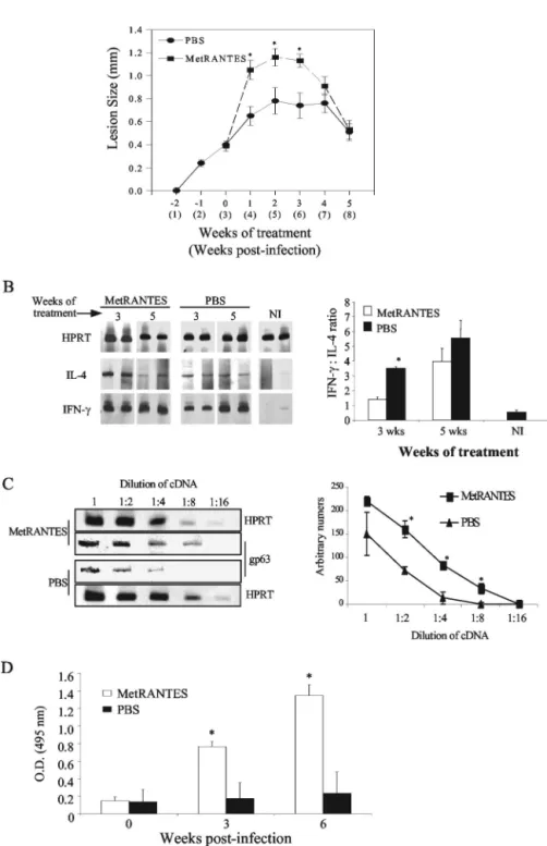
The results described above suggest that CCL5 may be rel-evant to resistance against L. major infection. To verify this possibility, C57BL/6 mice were treated daily subcutaneously with Met-RANTES (10 g/mouse; kindly provided by A. E. Proudfoot, Serono Pharmaceuticals, Geneva, Switzerland), a functional antagonist of CCR1 and CCR5 (1, 7, 12). Treatment started at week 2 of infection, when no significant increase in CCL5 mRNA expression was observed. Treatment with Met-RANTES led to a transitory increase in lesion size (Fig. 3A). Mice were sacrificed at weeks 3 and 5 after treatment. At 3 weeks, there was an increase in IL-4 mRNA but no change in IFN-␥expression in lesions (Fig. 3B). Met-RANTES also pro-moted an impressive down-regulation of the production of IFN-␥in draining lymph nodes, whereas IL-4 production was unchanged (Table 1). Tissue parasitism in these lesions was evaluated by PCR (19) and was higher in the Met-RANTES-treated group at week 3 of treatment (Fig. 3C), which was further confirmed in another experiment by serial dilution
* Corresponding author. Mailing address: Departamento de Bio-quı´mica e Imunologia, ICB-Universidade Federal de Minas Gerais, CP 486, 30161-970, Belo Horizonte MG, Brazil. Phone: 55-31-34992656. Fax: 55-31-3499-2614. E-mail: lqvieira@icb.ufmg.br.
4918
on June 13, 2017 by UNIVERSIDADE FEDERAL DE OURO PRETO
http://iai.asm.org/
FIG. 1. Kinetics of CXC and CC chemokine mRNA expression in the footpads of C57BL/6 and BALB/c mice infected withL. major. (A) Mice were infected withL. majorand sacrificed at 1 day, 2 days, 2 weeks, and 6 weeks postinfection, and the hind infected footpad was used in assays of mRNA expression by RT-PCR and ELISA. Representative gels are shown. (B) Densitometric analysis was performed, and quantification was normalized to the levels of hypoxanthine phosphoribosyltransferase (HPRT) expression. Results are expressed asn-fold increases over results with noninfected (NI) control animals. (C) ELISA for detection of chemokines at the site of infection was performed at 1 and 2 days and at 2, 4, 6, and 8 weeks postinfection. Briefly, footpad proteins were extracted at 50 mg of tissue/100l of phosphate-buffered saline to which 0.4 M NaCl, 0.05% Tween 20, and protease inhibitors (0.1 mM phenylmethylsulfonyl fluoride, 0.1 mM benzethonium chloride, 10 mM EDTA, and 20 KI aprotinin A/100 ml) were added. The samples were centrifuged for 10 min at 3,000⫻g, and the supernatant was immediately used for ELISAs. ELISA plates were coated overnight with sheep anti-mouse CCL2 (Pharmingen, San Diego, Calif.) or CCL5 (RD Systems, Minneapolis, Minn.), and ELISAs were performed as recommended by the manufacturer. The anti-CCL2 assay had a sensitivity of 16 pg/ml, and the anti-CCL5 assay had a sensitivity of 32 pg/ml. * and **, statistical differences (P⬍0.05) between results for C57BL/6 and BALB/c for each point, determined by Student’sttest. In panel B, each point represents the mean (⫾standard error [SE]) for at least three mice per time point of one experiment of three performed.
on June 13, 2017 by UNIVERSIDADE FEDERAL DE OURO PRETO
http://iai.asm.org/
analysis (data not shown). All changes had disappeared by week 5 of treatment. To evaluate why the effects of Met-RANTES were transient, we investigated the presence of anti-Met-RANTES antibodies in serum. Not only were there ele-vated titers of antibodies in serum (Fig. 3D), but there was also an increase in the expression of CCL5 mRNA following
Met-RANTES treatment (data not shown). Hence, it is possible that the effect of Met-RANTES (a competitive antagonist of CCL5 binding to CCR1 and CCR5) is transient due either (i) to the appearance of antibodies (as shown here) which recog-nize and, possibly, prevent the action of Met-RANTES, or (ii) to the increase in the levels of CCL5 (as suggested by the
FIG. 2. Course of infection, chemokine expression, and protein production at the site of infection in IL-12, IFN-␥, and IL-4 knockout (⫺/⫺) mice and their wild-type control. (A) Mice were infected in both hind footpads with 106stationary-phaseL. majorpromastigotes, and lesions were measured weekly. (B) Chemokine expression was determined at 2 and 6 weeks postinfection by semiquantitative real-time RT-PCR and compared with the expression in noninfected controls (NI). Results are expressed in arbitrary units normalized to results for noninfected control animals. (C) ELISA for chemokine production was performed with lesions at 2 and 6 weeks after infection, as described in the legend to Fig. 1. Each point represents the mean (⫾SE) for three mice per point. Different letters indicate aPvalue of⬍0.05 between results for different knockout mice at the same time point, determined by Student’sttest. Experiments were repeated, with similar results.
4920 NOTES INFECT. IMMUN.
on June 13, 2017 by UNIVERSIDADE FEDERAL DE OURO PRETO
http://iai.asm.org/
FIG. 3. (A) Effect of Met-RANTES on the course of infection byL. major; (B) cytokine mRNA expression by RT-PCR at the site of infection; (C) quantification ofL. majorgp63 mRNA expression at the lesion site; (D) production of anti-MetCCL5 antibody in sera from treated and nontreated animals. (A) C57BL/6 mice were infected withL. majorin the hind footpads and treated daily from week 3 to 8 postinfection with intralesion injections of Met-RANTES (10g/animal) or with phosphate-buffered saline (PBS; vehicle). Lesions were measured weekly, and each point represents the mean result (⫾SE) for four to eight animals per time point of one experiment of two performed. (B) Animals were sacrificed at weeks 3 and 5 of treatment, and the infected footpads were used to assay mRNA expression by RT-PCR. Representative gels are shown. Densitometric analysis was performed, and the quantification was normalized to the levels of HPRT expression. To determine the Th1/Th2 balance, the densities of the bands were compared by dividing values obtained for IFN-␥by values obtained for IL-4. Each point represents means (⫾SE) of results for three mice per time point of one of two experiments performed. (C) Parasite load was determined by RT-PCR forL. major
gp63 using various dilutions of the total lesion cDNA, at 3 weeks of treatment (6 weeks of infection). The densitometric analysis was performed and normalized by HPRT levels at the same dilution factor. Each point represents the mean (⫾SE) of results for three to four mice. (D) Anti-Met-RANTES antibodies were determined by ELISA of a 1:20 dilution of the serum at the end of the experiment (5 weeks of treatment). Bars represent means (⫾SE) of results for four to six mice from two different experiments for the absorbance of the serum. *, statistical differences (P⬍0.05), determined by Student’sttest. NI, noninfected; O.D., optical density.
on June 13, 2017 by UNIVERSIDADE FEDERAL DE OURO PRETO
http://iai.asm.org/
mRNA expression) following treatment with the drug. All re-sults were confirmed using anti-CCL5 antiserum (gift from Nicholas Lukacs, University of Michigan Medical School, Ann Arbor), which also rendered mice more susceptible and show-ing larger lesions, eightfold more parasites/mg of tissue by limiting dilution, and lower IFN-␥ production than mice treated with control antibody and no alteration of IL-4 pro-duction by draining lymph nodes (data not shown).
Resistance to leishmaniasis is highly dependent on a Th1 response (18). CCL5 up-regulates IL-12 (1), IFN-␥ (8), and migration of Th1 cells, particularly memory T cells (24). Treat-ment with Met-RANTES or anti-CCL5 rendered C57BL/6 an-imals more susceptible to L. majorand skewed the immune response from type 1 to type 2 by diminishing IFN-␥ produc-tion by draining lymph nodes and increasing IL-4 mRNA ex-pression in lesions. Such skewing may explain why the treat-ment with Met-RANTES or anti-CCL5 promoted increased susceptibility to infection. However, CCR5-deficient mice were not more susceptible toL. majorthan their wild-type counter-parts (our unpublished data and reference 20), and CCR1-deficient mice were more resistant than wild-type mice (17). The explanation for such differences is not immediately appar-ent. Possibly, a blockade of the action of CCL5 on CCR1 and CCR5 simultaneously is necessary for an increase in suscepti-bility to infection. The expression of CCL2 did not correlate with resistance or susceptibility to infection. This finding is in agreement with those of other studies that found no correla-tion between CCL2 and susceptibility to leishmaniasis (4, 15, 16, 22, 25). These data challenge the importance of CCL2 in determining susceptibility to L. major in models other than that of the wild-type BALB/c mouse. It is likely that the con-certed and timely actions of several chemokines and chemo-kine receptors are necessary to controlLeishmaniainfection, and their roles are just beginning to be understood.
Our results demonstrate a correlation between CCL5 ex-pression and resistance to infection. Altogether, these data support a role for CCL5 in the cascade of events leading to parasite control duringL. majorinfection.
We are indebted to Maria Helena Alves de Oliveira, Ronilda Maria de Paula (in memoriam), and Antoˆnio Vaz Mesquita for animal care,
to Eneida Paganini Valente for excellent technical assistance, and to Milton Adriano Pelli de Oliveira for useful discussions. H.C.S. is thankful to Gilton Santiago and Eli M. C. Santiago for encouragement and support.
This work was supported by PRONEX/CNPq and CAPES. H.C.S., C.F.O., L.S., L.A.R.F., M.M.T., R.T.G., and L.Q.V. are research fel-lows from CNPq. H.C.S. was also a research fellow from FAPEMIG.
REFERENCES
1. Aliberti, J., C. R. Souza, M. Schito, S. Hieny, T. Wells, G. B. Huffnagle, and A. Sher.2000. CCR5 provides a signal for microbial induced production of IL-12 by CD8␣⫹dendritic cells. Nat. Immunol.1:83–87.
2. Brenier-Pinchart, M. P., H. Pelloux, D. D. Guergour, and P. A. Tomas.2001. Chemokines in host-protozoan-parasite interaction. Trends Parasitol.17:
292–296.
3. dos Santos, P. V. A., E. Roffeˆ, H. C. Santiago, R. A. Torres, A. P. M. P. Marino, C. N. Paiva, A. A. Silva, R. T. Gazzinelli, and J. Lannes-Vieira.2001. Prevalence of CD8⫹␣T cell inTrypanosoma cruzi-elicited myocarditis is
associated with acquisition of CD62LLowLFA-1HighVLA-4High activation
phenotype and expression of IFN-␥-inducible adhehsion and chemoattrac-tant molecules. Microbes Infect.3:971–984.
4. Gu, L., S. Tseng, R. M. Horner, C. Tam, L. Massimo, and J. R. Barrett.2000. Control of Th2 polarization by the chemokine monocyte chemoattractant protein-1. Nature404:407–411.
5. Ji, J., J. Sun, and L. Soong.2003. Impaired expression of Inflammatory cytokines and chemokines at early stages of infection withLeishmania ama-zonensis. Infect. Immun.71:4278–4288.
6. Kopf, M., F. Brombacher, G. Kohler, G. Kienzle, K. H. Widmann, K. Le-frang, C. Humborg, B. Ledermann, and W. Solbach.1996. IL-4 deficient BALB/c mice resist infection withLeishmania major. J. Exp. Med.184:1127– 1136.
7. Lloyd, C. M., A. W. Minto, M. E. Dorf, A. Proudfoot, T. N. C. Wells, D. J. Salant, and J. C. Gutierrez-Ramos.1997. RANTES and monocyte chemoat-tractant protein-1 (MCP-1) play an important role in inflammatory phase of crescentic nephritis, but only MCP-1 is involved in crescent formation and interstitial fibrosis. J. Exp. Med.185:1371–1380.
8. Makino, Y., D. N. Cook, O. Smithies, O. Y. Hwang, E. G. Neilson, L. A. Turka, H. Sato, A. D. Wells, and T. M. Danoff.2002 Impaired T cell function in RANTES-deficient mice. Clin. Immunol.102:302–309.
9. Matte, O., and M. Oliver.2002.Leishmania-induced cellular recruitment during the early inflammatory response: modulation of proinflammatory mediators. J. Infect. Dis.185:673–681.
10. Mattner, F., J. Magram, P. Launois, K. Di Padova, R. Behin, M. K. Gately, J. A. Louis, and G. Alber.1996. Genetically resistant mice lacking interleu-kin-12 are susceptible to infection withLeishmania majorand mount a polarized Th2 cell response. Eur. J. Immunol.26:1553–1559.
11. Oliveira, M. A. P., H. C. Santiago, C. R. Lisboa, I. P. Ceravollo, G. Trinch-ieri, R. T. Gazzinelli, and L. Q. Vieira.2000.Leishmaniasp: comparative study with Toxoplasma gondii andTrypanosoma cruziin their ability to initialize IL-12 and IFN-␥synthesis. Exp. Parasitol.95:96–105.
12. Proudfoot, A. E. I., C. A. Power, A. J. Hoogewerf, M. O. Montjovent, F. Borlat, R. E. Offord, and T. N. C. Wells.1996. Extension of recombinat human RANTES by the retention of the initiating methionine produces a potent antagonist. J. Biol. Chem.271:2599–2603.
13. Racoosin, E. L., and S. M. Beverley.1997.Leishmania major: promastigotes induce expression of chemokine genes in murine macrophages. Exp. Para-sitol.85:283–295.
14. Ritter, U., and H. Ko¨rner.2002. Divergent expression of inflammatory der-mal chemokines in cutaneous leishmaniasis. Parasite Immunol.24:295–301. 15. Ritter, U., and H. Moll.2000. Monocyte chemotactic protein-1 stimulates the killing ofLeishmania majorby human monocytes, acts synergistically with IFN-␥and is antagonized by IL-4. Eur. J. Immunol.30:3111–3120. 16. Ritter, U., H. Moll, T. Laskay, E. B. Bro¨cker, O. Velazco, I. Becker, and R.
Gillitzer.1996. Differential expression of chemokines in patients with local-ized and diffuse cutaneous American leishmaniasis. J. Infect. Dis.173:699– 709.
17. Rodriguez-Sosa, M., L. E. Rosas, L. I. Terrazas, B. Lu, C. Gerard, and A. R. Satoskar.2002. CC chemokine receptor 1 enhances susceptibility to Leish-mania majorduring early phase of infection. Immunol. Cell Biol.81:114–120. 18. Sacks, D., and N. Noben-Trauth.2002. The immunology of susceptibility and
resistance to Leishmania major in mice. Nat. Rev. Immunol.2:845–858. 19. Santiago, H. C., M. A. P. Oliveira, E. A. Bambirra, A. M. Faria, L. C. C.
Afonso, L. Q. Vieira, and R. T. Gazzinelli.1999. Coinfection with Toxo-plasma gondiiinhibits antigen-specific Th2 immune responses, tissue inflam-mation, and parasitism in BALB/c mice infected withLeishmania major. Infect. Immun.67:4939–4944.
20. Sato, N., S. K. Ahuja, M. Quinones, V. Kostecki, R. L. Reddick, P. C. Melby, W. A. Kuziel, and S. S. Ahuja.2000. CC chemokine receptor (CCR)2 is required for Langerhans cell migration and localization of T helper cell type 1 (Th1)-inducing dendritic cells: absence of CCR2 shifts theLeishmania major-resistant phenotype to a susceptible state dominated by Th2 cytokines, TABLE 1. IFN-␥and IL-4 production by culture of lymph node
cells from C57BL/6 mice infected withL. majorand treated with Met-RANTES or not
Lymph node treatment
Time of treatmentb
(wk)
Cytokine productiona
IFN-␥(ng/ml) IL-4 (pg/ml)
PBS 3 59.9⫾7.3 39.1⫾3.0
5 66.0⫾0.9 48.0⫾6.4
Met-RANTES 3 12.1⫾5.3* 52.5⫾14.6
5 47.1⫾4.6 50.2⫾15.5
aCytokines were measured by ELISA of culture supernatants. Data represent
mean results for three mice per group (⫾standard error). Levels of cytokine in supernatants from unstimulated cultures were below detection limits. Repre-sented are the data for one experiment of two performed with similar results. An asterisk indicates statistical difference between results for phosphate-buffered saline (PBS)- and Met-RANTES-treated groups at the same time point.
bTreatment began at the third week of infection; Met-RANTES was injected
subcutaneously at 10g/mouse.
4922 NOTES INFECT. IMMUN.
on June 13, 2017 by UNIVERSIDADE FEDERAL DE OURO PRETO
http://iai.asm.org/
B cell outgrowth, and sustained neutrophilic inflammation. J. Exp. Med.
192:205–218.
21. Souza, D. G., V. Pinho, G. D. Cassali, S. Poole, and M. M. Teixeira.2002. Effect of a BLT receptor antagonist in a model of severe ischemia and reperfusion injury in the rat. Eur. J. Pharmacol.440:61–69.
22. Vester, B., K. Mu¨ller, W. Solbach, and T. Laskay.1999. Early gene expres-sion of NK cell-activating chemokines in mice resistant toLeishmania major. Infect. Immun.67:3155–3159.
23. Wang, Z. E., S. L. Reiner, S. Zheng, D. K. Dalton, and R. M. Locksley.1994.
CD4⫹effector cells default to the Th2 pathway in interferon␥-deficient mice
infected withLeishmania major. J. Exp. Med.179:1367–1371.
24. Weber, C., K. S. Weber, C. Klier, S. Gu, R. Wank, R. Horuk, and P. J. Nelson.2001. Specialized roles of the chemokine receptors CCR1 and CCR5 in the recruitment of monocytes and T(H)1-like/CD45RO(⫹) T cells. Blood
97:1144–1146.
25. Zaph, C., and P. Scott.2003. Interleukin-12 regulates chemokine gene ex-pression during the early immune response toLeishmania major. Infect. Immun.71:1587–1589.
Editor:J. F. Urban, Jr.