Research Articles
Combination and monotherapy of
Leishmania major
infection in BALB/c
mice using plant extracts and herbicides
Judith A. Makwali
1, Frederick M.E. Wanjala
1, Josyline C. Kaburi
2, Johnstone Ingonga
2,
Wabwoba W. Byrum
3& Christopher O. Anjili
21School of Science, Moi University, Eldoret; 2Center for Biotechnology Research and Development (CBRD), Kenya Medical Research
Institute (KEMRI), Nairobi; 3Mombasa Technical Training Institute, Department of Applied Sciences, Mombasa, Kenya ABSTRACT
Background & objectives: Leishmaniasis is a growing health problem in many parts of the world. Efforts to find new chemotherapeutics for leishmaniasis remain a priority. This study was carried out to determine the effect of combination and monotherapies using plant extracts and herbicides on Leishmania major infection in BALB/c mice.
Methods: The herbicides and saponin extract were purchased from Sigma. Roots of Plumbago capensis were collected from Karura forest, Nairobi, Kenya. Plant extractions were done in KEMRI at Center for Traditional Medicines and Drugs Research.
Results: Lesion sizes after infection of BALB/c mice were similar in all the experimental groups till the onset of therapeutic treatments (p >0.05). At 15 days post-treatment, significant differences (p < 0.05) were discerned in the lesion sizes of the BALB/c mice in all the mono- and combined-treated groups. However, the combined therapies caused total elimination of the parasites from the lesions and significantly reduced parasite burden in liver and spleen compared to the untreated controls at the end of the experiment.
Interpretation & conclusion: The results of this study demonstrate that combination therapy using alternative administration of saponin, acriflavine, trifluralin and plumbagin is effective in treating L. major infection in mice. In this regard, an investigation into the efficacy of these combined therapies against other Leishmania
strains should be explored further. Furthermore, studies with these combination therapies should be done on non-human primates such as the vervet monkey (Cercopithecus aethiops).
Key words Antileishmanial activity; herbicides; Leishmania major; plant extracts
INTRODUCTION
Leishmaniasis is a zoonotic disease of major public health and veterinary importance, affecting 88 countries with at least two million new cases each year and 367 million people at risk1. It is caused by the protozoan
para-site of the genus Leishmania and transmitted to the mam-malian host by the bite of the phlebotomine sandfly. Com-plicated clinical symptoms range from the skin lesions of cutaneous leishmaniasis (CL) to the fatal visceral leish-maniasis (VL), making the disease an important health
problem in the world. Leishmania parasite has a complex
life cycle that includes two different developmental forms. The parasites replicate as promastigotes and develop into infective metacyclic promastigotes in the sandfly vector, and the infective promastigotes invade the macrophages and differentiate into amastigotes in the mammalian host2.
Since vaccines against leishmaniasis are not available,
the control of this disease depends on prompt diagnosis and chemotherapy on infected humans and dogs. Chemo-therapy of this disease has relied mainly on pentavalent
antimonials, amphotericin B (AMB), and pentamidine3.
These drugs are toxic and difficult to administer because of their long-term treatment and high cost4,5. Though
tar-geting of antileishmanial agents in liposomes, microspheres, and nanoparticles can successfully increase the efficacy and reduce toxicity of antileishmanial drugs, the limitations of these delivery strategies include non-spe-cific accumulation of reticuloendothelial system (RES), instability of the liposomal formulations, and the incom-plete degradation of polymeric delivery systems6.
There-fore, investigations are still in progress to discover new antileishmanial drugs.
cells and are inexpensive to produce. Trifluralin acts di-rectly with the major microtubule protein, tubulin, lead-ing to disruption of mitosis. Leishmania species also ap-pear to have the same biochemical mechanism of inhibition as plants, since trifluralin was found to specifically in-hibit polymerization of Leishmania tubulin, but not mouse tubulin, in vitro7. Thus, leishmanial tubulin appears to be
more similar to plant tubulin than to mammalian tubulin, with distinct structures that can be targeted by drugs. Al-though, the antimicrobial dinitroaniline herbicides show great potential as antiprotozoal compounds, disputed in-dications of potential carcinogenecity will probably keep trifluralin from being developed for human use. However, in 1993 Chan and his co-workers indicated that an impu-rity contaminant in trifluralin, not trifluralin itself, may be responsible for the observed in vitro activity against
Leishmania species8. The active impurity in trifluralin is
chloralin, an intermediate in the synthesis of trifluralin. However, there is no commercial impetus to reformulate this drug.
On the basis of ultrastructural morphology and se-quence comparison of small subunit ribosomal RNAs,
Leishmania and Trypanosoma are phylogenetically related
and both species have a kinetoplast that contains superhe-lical DNA9. As early as 1968, Simpson10 showed that
ac-riflavine in low concentrations, through selective inhibi-tion of replicainhibi-tion of kinetoplast DNA and not nuclear
DNA is able to induce dyskinetoplasia in cultures of L.
tarentolae. During growth in acriflavine cultures, cells
were seen to exhibit an increasing impairment of colony-forming ability and rate of respiration. It was suggested that the mechanism of action could be photo-oxidization of the kinetoplast DNA. It is, therefore, possible that acri-dine compounds, either modified or used in combination with other drugs can be used to treat the leishmaniases.
Saponins on the other hand are high molecular weight glycosylated plant secondary metabolites, consisting of a sugar moiety linked to a triterpene or steroid aglycone that show various kinds of biological activity and have been used widely as anti-inflammatory, antitumor, anti-HIV, and antifungal agents11. Consequently, triterpenoid
sapo-nin structures have become the synthetic targets of many research groups12. Detergent properties are the typical
characteristics of saponins. These produce stable foam when dissolved in water, which is why some saponin-con-taining plants have been used as soaps for hundreds of years. Saponin containing plants are used as traditional medicines, especially in Asia, and are intensively used in food, veterinary and medical industries13. Antileishmanial
activity has been reported for Hedera, Dracaena and Yucca saponins14, but their value as drug candidates has not been
fully assessed. Saponins inhibit the growth of the promastigote forms by acting on the membrane of the para-site where they induce a drop in membrane potential15.
Plumbagin (2-methoxy-5-hydroxy-1,4-naphtho-quinone) is a natural naphthoquinone possessing various pharmacological activities such as antimalarial16,
antimi-crobial17, anticancer18, cardiotonic19 and antifertility
ac-tion20. At present, the most exploited source of plumbagin
is the roots of Plumbago spp. (Plumbago europea, P.
rosea, P. zeylanica and P. capensis). However, these plants
grow quite slowly and it takes long before the roots are suitable for use21. In this study, the roots of P. capensis
were used for plumbagin extraction. Therefore, there is need to search for alternative sources of plumbagin that grow quickly.
Advances in antileishmanial chemotherapy including development of new drugs have made combination che-motherapy a real possibility. Drug combinations aim to delay or prevent the emergence of resistance, shorten the course of treatment and lower required doses. Other po-tential advances include convenience, better compliance and lower costs22. In this study, trifluralin, acriflavine,
saponin and plumbagin were used in alternate combina-tion of two drugs against L. major infection BALB/c mice.
MATERIAL & METHODS
Leishmania parasites
Metacyclic promastigotes of L. major (IDUB/KE/83=
NLB-144 strain) were used. This strain has been main-tained by cryopreservation and in vitro culture, and peri-odic passage in BALB/c mice at Kenya Medical Research
Institute (KEMRI). Parasites were grown at 26oC in
Schneider’s Drosophila medium supplemented with 10% (v/v) heat-inactivated fetal calf serum (FCS).
Mice
Female BALB/c mice ranging from 8-12 wk old were used in this study. They were strictly maintained under the rules and regulations of the Animal Care and Use Com-mittee-KEMRI (ACUC-KEMRI, Kenya) in specific patho-gen-free conditions with laminal air flow. All mice were inoculated intramuscularly with 2 × 10⁶ stationary phase
Farrell23. Briefly, the lesion size was measured in two
di-mensions (D and d) at right angles to each other with a caliper gauge, and the lesion size(s) determined by the fol-lowing formula: S = (D × d)/2. The mice were anesthe-tized with sodium thiopental (50 g/g of body weight) and sacrificed at the end of the experiment and their spleen and liver were removed for parasite load determination.
This was done because L. major infection in BALB/c
visceralises.
Drug administration
After the skin lesion was confirmed (two months after the inoculation), the infected mice were randomly assigned to seven groups of 10 mice each. The drugs were subcuta-neously injected into the mice twice daily for seven days in different schemes of drug combinations with a drug con-centration equivalent to the IC50/24 h of each of the six drugs used. In some cases the treatment was withheld up to four months to evaluate the efficacy of the treatment in advanced infection. For the experimental handling, the groups had been divided as follows: Group 1 received a dose each of acriflavine and saponin; Group 2 received a dose each of trifluralin and acriflavine; Group 3 received a dose each of trifluralin and plumbagin; Group 4 received a dose each of saponin and plumbagin; Group 5 received a dose each of trifluralin and saponin; and Group 6 re-ceived a dose each of acriflavine and plumbagin. The con-trol group was left untreated. Similar groupings were set up for monotherapy treatments.
Drugs and plant extracts
Trifluralin, acriflavine and saponin extract were
pur-chased from Sigma. Roots of P. capensis were collected
from Karura forest, Nairobi, Kenya. Plant extractions were done in KEMRI at Center for Traditional Medicines and Drugs Research. Reference drugs included sodium stibogluconate (Pentostam; Sigma), pentamidine isethionate (Sigma), and amphotericin B (AMB) (Fungizone; Squib).
Extract preparation
Roots of P. capensis were oven dried at 40°C and
powdered (5 mm of particle dia). The plant (9.5 g) was extracted with 300 ml of chloroform in the Soxhlet appa-ratus for 5 h to obtain high percentage of plumbagin. All the solutions were evaporated to dryness under reduced pressure. The extraction efficiency was defined as follows:
Mass of extracts
Mass of extraction = × 100
Mass of dried material roots
Compounds used in this study
Plant extract solutions for biological testing and the reference drugs were prepared in 100% dimethyl sulfox-ide (DMSO) at 20 mg/ml. Reference drugs included so-dium stibogluconate (Pentostam; Sigma), amphotericin B (Fungizone; Squib), pentamidine isethionate (Sigma), tri-fluralin (Sigma) and acriflavine (Sigma). All the prepared drugs were stored at 4oC and retrieved only during use. In
most experiments, pentostam was used as a reference drug.
Determination of parasite burdens
The assessment of the amastigote burden was carried out by blinded microscopic enumeration with Giemsa-stained liver and spleen touch prints. The total amastigote burdens were calculated as Leishman Donovan units (LDU, number of amastigotes per 1000 nucleated cells × organ weight [in grams] × 2 × 10⁵), according to Stauber’s
formula24. The percent efficacy was calculated as [1–
(mean amastigote load in treated mice/mean amastigote load in NTC)]×100. An LDU reduction of at least 80% was adopted as the minimal criterion for drug efficacy. Collection of target organs and determination of amastigote burdens were not done for mice that succumbed due to severe clinical disease.
Statistical analysis
All the experiments were carried out thrice. The mean
and standarddeviation of at least three experiments were
determined. Statisticalanalysis of the differences between
mean values obtained forthe experimental groups was done
by analysis of variance (ANOVA) and Student’s t-test. P -valuesof 0.05 or less were considered significant.
RESULTS
Effect of monotherapy on L. major lesion development
BALB/c mice infected on the LHFD with L. major
Table 1. Effects of monotherapy treatment on L. major infection in BALB/c mice
Body wt (g) Days post- Relative weight (%)† LDU(106) (% efficacy)‡ infection
Liver Spleen Liver Spleen
Treatment groupa
Plumbagin 25.8 ± 0.7 8 4.3 ± 0.29 0.46 ± 0.07 11.72 ± 0.2 (98.8) 0.53 ± 0.08 (97.6)
Saponin 24.5 ± 0.5 8 4.2 ± 0.31 0.43 ± 0.06 13.23 ± 0.3 (97.8) 0.52 ± 0.07 (97.6)
Trifluralin 25.9 ± 0.8 8 5.2 ± 0.17 0.58 ± 0.1 20.25 ± 0.44 (86.4) 0.63 ± 0.5 (84.1)
Acriflavin 25.5 ± 0.5 8 5.6 ± 0.12 0.59 ± 0.25 22.06 ± 0.35 (85.4) 0.69 ± 0.3 (83.3)
Pentostam (250 mg/kg) 26 ± 2.8 8 4.4 ± 0.24 0.45 ± 0.23 11.98 ± 0.52 (98.6) 0.56 ± 0.11(95.6)
NTC 24.5 ± 1.4 8 6.75 ± 2.19 0.69 ± 0.08 149.3 ± 12.84 3.18 ± 0.25
Treatment groupb
Plumbagin 17.4 ± 0.7 35 6.45 ± 0.55 0.69 ± 0.09 5.63 ± 0.48 (99.6) 0.51 ± 0.08 (97.4)
Saponin 18.1 ± 0.7 35 5.69 ± 0.59 0.58 ± 0.08 3.23 ± 0.28 (99.9) 0.56 ± 0.07 (97.6)
Trifluralin 16 ± 0.8 35 9.60 ± 0.55 1.10 ± 0.18 102.59 ± 0.45 (93.1) 0.48 ± 0.54 (84.1) Acriflavin 15.5 ± 0.5 35 9.26 ± 0.31 1.18 ± 0.05 13.66 ± 0.30 (90.8) 0.37 ± 0.14 (78.6) Pentostam (250 mg/kg) 17.2 ± 0.5 35 4.52 ± 0.22 0.45 ± 0.22 0 ± 0 (100)
NTC 17.5 ± 0.6 35 7.01 ± 0.4 1.3 ± 0.3 746.5 ± 80.54
Plumbagin 17 ± 0.56 56 5.17 ± 0.9 0.39 ± 0.12 11.28 ± 1.3 (92.44) 1.51 ± 0.14 (83.9)
Saponin 18.3 ± 1.54 56 5.32 ± 0.63 0.52 ± 1.4 27.24 ± 2 (92.06) 1.48 ± 0.21 (86.8)
Trifluralin 16 ± 1.67 56 5.3 ± 0.85 0.32 ± 0.05 29.47 ± 3.1(82.40) 2.73 ± 0.11 (77.0)
Acriflavin 15 ± 2.09 56 4.18 ± 0.30 0.39 ± 0.08 38.4 ± 4.25 (80.44) 3.06 ± 0.18 (66.7) Pentostam (250 mg/kg) 17 ± 0.57 56 6.25 ± 1.68 0.82 ± 0.88 11.85 ± 3.65 (92.1) 2.84 ± 0.68 (73.6)
NTC 17.5 ± 1.2 56 8.03 ± 0.91 2.01 ± 0.41 597.2 ± 75.7 12.51 ± 6.01
Plumbagin 14 ± 0.98 – ND
Saponin 15 ± 0.78 63 ND
Trifluralin 14 ± 0.65 63 ND
Acriflavin 15 ± 0.74 63 ND
Pentostam (250 mg/kg) 15 ± 0.54 84 6.31 ± 0.92 1.25 ± 0.58 ND ND
NTC 13 ± 0.75 84 –
aTreatment of acute infection on Day 1 post-infection at 0.2 mg/kg; bTreatment of chronic infection on Day 28 post-infection at 1.25 mg/kg;
†Organ weight/body weight; ‡[1-(mean amastigote load in treated mice/mean amastigote load in NTC)] × 100; NTC —Non-treated control; LDU—Leishman Donovan units; ND — Not done.
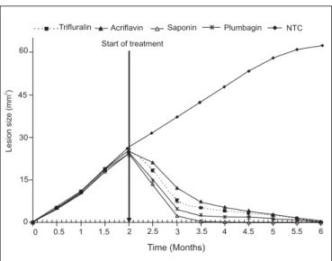
Fig. 1: Lesion sizes of mice during infection, pre- and post-monotherapeutic treatments.
had differentiated (p < 0.05) with saponin maintaining sig-nificantly lower lesion size in the BALB/c mice. Lesions
were completely eliminated after monotherapeutic treat-ment using saponin after two months post-treattreat-ment while all the other monotherapy treatments reduced the lesions after four months post-treatment.
Effects of monotherapy on liver and spleen parasite burdens
A single 0.2 mg/kg dose of saponin, plumbagin, tri-fluralin and acriflavine resulted in 97.8, 98.8, 86.4 and 85.4% reductions in liver amastigote burden on Day 7 post-treatment, respectively, whereas on Day 56 post-treat-ment the liver amastigote burden in mice-treated with sa-ponin, plumbagin, trifluralin and acriflavine were 92.06, 92.44, 82.42 and 80.44%, respectively. The severity of infection in the non-treated controls (NTC) increased dra-matically between Day 7 and Day 56, as illustrated by a 4-fold increase in the liver amastigote burden (from 149.3 × 10⁶ LDU on Day 7 to 597.2 × 10⁶ LDU on Day 56) (Table
1). Despite the apparently adequate parasite control by
0 15 30 45 60
0 0.5 1 1.5 2 2.5 3 3.5 4 4.5 5 5.5 6
Time (Months)
Lesion
size
(mm
)
2.
Trifluralin Acriflavin Saponin Plumbagin NTC
the test compounds at 0.2 mg/kg (80.44–92.44% reduc-tion in liver amastigote burden), deaths did occur and the survivors showed considerable weight loss (52%). Treat-ment with the test compounds at 1.25 mg/kg resulted in a high reduction in liver amastigote burden. However, one animal died on Day 46 and the overall body weight gain was low (5–7 g). By Day 35, control mice already showed liver infection (746.5 × 10⁶ LDU), poor health status, and
a body weight loss of about 25%. Control of liver infec-tion was obtained with saponin at 0.8 mg/kg and pentostam at 250 mg/kg/body weight, with 99.9 and 99.4% reduc-tions within 7 days of treatment, respectively; and control of liver infection was obtained with plumbagin, trifluralin and acriflavine at 1.25 mg/kg with 99.6, 93.1 and 90.8% reductions within 7 days of treatment, respectively. How-ever, a net body weight loss was noted in all the animals, and the spleen remained positive for amastigotes.
Effect of combination therapy on L. major lesion devel-opment
In the initial testing for the antileishmanial activity of independent drugs the order of potency was as follows in decreasing strength: saponin (Sap) >Plumbagin (Plgn) > Trifluralin (Tri) > Acriflavine (Acri). After 7 days of treatment, twice daily with saponin, the parasites in all the infected animals disappeared from the lesion area after 14 accumulated drugging, and the open lesions completely healed in a period of 30 days (Fig. 1). In the independent drug therapy (monotherapy), the parasites reappeared in the skin at the site of infection and in the surrounding skin area 90 to 150 days after the end of the treatment. It was a result of a successful combination of acriflavine and saponin that prompted the following series of experiments. Each drug combination was examined. In this case the lesions healed, with only the untreated mice dying.
Loss of weight and discoloration was apparent in un-treated mice and mice un-treated with the drugs of choice namely AMB and pentostam. The treated mice, on the other hand, increased in weight. Of the six drugs of choice tested against the disease, sodium stibogluconate and pen-tamidine were effective but some mice treated with these drugs of choice died as a result of toxicity. None of the mice treated with combination therapy died. No formula-tion was toxic to the experimental mice. Combinaformula-tion treat-ment comprising of alternate administration of test com-pounds caused total elimination of the parasites from the lesion at the end of the treatment (Fig. 2). After 7 days of treatment, twice daily, with these combinations, the para-sites in all the infected animals disappeared from the le-sion area, and the open lele-sion completely healed in a pe-riod of 30 days.
Efficacy of combined treatments on the development of lesion sizes in BALB/c mice is presented in Fig. 2. Le-sion sizes after infection of the BALB/c were similar in all the treatment groups till the onset of therapeutic treatments (p >0.05). Treatment was started after two months of infection. At 15 days post-treatment, significant differ-ences (p <0.05) were discerned in the lesion sizes of the BALB/c mice. BALB/c mice being treated using acrifla-vine/plumbagin produced significantly larger lesion sizes than those being treated using acriflavine/saponin. How-ever, after one month of monotherapy treatment, lesion sizes in all the BALB/c mice had differentiated (p < 0.05) with acriflavine/saponin maintaining significantly lower lesion size in the BALB/c mice. Lesions were completely eliminated after combination treatment using acriflavine/ saponin after one and half months post-treatment, while all the rest of the combinations cleared the lesions after two months post-treatments, except for acriflavine/plum-bagin treatment that reduced lesions after four months post-treatment.
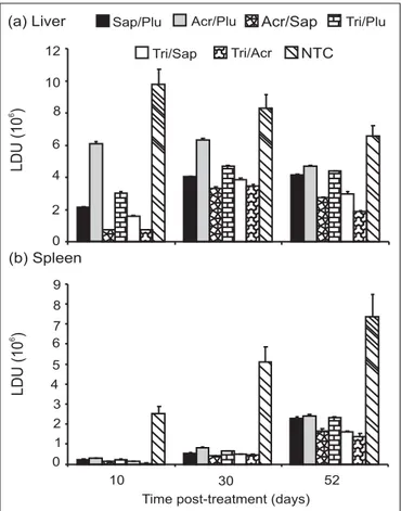
Effects of combined treatments on the amastigote liver and spleen burdens
Results of therapeutic treatments on the liver and spleen LDU are presented in Fig. 3. For both the liver and spleen, the non-treated controls (NTC) had the highest LDU whereas there was a decline in the LDU of the liver from Day 10 to 52, the LDU increased during the same time period. After 10, 30 and 52 days of treatments, sig-nificantly (p <0.05) higher LDU in the liver and spleen occurred in acriflavine/plumbagin combination followed by trifluralin/plumbagin and the lowest LDU occurred in the trifluralin/acriflavine combination therapies.
0 10 30 60
Time (Months) Start of treatment
Lesion
size
(mm
)
2
Acri/Sap Sap/Plum. Trif/Acr.
Trif/Sap Trif/Plum. Acri/Plum.
NTC 70
50
40
20
0 1 2 3 4 5 6
DISCUSSION
Leishmania major, the aetiological agent of CL on
humans, is a parasite of the skin. However, in the BALB/c mice, L. major produces visceral infection in addition to the local lesion at the point of inoculation. Thus, it was a suitable model for this study. The fact that a spontaneous healing cannot be achieved in these mice indicates that the complete healing of the lesions is due to the combination therapy administered only. Therefore, to better evaluate if the drugs effectively reduced the parasites burden in the liver and spleen after treatment, short-term and long-term efficacies for the mono and combination therapies were determined. For both the liver and spleen, the non-treated controls had the highest LDU. Significantly (p <0.05) higher parasite load in the liver and spleen occurred in acriflavine/plumbagin combination followed by triflura-lin/plumbagin and the lowest LDU occurred in the triflu-ralin and acriflavine combination therapies.
These results indicate that acridine and dinitroaniline herbicides with the plant extracts are promising lead com-pounds for development as novel antileishmanial agents. DMSO was used in all the drug formulations because it
has been reported to increase drug penetration25.
Com-bined therapy of saponin and acriflavine was the most ef-fective in reducing both the lesion sizes and hepatic para-site load; followed by saponin & plumbagin; trifluralin & acriflavine; trifluralin & saponin; trifluralin & plumbagin and acriflavine & plumbagin combinations. These com-binations were used in two doses injected in mice previ-ously infected with L. major and having pentostam as ref-erence drug. The significant decrease in the lesion sizes in the combined therapy is indicative of the occurrence of a synergistic mechanism between the drug combinations. It is interesting to note that the combined administration of saponin and other test drugs significantly (p <0.05) re-duced footpad size and concomitantly significantly inhib-ited parasite growth. This is attributable to the main bio-logical activity ascribed to saponins: their membrane permeabilizing property. Saponin enhanced the uptake of the other drugs, thus, increasing their activity against the parasites.
There was a remarkable long-term in vivo activity of trifluralin, acriflavine, saponin and plumbagin at 1.25 mg/ kg dosage administered subcutaneously against established
L. major infection in BALB/c mice. The optimal dose used
with the test compounds was 500 g/ml. At 500 g/ml
per kg of body weight daily for seven days showed a high short-term efficacy against recent infection of L. major. For the sake of completeness, since a direct extrapolation of the drug doses from mice to humans is unreliable, it
should be noted that in Leishmania therapy the
recom-mended therapeutic dose of pentostam on humans has been established to be 20 mg/kg of body weight/day for 20
days26. The short-term efficacy of the test compounds
against a Leishmania infection which evolved for two
months prior to drug administration was demonstrated. This phase of murine cutaneous leishmaniasis caused by
L. major corresponds to the end of the acute phase of the
disease27 and was characterized in this study as
demon-strated by other researchers by a spontaneous decrease of the hepatic and a steady increase of the splenic parasite burden (Fig. 3)28.
The amastigote loads were significantly lower in de-creasing order in the spleens and livers of saponin, plum-bagin, trifluralin and acriflavine-treated mice respectively than in those of the controls at all the time points tested. Moreover, the progression of the splenomegaly and the increase of the splenic load were found to be significantly lower in treated mice than in controls, demonstrating that parasite suppression and the inhibition of growth persisted for at least 7 to 8 wk after the end of the drug uptake.
Although data in this study do not provide a direct measure of the parasite killing 1-[amastigote load at the end of the treatment/amastigote load before the start of
Fig. 3: Liver and spleen LDU on Day 10, 30 and 52 post-treatment.
a aa
a a a a a aa aa aa aa aa aa aa aa aa aa aa aa aa aa aa aa aa aa aa aa aa aa aa aa aa aa aa aa aa aa a aa aa aa aa 0 2 4 6 8 10 12
a a aa
a
aa aaaa aaaa
aa aa aa aa a aa aa aa aa 0 1 2 3 4 5 6 7 8 9
10 30 52
Time post-treatment (days)
Sap/Plu Acr/Plu aAcr/Sap aaaaTri/Plu
aa aa
Tri/Sap Tri/Acr NTC
the treatment], these show induction of effective leishmanicidal activity. The spleen is a major site of
Leish-mania multiplication in the natural infection in
suscep-tible hosts. In BALB/c mice, the splenic parasite burden is initially quite low, but it increases steadily for at least three months, and unlike the hepatic burden, it does not decline spontaneously without treatment29. The splenic
ef-ficacy of the test compounds should be emphasized, since until recently splenectomy was performed as the last re-course for cases of antimony resistant leishmaniasis. The efficacy of the test compounds in the spleen is compatible with the available data on its tissue distribution30. The
prolonged effect of the test compounds on parasite growth suggests a low catabolism and/or slow clearance, result-ing in a long biological half-life of the drugs.
From the results of this study, it is evident that combi-nation therapy was highly effective in both healing the lesion and eliminating the parasites from the liver and spleen at low drug concentration (500 g/ml) as opposed to monotherapy at 1.25 mg/kg. Total elimination of the parasites was achieved after 7 days of treatment of both early and advanced infections. Furthermore, this duration of treatment was sufficient to clear the lesion of both the leishmanial parasites and the secondary infection, mainly bacterial, accompanying this disease. In monotherapy treat-ment, parasites reappeared at the site of inoculation many months later after the termination of treatment. On the other hand, in combination treatments, no reappearance of parasites at the site of infection ever occurred. This implies that monotherapy did not achieve total clearance of parasites, hence, the relapse. In contrast, combination therapy caused elimination of parasites in the internal or-gans. It can, therefore, be concluded that administration of the combination of drugs is effective and superior than individual drugs. The synergism shown with the combined drugs brings us to the concept of structure-function ap-proach in fighting leishmaniasis. In 1993, Marion and oth-ers reported that higher efficacy may be correlated with molecular structural features of compounds under study31.
This is the first time a novel antileishmanial activity in the plumbaginacea extract and triterpenoid saponin extract in combination with acridine and dinitroaniline herbicides has been reported.
CONCLUSION
Combination therapy with plumbaginacea extract and triterpenoid saponin extract in combination with acridine and dinitroaniline herbicides resulted in complete clear-ance of parasitemia from both the lesion site and internal organs (the livers and spleens) of L. major-infected BALB/c
mice. On the basis of these results and considered together with existing studies that have been reported on the safe doses and side effects of combination drug therapy, and its administration by the subcutaneous route, combina-tion therapy is a promising approach for the treatment of
L. major infection.
ACKNOWLEDGEMENTS
This study was funded by Deutsche Akademische Austauschdienst (DAAD) as part of Ph.D. Scholarship for J.A. Makwali. We wish to appreciate the support given by Moi University as the host University and Center for Biotechnology Research and Development (CBRD), KEMRI for providing the laboratory space to carry out the investigations.
REFERENCES
1. Desjeux P. Leishmaniasis: Current situation and new perspec-tives. Comparative Immunol Microbiol Infect Dis J 2004; 27:
305–18.
2. Bañuls A, Hide M, Prugnolle F. Leishmania and the leishma-niases: A parasite genetic update and advances in taxonomy, epidemiology and pathogenicity in humans. Ad Parasitol J 2007;
64: 2–70.
3. Murray HW. Clinical and experimental advances in treatment of visceral leishmaniasis. Antimicrob Agents Chemother 2001;
45: 2185–97.
4. Melby Peter C. Vaccination against cutaneous leishmaniasis: Current status. Am J Clin Dermatol 2002; 3(8): 557–70. 5. Mishra J, Saxena A, Singh S. Chemotherapy of leishmaniasis:
Past, present and future. Curr Med Chem J 2007; 14: 1153–69. 6. Basu MK, Lala S. Macrophage specific drug delivery in
experi-mental leishmaniasis. Curr Mol Med J 2004; 4(6): 681–9. 7. Chan MM, Tremer RE, Fong D. Effect of the anti-microtubule
drug oryzalin on growth and differentiation of the parasitic pro-tozoan Leishmania mexicana. Differentiation (Berlin) 1991;
46(1): 15–21.
8. Chan MM, Grogl M, Chen CC, Bienen EJ, Fong D. Herbicides to curb human parasitic infections: In vitro and in vivo effects of trifluralin on the trypanosomatid protozoans. Proc Nat Acad Sci USA 1993; 90: 5657–61.
9. Agbe A, Yielding KL. Kinetoplasts play an important role in the drug responses of Trypanosoma brucei. J Parasitol 1995;
6: 968–73.
10. Simpson L. Effect of acriflavine on the kinetoplast of Leishma-nia tarentolae. Mode of action and physiological correlates of the loss of kinetoplast DNA. J Cell Biol 1968; 37: 660–82. 11. Mahato SB, Garai S. Triterpenoid saponins. Progr Chem Org
Nat Prod 1998; 74: 1–196.
12. Gauthier C, Legault J, Pichette A. Recent progress in the syn-thesis of naturally occurring triterpenoid saponins. Mini-Rev Org Chem 2009; 6: 321–44.
13. Hostettmann K, Marston A. Chemistry and pharmacology of natural products, Saponins. Cambridge, United Kingdom: Cam-bridge University Press 1995; p. 239–84.
Correspondence to: Dr Judith A. Makwali, School of Science, Department of Biological Sciences, Chepkoilel Campus, Moi University, P.O. Box 1125, Eldoret 30100, Kenya.
E-mail: [email protected]
Received: 3 May 2011 Accepted in revised form: 15 July 2012 cytometry and microscopical methods to characterize the effect
of herbal drugs on Leishmania spp. Exp Parasitol 2001; 97:
141–53.
15. Delmas F, Giorgio CD, Elias R, Gasquet M, Azas N, Mshvildadze V, Dekanosidze G, Kemertelidze E, Timon-David P. Antileishmanial activity of three saponins isolated from ivy, alpha-hederin, beta-hederin and hederacolchiside A1. Planta Medica 2000; 66: 343–7.
16. Likhitwitayawuid K, Kaewamatawong R, Ruangrungsi N, Krungkrai J. Antimalarial naphthoquinones from Nepenthes thorelii.Planta Medica 1998; 64(3): 237–41.
17. Didry N, Dubrevil L, Pinkas M. Activity of anthroquinonic and naphthoquinonic compounds on oral bacteria. Pharmazie 1994;
49: 681–3.
18. Parimala R, Sachdanandam P. Effect of plumbagin on some glu-cose metabolizing enzymes studied in rats in experimental hepatoma. Mole Cell Biochem 1993; 12(1): 59–63.
19. Itoigawa M, Takeya K, Furukawa H. Cardiotonic action of plum-bagin on guinea-pig papillary muscle. Planta Medica 1991;
57(4): 317–9.
20. Bhargava SK. Effects of plumbagin on reproductive function of male dog. Indian J Exp Biol 1984; 22(3): 153–6.
21. Kitanov GM, Pashankov PP. Quantitative investigation on the dynamics of plumbagin in Plumbago europea L. roots and herb by HPLC. Pharmazie 1994; 49(6): 462–4.
22. Sundar S, Rai M, Chakravarty J, Agarwal D, Agrawal N, Vaillant M, Olliaro P, Murray HW. New treatment approach in Indian vis-ceral leishmaniasis: Single-dose liposomal amphotericin B followed by short-course oral miltefosine. Clin InfectDis J 2008; 47: 1000–6. 23. Nolan TJ, Farrel JP. Experimental infections of the
multimmamate rat (Mastomys natalensis) with Leishmania donovani and Leishmania major. Am J Trop Med Hyg 1987;
36(2): 264–9.
24. Stauber LA, Franchino EM, Grun J. An eight-day method for screening compounds against Leishmania donovani in golden hamsters. J Protozool 1958; 5: 269–73.
25. Idson B. Percutaneous absorption. J Pharmaceut Sci 1975; 64(6): 901–23.
26. Barbara HL, Jonathan BD. Recommendations for treating leish-maniasis with sodium stibogluconate (Pentostam) and review of pertinent clinical studies. Am J Trop Med Hyg 1992; 46(3): 296–306.
27. Bradley DJ. Genetics of susceptibility and resistance in the ver-tebrate host. The leishmaniasis in biology and medicine. Lon-don, United Kingdom : Academic Press 1987; p. 551–77. 28. Rousseau D, Le Fichoux Y, Stien X, Suffia I, Ferrua B, Kubar
J. Progression of visceral leishmaniasis due to Leishmania infantum in BALB/c mice is markedly slowed by prior infection with Trichinella spiralis. Infect Immunol 1997; 65: 4978–83. 29. Croft SL, Yardley V, Coombs GH. Antiprotozoal activities of
phospholipids analogues. Mol Biochem Parasitol 2003; 126:
165–72.
30. Marschner N, Kotting J, Eibl H, Unger C. Distribution of hexadecaphosphocholine and octadecyl-methyl-glycerol-3-phosphocholine in rat tissues during steady-state treatment.
Cancer Chemother Pharmacol 1992; 31: 18–22.