Assessing the Threat of Amphibian Chytrid
Fungus in the Albertine Rift: Past, Present and
Future
Tracie A. Seimon1*, Samuel Ayebare1, Robert Sekisambu2, Emmanuel Muhindo1, Guillain Mitamba1, Eli Greenbaum3, Michele Menegon4, Fabio Pupin4, Denise McAloose1, Alyssa Ammazzalorso1, Danny Meirte5, Wilbur Lukwago2, Mathias Behangana2,
Anton Seimon1¤, Andrew J. Plumptre1
1Wildlife Conservation Society, Bronx, NY, United States of America,2Department of Environmental Sciences, Makerere University, Kampala, Uganda,3Department of Biological Sciences, University of Texas at El Paso, El Paso, TX, United States of America,4Tropical Biodiversity Section, MUSE, The Science Museum of Trento, Trento, Italy,5Department of African Biology, Royal Museum for Central Africa, Tervuren, Belgium
¤ Current address: Dept. of Geography and Planning, Appalachian State University Boone, NC, United States of America
*tseimon@wcs.org
Abstract
Batrachochytrium dendrobatidis(Bd), the cause of chytridiomycosis, is a pathogenic fungus that is found worldwide and is a major contributor to amphibian declines and extinctions. We report results of a comprehensive effort to assess the distribution and threat ofBdin one of the Earth’s most important biodiversity hotspots, the Albertine Rift in central Africa. In herpetological surveys conducted between 2010 and 2014, 1018 skin swabs from 17 amphibian genera in 39 sites across the Albertine Rift were tested forBdby PCR. Overall, 19.5% of amphibians tested positive from all sites combined. Skin tissue samples from 163 amphibians were examined histologically; of these two had superficial epidermal intracorneal fungal colonization and lesions consistent with the disease chytridiomycosis. One amphibian was found dead during the surveys, and all others encountered appeared healthy. We found no evidence forBd-induced mortality events, a finding consistent with other studies. To gain a historical perspective aboutBdin the Albertine Rift, skin swabs from 232 museum-archived amphibians collected as voucher specimens from 1925–1994 were tested forBd. Of these, one sample was positive; an Itombwe River frog (Phrynobatrachus asper) collected in 1950 in the Itombwe highlands. This finding represents the earliest record ofBdin the Democratic Republic of Congo. We modeled the distribution ofBdin the Albertine Rift using MaxEnt soft-ware, and trained our model for improved predictability. Our model predicts thatBdis currently widespread across the Albertine Rift, with moderate habitat suitability extending into the low-lands. Under climatic modeling scenarios our model predicts that optimal habitat suitability of
Bdwill decrease causing a major range contraction of the fungus by 2080. Our baseline data and modeling predictions are important for comparative studies, especially if significant changes in amphibian health status or climactic conditions are encountered in the future. OPEN ACCESS
Citation:Seimon TA, Ayebare S, Sekisambu R, Muhindo E, Mitamba G, Greenbaum E, et al. (2015) Assessing the Threat of Amphibian Chytrid Fungus in the Albertine Rift: Past, Present and Future. PLoS ONE 10(12): e0145841. doi:10.1371/journal. pone.0145841
Editor:Jake Kerby, University of South Dakota, UNITED STATES
Received:August 21, 2015
Accepted:December 9, 2015
Published:December 28, 2015
Copyright:© 2015 Seimon et al. This is an open access article distributed under the terms of the
Creative Commons Attribution License, which permits unrestricted use, distribution, and reproduction in any medium, provided the original author and source are credited.
Data Availability Statement:All relevant data are within the paper and its Supporting Information files.
Funding:This study was made possible by the generous support from the United States Fish and the Wildlife Service’s (USFWS) Amphibian in Decline Fund (F12AP01162; F13AP00871) and the IUCN Save our Species Fund to AP (2011A–012), the John D. and Catherine T. MacArthur Foundation to AS (09–
Introduction
The Albertine Rift region in central Africa is a hotspot for biodiversity, the richest area for vertebrates in Africa, and one of the most threatened [1,2]. Intensive agriculture, land and resource pressures, and high rates of habitat loss and land conversion make it a high priority area for conservation [1]. The region, which extends across parts of western Uganda, Rwanda, Burundi and eastern Democratic Republic of Congo (DRC), contains more than 145, or approximately 23%, of all known amphibian species in Africa. At least 42 of these species (29%) are endemic to the Albertine Rift and work over the past decade has led to the discovery of several new species [3–7]. At least 13 amphibian species in the Rift are listed as Threatened by the International Union for the Conservation of Nature (IUCN), 27 are listed as Data Defi-cient, and 8 species have not been classified, and are likely data deficient or threatened (Plump-tre, unpublished data).
Studies focusing on the distribution and abundance of amphibians have identified that
Batrachochytrium dendrobatidis(Bd), alternatively called chytrid, is responsible for the appar-ent decline and extinction of amphibians in many parts of the world [8–14].Bd, a fungal patho-gen that causes chytridiomycosis, is considered to be one of the greatest infectious disease threats to ever face any taxonomic group [12].Bdis associated with infections, die-offs or extinctions in more than 200 amphibian species [14] and is found on every continent except Antarctica [15]. On the African continent,Bdhas been identified in Ethiopia [16], Tanzania [17], Nigeria [18,19], Kenya [20], Cameroon [21,22], South Africa [23], Gabon [24], and most recently in Madagascar [25]. Environmental niche modeling has predicted widespread occur-ance ofBdin the Albertine Rift [26–28], and to date this fungus has been detected in Kibale Forest in Uganda, [29], the eastern DRC including the Katanga province, Itombwe Natural Reserve, and Kahuzi-Biega National Park [30–32]. The earliest knownBd-positive amphibian from the Albertine Rift is a specimen collected in Uganda in 1934 [33]. However, despite recent PCR testing forBdand its known presence in the region, little information is available about whether Albertine Rift amphibians have historically or currently develop the disease chytridio-mycosis [30].
In addition to natural spread or anthropogenic introductions ofBd, both of which are docu-mented as contributors to infection and disease transmission, climate change is a factor that may impact the presence, persistence, and transmission patterns ofBd[15,27,34]. A changing climate can significantly alter or or provide new environmental niches for amphibians, other plant and animal species, and microbial pathogens. For example, it is known that amphibians that inhabit high elevations adapt to climatic warming by upward expansion of their range into new habitat [35,36]. As climate change affects host and pathogen range contraction or expan-sion, it may also result in new pathogen-host interactions or transmission ofBdfrom carrier to naïve hosts that in either case, can lead to disease emergence [35–37]. Additionally, there is evi-dence thatBdoutbreaks occur more frequently when the environment cycles to drier condi-tions [38,39], as pools become smaller, streamflow is reduced and amphibians are likely to cluster in higher numbers around fewer and diminished water sources that have increased con-centrations of infective, fungal zoospores [38]. In the Albertine Rift, climatic models predict significant temperature and precipitation increases by the end of the 21stcentury [40–42], and recent climate models predict that the worldwide habitat suitability and overall risk ofBd infec-tion could be diminished rather than increased by anthropogenic climate change [27]. How-ever, interpretions of species distribution models are complex, and additional factors should be taken into consideration that influence the epidemiology ofBdsuch as the virulence of the pathogen or strain type, the host response to the particular strain ofBd, and whether there has been any coevolution of the amphibian species withBd[43].
study design, data collection and analysis, decision to publish, or preparation of the manuscript.
The main objectives of this study were to document the current distribution and prevalence of chytrid fungus in amphibians in existing and proposed protected areas of the Albertine Rift, determine whether fungal presence is associated with chytridiomycosis, and predict what the effect of climate change will be onBddistribution. We mapped areas of potentialBd distribu-tion using data collected during field surveys between 2010 and 2012 and MaxEnt modeling software. We then tested our model using additional data collected in 2013–14 and used this information to retrain the model. We also identified which species are infected byBd, and determined if any had histologic evidence of chytridiomycosis. Focusing on the ITS1-5.8S-ITS2 region of theBdgenome, we determined if DNA sequence variation exists within or between protected areas, and looked forBdpresence in archived, whole animal voucher speci-mens from museum collections dating back to 1925. Finally, we used our distribution model to predict the habitat suitability and future distribution ofBdover the next century.
Materials and Methods
Ethics statement
All work complies with guidelines for the use of live amphibians in field research by the Ameri-can Society of Ichthyologists and Herpetologists, the Society for the study of Amphibians and Reptiles and The Herpetologists’League. Swab samples were collected non-invasively from the skin of amphibians that were encountered in the environment and animals were released shortly after handling. Recommended biosecurity practices for minimizing risk of disease transmission between animals and field sites were followed at all sites where amphibian swab samples were collected [44]. No animals were sacrificed specifically for this project. In some cases both swab and tissue samples were taken from voucher amphibian specimens previously collected in the DRC under a separate and unrelated project. IACUC approval for this project was not obtained because WCS institutional requirements for IACUC review do not include field projects that take place outside of our facilities, however non-invasive skin swabbing, cap-ture and release of wild amphibians, collecting dead animals found in the environment, or sam-pling after euthanasia are all standard techniques and procedures for clinical and pathology examinations or investigations. Trained herpetologists were primarily responsible for collect-ing swabs from the animals. Sample collection and export permits were obtained from the Royal Museum of Central Africa, Belgium; Ugandan Wildlife Authority and Uganda National Council for Science and Technology, and Makerere University, Uganda; Institut Congolais Pour la Conservation de la Nature (ICCN), DRC; and the Rwandan Development Board, Rwanda (Permits: B2014–001 (Belgium), NS378 (Uganda), and Nos. 31/RDB–T&C/V.U/10 (Rwanda), 02/ICCN/PNKB2011, 01/ICCN/PKNP/2012, 120/ICCN/PKNP/2012, 02/ICCN/ PKNP/2013, 074/ICCN/PKNP/2013, 3/ICCN/PKNB/2014, 7/ICCN/PNKB/2014 (DRC). IACUC approval, review of, and approval of sampling procedures were not required by the government agencies for obtaining the sample collection and field research permits. Field sur-veys were conducted in accordance with the Declining Amphibian Task Force Fieldwork Code of Practice (http://www.amphibianark.org/pdf/Husbandry/The%20DAPTF%20Fieldwork% 20Code%20of%20Practice.pdf) and previously described methods [44,45].
Sample collection and storage
DRC from 18–27 March, 2013; in Kisimba Ikobo Community Reserve, DRC from 14 April, 2013; in Reserve des Gorillas de Punia (Punia Gorilla Reserve)–west of Kahuzi-Biega National Park, DRC from 28 August–17 September, 2013; in lowland Kahuzi-Biega National Park– Nzovu, DRC from 3–27 September, 2013; in lowland Kahuzi Biega Park-Kasese and Itebero, DRC from 5–27 December, 2013; and in the North Balala Forest, DRC from 24 February–15 March, 2014. Herpetological surveys were cut short in Kisimba-Ikobo Reserve, west of Lake Edward in DRC due to insecurity occurring in the area. Therefore very few samples were obtained from this area forBdtesting.
Swab samples were collected using BBL 1/8”diameter sterile rayon tipped culture swabs (Fisher Scientific, Hampton NH). Each frog was swabbed 4–5 times each on the underside of the hind feet, thighs, abdomen, and forefeet. Swab samples were dried and stored in airtight plastic containers at room temperature. Handlers changed non-powdered latex or nitrile gloves, or washed hands with soap and water (Nyungwe surveys only) between each animal to prevent contamination. Dead animals that were encountered were collected for chytrid analysis and stored in 70% ethanol. Amphibians were identified to genus or genus and species level dur-ing surveys and were released, except for a subset of 163 that were collected as voucher speci-mens for additional taxonomic identification and histological analysis. A sample of skin from voucher specimens, which had also been swabbed forBdPCR, was collected as either a whole foot or sections from the thigh and preserved in 70% ethanol. Tissue samples were held at room temperature until export forBdtesting and histologic processing.
Skin swab samples were also collected from voucher specimens from the Albertine Rift housed in the collections of the Royal Museum for Central Africa (RMCA) in Tervuren, Bel-gium and Markerere University, Uganda. Amphibians at the RMCA were collected between 1926–1951 by G.F. de Witte, R. F. Laurent, and H. Schouteden. Those from Makerere Univer-sity were collected by J.B. Goodman, D.F. Oren, M. Behangana and W. Lukwago between 1965–2013. Historical notes on the RMCA voucher specimens indicate that samples from the de Witte collection were preserved in ~5% neutral buffered formalin, while samples from the Laurent and Schouteden collections were also preserved in formalin, however details on the percentage of formaldehyde, or if it was buffered, are not known. Amphibians from these col-lections were transitioned from formalin to ethanol upon arrival to the RMCA in the following years: Laurent (I.R.S.A.C.) specimens, 1951–1953; Schouteden specimens, 1925, de Witte speci-mens, 1941. Historical notes on preservation methods in the Makerere University voucher specimens are presented inS2 Table. Gloves were worn and changed between handling indi-viduals from these collections, and when swabbing these specimens for this study to avoid potential cross contamination. However, the herpetologists that collected samples between 1926–2013 for the RMCA and Makerere collections did not wear gloves. Thus ourBd preva-lence, particularily regarding samples collected from the Bonongo Forest in 2013, may be over-estimated because of the possibility of false positives.
Sampled taxa
Skin swab and/or skin samples were collected from the following genera: Caeciliidae, Boulen-gerula; Dicroglossinae,Hoplobatrachus; Hyperoliidae,Afrixalus spp.,Hyperolius spp.,Kassina spp.,Phlyctimantis spp.; Pyxicephalidae,Amietia spp.; Ranidae,Hylarana (Amnirana) spp.; Bufonidae,Amietophrynus sp.; Arthroleptidae,Arthroleptis spp.,Cardioglossa spp.,Leptopelis spp.; Hemisotidae,Hemisus spp.; Rhacophorinae,Chiromantis spp., Phrynobatrachidae, Phry-nobatrachus spp.; Ptychadenidae,Ptychadena spp.; Pipidae,Xenopus spp. In addition, we col-lected samples from the following species classified as Vulnerable to extinction by the IUCN:
Histology
Tissue samples from dead amphibians were preserved in 70% ethanol for histologic examina-tion. Following ethanol fixation, skin samples (n = 163 voucher specimens) and multiple tis-sues, including skin, from one frog found dead during the current surveys, were processed routinely, paraffin-embedded, sectioned at 5μm, stained with hematoxylin and eosin, and examined by a certified pathologist. A diagnosis of chytridiomycosis was based on identifica-tion of characteristic changes associated with the disease, including marked thickening of the epidermis and stratum corneum (epidermal hyperplasia and hyperkeratosis, respectively) and numerous intracorneal chytrid fungal thalli.
PCR testing
PCR testing of field samples. Skin swab samples were air dried and stored in individual sterile cryovial tubes (Fisher Scientific, Hampton, NH, USA) at room temperature. ForBd anal-ysis, DNA was extracted using 150μl of PrepMan (Life Technologies; Grand Island, NY, USA) and extracts were diluted 1:10 in RNAse/DNAse-free water. The samples were then analyzed by real-time quantitative PCR amplification of the internal transcribed spacer (ITS1) and 5.8S rDNA region using established methods [47]. Taqman PCR assays were conducted using a Bio-Rad Mini-Opticon Real–Time PCR detection system. Reaction tubes contained a total of 20μl consisting of 10μl of 2X Taqman Environmental Master Mix (Life Technologies), 900 nM of each primer (ITS–1 Chytr3 and 5.8S Chytr), 250 nM of Chytr MGB TaqMan probe (Life Tech-nologies), 2.5μl of 10X exogenous internal positive control primers and probe, 0.5μl of 50X exogenous internal positive control DNA, (TaqMan Exogenous Internal Positive Control kit; Life Technologies), and DNase/RNase–free water containing 5μl of diluted DNA. The exoge-nous internal positive control reagents served as inhibition controls in the PCR reactions. PCR amplification conditions were: 2 minutes at 50°C, 10 minutes at 95°C, followed by 50 cycles of 15 seconds at 95°C and 1 minute at 60°C. Purified genomicBdDNA orBdplasmid carrying the ITS1–5.8S–ITS2 region was provided by Dr. Allan Pessier (San Diego Institute for Conservation Research, CA) and was diluted to a range of concentrations to generate a standard curve for quantifying copy number in all samples and for use as a positive control. Each sample was run in triplicate. The copy number per swab was calculated by taking the copy number obtained in each triplicate PCR reaction from the standard curve, and then multiplying that number times by its relative proportion of the DNA extract, and then multiplying by the dilution factor. The calculated copy numbers/swab were then averaged and graphed with the standard deviations. Confidence limits for prevalence were calculated based on the Wilson score interval [48].
All samples collected from the RMCA collection were tested forBdin triplicate with internal inhibition controls. For the samples collected from Makerere University, samples were initially tested forBdin singlicate, and any positives were retested in triplicate. All positives from his-torical samples were retested using 0.2μl of AmpErase per reaction (UNG, Life Technologies), which was added to the PCR reaction to prevent amplification of contaminating PCR product that could potentially lead to false positives. Because DNA degradation was a concern, an amplification control for the amphibian mitochondrial 16S (SSU rRNA) gene was also run for each sample. For amplification of the 16S, we developed consensus primers (Amph16S–F,
GGGATAACAGCGCAATCWAYTTand Amph16S–R,CCCYGATCCAACATCGAGGTCG) to a
conserved 72 bp region chosen from a region in an alignment that would broadly amplify the following African species that were represented in Genbank: AF215430 (Afrixalus laevis), KC756288 (Amietia angolensis), HM770015 (Amietophrynus regularis), HQ882846 ( Amieto-phrynus superciliaris), DQ283237 (Arthroleptis schubotzi), FJ151072 (Cardioglossa elegans), GU444000 (Hyperolius castaneus), KF447811 (Hyperolius cinnamomeoventris), KF562041 (Hyperolius discodactylus), KF447815 (Hyperolius kivuensis), FJ594084 (Hyperolius marmora-tus), HQ130782 (Leptopelis karissimbensis), HQ130767 (Leptopelis kivuensis), FJ829265 ( Phry-nobatrachus graueri), FJ829318 (Phrynobatrachus versicolor), and HQ225699 (Xenopus wittei). Samples tested for 16S were run in singlicate under the following cycling conditions: 95°C for 5 minutes, followed by 45 cycles of 95°C for 45 seconds, 55°C for 30 seconds, and 72°C for 45 seconds, and a final elongation step of 72°C for 5 minutes. Samples were analyzed using SYBR green on a 2% agarose gel by electrophoresis.
DNA sequence analysis
Samples positive forBdby quantitative real-time PCR were retested using conventional PCR using primers for the ITS1-5.8S-ITS2 region as previously described [50]. Positive bands were purified using ExoSAP-IT (Affymetrix; Santa Clara, CA, USA) and sequenced in both the for-ward and reverse directions (Genewiz, Inc. South Plainfield, NJ). Sequences were analyzed, trimmed and aligned using Geneious software (Geneious Pro 6.0, Biomatters LTD. Auckland NZ) and sequence comparisons were performed using the GenBank database and BLASTn. Sequences with>98% identity to those in Genbank were used in the comparative DNA sequence alignments.
Modeling and statistical analysis
(obtained from Bd-maps.net: accessed October 2014). Default MaxEnt model parameter set-tings (auto features, convergence threshold of 0.00001, maximum number of background points = 10,000, regularization multiplier = 1) were used [52]. Models were fitted using 100 bootstrap model runs with 70/30 partition percentage for the training/testing of the data sets accordingly. We assessed the predictive performance of the MaxEnt models with the receiver operating characteristic (ROC) plots [55,56]. A logistic format output was used that produces continuous, linear-scaled maps giving an index of habitat suitability, and hence potential distri-bution ofBd, which range from 0 (unsuitable) to 1 (highly suitable). Model validation was per-formed using the area under the curve (AUC), and the average training ROC plots AUC for the 100 bootstrap replicate runs was 0.941, with a standard deviation of 0.009. A MaxEnt logis-tic format output that estimates an index of habitat suitability was used to quantify the risk of
Bdinfection in amphibians. Maps were generated using ArcGIS software developed by ESRI (Environmental Systems Resource Institute). Redlands, California (www.esri.com). The follow-ing Shuttle Radar Topography Mission (SRTM; USGS, 2006) Digital Elevation Model (DEM), was sourced from the Global Land Cover Facility, (www.landcover.org) and used as a back-ground inFig 1: USGS (2006), Shuttle Radar Topography Mission, 3 Arc Second scene Africa, “Filled Finished 2.0, Global Land Cover Facility, University of Maryland, College Park, Mary-land, February 2000.
Predictor variables. A total of 19 bioclimatic variables at a spatial resolution of ~1 km2 were obtained from the WorldClim database (http://www.worldclim.org) [57]. The bioclimatic variables are derived from monthly temperature and rainfall values to produce biologically meaningful variables. Pairwise Pearson correlations between bioclimatic variables were calcu-lated, and only variables with less than (+/-0.75) correlation were retained. For the final model, we used seven variables that are known to have a limiting effect on the distribution of chytrid fungus in the MaxEnt model parameterization, as reported in previous studies [9,23,26–
28,58,59]. The covariates used for modeling the distribution of chytrid fungus in the Albertine Rift are as follows: Bio2, Mean diurnal temperature range; Bio5, Maximum temperature of warmest month; Bio6, Minimum temperature of coldest month; Bio7, Annual temperature range; Bio12, Annual precipitation; Bio16, Precipitation of wettest quarter, and Bio17, Precipi-tation of driest quarter.
Estimation ofBdoccurrence in 2080. To estimate future distributional areas in the
Albertine Rift where amphibians are likely to be at risk forBd, output from three General Cir-culation Models for the year 2080 under the A2a anthropogenic climate change scenario were used in the MaxEnt modeling; 1. CCCMA: CGCM2, from the Canadian Centre for Climate Modeling and Analysis, 2. CSIRO: MK2, from the Commonwealth Scientific and Industrial Research Organization, and 3. HADCM3, from the Hadley Centre for Climate Prediction and Research. The Special Report on Emissions Scenarios describes images of the future using four storylines (A1, A2, B1 and B2) in relation to a wide range of demographic, economic and tech-nological driving forces and how green house gas emissions are likely to vary [60]. We modeled
Bdusing the A2a storyline, a high emissions scenario, which describes a very heterogeneous world that is self-reliant, with a high rate of population growth, slow economic development that is regionally oriented and slow technological change compared to other storylines [61]. It also seems to be the storyline that is currently tracking actual climate changes.
Results
PCR testing for
Bd
Fig 1. Map of the Albertine Rift region showing the study sites where amphibian skin swab samples have been collected for PCR analysis ofBd.
Water bodies, country boundaries, protected area boundaries and elevation brackets (meters above sea level) are shown. Yellow circles mark the locations of the sites where samples were collected for this study. The yellow asterick and green circles mark 3Bd-positive sample locations previously identified in DRC and Uganda respectively (data obtained from Bd-maps.net), and the purple hexagons mark sites where samples have been collected from and tested forBdfrom previous studies [30–32].
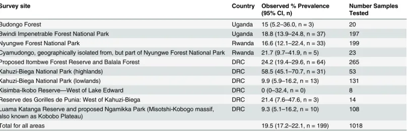
samples have been collected is shown inFig 1. The prevalence ofBdin each of the current or proposed protected areas is listed inTable 1. Overall, 19.5% (199/1018) of the samples were PCR positive forBd.
Of the 17 genera (Afrixalus,Amietia,Amietophrynus,Arthroleptis,Boulengerula, Cardio-glossa,Chiromantis,Hemisus,Hylarana,Hoplobatrachus,Hyperolius,Kassina,Leptopelis,
Phlyctimantis,Phrynobatrachus,Ptychadena,and Xenopus) that were sampled for the presence of chytrid fungus in the Albertine Rift, 10 genera had samples that wereBdpositive and 7 gen-era were negative (Boulengerula,Cardioglossa,Chiromantis,Hemisus,Hylarana, Hoplobatra-chus, andKassina). Very few samples were collected fromBd-negative genera (n = 25 swabs for 7 genera combined). Shown inTable 2is the prevalence for each genus that was positive forBd. Amphibians from the genusAfrixalusandLeptopelishad the highest prevalence ofBd(29.9 and 29.4% respectively).
Analysis of the ITS1-5.8S-ITS2 region of
Bd
The ITS1-5.8S ofBdis a multi-copy region of theBdgenome and recent work has shown that copy number varies considerably (range of 10–144 copies with an average of 77 copies per zoo-spore) between differentBdstrains [62]. Shown inS1 Figare the number of copies/swab calcu-lated for all the positive samples. Overall, 63.3% (126/199) of theBdpositive samples had less than 10,000 copies of the ITS1-5.8S region per swab, which would indicate low levels of zoo-spores (69–1000 zoozoo-spores/swab range) assuming the above estimated (10–144) copies per zoospore [62]. We found that 24.1% of theBdpositive samples (48/199) had 10,000–100,000 copies, and 8.5% (17/199) had 100,000–1,000,000 copies. We found that 4.0% (8/199) of the positive samples had very high ITS1-5.8S copy numbers in the range of 1,000,000–10,000,000. Overall, 82.9% (165/199) of the positive samples had less than 50,000 copies/swab, which, depending on the types ofBdstrain(s) in each sample, would place these zoospore loads below the range of 347–5000 zoospores/swab assuming the above estimated range of copies per zoo-spore [62].
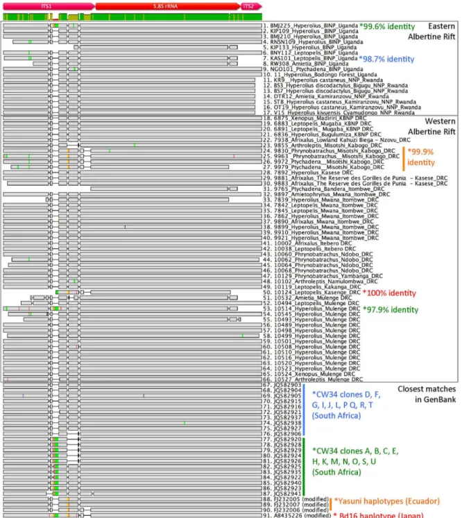
Samples positive forBdusing qPCR were retested by conventional PCR using primers that amplify a 300 bp region spanning the ITS1-5.8S-ITS2 region ofBd[50]. A subset of positive PCR products were directly sequenced and analyzed, and compared to each other and to knownBdsequences. Shown inFig 2is a DNA alignment of the ITS1-5.8S-ITS2 region ofBd
from 66Bd-positive samples separated by region (western and eastern Albertine Rift) and their matching sequences obtained from GenBank. Overall, 89.3% (59/66) of the sample sequences from both the western and eastern Albertine Rift were>99% identical to a Global Panzootic Lineage (Bd-GPL-2) strain CW34 isolated from aXenopussp. from South Africa, indicating very little variation inBdhaplotype diversity in this area of theBdgenome between the eastern and western side of the Albertine Rift region, and the region as a whole. We found that 87.7% (58/66) of the Albertine Rift sequences were>99% identical to CW34 group 1 (clones D, F, G, I, J, L, P Q, R and T) found in GenBank, and 1.5% (1/66) of the sequences were>99% identical to the CW34 group 2 (clones A, B, C, E, H, K, M, N, O, S and U). Only 3% (2/66) of the sequences were between 97.9% and 98.7% identical to CW34, which was the closest match found in GenBank. Although the majority of sequences closely matched the South AfricanBd
CW34 strain, we also found sequences that did not match, and were more closely related to strains found in other parts of the world. We found that 6% (4/66) of Albertine RiftBd
Balala, DRC, was 100% identical to the Bd16 strain found in aCeratophrys cranwelli, a South American species of frog that was that was bred in Japan from imported frogs [63].
Assessing amphibians for the disease chytridiomycosis
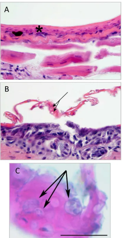
Histologic examination of skin samples was performed to identifyBdand chytridiomycosis. Of the 163 skin samples obtained from individual specimens, two (No. 9979,Ptychadena sp. from Ngamikka NP (Misotshi-Kobogo) and No. 7892,Hyperolius sp. from the lowlands of Kahuzi-Biega National Park, in DRC) had epidermal changes consistent with chytridiomycosis, includ-ing mild to moderate epidermal hyperplasia and hyperkeratosis, and numerous intracorneal zoospore-containing or empty chytrid thalli consistent withBdand suggestive of chytridiomy-cosis (Fig 3).
Both animals were also PCR-positive forBd; sample No. 9979 (Ptychadena) had 2843 copies (514 STDEV) and sample No. 7892 (Hyperolius) had 3,131,439 (1,228,693 STDEV) copies of the ITS1-5.8S region per swab (S1 Fig, red bars). Shown inTable 3are the results for all the PCR-positive animals that were also assessed by histology.
A full necropsy was performed on one amphibian that was found dead during our surveys (MUSE10068). This animal, a 6.07g adultPhrynobatrachussp. (puddle frog), was collected February 25th, 2014 from the Balala Forest in eastern DRC at 2171 m in elevation. A skin swab
Table 1. Bdprevalence in sampled amphibians in current or proposed protected areas throughout the Albertine Rift.
Survey site Country Observed % Prevalence
(95% CI, n)
Number Samples Tested
Budongo Forest Uganda 15 (5.2–36.0, n = 3) 20
Bwindi Impenetrable Forest National Park Uganda 18.8 (13.9–24.8, n = 37) 197
Nyungwe Forest National Park Rwanda 16.6 (12.1–22.4, n = 33) 199
Cyamudongo, geographically isolated from, but part of Nyungwe Forest National Park Rwanda 21.7 (9.7–41.9, n = 5) 23 Proposed Itombwe Forest Reserve and Balala Forest DRC 24.2 (19.4–29.6, n = 64) 265
Kahuzi-Biega National Park (highlands) DRC 58.5 (45.1–70.7, n = 31) 53
Kahuzi-Biega National Park (lowlands) DRC 9.9 (5.9–16.2, n = 13) 131
Kisimba-Ikobo Reserve—West of Lake Edward DRC 0 (0–32.4, n = 0) 8
Reserve des Gorilles de Punia: West of Kahuzi-Biega DRC 21.4 (7.6–47.6, n = 3) 14 Luama Katanga Reserve and proposed Ngamikka Park (Misotshi-Kobogo massif,
also known as Kobobo Plateau)
DRC 9.3 (5.1–16.2, n = 10) 108
Total for all areas 19.5 (17.2–22.1, n = 199) 1018
doi:10.1371/journal.pone.0145841.t001
Table 2. Prevalence ofBdpositive genera in tested samples (CI, Wilsons score).
Genera Number Sampled Positive Negative Observed % Prevalence (95% CI)
Afrixalus 87 26 61 29.9 (21.3–40.2)
Amietia 45 10 35 22.2 (12.5–36.3)
Amietophrynus 30 1 29 3.3 (0.6–16.7)
Arthroleptis 86 7 79 8.1 (4.0–15.9)
Hyperolius 433 89 344 20.6 (17.0–24.6)
Leptopelis 126 37 89 29.4 (22.1–37.8)
Phlyctimantis 5 1 4 20.0 (3.6–62.4)
Phrynobatrachus 82 13 69 15.9 (9.5–25.3)
Ptychadena 53 8 45 15.1 (7.1–27.1)
Xenopus 46 7 39 15.2 (7.6–28.2)
from this animal was positive forBd. Histologic evidence of chytridiomycosis was not seen in skin from multiple areas including the feet. Pathologic processes to explain the death of the frog were not identified in any of the examined tissues. No zoospores were identified in exam-ined pieces of skin, and the calculated copies of the ITS1-5.8S region per swab was 294,562
Fig 2. DNA sequence alignment of the ITS1-5.8S-ITS2 region ofBd.The color labels on the bottom right indicate theBdstrain and haplotypes found in GenBank, and sequences marked with an asterick with matching color indicates the percent identity of that sequence to the corresponding GenBank haplotype. Colors within the sequences denote differences in base pairs between sequences in the alignment (Adenine = red, Thymine = green,
Guanine = yellow and Cytosine = blue). Grey regions indicate nucleotides that are identical in the alignment. Sample identification and GenBank accession numbers are shown.
Fig 3. Skin histology.A–C. Skin section from the ventral hind limb skin ofPtychadena sp. 9977 (A) and 9979 (B, C). A. Normal skin, including thin epidermis (asterisk) and underlying skeletal muscle (1000X). B–C. Abnormal skin. Histological findings include moderate epidermal thickening and cellular disorganization (hyperplasia), mild hyperkeratosis with moderate numbers of intracorneal empty or zoospore-containing chytrid thalli (B, C; arrows; scale bar = 20μm).
(21,613 STDEV). These combined results suggest a subclinical infection and/or possible carrier state.
Assessing historical presence of
Bd
We analysed 209 voucher specimens collected between 1925–1951 to investigate the historical presence ofBdin the western Albertine Rift. Overall 88% (184/209) of the samples had amplifi-able 16S rDNA, and 25 samples were deemed indeterminate because amplification failed. Of 68 samples collected between 1925–1934, 67 were successfully amplified for 16S rDNA (98.5% success in DNA amplification). Of 141 samples collected between 1949–1951, 117 were suc-cessfully amplified for 16S (83.0% success in DNA amplification). Interestingly, we found higher success in rDNA amplification in the older samples. Results are shown inS1 Table. Of the 184 16S-positive samples, only 1 (AM130; ID108878) was positive forBd. DNA sequencing and BLAST analysis of this 97bp fragment was 98% identical toBdstrain CW34 (Clones G, I, J, P and T). Attempts to amplify larger segments of the ITS1-5.8S-ITS2 region were unsuccessful. The positive sample was collected from an Itombwe river frog (Phrynobatrachus asper) col-lected by R. Laurent in August 1950, near the Makenda River in the Itombwe highlands. This finding represents the oldest record ofBdin the DRC, and it confirms its presence in this region as early as 1950.
Skin swab samples were also collected from 49 amphibians collected between 1965–2013 that were archived at Makerere University. Overall, amphibian mitochondrial 16S rDNA was amplified in 78% (38/49) of these samples; 11 samples had indeterminate results due to failure to amplify the 16S gene fragment. Of the 16S-positive samples, 7.9% (3/38) were positive for
Bd. All positive samples were from amphibians collected in the Budongo Forest in 2013 (S2 Table).
Modeling the present and future distribution of
Bd
in the Albertine Rift
Species distribution models (SDM’s) have been used to predict the potential distribution ofBdover large regions [9,23,26–28,64,65]. We used our PCR data gathered from herpetological sur-veys conducted between 2010–2015 and gridded WorldClim interpolated climate surfaces to model which parameters exert the greatest control overBddistribution. The host-pathogen interaction ofBdhas a high degree of climatic sensitivity, particularly to temperature [58,66–
71]. The results of the MaxEnt modeling suggest thatBddistribution is mostly affected by tem-perature and rainfall, which together account for 70% of the model. Variable contributions for each predictor after 100 bootstrap runs were as follows: Bio5 (maximum temperature of the warmest month), 52.5%; Bio12 (mean annual precipitation), 17.5%; Bio2 (mean diurnal
Table 3. Genera assessed for the disease chytridiomycosis that were alsoBdpositive by PCR.
Genus and Sample (Sample IDs) Number ofBdPCR Positive
Samples (copy number range/ swab)
Number Positive for
Chytridiomycosis (by histology)
% Positive for Chytridiomycosis
Afrixalussp. (9734, 9774, 9782, 9883) 4 (308–195,005) 0
-Hyperoliussp. (7862, 7892, 9742, 9752, 9784, 9790, 9792, 9847, 9973)
9 (144–3,131,439) 1 11.1
Leptopelissp. (9770) 1 (190) 0
-Phlyctimantissp. (9738) 1 (186) 0
-Phrynobatrachussp. (MUSE 10068, 9830) 2 (7580–294,562) 0
-Ptychadenasp.(9732, 9979, 9765, 9733) 4 (144–119,378) 1 25.0
21 2 9.5
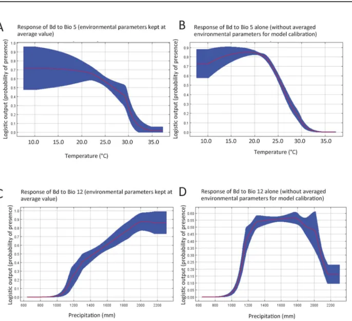
range), 11.1%; Bio17 (precipitation of driest quarter), 9.8%; Bio6 (min temperature of coldest month), 3.3%; Bio16 (precipitation of wettest quarter), 3.2%; and Bio7 (temperature annual range), 2.5%. We found that Bio5 produces the greatest contribution to model performance when used in isolation, which was similar to results obtained with Bio5 when the other envi-ronmental parameters were kept at average value (Fig 4A and 4B). This result shows that Bio5 contains information by itself that is critical in defining the distribution ofBdoccurrence in the Albertine Rift. Bio12 reduces model performance the most if omitted, indicating that it has infomation not present in the other variables used in the model. Based on our model predic-tions, the optimal range of maximum temperature of the warmest month for the probability of presence ofBdis between 10–21°C, and the probability of occurrence decreases rapidly above 22°C (Fig 4A and 4B). The MaxEnt results also indicate that the probability ofBdpresence in the study area increases with higher mean annual precipitation when all other environmental parameters are kept at the average value (Fig 4C). When Bio12 is used for model calibration without other environmental parameters, we found that likelihood ofBdpresence decreases sharply when annual rainfall exceeds 1800mm (Fig 4D). Based on our model, optimal annual precipitation for the habitat suitability ofBdranges between 1300mm to 1800mm. (Fig 4D).
In our initial MaxEnt SDM run, we used presence-only datapoints from a total of 634 indi-viduals that were tested forBd. Samples were collected from Kahuzi-Biega (n = 53), Itombwe (n = 54), Ngamikka (n = 108), Nyungwe (n = 222) and Bwindi (n = 197). The scale of the modeling was set at 1 km2, which meant that some presence records were found in the same cells as others. Therefore, a total of 40 chytrid presence records were used in the MaxEnt model fitting. The results of the outputs are given inS2A Figand show the predicted extent ofBd
using our original data.
We then tested the model on samples from areas that had not been previously surveyed, focusing on the northern part of the Albertine Rift and the lowlands. We used as inputs to test our model samples from six field expeditions were that were conducted in 2013 where 384 indi-viduals were tested forBd: Itombwe and North Balala Forest (n = 211), Kisimba-Ikobo Com-munity Reserve (n = 8), Punia Gorilla Reserve (Kasese-west of Kahuzi-Biega National Park, n = 14), lowland Kahuzi-Biega National Park (Nzovu, Kasese, Itebero n = 131), and Budongo Forest (n = 20). Chytrid presence locations from these samples were combined with locations provided by E. Greenbaum (S4 Table[30–32]) and from Uganda (Bd-maps.net, [29]), then analyzed through MaxEnt modeling. A total of 48 chytrid presence records were used for test-ing the previous model. The model results show that our original data correctly predicted 34 out of the 48 (70.8%) new locations, but did not successfully predict areas in Kahuzi-Biega low-land sector and also in the Katanga province in the southern Albertine Rift near the Zambian border (S2A Fig). However, by incorporating this new set of observations from a more diverse set of localities whereBdpresence has been confirmed we have rerun and trained this model, and thus the range of predictedBddistribution has expanded (S2B Fig). TheBd-positive data-points used in the predictive modeling are shown inS3andS4Tables. Some caution should be used with interpretation of these results as very few data points were collected from lower ele-vations. Further work is therefore needed to assess the prevalence of this disease in tropical Africa at elevations below 600m ASL.
Discussion
Bdis listed by the World Organization for Animal Health (OIE) as a reportable disease [72] and is considered a significant conservation threat in many amphibian species around the world. Our results identifyingBdin and across the Albertine Rift are consistent with results found by others, and also expand the knownBddistribution in the Albertine Rift [29–32]. In our study, 19.5% of the 1018 amphibians from the Albertine Rift sampled between 2010–2014 tested positive forBd. This is somewhat lower than previous observations, which found an overall prevalence of 34.9% in the eastern DRC samples collected between 2008–2011 [31]. Across other regions in Africa, researchers have found a 31.5% prevalence in Kenya [20], 19– 36% in Gabon [24], and 14.8% in South Africa [23]. Most of these studies, where zoospore genomic equivalents were measured, showed low to moderate zoospore loads (<1375 GE); and evidence to assess the disease chytridiomycosis by histopathology in wild African species is
Fig 4. MaxEnt response curves illustrate how the likelihood forBdoccurrence is influenced by maximum temperature of warmest month (Bio5) and the mean annual precipitation (Bio12).A. Shows the probability of presence ofBdas it relates to Bio5 when all other environmental variables are kept at their average values. B. Shows the same as in A, except that Bio5 alone was used for model calibration. C. Shows the probability of presence ofBdas it relates to Bio12 when all other environmental variables are kept at their average values. D. Shows the same except that Bio12 alone was used for model calibration. The red line is the mean of 100 bootstrap runs with +/- one standard deviation (blue).
limited [28,30,73]. Histopathology is a critical addition in any retrospective or prospective study to confirm the presence of chytridiomycosis, asBdinfection alone does not always result in disease or mortality, and because identification of clinically healthy carriers through histol-ogy can inform recovery, reintroduction, and translocation of individuals or groups of animals in conservation projects.
Several species, such as the bullfrog (Lithobates catesbeianus) and African clawed frog (Xenopus laevis)have been shown to have low susceptibility to the disease chytridiomycosis when infected withBd. These, and other low susceptibility species, may act as carriers and be a source of exposure to naïve and/or highly susceptible species [74–77]. Gross examination and histopathology of skin samples from voucher specimens combined with the low to moderate
Bdlevels in the majority of the positive samples tested using PCR (63.8% of all positive samples had less than 10,000 copies of the ITS1-5.8S region per swab), suggest that, with the exception of specimens 9979 (Ptychadena) and 7892 (Hyperolius), the majority of the animals that were
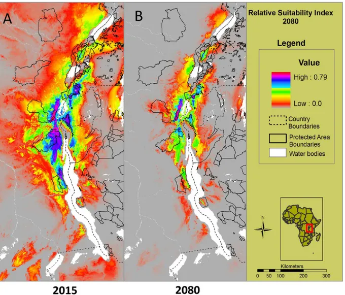
Fig 5. Predicted future habitat suitability andBddistribution in 2080.A. Illustrates the predicted current distribution and risk ofBdto amphibians in 2015 using all locations in the modeling (Bdrecords obtained in this study and previous studies [29–32]). B. An average of the model output from the three General Circulation Models that shows the future distribution of where amphibians are likely to be at risk forBdinfection in 2080 under the A2a scenario. Our results predict a large range contraction of suitable habitat forBdwith future climate change.
Bdpositive did not develop the disease chytridiomycosis despite being infected withBd. In addition, over our four-year study, only one dead amphibian was encountered and none of the animals handled exhibited clinical signs suggestive of chytridiomycosis (lethargy, lack of right-ing reflex, excessive sheddright-ing of skin). It is therefore our presumption that no significant mortality events have occurred in this study area during the current study period. However, confidence in this conclusion is limited by the relatively small sample size relative to the geo-graphic region of study and lack of continuous monitoring and testing. Mortality events may also be cryptic depending on the species and geographic location, and how quickly the car-casses decay before the event is noticed. In addition, this conclusion cannot be applied to amphibians in areas outside of the study sites or species. Based on the current state of our knowledge and when taken together with other studies looking at species within the same gen-era across Africa (Afrixalus,Arthroleptis,Hyperolius,Leptopelis,Phlyctimantis,Ptychadena, andPhrynobatrachus) [19,20,23,24,27], there is no current evidence to suggestBdinfection or chytridiomycosis is causing large die-offs or significant population effects across the Albertine Rift [29–32]. Our field surveys and these other studies however, provide only a snapshot ofBd -prevalence at a certain point in time and time of year, and do not rule out the possibility ofBd -induced mortality as a possible contributing significant factor in animal health or as a factor that could negatively affect population dynamics in the past or future. Many factors, including introductions of new strains ofBdor pathogen mutation towards increased virulence, could result in mortality events within or across species, or situations in which sub-clinical infections may cross a threshold for terminal disease [78–81]. Our data provide important baseline infor-mation for comparative studies should any mortality events be encountered in the future.
The earliest records ofBdin historical specimens are from Brazil in 1894 [82], Illinois in the United States in 1888 [83], and from Africa inXenopus laeviscollected in Cameroon in 1933, Uganda in 1934, and South Africa in 1938 [33,74,84], and our new finding ofBdin an Itombwe river frog (Phrynobatrachus asper) from the DRC in 1950. The Itombwe river frog is currently listed as data-deficient by the IUCN and was thought to have vanished until it was rediscovered in the Itombwe highlands in 2009. When rediscovered, one of two liveP.aspertested was posi-tive forBd[31,85]. These results suggest thatBdhas existed in the Albertine Rift for at least 65–80 years in DRC and Uganda respectively [33].
DNA sequence analysis of the ITS1-5.8S-ITS2 region ofBdshows that the Albertine Rift strains closely matchBdsequences from South Africa (CW34), Ecuador (Yasuni) and Japan (Bd-16). Given the wide, intercontinental distribution of these strains, questions remain as about their origin. ITS1-5.8S-ITS2 is a multicopy region, and many haplotypes have been found to exist withinBdstrains such as the CW34 strain [86]. Our PCR products were not cloned and sequenced, and we therefore cannot speculate on how many haplotypes may exist within individual strains ofBdfound in the Albertine Rift. However, our results suggest that sequences we have identified are closely related to haplotypes previously recovered from the CW34Bdstrain and that the vast majority match one particular group of clones. CW34 is thought to have originated in Namaqualand, South Africa and was isolated from aXenopus lae-visin 2005 [86]. The lineage groups phylogenetically within the globally dispersed clade called
Bd-GPL-2 (Global Panzootic Lineage)[86], and this cluster contains the most geographically diverse and genetically similar group of knownBdstrains.Bd-GPL-2 is found in Africa, Aus-tralia, Asia, and North and Central America [79,86,87]. Molecular clock analysis places an esti-mate of the emergence of the Global Panzootic Lineage strains at 10,000–40,000 years ago [79].
Africa. Future investigations into potential strain variations ofBdin these species and their populations through genomic analysis will be critical to understand if co-evolutionary relation-ships exist with the Albertine RiftBdstrains with their respective host genera, and may provide new insight into the complex and controversial origin of this organism, and the emergence and spread of it’s various lineages [43].
Earlier work by other research groups have used a MaxEnt approach to model the habitat suitability ofBdacross large areas, and predicted thatBdshould be widespread in the Albertine Rift region [11,27,28]. However, at the time these studies were conducted very little testing in the region was available to validate these models. Our results provide valuable data points that increase the resolution of this type of modeling, and by sampling new areas allowed us to test the certainty and train the model for improved predictability. However, with any model come certain caveats. WorldClim variables are based on interpolation of long-term climatological records onto a high-resolution (1 km) grid. Such records are especially sparse across the Alber-tine Rift, which makes accurate representation of WorldClim climatic variables away from observing sites questionable. This is more a concern for precipitation and other moisture vari-ables than it is for temperature. The latitudinal belt of the Albertine Rift falls within the inner tropics, which means annual temperature is strongly influenced by surface elevation, and to lesser degrees by proximity to major water bodies and land surface type. Thermal conditions in WorldClim reflect grid point elevations, which are quite accurate, so the WorldClim tempera-ture fields in the Rift corridor likely represent a fair estimation of reality. The Bio5 variable, maximum temperature of the warmest month, explains 52.5% of the variance inBdoccurance, and is unsurprising given the recognized thermal constraints overBddistribution.
Another limitation of the MaxEnt approach is the use of annual precipitation sum to char-acterize rainfall rather than variables that represent seasonality. Mean annual precipitation (Bio12) was the second variable that highly influenced our model (17.5% of the explanatory power). The timing and duration of wet and dry seasons are heterogeneous across the Albertine Rift [40,88]. It is unknown how infection rates or pathogen loads might similarly vary with pre-cipitation seasonality across the Albertine Rift because our sites were visited once, and whether such a factor might have influenced the results from the various field sampling initiatives. For example, recent studies found that amphibianBdinfection loads increase during dry seasons, likely due to the densification of populations and reduced water flow when ephemeral water bodies disappear with seasonal desiccation [38,89]. Our surveys also focused on collecting adult and juvenile amphibians, and we did not test forBdin mouthparts of tadpoles. Tadpoles have been shown to act as a host for maintainingBdin the environment [38]. If such informa-tion could be ascertained, it should become possible to develop more nuanced predictive mod-els that also incorporate factors like amphibian distributions, whether or not tadpoles could be maintainingBdin the environment, reproductive aggregations and behavior, aquatic parame-ters such as proximity to drainage, and vegetation parameparame-ters in the wet and dry seasons.
Our results using MaxEnt modeling indicate that the highest habitat suitability forBdis found in the highlands of the Albertine Rift, with moderate to low habitat suitability extending into the lowlands. Although our modeling results are also consistent with the known environ-mental preferences ofBdrelative to temperature and rainfall based on previous studies [9,35,38,59,67,90], the results we obtained from testing and training this model indicates that more work needs to be done in the lowlands of the Congo Basin (<600m) to better refine our model and understand the lower elevation and higher temperature limits onBdinfectivity in its natural environment.
resolution, demonstrates relatively monotonic thermal increases across the Albertine Rift throughout the 21st century under the moderate B1 and more severe A2 global emissions sce-narios, with little seasonal or spatial variation evident [40–42]. In contrast, precipitation changes are much more complex. Averaged across the domain, mean annual precipitation is projected to increase by 17.26% (1199 mm vs. 1406 mm) by 2090 relative to the baseline year 1990 [40]. These increases are not distributed evenly throughout the year. Most notably, in the southern sections of the Rift large precipitation increases in the November–January months shorten the duration of the dry season. Under our current model, the likelihood ofBd occur-rence is predicted to decrease during warmer periods, and when precipitation exceeds an annual rainfall threshold above 1800mm per year, so we infer thatBdprevalence may decrease as a result of climate change. This is borne out by the MaxEnt modeling we have presented here, which indicates a major range contraction of habitat suitability for this fungus by the end of the century.
Our database and sample archive can now be used as a reference when monitoring for new strains ofBdover time, and to determine if the occurrence of this pathogen changes seasonally or under wetter and warmer climatic conditions. More fundamentally, we still need to deter-mine if amphibians from the Albertine Rift possess innate resistance or tolerance toBd. If these amphibian species are indeed naturally immune to disease fromBdinfection, there is much to be learned from exploratory research focused on amphibian metagenomics, understanding the skin microbiome diversity, and the innate immunity of African species. The Albertine Rift is one of the world’s hotspots for amphibian biodiversity, and is also one of the most threatened. Baseline data onBdcan help to form a more complete picture of the presence and significance of this fungus and help guide and inform discussions on climate and species-related conserva-tion strategies at both the local and global levels.
Supporting Information
S1 Dataset. FASTA file.DNA sequence alignment of partial ITS1-5.8S-ITS2 sequences used inFig 2.
(FASTA)
S1 Fig. Calculated copy numbers in positive samples.Log-scale graph showing allBd-positive samples plotted against the calculated number of copies of the ITS1-5.8S region per swab. Error bars indicate the standard deviation (positive only) for each triplicate sample. Red bars indicate which two samples had corresponding skin tissue analyzed and had histological changes consistant with the disease chytridiomycosis.
(TIFF)
S2 Fig. Trained model of the current relative habitat suitability and distribution ofBd.A.
(Initial model). Current distribution of areas where amphibians are likely to be at risk forBd
infection using original records (maroon hexagons). All areas predicted as suitable where the new positive localities (green hexagons) appear indicate areas where we had not sampled but the model predicted as a potential suitable habitat. These areas include (Budongo, Kibale, Kamengo, Lake Bunyonyi, North Balala Forest, parts of Itombwe Massif). The model didn’t predict the lowlands of Kahuzi-Biega National Park and Katanga province. B. An updated model showing the current distribution of areas where amphibians are likely to be at risk for
Bdinfection when using both original (maroon hexagons) and new occurrence (green hexa-gons) records for training.
S1 Table. Summary ofBdresults from historical specimens.Elevations are in meters above
sea level. (DOCX)
S2 Table. PCR results of historical samples collected from Makerere University in Uganda. Elevations are in meters above sea level.
(DOCX)
S3 Table. Sample ID, Genus, Date of collection, and GPS location of allBdpositive samples
included in the modelling analysis.Elevations are in meters above sea level. (DOCX)
S4 Table. AdditionalBd-positive localities provided by Bd-maps.net and E. Greenbaum
that went into the modelling analysis [29–32,49].Elevations are in meters above sea level.
(DOCX)
Acknowledgments
We are grateful to the Institut Congolais pour la Conservation de la Nature (ICCN), Ugandan Wildlife Authority, and Rwandan Development Board for support for this research and help with the export of specimens for testing. We are grateful to WCS Rwanda, Uganda and DRC Programs, particularily Simon Nampindo, Deo Kujirakwinja, Doug Sheil, Miriam van Heist, who provided intellectual and logistical support, or helped arrange permits, and Mwenebatu M. Aristote and Wandege M. Moninga, who helped our teams collect specimens.
Author Contributions
Conceived and designed the experiments: TAS SA D. McAloose D. Meirte RS MM FP AS AP. Performed the experiments: TAS SA RS GM EM MM FP WL MB EG AA D. McAloose D. Meirte AS AP. Analyzed the data: TAS SA RS AA D. McAloose D. Meirte FP MM AP. Wrote the paper: TAS SA AP D. McAloose AS EG.
References
1. Brooks T, Balmford A, Burgess N, Fjeldsa J, Hansen LA, Moore J, et al. Towards a blueprint for conser-vation in Africa. Bioscience. 2001; 51: 613–624.
2. Plumptre AJ, Davenport TRB, Behangana M, Kityo R, Eilu G, Ssegawa P, et al. The Biodiversity of the Albertine Rift. Biol Conserv. 2007; 134: 178–194.
3. Portillo F, Greenbaum E. At the edge of a species boundary: A new and relatively young species of Lep-topelis (Anura: Arthroleptidae) from the Itombwe Plateau, Democratic Republic of the Congo. Herpeto-logica. 2014; 70: 100–119.
4. Portillo F, Greenbaum E. A new species of the Leptopelis modestus complex (Anura: Arthroleptidae) from the Albertine Rift of central Africa. J Herpetol. 2014; 48: 394–406.
5. Evans B, Carter T, Tobias M, Kelley D, Hanner R, Tinsley R. A new species of clawed frog (genus Xenopus) from the Itombwe Massif, Democratic Republic of the Congo: Implications for DNA barcodes and biodiversity conservation. Zootaxa. 2008; 1780: 55–68.
6. Evans BJ, Greenbaum E, Kusamba C, Carter TF, Tobias ML, Mendel SA, et al. Description of a new octoploid frog species (Anura: Pipidae: Xenopus) from the Democratic Republic of the Congo, with a discussion of the biogeography of African clawed frogs in the Albertine Rift. J Zool. 2011; 283: 276– 290.
7. Maximilian Dehling J. An African glass frog: A new Hyperolius species (Anura: Hyperoliidae) from Nyungwe National Park, southern Rwanda. Zootaxa. 2012; 53–64.
9. Puschendorf R, Carnaval AC, Vanderwal J, Zumbado-Ulate H, Chaves G, Bolaños F, et al. Distribution models for the amphibian chytrid Batrachochytrium dendrobatidis in Costa Rica: Proposing climatic ref-uges as a conservation tool. Divers Distrib. 2009; 15: 401–408.
10. Rosenblum EB, Voyles J, Poorten TJ, Stajich JE. The deadly chytrid fungus: A story of an emerging pathogen. PLoS Pathog. 2010; 6: 4–6. doi:10.1371/journal.ppat.1000550
11. Olson DH, Aanensen DM, Ronnenberg KL, Powell CI, Walker SF, Bielby J, et al. Mapping the global emergence of Batrachochytrium dendrobatidis, the amphibian chytrid fungus. PLoS One. 2013; 8. doi:
10.1371/journal.pone.0056802
12. Stuart SN, Chanson JS, Cox N a, Young BE, Rodrigues ASL, Fischman DL, et al. Status and trends of amphibian declines and extinctions worldwide. Science. 2004; 306: 1783–1786. doi:10.1126/science. 1103538PMID:15486254
13. Voyles J, Young S, Berger L, Campbell C, Voyles WF, Dinudom A, et al. Pathogenesis of chytridiomy-cosis, a cause of catastrophic amphibian declines. Science. 2009; 326: 582–585. doi:10.1126/science. 1176765PMID:19900897
14. Skerratt LF, Berger L, Speare R, Cashins S, McDonald KR, Phillott AD, et al. Spread of chytridiomyco-sis has caused the rapid global decline and extinction of frogs. Ecohealth. 2007; 4: 125–134. doi:10. 1007/s10393-007-0093-5
15. Fisher MC, Garner TWJ, Walker SF. Global emergence ofBatrachochytrium dendrobatidisand amphibian chytridiomycosis in space, time, and host. Annu Rev Microbiol. 2009; 63: 291–310. doi:10. 1146/annurev.micro.091208.073435PMID:19575560
16. Gower DJ, Doherty-bone TM, Aberra RK, Mengistu A, Menegon M, Sá De R, et al. High prevalence of the amphibian chytrid fungus (Batrachochytrium dendrobatidis) across multiple taxa and localities in the highlands of Ethiopia. Herpetological Journal. 2012; 22: 225–233.
17. Channing A, Finlow-Bates KS, Haarklau SE, Hawkes PG. The biology and recent history of the Criti-cally Endangered Kihansi Spray Toad Nectophrynoides Asperginis in Tanzania. J East African Nat Hist. 2006; 95: 117–138. doi:10.2982/0012-8317(2006)95[117:TBARHO]2.0.CO;2
18. Imasuen AA, Weldon C, Aisien MSO, Dupreez LH. Amphibian chytridiomycosis: first report in Nigeria from the skin slough of Chiromantis rufescens. Froglog. 2009; 90: 6–8.
19. Reeder NMM, Cheng TL, Vredenburg VT, Blackburn DC. Survey of the chytrid fungus Batrachochy-trium dendrobatidis from montane and lowland frogs in eastern Nigeria. Herpetol Notes. 2011; 4: 83–86.
20. Kielgast J, Rödder D, Veith M, Lötters S. Widespread occurrence of the amphibian chytrid fungus in Kenya. Anim Conserv. 2010; 13: 36–43. doi:10.1111/j.1469-1795.2009.00297.x
21. Doherty-Bone TM, Gonwouo NL, Hirschfeld M, Ohst T, Weldon C, Perkins M, et al. Batrachochytrium dendrobatidis in amphibians of Cameroon, including first records for caecilians. Dis Aquat Org. 2013; 102, 187–194. doi:10.3354/dao02557
22. BalážV, Kopecký O, Gvoždík V. Presence of the amphibian chytrid pathogen confirmed in Cameroon. Herpetol J. 2012; 22: 191–194.
23. Tarrant J, Cilliers D, du Preez LH, Weldon C. Spatial assessment of amphibian chytrid fungus (Batra-chochytrium dendrobatidis) in South Africa confirms endemic and widespread infection. PLoS One. 2013; 8. doi:10.1371/journal.pone.0069591
24. Bell RC, Gata Garcia AV, Stuart BL, Zamudio KR. High prevalence of the amphibian chytrid pathogen in Gabon. Ecohealth. 2011; 8: 116–120. doi:10.1007/s10393-010-0364-4PMID:21210295
25. Bletz M, Rosa G, Crottini A, Courtois E, Schmeller D, Rabibisoa N, et al. Widespread presence of the pathogenic fungus Batrachochytrium dendrobatidis in wild amphibian communities in Madagascar. Nat Commun. 2015; 5: 1–10. doi:10.1038/srep08633
26. Rödder D, Kielgast J, Bielby J, Schmidtlein S, Bosch J, Garner TWJ, et al. Global amphibian extinction risk assessment for the panzootic chytrid fungus. Diversity. 2009; 1: 52–66. doi:10.3390/d1010052 27. Rödder D, Kielgast J, Lötters S. Future potential distribution of the emerging amphibian chytrid fungus
under anthropogenic climate change. Dis Aquat Organ. 2010; 92: 201–207. doi:10.3354/dao02197
PMID:21268982
28. Penner J, Adum GB, McElroy MT, Doherty-Bone T, Hirschfeld M, Sandberger L, et al. West Africa—A Safe Haven for Frogs? A Sub-Continental Assessment of the Chytrid Fungus (Batrachochytrium den-drobatidis). PLoS One. 2013; 8.e56236. doi:10.1371/journal.pone.0056236PMID:23426141 29. Goldberg TL, Readel AM, Lee MH. Chytrid fungus in frogs from an equatorial African montane forest in
western Uganda. J Wildl Dis. 2007; 43: 521–524. PMID:17699093
31. Greenbaum E, Meece J, Reed KD, Kusamba C. Extensive occurrance of the amphibian chytrid fungus in the Albertine Rift, a Central African amphibian hotspot. Herpetol J. 2015; 25: 91–100.
32. Greenbaum E, Meece J, Reed KD. Amphibian chytrid Infections in non-forested habitats of Katanga, Democratic Republic of the Congo. 2014; 45: 610–614.
33. Soto-Azat C, Clarke BT, Poynton JC, Cunningham A. Widespread historical presence of Batrachochy-trium dendrobatidis in African pipid frogs. Divers Distrib. 2010; 16: 126–131. doi:10.1111/j.1472-4642. 2009.00618.x
34. Rohr JR, Raffel TR, Romansic JM, McCallum H, Hudson PJ. Evaluating the links between climate, dis-ease spread, and amphibian declines. Proc Natl Acad Sci U S A. 2008; 105: 17436–17441. doi:10. 1073/pnas.0806368105PMID:18987318
35. Seimon T, Seimon A, Daszak P, Halloy S. P, Schloegel L, Aguilar C, et al. Upward range extension of Andean anurans and chytridiomycosis to extreme elevations in response to tropical deglaciation. Glob Chang Biol. 2007; 13: 288–299. doi:10.1111/j.1365-2486.2006.01278.x
36. Raxworthy CJ, Pearson RG, Rabibisoa N, Rakotondrazafy AM, Ramanamanjato JB, Raselimanana AP, et al. Extinction vulnerability of tropical montane endemism from warming and upslope displace-ment: A preliminary appraisal for the highest massif in Madagascar. Glob Chang Biol. 2008; 14: 1703– 1720. doi:10.1111/j.1365-2486.2008.01596.x
37. Pounds JA, Fogden MPL, Campbell JH. Biological response to climate change on a tropical mountain. Nature. 1999; 398: 611–615.
38. Catenazzi A, von May R, Vredenburg VT. High prevalence of infection in tadpoles increases vulnerabil-ity to fungal pathogen in high-Andean amphibians. Biol Conserv. 2013; 159: 413–421. doi:10.1016/j. biocon.2012.11.023
39. McMenamin SK, Hadly E a, Wright CK. Climatic change and wetland desiccation cause amphibian decline in Yellowstone National Park. Proc Natl Acad Sci U S A. 2008; 105: 16988–16993. doi:10. 1073/pnas.0809090105PMID:18955700
40. Seimon A, Picton-Phillipps GP. Regional climatology of the Albertine Rift. Long-term changes in Afri-ca’s Rift Valley. In: Plumptre A, editor. New York: Nova Science Publishers; 2012.
41. Seimon A, Plumptre A. The Albertine Rift., Conservation and climate disruption: Landscape science and practice in a changing climate. In: Cross M, Hilty J, Chester C, editor. Island Press; 2012.
42. Seimon A, Ingram C, Watson J. Climatology of the East African Great Lakes Region and potential impacts of climate change on its biodiversity and ecosystem services. In: Gordon I, editor. Conservation Strategy in the Great Lakes Region. Chicago; 2013. pp. 103–134.
43. James TY, Toledo LF, Rödder D, da Silva Leite D, Belasen AM, Betancourt-Román CM, et al. Disen-tangling host, pathogen, and environmental determinants of a recently emerged wildlife disease: les-sons from the first 15 years of amphibian chytridiomycosis research. Ecol Evol. 2015; 5: 4079–4097. doi:10.1002/ece3.1672PMID:26445660
44. Pessier AP, Mendelson J. R. III. A manual for control of infectious diseases in amphibian survival assur-ance colonies and reintroduction programs. In: Pessier AP and M JR I, editor. 2009.
45. Brem F., Mendelson J.R. III, and Lips KR. Field-sampling protocol for Batrachochytrium dendrobatidis from living amphibians, using alcohol preserved awabs. Version 1. Arlington, Virginia, USA: Conserva-tion InternaConserva-tional; 2007. Available: Version 1.0http://www.amphibians.org
46. Greenbaum EJ, Sinsch U, Lehr E, Valdez F, Chifundera Z. Phylogeography of the reed frog Hyperolius castaneus (Anura: Hyperoliidae) from the Albertine Rift of central Africa: Implications for taxonomy, bio-geography and conservation. Zootaxa. 2013; 3731: 473–494. doi:10.11646/zootaxa.3731.4.3PMID:
25277586
47. Boyle DG, Boyle DB, Olsen V, Morgan J a T, Hyatt A. D. Rapid quantitative detection of chytridiomyco-sis (Batrachochytrium dendrobatidis) in amphibian samples using real-time Taqman PCR assay. Dis Aquat Organ. 2004; 60: 141–148. doi:10.3354/dao060141PMID:15460858
48. Brown L, Cat T, DasGupta A. Interval estimation for a proportion. Stat Sci. 2001; 16: 101–133.
49. Soto-Azat C, Clarke BT, Fisher MC, Walker SF, Cunningham AA. Non-invasive sampling methods for the detection ofBatrachochytrium dendrobatidisin archived amphibians. Dis Aquat Organ. 2009; 84: 163–166. doi:10.3354/dao02029PMID:19476287
50. Annis SL, Dastoor FP, Ziel H, Daszak P, Longcore JE. A DNA-based assay identifiesBatrachochytrium dendrobatidisin amphibians. J Wildl Dis. 2004; 40: 420–8. PMID:15465708
51. Phillips SJ, Anderson RP, Schapire RE. Maximum entropy modelling of species geographic distribu-tions. Ecol Modell. 2006; 190: 231–259.
53. Elith J, Phillips SJ, Hastie T, Dudık M, Chee YE, Yates CJ. A statistical explanation of MaxEnt for ecolo-gists. Divers Distrib. 2011; 17: 43–57.
54. Phillips SJ, Dudik M, Schapire RE. A maximum entropy approach to species distribution modeling. Pro-ceeding of the twenty-first international conference on machine learning. New York: ACM Press, Banff, Canada; 2004. pp. 655–662.
55. Freeman EA, Moisen G. A comparison of the performance of threshold criteria for binary classification in terms of predicted prevalence and kappa. Ecol Model. 2008; 217: 48–58.
56. Manel S, Williams HC, Ormerod SJ. Evaluating presence–absence models in ecology: the need to account for prevalence. J Appl Ecol. 2001; 38: 921–931.
57. Hijmans RJ, Cameron SE, Parra JL, Jones PG, Jarvis A. Very high resolution interpolated climate sur-faces for global land areas. Int J Climatol. 2005; 25: 1965–1978.
58. Stevenson LA, Alford RA, Bell SC, Roznik EA, Berger L, Pike DA. Variation in thermal performance of a widespread pathogen, the amphibian chytrid fungus Batrachochytrium dendrobatidis. PLoS One. 2013; 8: 1–14. doi:10.1371/journal.pone.0073830
59. Muths E, Pilliod DS, Livo LJ. Distribution and environmental limitations of an amphibian pathogen in the Rocky Mountains, USA. Biol Conserv. 2008; 141: 1484–1492. doi:10.1016/j.biocon.2008.03.011 60. IPCC. Emmissions Scenarios. Nakicenovic N, Swart R, editors. United Kingdom: Cambridge
Univer-sity Press; 2000.
61. IPCC. Contribution of Working Group I to the Fourth Assessment Report of the Intergovernmental Panel on Climate Change. Solomon S, Qin D, Manning M, Chen Z, Marquis M, Averyt K, et al., editors. Cambridge, United Kingdom and New York, NY, USA.: Cambridge University Press; 2007.
62. Longo AV., Rodriguez D, da Silva Leite D, Toledo LF, Mendoza Almeralla C, Burrowes P a., et al. ITS1 copy number varies among Batrachochytrium dendrobatidis strains: Implications for qPCR estimates of infection intensity from field-collected amphibian skin swabs. PLoS One. 2013; 8: 1–10. doi:10.1371/ journal.pone.0059499
63. Goka K, Yokoyama J, Une Y, Kuroki T, Suzuki K, Nakahara M, et al. Amphibian chytridiomycosis in Japan: Distribution, haplotypes and possible route of entry into Japan. Mol Ecol. 2009; 18: 4757–4774. doi:10.1111/j.1365-294X.2009.04384.xPMID:19840263
64. Ron SR. Predicting the distribution of the amphibian pathogenBatrachochytrium dendrobatidisin the New World. Biotropica. 2005; 37: 209–221.
65. Hof C, Araújo MB, Jetz W, Rahbek C. Additive threats from pathogens, climate and land-use change for global amphibian diversity. Nature. 2011; 1–6. doi:10.1038/nature10650
66. Bustamante H. M., Livo L. J. and Carey C. Effects of temperature and hydric environment on survival of the Panamanian Golden Frog infected with a pathogenic chytrid fungus. Integr Zool. 2010; 5: 143–153. doi:10.1111/j.1749-4877.2010.00197.xPMID:21392332
67. Rowley JJL, Alford RA. Hot bodies protect amphibians against chytrid infection in nature. Sci Rep. 2013; 3: 1515. doi:10.1038/srep01515PMID:23519020
68. Woodhams DC, Alford RA, Marantelli G. Emerging disease of amphibians cured by elevating body tem-perature. Dis Aquat Organ. 2003; 55: 65–67. doi:10.3354/dao055065PMID:12887256
69. Berger L, Speare R, Hines HB, Marantelli G, Hyatt AD, McDonald KR, et al. Effect of season and tem-perature on mortality in amphibians due to chytridiomycosis. Aust Vet J. 2004; 82: 434–439. doi:10. 1111/j.1751-0813.2004.tb11137.xPMID:15354853
70. Forrest MJ, Schlaepfer MA. Nothing a hot bath won’t cure: Infection rates of amphibian chytrid fungus correlate negatively with water temperature under natural field settings. PLoS One. 2011; 6. e28444. doi:10.1371/journal.pone.0028444PMID:22205950
71. Piotrowski JS, Annis SL, Longcore JE. Physiology ofBatrachochytrium dendrobatidis, a chytrid patho-gen of amphibians. Mycologia. 2004; 96: 9–15. doi:10.2307/3761981PMID:21148822
72. Schloegel LM, Daszak P, Cunningham AA, Speare R, Hill B. Two amphibian diseases, chytridiomyco-sis and ranaviral disease, are now globally notifiable to the World Organization for Animal Health (OIE): An assessment. Dis Aquat Organ. 2010; 92: 101–108. doi:10.3354/dao02140PMID:21268971 73. Lane EP, Weldon C, Bingham J. Histological evidence of chytridiomycete fungal infection in a
free-ranging amphibian, Afrana fuscigula (Anura: Ranidae), in South Africa : Short communication. Journal of the South African Veterinary Association. 2003. pp. p.20–21. PMID:12836742
74. Weldon C, Du Preez LH, Hyatt AD, Muller R, Speare R. Origin of the amphibian chytrid fungus. Emerg Infect Dis. 2004; 10: 2100–2105. doi:10.3201/eid1012.030804PMID:15663845