Antagonistic and Cooperative Actions of Kif7 and Sufu
Define Graded Intracellular Gli Activities in Hedgehog
Signaling
Kelvin King Lo Law1,2, Shigeru Makino3, Rong Mo1, Xiaoyun Zhang1, Vijitha Puviindran1, Chi-chung Hui1,2*
1Program in Developmental & Stem Cell Biology, The Hospital for Sick Children, Toronto Medical Discovery Tower, Toronto, Ontario, Canada,2Department of Molecular Genetics, University of Toronto, Toronto, Ontario, Canada,3Mutagenesis and Genomics Team, RIKEN BioResource Center, Koyadai, Tsukuba, Ibaraki, Japan
Abstract
Graded Hedgehog (Hh) signaling governs the balance of Gli transcriptional activators and repressors to specify diverse ventral cell fates in the spinal cord. It remains unclear how distinct intracellular Gli activity is generated. Here, we demonstrate that Sufu acts universally as a negative regulator of Hh signaling, whereas Kif7 inhibits Gli activity in cooperation with, and independent of, Sufu. Together, they deter naı¨ve precursors from acquiring increasingly ventral identity. We show that Kif7 is also required to establish high intracellular Gli activity by antagonizing the Sufu-inhibition of Gli2. Strikingly, by abolishing the negative regulatory action of Sufu, diverse ventral cell fates can be specified in the absence of extracellular Hh signaling. These data suggest that Sufu is the primary regulator of graded Hh signaling and establish that the antagonistic and cooperative actions of Kif7 and Sufu are responsible for setting up distinct Gli activity in ventral cell fate specification.
Citation:Law KKL, Makino S, Mo R, Zhang X, Puviindran V, et al. (2012) Antagonistic and Cooperative Actions of Kif7 and Sufu Define Graded Intracellular Gli Activities in Hedgehog Signaling. PLoS ONE 7(11): e50193. doi:10.1371/journal.pone.0050193
Editor:Ben Hogan, University of Queensland, Australia
ReceivedSeptember 7, 2012;AcceptedOctober 22, 2012;PublishedNovember 16, 2012
Copyright:ß2012 Law et al. This is an open-access article distributed under the terms of the Creative Commons Attribution License, which permits unrestricted use, distribution, and reproduction in any medium, provided the original author and source are credited.
Funding:This work was supported by a University of Toronto Open Fellowship to Kelvin Law and funded by the Canadian Cancer Society Research Institute (2010-700320 and 2011-700774) to Chi-chung Hui (http://www.cancer.ca/Research.aspx). The funders had no role in study design, data collection and analysis, decision to publish, or preparation of the manuscript.
Competing Interests:The authors have declared that no competing interests exist.
* E-mail: cchui@sickkids.ca
Introduction
Sonic hedgehog (Shh) acts as a classical morphogen forming a ventral-to-dorsal signaling gradient to specify diverse cell fates in the spinal cord [1–3]. Shh first emanates from the notochord to induce the formation of floor plate (FP) cells, which then serve as a secondary source of Shh, and patterns the ventral neural tube into five neuronal progenitor populations, p0, p1, p2, pMN and p3 [2– 4]. Increasing signaling activity, determined by the level and duration of Shh exposure, drives naı¨ve neuroepithelial cells to progressively more ventral neuronal cell fates. For example, Shh first induces Olig2+pMN precursors, which are programmed by additional Shh signaling to become Nkx2.2+ p3 cells. Further-more, a temporal requirement of Shh signaling is involved in the induction of the non-neuronal FP cells. While FP induction depends initially on attaining high levels of Shh signaling, subsequent down-regulation of signaling in presumptive Foxa2+ FP cells is required for their differentiation into mature Shh+/ Foxa2+FP cells; however if this down-regulation is blocked, these cells adopt a p3 fate [5].
Mutant studies in mice illustrate that Shh signaling activity is governed by the balance of Gli activators and repressors [6]. In the absence of Shh, Patched1 (Ptch1) inhibits Smoothened (Smo) to repress signaling through Gli3. Shh binding to Ptch1 alleviates Smo inhibition and initiates signaling to promote Gli-dependent transcription. Gli2 is the main transcriptional activator, whereas
Gli1 potentiates Shh signaling as a secondary activator. Proteolytic processing converts Gli3 into the major repressor of Shh signaling, though its full length form acts as an activator. In Shh2/2 or
Smo2/2neural tubes, FP, p3 and pMN fates are not specified due
to the absence of Shh signaling [7,8]. Elimination of Gli3 repressor function rescues the pMN, but not p3 or FP, fate in these mutants, indicating that the induction of p3 and FP fates depends on Gli activators rather than inactivation of Gli repressors [8,9]. Consistent with this notion, p3 and FP induction are largely compromised inGli22/2 and Gli12/2;Gli2+/2 embryos [10,11].
Conversely, ectopic FP and p3 cells are induced in the neural tube ofPtch12/2embryos with elevated levels of Gli activators [12,13].
These studies together with the in vivo analysis of intracellular Gli activity [3,5,14,15] clearly unveil the importance of both Gli activators and repressors in the interpretation of the Shh gradient during ventral neural tube patterning. However, the mechanisms by which Shh signaling is converted into graded intracellular Gli activity are poorly understood.
Sufu and Kif7 are two key conserved regulators of Gli proteins [16–20]. They interact directly with Gli and control their processing, stabilization, as well as subcellular distribution [21– 25].Sufu2/2 embryos exhibit a severely ventralized neural tube,
whereasKif72/2embryos display a subtle phenotype with a slight
the specification of graded Gli activity in the ventral neural tube. Strikingly, when Sufu function is eliminated, diverse ventral cell fates can be specified despite the absence of Shh signaling.
Results
Sufu and Kif7 possess overlapping negative regulatory roles in Shh-dependent ventral neural patterning
To investigate whether Kif7 and Sufu function together during ventral neural patterning, we generatedKif72/2;Sufu2/2mice and
examined the expression of Olig2 and Nkx2.2, which are markers of pMN and p3 cells respectively, at embryonic day (E) 9.5. While
Sufu2/2 embryos exhibit an increase of both Olig2+
cells (45% increase) and Nkx2.2+
cells (3-fold increase),Kif72/2embryos only
show a slight increase (25%) of Olig2+cells (Figures 1A–1C and 1M). These results are consistent with previous observations that both Sufu and Kif7 act as negative regulators of Shh-dependent ventral neural patterning. The fact that Kif7 inactivation only results in a slight increase of Olig2+
cells, but not Nkx2.2+ cells, suggests that Kif7 is a weaker negative regulator than Sufu. In
Kif72/2;Sufu2/2 embryos, we found a significant increase in the
number of Nkx2.2+
cells (33% higher than that of Sufu2/2
embryos) at the expense of Olig2+
cells (Figures 1D and 1M), suggesting that the removal of Kif7 in the Sufu2/2 background
drives more Olig2+
pMN precursors toward the Nkx2.2+ p3 fate. These results demonstrate that Sufu and Kif7 possess overlapping roles in the negative regulation of Shh signaling during ventral neural patterning.
Opposing roles of Kif7 and Sufu in FP development Kif7 appears to act positively in Shh-dependent FP induction [17,18]. The number of Shh+/Foxa2+FP cells is reduced by 50% in E9.5Kif72/2 embryos (Figures 1E–F and 1 M). In contrast,
Sufu2/2embryos exhibit a drastic increase (3-fold) in the number
of FP cells (Figures 1G–1G’ and 1M), revealing a key role for Sufu in limiting Shh-dependent FP induction. These results illustrate that Kif7 and Sufu play opposing roles in FP development. Importantly,Kif72/2;Sufu2/2embryos show a FP defect similar to
that of Sufu2/2 embryos (Figures 1G–H’ and 1M). In situ
hybridization analysis confirms that the expansion of the Shh Figure 1. Opposing roles of Kif7 and Sufu in FP development.(A–D) Nkx2.2 and Olig2 immunofluorescence labeling shows increasing neural tube ventralization in E9.5Kif72/2,Sufu2/2andKif72/2;Sufu2/2embryos. (E–H’) Shh and Foxa2 expression contrasts the FP deficienciesKif72/2 embryos to ectopic FP induction inSufu2/2andKif72/2;Sufu2/2embryos.Kif72/2;Sufu2/2andSufu2/2embryos exhibit similar FP defects, indicating thatKif7is epistatic toSufuin FP induction. Scale bar, 25mm. (I–L) In situ hybridization analysis ofShhRNA expression verifies FP phenotypes. (M) Graphs indicate the number of Olig2+
pMN, Nkx2.2+
p3 and Shh+ /Foxa2+
FP cells, represented as the mean6SEM, n$4, n.s., not significant. doi:10.1371/journal.pone.0050193.g001
Figure 2. Kif7 promotes ectopic FP induction by restricting Sufu inPtch1mutants.(A–D) Nkx2.2 and Olig2 expression shows an increased number of Olig2+pMN cells byKif7inactivation in thePtch12/2background. (E–H’) Shh and Foxa2 expression shows thatKif7inactivation abolishes ectopic FP induction inPtch12/2embryos, which is restored by reducing one gene dosage ofSufu. Arrowheads indicate the dorsal limit of the neural tube. Scale bar, 25mm. (I–L) In situ hybridization analysis ofShhRNA expression from the FP in anterior spinal cord sections. (M) Graphs indicate the number of FP cells, represented as the mean6SEM, n$5.
doi:10.1371/journal.pone.0050193.g002
domain in these mutants is due to increasedShhRNA expression (Figures 1I–1L). Although Kif7 is positively involved in FP induction, it exerts no effect on ectopic FP development in
Sufu2/2 embryos as revealed by the analysis ofKif72/2;Sufu2/2
embryos. These observations indicate thatKif7is epistatic toSufu
in FP induction and suggest that the positive action of Kif7 in FP induction is mediated through the restriction of Sufu.
Kif7is required for ectopic FP induction inPtch1mutants Next, we examined how Kif7 acts positively in ectopic FP induction in Ptch12/2 mice, where Smo is constitutively active.
Previously, we have shown that Shh+
FP cells are formed throughout the neural tube of E9.5 Ptch12/2 embryos [13] (see
Figures 2A–2B and 2E–2F’). Gli2 is the major activator of mammalian Shh signaling and is essential for FP development [10]. Consistent with this notion, removal of Gli2 function largely suppresses ectopic FP development in the Ptch12/2 neural tube
[13]. If Kif7 acts as a positive regulator of Shh signaling, we expect that Ptch12/2;Kif72/2 embryos should exhibit a FP phenotype
similar to that ofPtch12/2;Gli22/2embryos. Indeed, removal of Kif7 function drastically reduces the number of Shh+/Foxa2+FP cells by 90% in the Ptch12/2 background (Figures 2G–2G’ and
2M), suggesting that Kif7 is required to maintain robust Gli2 activator function for FP induction.
Our analysis indicates that Kif7 acts upstream of Sufu during FP induction (Figure 1). To test whether activated Smo regulates Sufu through Kif7, we examined the effects of reducedSufugene dosage on FP induction inPtch12/2;Kif72/2embryos. Consistent
with this, Ptch12/2;Kif72/2;Sufu+/2 embryos show a 6-fold
increase in the number of FP cells when compared with
Ptch12/2;Kif72/2 embryos (Figures 2H–H’, 2I–L, 2M). These
observations support the model that Sufu is the major negative regulator of FP induction and that activated Smo restricts the inhibitory function of Sufu through Kif7.
Although Kif7 appears to act negatively in the induction of neuronal progenitors (Figure 1), a positive regulatory role for Kif7 is also unveiled in ventral neural patterning ofPtch12/2;Kif72/2
and Ptch12/2;Kif72/2;Sufu+/2
embryos (Figure 2). When com-pared withPtch12/2embryos,Ptch12/2;Kif72/2embryos exhibit
a dramatic increase in the number of Olig2+ cells and fewer Nkx2.2+ cells (Figures 2B–2C), suggesting that activated Smo is less efficient at driving cells towards the Nkx2.2+ p3 fate in the absence of Kif7. The fact thatPtch12/2;Kif72/2;Sufu+/2embryos
show a reduction in the number of Olig2+
pMN cells further suggests the involvement of Sufu in limiting the conversion of pMN to p3 fate in Smo-active cells. Together, these observations indicate that when Smo is active, Kif7 functions positively in the induction of both non-neuronal FP and neuronal p3 cells. Figure 3. Kif7 alleviates Sufu inhibition of Gli2-dependent FP development.(A–E) Nkx2.2 and Olig2 expression shows thatKif7inactivation inGli2+/2mice results in a ventral shift of the Olig2+
pMN and Nkx2.2+
p3 domains, and reduced formation of p3 cells. The dorsalization and reduced p3 cell specification ofKif72/2;Gli2+/2embryos is restored to that ofKif72/2embryos by reducingSufugene dosage. (F–J’) Shh and Foxa2 expression showsKif7inactivation inGli2+/2embryos leads to FP defects, which is rescued inKif72/2;Gli2+/2;Sufu+/2embryos by the reduction inSufugene dosage. Scale bar, 25mm. (K) Graphs indicate the number of FP cells, represented as the mean6SEM, n = 3. (L–O) Whole mount in situ hybridization ofShhRNA expression from the notochord (Nc) and FP showing that FP defects inKif72/2;Gli2+/2embryos are rescued by the reducedSufugene dosage ofKif72/2;Gli2+/2;Sufu+/2embryos. (P) Model of Kif7 and Sufu regulation of Gli2 during FP development: Kif7 restricts the potent inhibitory action of Sufu on Gli2 to promote FP induction.
doi:10.1371/journal.pone.0050193.g003
Kif7 alleviates Sufu inhibition of Gli2-dependent FP development
Gli2 is essential for robust Shh signaling and FP induction [10]. We have previously demonstrated that reduction of one dose of
Gli2can exacerbate the FP phenotype ofKif72/2mice [17] (see
Figures 3A–C, 3F–H’, 3L–N and 3K). To determine whether the absence of FP cells inKif72/2;Gli2+/2embryos is due to elevated
inhibition by Sufu, we reduced the gene dosage ofSufuto test the possibility of restoring FP development. Indeed, FP cells are readily detected along the neural tube ofKif72/2;Gli2+/2;Sufu+/2
embryos (Figures 3J–J’, 3O and 3K). Furthermore, when theSufu
dosage is reduced in Kif72/2;Gli2+/2 embryos, the number of
Nkx2.2+p3 cells is restored to a level comparable to that found in
Kif72/2embryos (Figures 3B–3E). These results indicate that the
requirement for Kif7 in promoting robust Gli2 activity can be bypassed by reducing the amount of Sufu and suggest that the primary function of Kif7 is to relieve the inhibitory effect of Sufu on Gli2 (Figure 3P).
Diverse ventral cell fates are specified despite the absence of graded Shh signaling when Sufu function is eliminated
Our results so far indicate that when Smo is activated, Kif7 regulation of Sufu’s inhibitory action on Gli2 is a critical step in Shh signaling. We next investigated the roles of Kif7 and Sufu in cells lacking Smo, in which the pathway is repressed and insensitive to Hh inputs. We have previously shown that Kif7
inactivation in the Smo2/2 background alleviates pathway inhibition, leading to the induction of Olig2+
pMN cells in
Smo2/2;Kif72/2 mice [17] (see Figures 4F and 4G). However,
despite the abundance of Gli2 proteins in Smo2/2;Kif72/2
embryos [17], Gli2 rarely specifies Nkx2.2+ p3 cells and is incapable of inducing FP without activated Smo (Figures 4G and 4K–K’). Strikingly, pMN, p3 and FP cells are all detected in
Smo2/2;Sufu2/2 embryos (Figures 4D, 4H and 4L–L’). Though
the number of pMN and p3 cells is higher than in wild type embryos (Figure 4M), the relative position of these neuronal progenitor and FP cells along the dorsoventral axis of the neural tube appears quite normal. Consistent with the observations that Figure 4. Diverse ventral fates form despite the absence of graded Shh signaling when Sufu function is abolished.(A–D) Nkx2.2 and Pax6 expression shows thatKif7orSufuinactivation rescues dorsalization of theSmo2/2neural tube. (E–H) Nkx2.2 and Olig2 expression shows that in Smo2/2embryos,Kif7inactivation rescues Olig2+pMN cell specification, but rarely that of Nkx2.2+p3 cells, whileSufuinactivation leads to robust induction of both cell fates. (I–L’) Shh and Foxa2 expression shows that onlySufuinactivation, but not that ofKif7, rescues FP development inSmo2/2 embryos. Scale bar, 25mm. (M) Graphs indicate the number of pMN, p3 and FP cells, represented as the mean6SEM, n$4. (N) Western blot analysis shows reduced Gli2 expression inSufu2/2embryos are restored whenSmois simultaneously inactivated; little to no Gli383kDrepressor is expressed in Sufu2/2andSmo2/2;Sufu2/2embryos.
doi:10.1371/journal.pone.0050193.g004
Sufu controls Gli2 degradation and Gli3 processing [22,23,25], we found that Sufu2/2 embryos exhibit low levels of Gli2185kD
and little or no Gli383kD(Figure 4N). Importantly, Gli2185kDis restored to higher levels in Smo2/2;Sufu2/2embryos. These observations
indicate that, in the absence of Sufu, diverse cell fates (pMN, p3 and FP) can be specified despite the absence of graded Shh signaling. Thus, a Shh signaling gradient is no longer necessary to drive graded intracellular Gli activity responsible for the specifi-cation of distinct neuronal and non-neuronal cell fates when the inhibitory action of Sufu is eliminated.
Discussion
In this study, we establish that Kif7 and Sufu play distinct and overlapping functions in specifying graded Gli activity during Shh-dependent ventral neural tube patterning. We show that there are different requirements of Kif7 and Sufu for FP, p3 and pMN cell fate specification. Kif7 acts positively in Shh-dependent FP induction and negatively in the formation of pMN cells, while its dual functions are involved in p3 fate specification. In contrast, Sufu functions universally as a negative regulator of these Shh-induced cell fates. Importantly, in the complete absence of extracellular Shh signaling (i.e. inSmo2/2 embryos), inactivation
ofKif7orSufucan lead to specification of distinct ventral cell fates. We propose that, through acting on both Gli activators and repressors, Sufu and Kif7 function cooperatively to generate graded intracellular Gli activity (Figure 5).
Sufu possesses multiple molecular functions in the negative regulation of Shh signaling. It limits the nuclear translocation of Gli activators by forming inhibitory complexes in the cytoplasm [21,26,27] and also plays a major role in the processing of Gli3 into its repressor form [22,23]. Studies using cultured fibroblasts show that Smo activation promotes the dissociation of inhibitory cytoplasmic Sufu-Gli complexes [24,25]. The molecular action of Kif7, a member of the kinesin motor proteins, is not well understood. The motor domain of Kif7 is important for its Shh-dependent translocation to the primary cilium [19]. When Smo is inactive or absent, Kif7 is critical for Gli3 repressor function as loss of Kif7 restores the formation of pMN cells inSmo2/2embryos
similar to those observed inGli32/2;Smo2/2embryos [8,17]. Our
recent studies suggest that Kif7 restricts the ciliary localization of Sufu-Gli complexes in chondrocytes [28]. Here, we provide genetic evidence that Kif7 acts positively in Shh-dependent FP induction by alleviating the inhibitory action of Sufu on Gli2. We speculate that Kif7 mediates the action of activated Smo to promote the dissociation of inhibitory Sufu-Gli complexes. Contrary to this Sufu-dependent positive regulatory function, Kif7 acts negatively in Shh signaling via both Sufu-dependent and -independent mechanisms. We show here that more neuronal precursors adopt a p3 fate inKif72/2;Sufu2/2embryos than those
in Sufu2/2 embryos, indicating that loss of Kif7 augments Hh
pathway activity in a Sufu2/2 background. Further studies are
needed to decipher these Sufu-dependent and -independent functions of Kif7 in the control of Gli activator and repressor.
Strikingly, inactivation of Sufu leads to the specification of diverse ventral cell fates in the absence of extracellular Shh signaling. InSmo2/2;Sufu2/2embryos, not only pMN, but p3 and
FP fates are specified with relatively normal positions along the dorsoventral axis of the neural tube. Thus, positional information and distinct cell fates in the ventral neural tube can be conveyed without Smo-dependent signaling when Sufu is absent. These observations suggest that Sufu is the key regulatory target of Shh signaling and that its regulated activity leads to distinct intracel-lular Gli activity and cell fate. In the absence of Sufu, Gli3 repressor is not formed and Gli activators are not restricted by cytoplasmic sequestration. In this situation, Smo activation is no longer required to promote Gli2-dependent formation of FP and p3 cells. In addition to Shh, retinoic acid and Tcf signaling also contribute to the patterning of the ventral neural tube [29–33]. It remains to be determined how these and other signaling pathways contribute to the regulation of Gli activators. Perhaps once Sufu regulation is alleviated, alternate pathways normally obscured by Shh can assume more prevalent roles in patterning, and through the cross-regulatory interactions of theNkx2.2,Olig2andPax6gene networks, can refine the precise patterning of these ventral progenitor domains [15].
Materials and Methods
Ethics Statement
All experimental procedures performed were approved by The Hospital for Sick Children Animal Care Committee.
Mice
Sufu mutant mice were generated by crossing NLS-Cre mice with ubiquitous Cre expression (provided by C. Lobe, University of Toronto) toSufu-floxed mice [34].Kif7[17],Smo[35],Gli2[36] andPtch1[12] mutant mice were maintained in a CD-1 outbred background and genotyped as described.
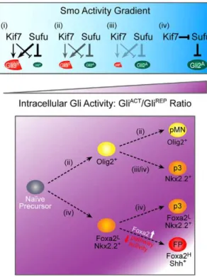
Figure 5. A model of Kif7 and Sufu regulation of graded Gli activity in ventral cell fate specification.The concerted actions of Kif7 and Sufu alter the Gli activator to repressor ratio to regulate intracellular Gli activity for the specification of ventral cell fates (pMN, p3 and FP) from naı¨ve neuroepithelial precursor cells. As Shh signaling increases Smo activity, Gli repressor formation and the inhibition of activators become less efficient as denoted (i–iii), while robust Smo activation results in Kif7 antagonizing the inhibition of Gli activators by Sufu for p3 and FP induction (iv).
doi:10.1371/journal.pone.0050193.g005
Immunofluorescence
Embryos were harvested at E 9.5 and fixed in 4% paraformal-dehyde (PFA) in PBS at 4uC overnight, then subjected to ethanol series dehydration, paraffin embedding and sectioning at 7mm. Immunohistochemistry on anterior spinal cord sections was performed as described [13]. Mouse anti-Nkx2.2 and anti-Foxa2 (HNF-3ß) (1:20, Developmental Studies Hybridoma Bank), rabbit anti-Olig2 (1:300, Chemicon), rabbit anti-Pax6 (1:300, Covance), and rabbit anti-Shh (1:50, Santa Cruz) antibodies were used. Images were acquired with a Zeiss LSM510 META laser scanning confocal microscope.
Statistical Analysis
All analysis was performed using anterior spinal cord sections at the forelimb bud level in somite-matched E9.5 embryos (2461 somite pairs), exceptPtch1single mutant embryos which arrest at 16–20 somite pairs. Cell fate quantification data were expressed and plotted as means 6 standard error of the mean (SEM). Statistical analysis for multiple comparisons was completed using one-way ANOVA followed by the Bonferroni post-test (Figures 1M, 2M and 3K). Analysis for pair-wise comparisons was completed using the Student’st-Test (Figure 4M). The sample size and p-value for each Bonferroni comparison or Student’st -Test are provided in the respective legends and figures.
In situ hybridization
Whole-mount or section in situ hybridization was performed as described [10]. Embryos were fixed in 4% PFA in PBS at 4uC overnight, then subjected to methanol series dehydration. In situ hybridization was carried out with digoxigenin-dUTP-labeled RNA probes forShh[36].
Western blot analysis
Embryos were snap-frozen, and sonicated in RIPA buffer as described [17]. Immunoblotting was performed with rabbit anti-Gli3 (Santa Cruz), and rabbit anti-Gli2 (amino acids 327–442) generated using standard protocols.
Acknowledgments
We thank Olena Zhulyn, Zhu Juan Li and Kyoung-Han Kim for helpful discussions and critical reading of the manuscript.
Author Contributions
Conceived and designed the experiments: KL CCH. Performed the experiments: KL SM RM XZ VP. Analyzed the data: KL SM RM XZ. Wrote the paper: KL CCH.
References
1. Ericson J, Rashbass P Schedl A, Brenner-Morton S, Kawakami A, et al. (1997) Pax6 controls progenitor cell identity and neuronal fate in response to graded Shh signaling. Cell 90: 169–180.
2. Briscoe J, Pierani A, Jessell TM, Ericson JA (2000) Homeodomain protein code specifies progenitor cell identity and neuronal fate in the ventral neural tube. Cell 101: 435–445.
3. Dessaud E, McMahon AP, Briscoe J (2008) Pattern formation in the vertebrate neural tube: a sonic hedgehog morphogen-regulated transcriptional network. Development 135: 2489–2503.
4. Placzek M, Briscoe J (2005) The floor plate: multiple cells, multiple signals. Nat Rev Neurosci 6: 230–240.
5. Ribes V, Balaskas N, Sasai N, Cruz C, Dessaud E, et al. (2010) Distinct Sonic Hedgehog signaling dynamics specify floor plate and ventral neuronal progenitors in the vertebrate neural tube. Genes Dev 24: 1186–1200. 6. Hui C-c, Angers S (2011) Gli Proteins in Development and Disease. Annu Rev
Cell Dev Biol 27: 513–527.
7. Chiang C, Litingtung Y, Lee E, Young KE, Corden JL, et al. (1996) Cyclopia and defective axial patterning in mice lacking Sonic hedgehog gene function. Nature 383: 407–413.
8. Wijgerde M, McMahon JA, Rule M, McMahon AP (2002) A direct requirement for Hedgehog signaling for normal specification of all ventral progenitor domains in the presumptive mammalian spinal cord. Genes Dev 16: 2849–2864. 9. Litingtung Y, Chiang C (2000) Specification of ventral neuron types is mediated by an antagonistic interaction between Shh and Gli3. Nat Neurosci 3: 979–985. 10. Ding Q, Motoyama J, Gasca S, Mo R, Sasaki H, et al. (1998) Diminished Sonic hedgehog signaling and lack of floor plate differentiation in Gli2 mutant mice. Development 125: 2533–2543.
11. Park HL, Bai C, Platt KA, Matise MP, Beeghly A, et al. (2000) Mouse Gli1 mutants are viable but have defects in SHH signaling in combination with a Gli2 mutation. Development 127: 1593–1605.
12. Goodrich LV, Milenkovic´ L, Higgins KM, Scott MP (1997) Altered neural cell fates and medulloblastoma in mouse Patched mutants. Science 277: 1109–1113. 13. Motoyama J, Milenkovic´ L, Iwama M, Shikata Y, Scott MP, et al. (2003) Differential requirement for Gli2 and Gli3 in ventral neural cell fate specification. Dev Biol 259: 150–161.
14. Stamataki D, Ulloa F, Tsoni SV, Mynett A, Briscoe J (2005) A gradient of Gli activity mediates graded Sonic Hedgehog signaling in the neural tube. Genes Dev 19: 626–641.
15. Balaskas N, Ribeiro A, Panovska J, Dessaud E, Sasai N, et al. (2012) Gene regulatory logic for reading the Sonic Hedgehog Signaling gradient in the vertebrate neural tube. Cell 148: 273–284.
16. Cooper AF, Yu KP, Brueckner M, Brailey LL, Johnson L, et al. (2005) Cardiac and CNS defects in a mouse with targeted disruption of Suppressor of fused. Development 132: 4407–4417.
17. Cheung HO-L, Zhang X, Riberio A, Mo R, Makino S, et al. (2009) The kinesin protein Kif7 is a critical regulator of Gli transcription factors in mammalian hedgehog signaling. Sci signal 2: ra29.
18. Endoh-Yamagami S, Evangelista M, Wilson D, Wen X, Theunissen J-W, et al. (2009) The mammalian Cos2 homolog Kif7 plays an essential role in modulating Hh signal transduction during development. Curr Biol 19: 1320–1326. 19. Liem KF, He M, Ocbina PJR, Anderson KV (2009) Mouse Kif7/Costal2 is a
cilia-associated protein that regulates Sonic hedgehog signaling. Proc Natl Acad Sci USA 106: 13377–13382.
20. Sva¨rd J, Heby-Henricson K, Henricson KH, Persson-Lek M, Rozell B, et al. (2006) Genetic elimination of Suppressor of fused reveals an essential repressor function in the mammalian Hedgehog signaling pathway. Dev Cell 10: 187–197. 21. Barnfield PC, Zhang X, Thanabalasingham V, Yoshida M, Hui C-c (2005) Negative regulation of Gli1 and Gli2 activator function by Suppressor of fused through multiple mechanisms. Differentiation 73: 397–405.
22. Chen M-H, Wilson CW, Li YJ, Law KKL, Lu C-S, et al. (2009) Cilium-independent regulation of Gli protein function by Sufu in Hedgehog signaling is evolutionarily conserved. Genes Dev 23: 1910–1928.
23. Wang C, Pan Y, Wang B (2010) Suppressor of fused and Spop regulate the stability, processing and function of Gli2 and Gli3 full-length activators but not their repressors. Development 137: 2001–2009.
24. Tukachinsky H, Lopez LV, Salic A (2010) A mechanism for vertebrate Hedgehog signaling: recruitment to cilia and dissociation of SuFu-Gli protein complexes. J Cell Biol 191: 415–428.
25. Humke EW, Dorn KV, Milenkovic´ L, Scott MP, Rohatgi R (2010) The output of Hedgehog signaling is controlled by the dynamic association between Suppressor of Fused and the Gli proteins. Genes Dev 24: 670–682. 26. Ding Q, Fukami S-i, Meng X, Nishixaki Y, Zhang X, et al. (1999) Mouse
suppressor of fused is a negative regulator of sonic hedgehog signaling and alters the subcellular distribution of Gli1. Curr Biol 9: 1119–1122.
27. Merchant M, Vajdos FF, Ultsch M, Maun HR, Wendt U, et al. (2004) Suppressor of fused regulates Gli activity through a dual binding mechanism. Mol Cell Biol 24: 8627–8641.
28. Hsu S-HC, Zhang X, Yu C, Li ZH, Wunder JS, et al. (2011) Kif7 promotes hedgehog signaling in growth plate chondrocytes by restricting the inhibitory function of Sufu. Development 138: 3791–3801.
29. Pierani A, Brenner-Morton S, Chiang C, Jessell TM (1999) A sonic hedgehog-independent, retinoid-activated pathway of neurogenesis in the ventral spinal cord. Cell 97: 903–915.
30. Diez del Corral R, Olivera-Martinez I, Goriely A, Gale E, Maden M, et al. (2003) Opposing FGF and retinoid pathways control ventral neural pattern, neuronal differentiation, and segmentation during body axis extension. Neuron 40:65–79.
31. Novitch BG, Wichterle H, Jessell TM, Sockanathan S (2003) A requirement for retinoic acid-mediated transcriptional activation in ventral neural patterning and motor neuron specification. Neuron 40: 81–95.
32. Wilson L, Gale E, Chambers D, Maden M (2004) Retinoic acid and the control of dorsoventral patterning in the avian spinal cord. Dev Biol 269: 433–446. 33. Wang H, Lei Q, Oosterveen T, Ericson J, Matise MP (2011) Tcf/Lef repressors
differentially regulate Shh-Gli target gene activation thresholds to generate progenitor patterning in the developing CNS. Development 138: 3711–3721.
34. Pospisilik JA, Schramek D, Schnidar H, Cronin SJF, Nehme NT, et al. (2010) Drosophila genome-wide obesity screen reveals hedgehog as a determinant of brown versus white adipose cell fate. Cell 140: 148–160.
35. Zhang XM, Ramalho-Santos M, McMahon AP (2001) Smoothened mutants reveal redundant roles for Shh and Ihh signaling including regulation of L/R asymmetry by the mouse node. Cell 106: 781–792.
36. Mo R, Freer AM, Zinyk DL, Crackower MA, Michaud J, et al. (1997) Specific and redundant functions of Gli2 and Gli3 zinc finger genes in skeletal patterning and development. Development 124: 113–123.