with the
lysA
Gene of Mycobacteriophage Ms6
Maria Joa˜o Catala˜o, Catarina Milho, Filipa Gil, Jose´ Moniz-Pereira, Madalena Pimentel*
Centro de Patoge´nese Molecular, Unidade dos Retrovı´rus e Infecc¸o˜es Associadas, Faculdade de Farma´cia, Universidade de Lisboa, Lisboa, Portugal
Abstract
Mycobacteriophages are dsDNA viruses that infect mycobacterial hosts. The mycobacteriophage Ms6 accomplishes lysis by producing two cell wall hydrolytic enzymes, Lysin A (LysA) that possesses a central peptidoglycan recognition protein (PGRP) super-family conserved domain with the amidase catalytic site, that cleaves the amide bond between the N -acetylmuramic acid and L-alanine residues in the oligopeptide crosslinking chains of the peptidoglycan and Lysin B (LysB) a mycolylarabinogalactan esterase that hydrolyzes the mycolic acids from the mycolyl-arabinogalactan-peptidoglycan complex. Examination of the endolysin (lysA) DNA sequence revealed the existence of an embedded gene (lysA241) encoded in the same reading frame and preceded by a consensus ribosome-binding site. In the present work we show that, even thoughlysAis essential for Ms6 viability, phage mutants that express only the longer (Lysin384) or the shorter (Lysin241) endolysin are viable, but defective in the normal timing, progression and completion of host cell lysis. In addition, both endolysins have peptidoglycan hydrolase activity and demonstrated broad growth inhibition activity against various Gram-positive bacteria and mycobacteria.
Citation:Catala˜o MJ, Milho C, Gil F, Moniz-Pereira J, Pimentel M (2011) A Second Endolysin Gene Is Fully Embedded In-Frame with thelysAGene of Mycobacteriophage Ms6. PLoS ONE 6(6): e20515. doi:10.1371/journal.pone.0020515
Editor:Eliana Saul Furquim Werneck Abdelhay, Instituto Nacional de Caˆncer, Brazil
ReceivedMarch 7, 2011;AcceptedMay 2, 2011;PublishedJune 9, 2011
Copyright:ß2011 Catala˜o et al. This is an open-access article distributed under the terms of the Creative Commons Attribution License, which permits unrestricted use, distribution, and reproduction in any medium, provided the original author and source are credited.
Funding:This work was supported by funds provided by Fundac¸a˜o para a Cieˆncia e Tecnologia (FCT) (http://alfa.fct.mctes.pt/index.phtml.en)- PTDC/SAU-FCF/ 73017/2006. Maria Joa˜o Catala˜o and Filipa Gil are the recipients of a fellowship from FCT (SFRH/BD/24452/2005) and (SFRH/BD/29167/2006). The funders had no role in study design, data collection and analysis, decision to publish, or preparation of the manuscript.
Competing Interests:The authors have declared that no competing interests exist.
* E-mail: [email protected]
Introduction
At the end of the replication cycle, bacteriophages must exit the host cell and disperse their newly formed progeny to infect new cells. The main barrier to host lysis is the peptidoglycan, a strong and stable structure that allows the bacterial envelope to withstand internal osmotic pressure [1]. With the exception of filamentous phages that as a result of their unique morphology and morphogenesis can extrude through the envelope without fatal consequences for the host, all other phages must either degrade or otherwise compromise the peptidoglycan to cause lysis [2,3]. Most of the tailed double-stranded DNA (dsDNA) phages achieve the proper time for lysis by the consecutive use of two lysis proteins – holin and endolysin. Holins are small hydrophobic membrane proteins that during the late phase of phage development progressively accumulate in the cytoplasmic membrane of the host and while the proton-motive force is maintained assemble into oligomers and rafts of intrinsic stability [2,4]. At a precise time programmed into its primary structure and upon a specific trigger event (holin effector concentration and partial depolarization of the membrane), the holin suddenly causes disruption of the membrane with non-specific hole formation and collapse of the membrane potential which sets the time of lysis by allowing the destruction of the cell wall by the released or activated phage encoded muralytic enzymes, the endolysins [5,6]. The term endolysin is used to describe the dsDNA bacteriophage-encoded peptidoglycan hydrolases, which are synthesized in phage-infected cells at the end of the multiplication cycle. They are characterized by the ability to directly target bonds in the peptidoglycan layer of
been described. The endolysin ofOenococcus oeni phage fOg44 is endowed with a bona-fide SP that is processed by the leader peptidase during infection and is exported by thesecmachinery [13]. A survey of orthologous endolysins from other phages of Gram-positive hosts suggested that some of these have N-terminal sequences resembling secretory signals, although in every case an adjacent holin gene was also present [6,13]. In addition, the endolysin ofLactobacillus plantarumphagewg1e has been reported as being exported inE. coliby the Sec machinery [14]. Particularly remarkable cases are the endolysins ofE. coliphages P1 and 21, which feature an N-terminal signal-arrest-release (SAR) sequence that allows the enzyme to be exported to the membrane where it is arrested, and to be released as a soluble active enzyme in the periplasm [15–17].
The mycobacteriophage Ms6 is a temperate bacteriophage with an unusual lytic cassette: in addition to the endolysin-holin lysis system, encoded by geneslysA(gp2) andgp4/gp5, respectively, the Ms6 lytic cassette comprises two additional lysis proteins encoded by genes gp1 and gp3 (lysB) [18,19]. The lysB gene has been previously characterized: it encodes an enzyme with lipolytic activity that hydrolyzes the mycolic acids from the mycolyl-arabinogalactan-peptidoglycan complex [20,21] acting at a later stage of infection to facilitate lysis by compromising the integrity of the mycobacterial outer membrane linkage to the arabinogalac-tan-peptidoglycan layer [22]. The Ms6 lysA gene was shown to encode a 384 amino acid polypeptide (LysA) with significant similarity to some bacteriophage encoded lysins withN -acetylmur-amoyl-L-alanine amidase activity [18]. Several types of cell wall hydrolases seem to be produced by mycobacteriophages. While phages Ms6 and TM4 encode enzymes with an amid-2 type domain [23], others such as D29 and Bxb1 employ hydrolases with lysozyme-like activity to bring about host cell lysis. Mycobacteriophage endolysins containing SP or SAR domains that allow secretion of the endolysin into the periplasmic space have not yet been described. Interestingly, however, our group has recently identified the product of gp1 gene as a chaperone-like protein that specifically interacts with the N-terminal region of the Ms6 endolysin. Gp1 is involved in the endolysin translocation across the cytoplasmic membrane independently of the holin function and is required for efficient phage release [24]. During an attempt to purify LysA as a C-terminal histidine-tagged fusion product, we detected the synthesis of two proteins, rather than a single polypeptide. Further examination oflysA(lysA384) nucleotide
sequence revealed a second possible gene (lysA241) in the same
reading frame and preceded by a potential ribosome-binding site (RBS). Here, we report studies directed at dissecting the precise role of the lysA-encoded gene products during Mycobacterium smegmatis infection by the mycobacteriophage Ms6. In addition, the lytic activity spectrum of both proteins was also examined in both Gram-positive and Gram-negative bacteria and also in mycobacteria regarding the potential application of mycobacter-iophage lysins.
Materials and Methods
Bacterial strains, phages, plasmids and culture conditions Bacterial strains, phages and plasmids used throughout this study are listed in Table 1.E. colistrains were grown at 37uC, in Luria-Bertani (LB) broth or agar supplemented with 100mg ml21
ampicillin or 30mg ml21 kanamycin, when appropriate. M.
smegmatisrecombinant strains were grown at 37uC in 7H9 medium (Difco) supplemented with 0.05% Tween 80, with shaking or Middlebrook 7H10 (Difco), containing 15mg ml21 kanamycin.
For induced conditions 0.2% succinate and 0.2% acetamide were also added to media.
Plasmid construction
Unless otherwise indicated, DNA fragments obtained by PCR were amplified using Ms6 genomic DNA as template. DNA amplification, plasmid isolation and electrophoresis were carried out using standard techniques [25]. E. coli and M. smegmatis mc2155 cells were transformed as described previously [25,26]. Restriction enzymes and T4 DNA ligase (New England Biolabs) were used according to the supplier’s recommendations. All oligonucleotides were purchased from Thermo Scientific and are listed in Table S1.
In order to construct plasmids pMJC40 and pMJC42,lysA241 was amplified using primerslysA241fwd-1/plysA-c3 or lysA241 fwd-2/plysA-c3 and the resulting DNA fragments were introduced into the BamHI/HindIII sites of vector pQE30, allowing a fusion to a hexahistidine tag at the N-terminus, or pET29b allowing a C-terminal hexahistidine tag fusion, respectively. To obtain plasmid pMJC41, the DNA fragment containing lysA, was amplified by PCR with primers gp2A/plysA-c3 and cloned into BamHI/ HindIII sites of pET29b fused to a C-terminal His6tag. Plasmid
pMJC43 was constructed by PCR amplifyinglysA384(lysA) lacking
the GTG start codon with primers plysADGTGfwd/plysA-c3 and cloning the DNA fragment, after restriction with XbaI/HindIII which removed the vector translational signals (RBS and start codons), in pET29b. All constructs were validated by sequencing the insert nucleotide sequence.
Construction of Ms6 mutant phages
Construction of Ms6 mutant phages was performed using the Bacteriophage Recombineering of Electroporated DNA (BRED) as described previously [24,27]. Briefly, for Ms6lysAdeletion, a 100 bp oligonucleotide, PrDlysA, that has 50 bp of homology upstream and downstream of the region to be deleted was extended by PCR using two 75 bp extender primers, PrExtD ly-sAfwd/ PrExtDlysArv, which have 25 bp of homology to the ends of the 100-mer and add additional 50 bp of homology on either end. The final 200 bp dsDNA product was purified using MinElute PCR Purification Kit (QIAGEN) and co-electroporated with Ms6wtgenomic DNA into electrocompetent recombineering cells of M. smegmatis mc2155:pJV53. To abolish synthesis of Lysin384 and Lysin241 we designed two complementary 73 bp
oligonucleotides (PrlysATGAHindIIIfwd/ PrlysATGAHindIIIrv) that
introduce a stop codon and a HindIII restriction site downstream of the start codon of lysA384, or two complementary 86 bp oligonucleotides (PrlysAGTGRTGGMscIfwd/ PrlysAGTGRTGG
Ms-cIrv) that modify thelysA241 GTG start codon (valine) to TGG (tryptophan) and introduce an MscI restriction site, respectively. Complementary oligonucleotides were co-transformed with Ms6-LysAHis6genomic DNA into recombineering cells ofM. smegmatis
mc2155:pJV53. Cells were resuspended in 7H9 supplemented with 0.5% glucose and 1 mM of CaCl2, incubated at 37uC for 2 hours
(prior to lysis) and plated as top agar lawns with M. smegmatis mc2155. Phage plaques were picked into 100ml phage buffer (10 mM Tris-HCl, pH 7.5; 10 mM MgSO4; 68.5 mM NaCl;
1 mM CaCl2), eluted for two hours at room temperature and
smegmatis. Individual secondary plaques or lysates were screened by DADA-PCR forlysAdeletion or by PCR and restriction with the same primers referred above to identify pure mutant phages. Construction of phage Ms6Dgp1-LysAHis6was done as described
previously [24] using Ms6Dgp1genomic DNA.
Lysin expression in M. smegmatis-infected cells
Examination of Lysin384and Lysin241synthesis inM. smegmatis
was performed as previously described [13]. An exponentially growing culture of M. smegmatismc2155 was infected with Ms6-LysAHis6, Ms6Dgp1-LysAHis6, Ms6-Lysin241His6or Ms6-Lysin384
-His6at an approximate multiplicity of infection (MOI) of 10 and
incubated at 37uC for 30 minutes. Ten-mililiter samples were withdrawn at 30-min intervals; cells were pelleted by centrifuga-tion and frozen at220uC. After thawing, cells were concentrated 100-fold in phosphate-buffered saline supplemented with 20 mg of lysozyme ml21. After an incubation period at 37uC for 1 hour, 25ml of 56SDS-PAGE sample buffer were added followed by incubation at 100uC for 5 minutes to complete cell lysis. M. smegmatis extracts were analysed by Western blotting and lysin immunodetection was performed using horseradish-peroxidase-conjugated anti-His monoclonal antibody (Roche).
One-step growth curves
One-step growth curve and burst-size determination were previously described [24]. The one step assays were carried out inM. smegmatisexponential growth cells using an MOI of 1. Cells were pelleted and resuspended in 1 ml of a phage suspension (Ms6wt, Ms6-Lysin241His6 or Ms6-Lysin384His6) supplemented
with 1 mM CaCl2. The mixture was incubated 50 min at 37uC
to allow adsorption of the phages. 100ml of 0.4% H2SO4 was
added to inactivate the non-adsorbed phages and the incubation continued for five min. The suspension was neutralized with 100ml of 0.4% NaOH and diluted 1:100 in 7H9 supplemented with 0.5% glucose and 1 mM CaCl2. 1 ml samples were
withdrawn every 30 min until reaching 300 min. 100ml of serial
dilutions of each sample were plated with 200ml ofM. smegmatis
cells, on 7H10 as top agar lawns and the phage titer for each sample was determined after 24 h incubation at 37uC. Results are averages of three independent experiments.
Expression of Lysin384and Lysin241 proteins inE. coli
E. coliBL21 (DE3):pMJC41 orE. coliBL21 (DE3):pMJC42 were grown in LB medium to an OD600 nmof 0.6, and expression of the
recombinant Lysin384-His6or Lysin241-His6was induced for 4 h
following the addition of isopropylb-D-1-thiogalactopyranoside to a final concentration of 1 mM. Bacterial cells were harvested by centrifugation, washed, resuspended in 50 mM Tris-HCl (pH 7.5) supplemented with a cocktail of protease inhibitors (Calbiochem), and disrupted by passage through a French pressure cell. Cell debris were removed by centrifugation, and the recombinant proteins present in the supernatant were analysed by SDS-PAGE, followed by Coomassie blue staining and Western blotting and detected as described above.
Assay of the antibacterial activity of Lysin384and Lysin241 The antibacterial activity was screened using a sensitivity test [28,29] with some modifications. 100ml of an exponential growing culture of the test strain was plated on LB or 7H10+OADC (for mycobacteria) as top agar lawns. 20ml of inducedE. coli:pMJC41
Table 1.Strains, bacteriophages, plasmids used in this study.
Strains, bacteriophages or plasmids Description Reference or source
Bacteria
Escherichia coli
JM109 recA1 endA1 gyr96 thi hsdR17 supE44 relA1D(lac-proAB) [F9traD36 proAB lacIqZDM15]
Stratagene
BL21 (DE3) F2ompT hsdS
B(rB2mB2)gal dmc(DE3) Novagen
Mycobacterium smegmatis
mc2155 High-transformation-efficiency mutant ofM. smegmatisATCC 607 [26]
Bacteriophages
Ms6wt Temperate bacteriophage fromM. smegmatis [70]
Ms6-LysAHis6 His6tag insertion at the 39end of Ms6lysA [24]
Ms6Dgp1-LysAHis6 His6tag insertion at the 39end of Ms6lysAin Ms6Dgp1 This study
Ms6-Lysin384His6 GTGRTGG change in codon 144 oflysAin Ms6-LysAHis This study
Ms6-Lysin241His6 Stop codon introduced in thelysAgene of Ms6-LysAHis6 This study
Plasmids
pQE30 Expression vector; T5 promoter; Ampr QIAGEN
pET29b(+) Expression vector, T7 promoter; Kanr Novagen
pJV53 Derivative of pLAM12 with Che9c60and61under control of the acetamidase promoter; Kanr
[71]
pMJC40 lysA241Ms6 cloned into pQE30 This study
pMJC41 lysA384Ms6 cloned into pET29b(+) This study
pMJC42 lysA241Ms6 cloned into pET29b(+) This study
pMJC43 lysA384DGTGcloned into pET29b(+)DRBS This study
Ms6 lysis genes Accession No. AF319619.
orE. coli:pMJC42 extracts containing Lysin384 or Lysin241 were
spotted onto the bacterial lawn of the test strain and incubated overnight at 37uC. After overnight incubation, the presence of a clear zone was examined.E. coli:pET29b induced extract was used as a negative control. Activity assays were performed in triplicate. Several bacterial strains were used to test the range of antibacterial activity and were obtained from the American Type Culture Collection (ATCC) or from the Institute Pasteur Collection, Paris.
Zymogram analysis
Sodium dodecyl sulfate (SDS)-polyacrylamide gel electrophore-sis was performed as described by Laemmli (1970) [30], and the zymogram assays were carried out as outlined by Piuri and Hatfull (2006) [31]. Briefly, 0.2% autoclaved and lyophilized Micrococcus luteuscells were included in 15% polyacrylamide gels for detection of bacteriolytic activity. Protein samples were heated for 3 min at 100uC in sample buffer (62.5 mM Tris-HCl, pH 6.8; 2% SDS; 5% mercaptoethanol; 20% glycerol; 0.01% bromophenol blue), and then separated on SDS-gels containing the autoclaved cells. After electrophoresis, the zymograms were washed for 30 min with distilled water at room temperature and then transferred into renaturation buffer containing 25 mM Tris-HCl (pH 7.5) and 0.1% Triton X-100 followed by further incubation for 16 h at 37uC. The zymograms were rinsed with distilled water, stained with 0.1% methylene blue in 0.01% KOH for 2 h at room temperature, and then destained with distilled water. Peptidogly-can hydrolase activity was detected as a clear zone on a dark blue background of stained peptidoglycan. Gels not containing peptidoglycan were stained with Coomassie Brilliant Blue. Lysozyme and bovine serum albumin (BSA) were used as positive and negative controls, respectively. Molecular masses were determined by comparison with prestained molecular weight standards that were electrophoresed on the same gel.
Results
Identification of two gene products from Ms6lysA
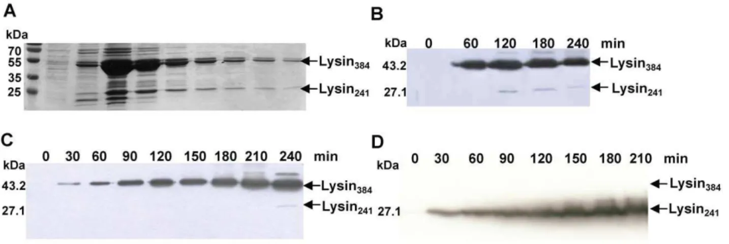
The 1155 bplysAgene of mycobacteriophage Ms6 starts at a GTG codon that overlaps thegp1TGA stop codon in a different reading frame, and is preceded in four nucleotides by a RBS sequence (59-GGGAGCA-39) (Fig. S1). It encodes a 384 amino acid polypeptide with significant similarity to bacteriophage encoded N-acetyl-muramoyl-L-alanine amidases [18]. During an attempt to purify LysA as a C-terminal histidine-tagged fusion product (LysA-His6), we detected the production of two proteins of ,27 kDa (Lysin241) and,43 kDa (Lysin384), rather than a single
polypeptide of 43 kDa, inE. colicrude extracts (Fig. 1A). We also observed that when the protein was tagged in the N-terminal domain, only the larger product (Lysin384) reacted with the
anti-His6antibody (data not shown). The fact that a time-dependent
decrease of the 43 kDa form with a concomitant increase of the labelled 27 kDa form was not observed, rather both proteins seemed to be produced independently over the time (Fig. 1B), led us to consider that the smaller protein was not an N-terminal processed form of a larger precursor, but rather two independent translated products. Also supporting this notion, is the fact that analysis of the amino acid sequence of Ms6 LysA did not predict an amino-terminal signal sequence or a peptidase cleavage site [24]. Further examination of thelysAnucleotide sequence revealed a second potential gene entirely embedded in the same reading frame, preceded by a putative Shine-Dalgarno sequence (59 -TGGAGGT-39) utilized by Gram-positive bacteria and mycobac-teria [32], 6 nucleotides upstream of the GTG start codon (Fig. S1), and designated lysA241. Occurrence of an additional
translation event at the predicted location would be compatible with the observed molecular masses (,43 and 27 kDa) of the two
proteins, considering that an additional His6tag was C-terminally
fused to both proteins. This was experimentally confirmed by sequencing the amino-terminal region of the 27 kDa protein (Lysin241). This protein was obtained by expressing LysA inE. coli,
followed by isolation of the smaller lysin from a polyvinylidene difluoride membrane. The obtained N-terminal sequence, MPDEPRPD, matched the deduced sequence from residues 144 to 151. Of note is the fact that, following expression inE. coli, the larger product (Lysin384) is in great excess when compared to the
smaller protein (Lysin241) and frequently Lysin241synthesis was not
observed (Fig. 1C). To further clarify these results,lysAlacking its own GTG start codon was cloned into the XbaI/HindIII sites of pET29 which removes the Shine-Dalgarno sequence and the start codons of the expression vector. We were expecting that if Lysin241
results in fact from a new translation event and is not the result of cleavage of Lysin384, it should be synthesized from this
construction. Western blotting analysis revealed the production of a single polypeptide with 27 kDa corresponding to Lysin241
(Fig. 1D). This result unambiguously proves that Lysin241results
from a new translation event and is independent of Lysin384
synthesis.
To follow mycobacteriophage Ms6 LysA production in the course ofM. smegmatisinfection, infected cells were examined for lysin synthesis.M. smegmatis was infected with a Ms6 derivative mutant, Ms6-LysAHis6phage, where the 39end oflysAgene was
fused to a sequence coding for a hexahistidine tag, allowing the production of a LysA-His6 tagged protein [24]. Samples were
collected immediately before and every 30 minutes following infection until near the end of the Ms6 infection cycle. Protein extracts were prepared from such samples as described in materials and methods and checked for the presence of Histagged proteins by immunoblotting. Two proteins rather than a single lysin band, with 27 kDa and 43 kDa were detected, corresponding to the predicted molecular masses of Lysin241-His6and Lysin384
-His6, respectively (Fig. 2A). Both proteins were first detected at
90 minutes postinfection with mobilities indistinguishable from that exhibited by the protein forms of theE. coliexpressed lysin. Taking into consideration that Lysin384interacts with Ms6 Gp1,
we followed the production of both proteins in an infection assay with an Ms6 mutant lackinggp1[24]. In this assay, we observed a decrease in Lysin384although Lysin241amount was comparable to
the levels detected for the wild-type phage (Fig. 2B). Similarly to what was observed during Ms6 infection, both endolysins were detected 90 minutes postinfection (Fig. 2A).
These results suggest that during M. smegmatis Ms6 infection, different products of thelysAgene are synthesized and result from the existence of two translational events that direct the production of a smaller (Lysin241) and a larger (Lysin384) endolysin rather than
a processing event. In addition, synthesis and/or stability of Lysin384seem to be dependent on the Gp1 chaperone-like protein
as already proposed [24].
Occurrence of Ms6 LysA-like proteins in mycobacteriophages
glycosidases and peptidases, as well as peptidoglycan-binding motifs [35]. Phages Ms6 and TM4 encode enzymes with an amid-2 type domain, while others such as Damid-29 and Bxb1 employ hydrolases with lysozyme-like domains to bring about host cell lysis [23,33]. A search for conserved domains showed that Ms6 LysA holds a central peptidoglycan recognition protein (PGRP) conserved domain (cd06583), localized between amino acid residues 168 and 312 (Fig. S2). PGRPs are pattern recognition receptors that bind, and in certain cases, hydrolyze peptidoglycan of bacterial cell walls. This family includes Zn-dependent N -acetylmuramoyl-L-alanine amidases (EC: 3.5.1.28) which cleave the amide bond between N-acetylmuramoyl and L-amino acids, preferentially D-lactyl-L-Ala, in bacterial cell walls.
The mycobacteriophage Ms6 lysis module is closely related to the lysis module of phages belonging to cluster F, subcluster F1,
which includes phages PMC, Llij, Che8, Boomer, Fruitloop, Pacc40, Ramsey and Tweety [33], Ardmore [36] and Wee (GenBank accession number YP004123853). A BLASTp search for Ms6 LysA homologues identified similar proteins amongst phages of this subcluster, with a high level of identity, except for the endolysin of mycobacteriophage Pacc40, and produced significant alignments with the N-terminal region of endolysins belonging to unclustered mycobacteriophages Corndog, Phyler, Phaedrus and Pipefish and with the endolysin of phage LeBron (GenBank accession number YP003857156) [37]. Examination of the endolysin nucleotide sequences of these bacteriophages revealed the existence in all of them of a second potential translation site positioned in close proximity to the beginning of the central PGRP domain (Fig. S2, Table 2). As recently described, the accessory lysis protein Gp1 binds the N-terminal Figure 1. LysA expression inE. coli. A.Purified LysA-His6fractions after SDS-PAGE analysis and Coomassie Blue staining. LysA-His6was produced
from pMJC41 inE. coliBL21 (DE3) after isopropylb-D-1-thiogalactopyranoside induction.B.Expression of LysA-His6from pMJC41: detection of
C-terminal His6tag LysA shows the production of Lysin384-His6and Lysin241-His6.C. LysA-His6synthesis from pMJC41 over the time is not always
followed by Lysin241-His6production.D.Synthesis of Lysin241-His6from pMJC43. Removal of pET29b and LysA (Lysin384-His6) translational signals
does not hinder Lysin241-His6synthesis. The molecular masses in kDa of Lysin384and Lysin241are indicated on the left; positions of both proteins are
indicated by an arrow on the right. Lysin384and Lysin241were detected by Western blotting with an anti-His6antibody, except for panel A.
doi:10.1371/journal.pone.0020515.g001
Figure 2. Time course of Lysin384and Lysin241synthesis during Ms6 infection ofM. smegmatis.Lysin production inM. smegmatiswas
analysed after infection with Ms6-LysAHis6or Ms6Dgp1-LysAHis6at an MOI of 10. Extracts were prepared from samples taken at 30-min intervals as
described in Material and Methods. Samples were analysed by Western blotting and Lysin384and Lysin241synthesis was detected with an anti-His6
monoclonal antibody.A.Lysin384and Lysin241synthesis is first detected 90 minutes postinfection both in Ms6-LysAHis6(upper panel) and Ms6Dgp1
-LysAHis6(lower panel) mutant phages. Only the results for 90 to 300 min postinfection are shown.B.Lysin384is synthesized to near undetectable
levels during Ms6Dgp1-LysAHis6infection ofM. smegmatis(lower panel) whereas Lysin241production is comparable to the wild-type phage. The
molecular masses in kDa of Lysin384and Lysin241are indicated on the left; positions of both proteins are indicated by an arrow on the right.
domain of LysA [24] andgp1was implicated in lysis because of its linkage to lysA and the demonstration of its requirement for mycobacteria efficient lysis [24]. gp1-like genes are present in many, but not all mycobacteriophage genomes and were grouped in the mycobacteriophage gene family Pham1480 [33]. In addition to subcluster F1, homologues of Ms6 Gp1 were also identified in all phages belonging to subcluster A1. However phages Lockley and Jasper did not reveal the existence of two potential translational sites in the lysA gene. Members of Pham1480 are mostly found adjacent to the Pham66-1 encoding genes, suggesting an even more intimate association between the gene products: while in some phages of subcluster A1 and of subcluster F1,gp1-like genes are overlapped with the lysin gene, in phages DD5, KBG and Solon (subcluster A1) and in phages Boomer and Ramsey (subcluster F1), Pham1480 is separated from the lysin gene by one intervening gene that code for putative homing endonuclease HNH motifs [33]. Although in these mycobacter-iophagesgp1andlysAare closely linked, several other
mycobacter-iophages seem to have somewhere in their genomes genes encoding proteins that could perform functions analogous to Gp1, while others simply lackgp1[33].
Role of Lysin384and Lysin241duringM. smegmatisphage
infection
For a better understanding of the contribution of the endolysin to the Ms6 infection cycle, we first investigated whether its hydrolase activity is an essential function for host lysis. We used the Bacteriophage Recombineering of Electroporated DNA (BRED) strategy [27] to deletelysAgene (gp2) from the mycobacteriophage Ms6 genome. Oligonucleotides were designed to introduce a 1089 bp internal-deletion in Ms6lysA, fusing 19 codons at the 59 and 39ends of the gene to maintain thelysB RBS and minimize effects on expression of the adjacentlysB gene, as well as avoiding genetic polarity. As expected,lysAwas essential for mycobacterial lysis and no viable phages could be recovered from a deletion of Ms6lysAgene (data not shown). The same result was also reported for mycobacteriophage Giles [27].
As a result of synthesis of Lysin241and Lysin384during phage
infection, we next investigated the influence of each endolysin form in phage growth parameters. Once more, we took advantage on the fact that the BRED recombineering strategy has already been described to efficiently introduce base changes that confer an amino acid substitution [27]. To eliminate synthesis of Lysin384or
Lysin241we designed oligonucleotides that introduce a stop codon
and a HindIII restriction site downstream of the start codon of lysA384, or two complementary oligonucleotides that modify the lysA241GTG start codon to TGG (tryptophan), and introduce an MscI restriction site, respectively. Both mutant phages Ms6-Lysin384His6(producing Lysin384) and Ms6-Lysin241His6
(produc-ing Lysin241) were readily isolated, demonstrating that for plaque
formation only one of the two LysA forms, Lysin384or Lysin241is
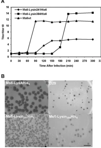
required (Fig. 3). Nevertheless, we considered whether the absence of one of the two endolysin forms during M. smegmatis phage infection (Fig. 4) could confer an altered lysis phenotype. To address this question one step growth curves and determination of phage growth parameters (latent period, rise period and burst size) were carried out to compare the phages infection cycle. The one step growth experiment (Fig. 5A) shows that in a phage Ms6-Lysin241His6infection, the latent period is prolonged 30 min in
comparison with the Ms6wtinfection and a decrease in the burst size was observed which means that a delay exists in the detection of phage release from cells infected with Ms6-Lysin241His6. In
agreement, absence of Lysin384leads to smaller size phage plaques
(Fig. 5B), meaning that Lysin384is important for infective particles
release. On the other hand, Ms6-Lysin384His6phage release starts
90 min later than with Ms6wt. This indicates that similarly to Lysin384, Lysin241has an obvious function in completion of lysis,
although it does not have a significant effect in the number of phage particles released. Examination of Ms6-Lysin384His6phage
plaques shows that although not larger in size, plaques are more turbid than wild-type Ms6 probably due to a partial host cell lysis (Fig. 5B). These results strongly suggest that even though only one of the two LysA forms, Lysin384 or Lysin241, is required to
accomplish host cell lysis, both enzymes are necessary for complete and efficient lysis ofM. smegmatis.
Lysin384and Lysin241are cell wall-degrading enzymes
with peptidoglycan hydrolase activity
LysA has been previously described as not affecting E. coli growth rate unless permeabilization of the plasma membrane by chloroform addition which results in immediate lysis [18]. To
Table 2.Occurrence of Ms6lysA-like genes in mycobacteriophages.
Mycobacteriophage E. coliconsensus
Start Codon Position*
SD
Start Codon
AGGAGGT 4-7 RATG
Cluster A/subcluster A1
Bxb1 CAAGGA tgcg ATG 397 (1434 bp)
U2 GAAGGA cgcg ATG 397 (1434 bp)
Bethlehem GAAGGA cgcg ATG 397 (1434 bp)
Solon AAAAGA tgcg ATG 379 (1395 bp)
KBG AAAAGA tgcg ATG 379 (1395 bp)
DD5 AAAAGA tgcg ATG 379 (1395 bp)
SkiPole AAAAGA ccca ATG 397 (1413 bp)
ClusterB/subcluster B3
Pipefish AGGAGG acagctccc GTG 478 (1224 bp)
Phaedrus AGGAGG acagcgccc GTG 322 (1068 bp)
Phyler AGGAGGC acagcgccc GTG 520 (1266 bp)
Cluster F/subcluster F1
Ms6 TGGAGGT accgcc GTG 430 (1155 bp)
Che8 CGGAGGA acct GTG 514 (1275 bp)
Tweety TGGAGGT accgcc GTG 430 (1212 bp)
PMC TGGAGGT accgcc GTG 430 (1194 bp)
Llij TGGAGGT accgcc GTG 430 (1194 bp)
Boomer CGGAGGT tccc ATG 523 (1302 bp)
Fruitloop TGGAGGT accgcc GTG 430 (1155 bp)
Ramsey CGGAGGA acct GTG 514 (1248 bp)
Pacc40 AGGAGAG gacgcaaac GTG 442 (1209 bp)
Wee TGGAGGT accgcc GTG 430 (1155 bp)
Ardmore TGGAGGT accgcc GTG 430 (1155 bp)
Unclustered
LeBron TGAGGT aatc GTG 448 (1173 bp)
Corndog GGGAGGA aca GTG 439 (1221 bp)
*Numbers refer to nucleotide positions. The gene size is indicated in parenthesis.
follow growth and viability ofE. colistrains expressing Lysin241, the
lysA DNA fragment corresponding to lysA241 was cloned into
pQE30 vector allowing expression of Lysin241under the control of
the regulated T5 bacteriophage promoter. Although induction of Lysin241did not result inE. colilysis unless chloroform was added,
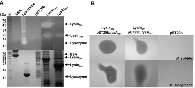
growth seems to be halted over the induction period (data not shown). Ms6 LysA holds a PGRP domain and its hydrolase activity was already demonstrated; the protein was shown to cleave the bond between L-Ala and D-muramic acid and to release up to 70% of the diaminopimelic acid present in isolated mycobacterial cell walls which confirmed the amidase activity of the enzyme (unpublished results). To more directly assess the enzymatic activity of Lysin384and Lysin241we tested their ability to generate
a zone of clearing in a zymogram assay. Lytic activity in lysin-producing E. coli extracts was checked by in situ protein renaturation after SDS-PAGE, using gel-incorporated autoclaved M. luteuscells as the substrate. As shown in Fig. 6A both Lysin384
and Lysin241 have hydrolase activity. Peptidoglycan hydrolysitic
activity in zymograms was already demonstrated for other three mycobacteriophage LysA proteins (Che8 Gp32, Bxz1 Gp236 and Corndog Gp69) [22] and TM4 Gp29 [38].
When Ms6 was first isolated, it was noticed that it forms plaques inM. smegmatislawns with halos surrounding them. Formation of the halo begins after two days of incubation, once the infected area is fully formed. Halo formation around phage plaques seems to be unusual among mycobacteriophages. Nevertheless, this phenom-enon has been described for, at least, mycobacteriophages Bxb1 [39] and Che12 [40] and has been observed for a number of other phages, particularly those infecting Gram-positive bacteria; plaques of phage A511 infecting Listeria monocytogeneswere shown to form clearly visible secondary lysis zones caused by release of Ply511, a hydrophilic amidase endolysin that diffuses from the
centre of the plaque and hydrolyses the surrounding cells ‘‘from without’’ [7].M. smegmatiscells in the exponential growth phase were infected with phages D29, Ms6wt, Ms6-Lysin241His6or
Ms6-Lysin384His6. Halo formation around D29 phage plaques was not
observed (Fig. 5B) while Ms6wtforms turbid plaques showing size variation with partially clear halos surrounding them (Fig. 5B). When we analysed halo formation inM. smegmatis infected with phages Ms6-Lysin241His6or Ms6-Lysin384His6, we observed that
both phages retain the ability to form halos, although smaller and much more turbid than the wild-type phage halo (Fig. 5B). It is possible that these halos might result from the diffusion of Lysin384
or Lysin241 from the Ms6wtphage infective centre, leading to a ‘‘lysis from without’’ in surrounding cells. As both endolysins forms of LysA are essential to obtain an effective lysis ofM. smegmatis, halo turbidity could result from partial lysis of cells surrounding the phage plaques. However, these results do not rule out the existence in the Ms6 genome of other genes whose products could be implicated in the halo formation phenomenon.
Despite the fact that Ms6 LysA is produced by a phage that infects a bacterium with a complex envelope and taking into consideration the above results, we tested the activity of E. coli crude extracts containing Lysin384 or Lysin241 in mycobacteria
and other bacterial species by spotting the extracts on lawns of exponential growing cells. The results, summarized in Table 3, showed that E. coli extracts containing Lysin384 or Lysin241
inhibited the growth of all mycobacteria tested exceptM. fortuitum, as well as the Gram-positive bacteria: M. luteus, M. pyogenes, B. subtilis and B. pumilus. A weak activity was observed against S. pyogenes,S. aureus,S. epidermidisand the Gram-negative bacteriumE. coli. For each strain tested, a crude extract of E. coli BL21 (DE3):pET29b was used as a control, and no inhibition was observed for any bacterial strain (Fig. 6B). Activity on mycobac-Figure 3. Construction of Ms6 lysAmutants.Two complementary oligonucleotides that modifylysA241GTG start codon (valine) to TGG
(tryptophan) and introduce an MscI restriction site, or two complementary oligonucleotides that introduce a stop codon and a HindIII restriction site downstream of the start codon oflysA384, were co-transformed with Ms6-LysAHis6genomic DNA; primary plaques were recovered and screened by
PCR and MscI or HindIII digestion to identify a mixed plaque containing wild-type and mutant phages DNA. The mixed primary plaque was diluted and plated; the lysate was screened to check for phage viability, and purified secondary plaques were screened to identify pure mutant phages of Ms6-Lysin384His6and Ms6-Lysin241His6, expressing only Lysin384or Lysin241, respectively.
teria cells was also confirmed by the ability of bacteriophages Ms6wt, Ms6-Lysin241His6and Ms6-Lysin384His6to form clearing
zones on lawns of growing cells withM. smegmatisand M. aurum showing to be the most sensitive mycobacteria (Table 3).
Discussion
In the present work, we provide evidence that two proteins (Lysin384and Lysin241) with endolysin activity are produced from
the mycobacteriophage Ms6 lysA gene. Our group has recently described that the N-terminal domain of Lysin384is necessary and
sufficient to directly interact with Gp1, a chaperone-like protein located upstream of LysA that assists the translocation of the endolysin across the cytoplasmic membrane in a holin-indepen-dent way [24]. Evidence for the presence of two forms of LysA raised the possibility that the shorter protein could be the mature form of Lysin384: if a cleavage event had occurred, it would
implicate the removal of the N-terminal 143 amino acids of the enzyme corresponding to 16 kDa. Although unusual, as generally mycobacterial SP length is 32 residues [41], similar to the lengths of SPs from Gram-positive bacteria [42], cleavage of a large segment of 143 amino acids that functions as a SP has already been described for the Staphylococcus simulans lysostaphin [43]. However, no sequence signals or cleavage sites were detected by bioinformatic analysis in the N-terminal region of LysA [24]. Examination of thelysAnucleotide sequence revealed the existence of a second possible lysin gene embedded withinlysAin the same reading frame and preceded by a consensus Shine-Dalgarno sequence (Fig. S1). The existence of two translation events was
clearly demonstrated when Lysin241 was still produced after
elimination of the translation signals from Lysin384, both inE. coli
and inM. smegmatisphage infection: if Lysin241was a processed
form of Lysin384it could not be detected. Thus, occurrence of a
cleavage event, as observed for the endolysin of bacteriophage fOg44 [13] was ruled out. Coding sequences entirely encompassed within other genes seem to be very rare among dsDNA bacteriophages: lrz and rz1 represent an example of two genes located in different reading frames in the same nucleotide sequence which encode different proteins, both required in the same physiological pathway [44]. Although very rare, some exceptions among bacteriophage endolysins were found: 1) the bacteriophage wvML3 endolysin gene encodes two proteins, a larger lysin that has homology with lysozymes and a smaller lysis protein that has some features resembling those of a holin [45]; 2) the streptococcal C1 bacteriophage lysin called PlyC is a multimeric lysin composed of two separate gene products, PlyCA and PlyCB responsible for the hydrolytic amidase activity and cell-wall-binding domain, respectively [46]; 3) examination of the Figure 4. Time course of endolysin synthesis during
Ms6-LysAHis6, Ms6-Lysin384His6or Ms6-Lysin241His6infection ofM.
smegmatis. Lysin production in M. smegmatis was analysed after infection at an MOI of 10. Extracts were prepared from samples taken at 30-min intervals as described in Material and Methods. Samples were analysed by Western blotting and Lysin384and Lysin241synthesis was
detected as already described. Both Lysin384 and Lysin241 synthesis
could be detected beginning 90 minutes postinfection in Ms6-LysAHis6
(upper panel). In Ms6-Lysin384His6and Ms6-Lysin241His6, (lower panels)
only Lysin384or Lysin241synthesis could be detected also beginning
90 min postinfection, respectively. Only the results for 90 to 270 min postinfection are shown. The molecular masses in kDa of Lysin384and
Lysin241 are indicated on the left; positions of both proteins are
indicated by an arrow on the right. doi:10.1371/journal.pone.0020515.g004
Figure 5. Both Lysin384and Lysin241are required for efficient
host cell lysis. A.One step growth curves of Ms6 and lysin-mutant derivatives. For each curve the titers measured were divided by the titer at t = 0 for normalization (titer/titer t0). B. Halo formation by
mycobacteriophage Ms6 and lysin-mutant derivatives. Serial dilutions of the bacteriophages stocks containing ,1010 particles ml21 were
prepared and 100ml of the 1028dilution was plated with 200ml of an
exponential growing culture of M. smegmatis as top agar lawns. Incubation was continued for 3 days at 37uC. Mycobacteriophage D29 was used as a negative control of halo formation [36]. Scale bar represents 1 cm.
nucleotide sequence of bacteriophage CMP1 endolysin gene revealed a possible Shine-Dalgarno sequence within the gene, four nucleotides upstream of a second ATG codon in the same reading frame which would correspond to a gene product consisting of 166 C-terminal amino acid residues that includes the binding domain of the enzyme [47].
A BLASTn search for Ms6lysAhomologues revealed that this peculiar endolysin gene arrangement is widespread in mycobac-teriophages, in particular amongst those that possess a gp1-like gene (Pham1480) [33], which would suggest that Gp1 may confer a selective advantage for host cell lysis under different environ-mental conditions: very small differences in lysis timing and Figure 6. Peptidoglycan hydrolysis byE. coli-produced Lysin384and Lysin241inM. luteuscells. A.Lytic activity of lysin extracts was
assessed byin siturenaturation after SDS-PAGE using a gel matrix containingM. luteuscells as substrate (upper panel). Peptidoglycan hydrolysis by renatured proteins within the gel produces clear zones that no longer stain with methylene blue. Lysozyme and bovine serum albumin (BSA) represent positive and negative controls, respectively. A cell-free control gel was run in parallel and stained with Coomassie blue (lower panel). The molecular masses in kDa of BSA, lysozyme, Lysin384and Lysin241are indicated on the left; positions of proteins are indicated by an arrow on the right. B.Effect of Lysin384or Lysin241activity on lawns ofB. subtilis(upper panel) andM. smegmatis(lower panel). 20ml ofE. coli:pMJC41 orE. coli:pMJC42
extracts containing Lysin384or Lysin241were spotted onto the bacterial lawn of the test strain and incubated overnight at 37uC. After overnight
incubation, the presence of a clear zone was examined.E. coli:pET29b induced extract was used as a negative control. doi:10.1371/journal.pone.0020515.g006
Table 3.Antibacterial activity of Ms6 and derivative mutants and its lysis proteins.
Bacteriophage activity* Protein activity*
Ms6wt Ms6-Lysin241His6 Ms6-Lysin384His6 Lysin384 Lysin241
Mycobacterium smegmatis ++ ++ ++ ++ ++
Mycobacterium vaccae + + 2 ++ ++
Mycobacterium aurum ++ ++ 2 ++ ++
Mycobacterium fortuitum + + 2 2 2
Enterococcus faecium 2 2 2 2 2
Enterococcus faecalis 2 2 2 2 2
Streptococcus pyogenes 2 2 2 + +
Micrococcus luteus 2 2 2 ++ ++
Micrococcus pyogenes 2 2 2 ++ ++
Bacillus subtillis 2 2 2 ++ ++
Bacillus pumilus 2 2 2 ++ ++
Staphylococcus aureus 2 2 2 + +
Staphylococcus epidermidis 2 2 2 + +
Escherichia coli 2 2 2 + +
*Designations refer to bacterial lawn clearance and are as follows:++, clearance observable at the site of bacteriophage or lytic protein application;+, partial clearance observable at the site of bacteriophage or lytic protein application;2, no clearance.
efficiency are strongly selective because of competition for hosts by newly released progeny [48]. However, two putative translational signals were also identified in endolysin genes belonging to five mycobacteriophages (Phyler, Phaedrus, Pipefish, Corndog and LeBron) that do not possess Gp1 homologues but possess related Ms6 LysA sequences. The lack of representation of Pham1480 upstream oflysAin these phages could result from loss ofgp1-like gene in these genomes. Furthermore, in three mycobacteriophages (TM4, Jasper and Lockley) that possess Gp1 similar proteins but unrelated Ms6 LysA enzyme, thislysAgene arrangement was not observed which suggests that Pham1480 in these mycobacter-iophages might result from recent acquisition by horizontal genetic exchange [33]. These data also support the idea that all of these genomes have been in genetic communication, as Pham1480 is restricted to mycobacteriophages [33], and reflect the highly sequence diversity and modular nature of mycobacteriophage genomes that are characteristically mosaic comprising modules (frequently containing just a single gene) or cassettes, many of which shared by other phage genomes [49,50]. Although we have clearly shown that Ms6 produces two endolysins and that both are required for lysis, a question remains to be answered: why mycobacteriophages need to produce two endolysins, or is this phenomenon only a consequence of gene transfer through evolution? More studies with other mycobacteriophages will certainly help to clarify the need for two endolysins and for Gp1 homologues.
Although a clear relationship between Gp1 and LysA does not seem to occur among all mycobacteriophages, our results indicate that in Ms6 a tight association between the two proteins exists. We observed that synthesis and/or stability of the larger endolysin (Lysin384) is highly dependent on Gp1 production. A reasonable
explanation is that in the absence of its chaperone, the endolysin becomes unstable in the cytoplasm, or that an efficient translation of LysA is more or less dependent upon translation of the adjacent gp1-coding region as suggested by overlapping stop/start codons: Lysin384expression may rely on the ribosome frameshifting at the
GTGA sequence joining thegp1andlysA reading frames. When analysing LysA production during the infective cycle of both Ms6-LysAHis6and Ms6Dgp1-LysAHis6phages, we observed a decrease
in the Lysin384 levels in cells infected with Ms6Dgp1, although Lysin241synthesis remains apparently unaffected. However, this is
not the result of a polar effect at the transcriptional level as infection ofM. smegmatiscells expressing the wild-type Gp1 protein in trans, with Ms6Dgp1, leads to a reversion of the lysis defect [24]. Construction of Ms6 mutant phages deleted inlysAor defective for Lysin384or Lysin241synthesis showed that LysA is essential for host
cell lysis. In fact, as pointed out by R. Young (2005) [48] endolysins are always essential (for dsDNA phages) in terms of plaque-forming ability, whereas holins may be not; indeed, for mycobacteriophage Ms6, LysA is the only lysis function that can not be suppressed and is indispensable for lysis, even though deletion of the additional lysis genes (gp1,lysB,gp4andgp5) may result in poor phage viability and severe lysis defects [19,24; Catala˜o and Gil, unpublished results]. Suppression of Lysin384or
Lysin241synthesis does not result in a non-lysis phenotype as both
proteins harbour the PGRP domain. However, lack of Lysin384or
Lysin241 in phage virion results in an altered lysis phenotype;
analysis of the Ms6-Lysin241His6(defective for Lysin384synthesis)
and Ms6-Lysin384His6 (defective for Lysin241 synthesis) phage
growth parameters revealed that, whereas Lysin384is necessary to
achieve a normal burst of infective phages, Lysin241 has an
important function in the progression and complete host cell lysis. At this time it is unknown if Lysin384activation is dependent on
holin function It is possible that Gp1 plays a role in maintenance
of Lysin384inactive state: Gp1 binding to the N-terminal domain
may alter the endolysin conformation and block substrate binding or Gp1 may allow Lysin384 to adopt an active conformation.
Indeed, the fact that Lysin384is detected almost exclusively in the
presence of Gp1 suggests that Gp1 might affect the stability of the endolysin. Chaperone-synthesis/stability dependence has been already described for some lipases [51–54]. The energized state of the cytoplasmic membrane was also described as being implicated both in autolysins activation [55–58] and secretory endolysins activation [13,15,59].
Remarkably, E. coli extracts containing Lysin384 or Lysin241
enzymes inhibited bacterial growth of most of the Gram-positive bacteria and mycobacteria tested (that includedM. smegmatis,M. vaccae,M. aurumandM. fortuitum) contrary to what was previously thought [22,36]. This data is in agreement with the ability of Ms6 to form turbid plaques surrounded by a clear zone of apparent bacterial growth inhibition. This phenomenon is widely observed among bacteriophages that infect Gram-positive hosts and results from ‘‘lysis from without’’ of bacteria as a result of endolysin diffusion from phage plaques that kills uninfected cells [7]. AlthoughMycobacteriumspp. are Gram-positive bacteria included in the suborder ofCorynebacterineae, the envelope of this bacterial group is composed of a typical plasma membrane surrounded by a cell wall core, which, in turn, is surrounded by an outer membrane layer [60,61].
Unexpectedly, we observed that different mycobacterial species are susceptible to exogenously added Lysin384 and Lysin241,
despite their mycolic-acid-rich outer membrane. Even though it is unlikely that Lysin384or Lysin241can diffuse through water-filled
channels, the porins [62,63], as typically only molecules with masses up to 600 Da can pass through the pores [64], it is possible that lysin access to the peptidoglycan may occur during cell division and septal peptidoglycan biogenesis. This is of interest as exogenously applied phage-encoded endolysins have been shown to possess effective antimicrobial activity [65] against Gram-positive bacterial pathogens [66–69]. The ultimate challenge will be engineer improved mycobacteriophage lysins with higher activity and test the synergistic effect with other enzymes as LysB or outer membrane permeabilizers that could facilitate the access of LysA to the peptidoglycan.
Supporting Information
Figure S1 Relevant features of the DNA sequence including and surrounding the Ms6 lysA gene. Putative RBS consensus sequences fromlysA384and lysA241 are shown in
bold and underlined. Translational start and stop codons are superscripted and/or in bold. Amino acids residues of LysA are indicated below the nucleotide sequence. Amino acid substitutions and insertions to construct Ms6 lysA mutant phages are highlighted; Ms6-Lysin241His6 has a stop codon and a HindIII
restriction site, downstream of the lysA start codon which eliminates synthesis of Lysin384; substitution of the GTG codon
by TGG at position 144 eliminates synthesis of Lysin241
(Ms6-Lysin384His6); Ms6-LysAHis6[23] and Ms6Dgp1-LysAHis6have a
five histidine insertion just before the TGA stop codon to generate a His6tag C-terminal fusion withlysA.
(TIF)
Gp32 (NP817370), Boomer Gp32 (YP002014248) and Ramsey Gp32 (YP002241819); the primary accession numbers of the UniProtKB/TrEMBL database are given in parenthesis. Identical (*), highly similar (:) and similar (.) amino acids are indicated. Dashes represent gaps introduced by CLUSTALW to optimize the alignment. The PGRP conserved domain is highlighted on a grey background. Numbers refer to amino acid positions. Predicted start codons are shown in bold.
(TIFF)
Table S1 Oligonucleotides used in this study. (DOC)
Acknowledgments
We would like to thank Dr. Graham Hatfull, Dr. Julia van Kessel and Dr. Laura Marinelli (University of Pittsburgh, USA) for supplying plasmid pJV53 and for technical assistance with the recombineering experiments.
Author Contributions
Conceived and designed the experiments: MJC JM-P MP. Performed the experiments: MJC CM FG. Analyzed the data: MJC CM FG JM-P MP. Wrote the paper: MJC MP.
References
1. Young R, Wang IN, Roof WD (2000) Phages will out: strategies of host cell lysis. Trends Microbiol 8: 120–127.
2. Young R (2002) Bacteriophage holins: deadly diversity. J Mol Microbiol Biotechnol 4: 21–36.
3. Sa˜o-Jose´ C, Nascimento J, Parreira R, Santos M (2007) Release of progeny phages from infected cells. In: McGrath S, van Sinderen D, eds. Bacteriophage: genetics and molecular biology Caister Academic Press. pp 309–336. 4. Gru¨ndling A, Manson MD, Young R (2001) Holins kill without warning. Proc
Natl Acad Sci U S A 98: 9348–9352.
5. Wang IN, Smith DL, Young R (2000) Holins: the protein clocks of bacteriophage infections. Annu Rev Microbiol 54: 799–825.
6. Young R, Wang IN (2006) Phage lysis. In: Calendar R, ed. The bacteriophages Oxford University Press, New York. pp 104–125.
7. Loessner MJ (2005) Bacteriophage endolysins: current state of research and applications. Curr Opin Microbiol 8: 480–487.
8. Borysowski J, Weber-Dabrowska B, Gorski A (2006) Bacteriophage endolysins as a novel class of antibacterial agents. Exp Biol Med (Maywood) 231: 366–377. 9. Pritchard DG, Dong S, Baker JR, Engler JA (2004) The bifunctional
peptidoglycan lysin ofStreptococcus agalactiaebacteriophage B30. Microbiology 150: 2079–2087.
10. Navarre WW, Ton-That H, Faull KF, Schneewind O (1999) Multiple enzymatic activities of the murein hydrolase from staphylococcal phagew11. Identification of a D-alanyl-glycine endopeptidase activity. J Biol Chem 274: 15847–15856. 11. Cheng Q, Nelson D, Zhu S, Fischetti VA (2005) Removal of group B
streptococci colonizing the vagina and oropharynx of mice with a bacteriophage lytic enzyme. Antimicrob Agents Chemother 49: 111–117.
12. Yokoi KJ, Kawahigashi N, Uchida M, Sugahara K, Shinohara M, et al. (2005) The two-component cell lysis genesholWMYandlysWMYof theStaphylococcus warneri M phage wWMY: cloning, sequencing, expression, and mutational analysis inEscherichia coli. Gene 351: 97–108.
13. Sa˜o-Jose´ C, Parreira R, Vieira G, Santos MA (2000) The N-terminal region of theOenococcus oeni bacteriophage fOg44 lysin behaves as a bona fide signal peptide inEscherichia coliand as acis-inhibitory element preventing lytic activity on oenococcal cells. J Bacteriol 178: 5823–5831.
14. Kakikawa M, Yokoi K, Kimoto H, Nakano M, Kawasaki K, et al. (2002) Molecular analysis of the lysis protein Lys encoded byLactobacillus plantarum
phagewg1e. Gene 299: 227–234.
15. Xu M, Struck DK, Deaton J, Wang I, Young R (2004) A signal-arrest-release sequence mediates export and control of the phage P1 endolysin. Proc Natl Acad Sci U S A 101: 6415–6420.
16. Xu M, Arulandu A, Struck DK, Swanson S, Sacchettini JC, et al. (2005) Disulfide isomerization after membrane release of its SAR domain activates P1 lysozyme. Science 307: 113–117.
17. Sun Q, Kuty GF, Arockiasamy A, Xu M, Young R, et al. (2009) Regulation of a muralytic enzyme by dynamic membrane topology. Nat Struct Mol Biol 16: 1192–1194.
18. Garcia M, Pimentel M, Moniz-Pereira J (2002) Expression of mycobacterioph-age Ms6 lysis genes is driven by two sigma (70)-like promoters and is dependent on a transcription termination signal present in the leader RNA. J Bacteriol 184: 3034–3043.
19. Catala˜o MJ, Gil F, Moniz-Pereira J, Pimentel M (2011) Functional analysis of the holin-like proteins of mycobacteriophage Ms6. J Bacteriol 193: 2793–2803. 20. Gil F, Catala˜o MJ, Moniz-Pereira J, Leandro P, McNeil M, et al. (2008) The lytic cassette of mycobacteriophage Ms6 encodes an enzyme with lipolytic activity. Microbiology 154: 1364–1371.
21. Gil F, Grzegorzewicz AE, Catala˜o MJ, Vital J, McNeil M, et al. (2010) The mycobacteriophage Ms6 LysB specifically targets the outer membrane of
Mycobacterium smegmatis. Microbiology 156: 1497–1504.
22. Payne K, Sun Q, Sacchettini J, Hatfull GF (2009) Mycobacteriophage Lysin B is a novel mycolylarabinogalactan esterase. Mol Microbiol 73: 367–381. 23. Sa˜o-Jose´ C, Parreira R, Santos MA (2003) Triggering of host-cell lysis by
double-stranded DNA bacteriophages: fundamental concepts, recent developments and emerging applications. Recent Res Dev Bacteriol 1: 103–130.
24. Catala˜o MJ, Gil F, Moniz-Pereira J, Pimentel M (2010) A chaperone-like protein is involved in the endolysin delivery to the peptidoglycan. Mol Microbiol 77: 672–686.
25. Sambrook J, Russell DW (2001) Molecular Cloning: a laboratory manual, 3rd ed Cold Spring Harbor Laboratory Press, Cold Spring Harbor, NY.
26. Snapper SB, Melton RE, Mustafa S, Kieser T, Jacobs WR (1990) Isolation and characterization of efficient plasmid transformation mutants of Mycobacterium smegmatis. Mol Microbiol 4: 1911–1919.
27. Marinelli LJ, Piuri M, Swigonova´ Z, Balachandran A, Oldfield LM, et al. (2008) BRED: a simple and powerful tool for constructing mutant and recombinant bacteriophage genomes. PLoS One 3: e3957.
28. Davison S, Couture-Tosi E, Candela T, Mock M, Fouet A (2005) Identification of theBacillus anthracis(gamma) phage receptor. J Bacteriol 187: 6742–6749. 29. Son JS, Jun SY, Kim EB, Park JE, Paik HR, et al. (2009) Complete genome
sequence of a newly isolated lytic bacteriophage, EFAP-1 ofEnterococcus faecalis, and antibacterial activity of its endolysin EFAL-1. J Appl Microbiol 108: 1769–1779.
30. Laemmli UK (1970) Cleavage of structural proteins during the assembly of the head of bacteriophage T4. Nature 227: 680–685.
31. Piuri M, Hatfull GF (2006) A peptidoglycan hydrolase motif within the mycobacteriophage TM4 tape measure protein promotes efficient infection of stationary phase cells. Mol Microbiol 62: 1569–1585.
32. Dale JW, Patki A (1990) Mycobacterial gene expression and regulation. In: McFaden J, ed. Molecular biology of mycobacteria Surrey University Press, London. pp 173–198.
33. Hatfull GF, Jacobs-Sera D, Lawrence JG, Pope WH, Russell DA, et al. (2010) Comparative genomic analysis of 60 mycobacteriophage genomes: genome clustering, gene acquisition, and gene size. J Mol Biol 397: 119–143. 34. Hatfull GF, Pedulla ML, Jacobs-Sera D, Cichon PM, Foley A, et al. (2006)
Exploring the mycobacteriophage metaproteome: phage genomics as an educational platform. PLoS Genetics 2: 0835–0847.
35. Hatfull GF (2010) Mycobacteriophages: genes and genomes. Annu Rev Microbiol 64: 331–56.
36. Henry M, O’Sullivan O, Sleator RD, Coffey A, Ross RP, et al. (2010)In silico
analysis of Ardmore, a novel mycobacteriophage isolated from soil. Gene 453: 9–23.
37. Pope WH, Jacobs-Sera D, Russell DA, Peebles CL, Al-Atrache Z, et al. (2011) Expanding the diversity of mycobacteriophages: insights into genome architec-ture and evolution. PLoS One 6: e16329.
38. Henry M, Begley M, Neve H, Maher F, Ross RP, et al. (2010) Cloning and expression of a mureinolytic enzyme from the mycobacteriophage TM4. FEMS Microbiol Lett 311: 126–32.
39. Mediavilla J, Jain S, Kriakov J, Ford ME, Duda RL, et al. (2000) Genome organization and characterization of mycobacteriophage Bxb1. Mol Microbiol 38: 955–970.
40. Kumar V, Loganathan P, Sivaramakrishnan G, Kriakov J, Dusthakeer A, et al. (2008) Characterization of temperate phage Che12 and construction of a new tool for diagnosis of tuberculosis. Tuberculosis (Edinb) 88: 616–623. 41. Wiker HG, Wilson MA, Schoolnik GK (2000) Extracytoplasmic proteins of
Mycobacterium tuberculosis- mature secreted proteins often start with aspartic acid and proline. Microbiology 146: 1525–1533.
42. von Heijne G, Abrahmse´n L (1989) Species-specific variation in signal peptide design. Implications for protein secretion in foreign hosts. FEBS Lett 244: 439–446.
43. Recsei PA, Gruss AD, Novick RP (1987) Cloning, sequence, and expression of the lysostaphin gene fromStaphylococcus simulans. Proc Natl Acad Sci U S A 84: 1127–1131.
44. Zhang N, Young R (1999) Complementation and characterization of the nested
RzandRz1reading frames in the genome of bacteriophage lambda. Mol Gen Genet 262: 659–667.
45. Shearman CA, Jury KL, Gasson MJ (1994) Controlled expression and structural organization of a Lactococcus lactis bacteriophage lysin encoded by two overlapping genes. Appl Environ Microbiol 60: 3063–3073.
46. Nelson D, Schuch R, Chahales P, Zhu S, Fischetti VA (2006) PlyC: a multimeric bacteriophage lysin. Proc Natl Acad Sci U S A 103: 10765–70.
48. Young R (2005) Phage lysis. In: Waldor MK, Friedman DI, Adhya S, eds. Phages: their role in bacterial pathogenesis and biotechnology ASM Press, Washington DC. pp 92–127.
49. Pedulla ML, Ford ME, Houtz JM, Karthikeyan T, Wadsworth C, et al. (2003) Origins of highly mosaic mycobacteriophage genomes. Cell 113: 171–182. 50. Hatfull GF (2005) Mycobacteriophages: pathogenesis and applications. In:
Waldor MK, Friedman DI, Adhya S, eds. Phages: their role in bacterial pathogenesis and biotechnology ASM Press, Washington DC. pp 238–255. 51. Hobson AH, Buckley CM, Aamand JL, Jørgensen ST, Diderichsen B, et al.
(1993) Activation of a bacterial lipase by its chaperone. Proc Natl Acad Sci U S A 90: 5682–5686.
52. Frenken LG, Bos JW, Visser C, Mu¨ller W, Tommassen J, et al. (1993) An accessory gene,lipB, required for the production of activePseudomonas glumae
lipase. Mol Microbiol 9: 579–589.
53. Frenken LG, de Groot A, Tommassen J, Verrips CT (1993) Role of thelipBgene product in the folding of the secreted lipase ofPseudomonas glumae. Mol Microbiol 9: 591–599.
54. Kok RG, van Thor JJ, Nugteren-Roodzant IM, Vosman B, Hellingwerf KJ (1995) Characterization of lipase-deficient mutants ofAcinetobacter calcoaceticus
BD413: identification of a periplasmic lipase chaperone essential for the production of extracellular lipase. J Bacteriol 177: 3295–3307.
55. Jolliffe LK, Doyle RJ, Streips UN (1981) The energized membrane and cellular autolysis inBacillus subtilis. Cell 25: 753–763.
56. Blackman SA, Smith TJ, Foster SJ (1998) The role of autolysins during vegetative growth ofBacillus subtilis168. Microbiology 144: 73–82.
57. Smith TJ, Blackman SA, Foster SJ (2000) Autolysins ofBacillus subtilis: multiple enzymes with multiple functions. Microbiology 146: 249–262.
58. Patton TG, Yang SJ, Bayles KW (2006) The role of proton motive force in expression of theStaphylococcus aureus cidandirgoperons. Mol Microbiol 59: 1395–1404.
59. Nascimento JG, Guerreiro-Pereira MC, Costa SF, Sa˜o-Jose´ C, Santos MA (2008) Nisin-triggered activity of Lys44, the secreted endolysin fromOenococcus oeniphage fOg44. J Bacteriol 190: 457–461.
60. Hoffmann C, Leis A, Niederweis M, Plitzko JM, Engelhardt H (2008) Disclosure of the mycobacterial outer membrane: cryo-electron tomography and vitreous sections reveal the lipid bilayer structure. Proc Natl Acad Sci U S A 105: 3963–3967.
61. Zuber B, Chami M, Houssin C, Dubochet J, Griffiths G, et al. (2008) Direct visualization of the outer membrane of mycobacteria and corynebacteria in their native state. J Bacteriol 190: 5672–5680.
62. Daffe´ M, Draper P (1998) The envelope layers of mycobacteria with reference to their pathogenicity. Adv Microb Physiol 39: 131–203.
63. Nikaido H (1994) Porins and specific diffusion channels in bacterial outer membranes. J Biol Chem 269: 3905–3908.
64. Draper P, Daffe´ M (2005) The cell envelope ofMycobacterium tuberculosiswith special reference to the capsule and outer permeability barrier. In: Cole ST, et al. (2005) Tuberculosis and the tubercle bacillus ASM Press, Washington DC. pp 261–272.
65. Fischetti VA (2008) Bacteriophage lysins as effective antibacterials. Curr Opin Microbiol 11: 393–400.
66. Loeffler JM, Nelson D, Fischetti VA (2001) Rapid killing of Streptococcus pneumoniaewith a bacteriophage cell hydrolase. Science 294: 2170–2172. 67. Schuch R, Nelson D, Fischetti VA (2002) A bacteriolytic agent that detects and
killsBacillus anthracis. Nature 418: 884–889.
68. Loeffler JM, Fischetti VA (2003) Synergistic lethal effect of a combination of phage lytic enzymes with different activities on penicillin-sensitive and -resistant
Streptococcus pneumoniaestrains. Antimicrob Agents Chemother 47: 375–377. 69. Loeffler JM, Djurkovic S, Fischetti VA (2003) Phage lytic enzyme Cpl-1 as a
novel antimicrobial for pneumococcal bacteremia. Infect Immun 71: 6199–6204.
70. Portugal I, Anes E, Moniz-Pereira J (1989) Temperate mycobacteriophage from
M. smegmatis. Acta Leprol 7: 243–244.