Chitin Recognition via Chitotriosidase
Promotes Pathologic Type-2 Helper T Cell
Responses to Cryptococcal Infection
Darin L. Wiesner1, Charles A. Specht2, Chrono K. Lee2, Kyle D. Smith1,
Liliane Mukaremera1, S. Thera Lee1, Chun G. Lee3, Jack A. Elias4, Judith N. Nielsen5, David R. Boulware6, Paul R. Bohjanen1,6, Marc K. Jenkins1, Stuart M. Levitz2, Kirsten Nielsen1*
1Department of Microbiology, Medical School, University of Minnesota, Minneapolis, Minnesota,2Division of Infectious Diseases and Immunology, Department of Medicine, University of Massachusetts Medical School, Worcester, Massachusetts,3Section of Pulmonary and Critical Care Medicine, Department of Internal Medicine, Yale University School of Medicine, New Haven, Connecticut,4Warren Alpert Medical School, Division of Biology and Medicine, Brown University, Providence, Rhode Island,5Department of Pathology and Laboratory Medicine, School of Medicine, University of North Carolina, Chapel Hill, Chapel Hill, North Carolina,6Division of Infectious Diseases and International Medicine, Medical School, University of Minnesota, Minneapolis, Minnesota
Abstract
Pulmonary mycoses are often associated with type-2 helper T (Th2) cell responses. Howev-er, mechanisms of Th2 cell accumulation are multifactorial and incompletely known. To in-vestigate Th2 cell responses to pulmonary fungal infection, we developed a peptide-MHCII tetramer to track antigen-specific CD4+ T cells produced in response to infection with the fungal pathogenCryptococcus neoformans. We noted massive accruement of pathologic cryptococcal antigen-specific Th2 cells in the lungs following infection that was coordinated by lung-resident CD11b+ IRF4-dependent conventional dendritic cells. Other researchers have demonstrated that this dendritic cell subset is also capable of priming protective Th17 cell responses to another pulmonary fungal infection,Aspergillus fumigatus. Thus, higher order detection of specific features of fungal infection by these dendritic cells must direct Th2 cell lineage commitment. Since chitin-containing parasites commonly elicit Th2 re-sponses, we hypothesized that recognition of fungal chitin is an important determinant of Th2 cell-mediated mycosis. UsingC. neoformansmutants or purified chitin, we found that chitin abundance impacted Th2 cell accumulation and disease. Importantly, we determined Th2 cell induction depended on cleavage of chitin via the mammalian chitinase, chitotriosi-dase, an enzyme that was also prevalent in humans experiencing overt cryptococcosis. The data presented herein offers a new perspective on fungal disease susceptibility, where-by chitin recognition via chitotriosidase leads to the initiation of harmful Th2 cell differentia-tion by CD11b+ convendifferentia-tional dendritic cells in response to pulmonary fungal infecdifferentia-tion. OPEN ACCESS
Citation:Wiesner DL, Specht CA, Lee CK, Smith KD, Mukaremera L, Lee ST, et al. (2015) Chitin Recognition via Chitotriosidase Promotes Pathologic Type-2 Helper T Cell Responses to Cryptococcal Infection. PLoS Pathog 11(3): e1004701. doi:10.1371/ journal.ppat.1004701
Editor:Anita Sil, University of California San Francisco, UNITED STATES
Received:October 15, 2014
Accepted:January 23, 2015
Published:March 12, 2015
Copyright:© 2015 Wiesner et al. This is an open access article distributed under the terms of the
Creative Commons Attribution License, which permits unrestricted use, distribution, and reproduction in any medium, provided the original author and source are credited.
Data Availability Statement:All relevant data are within the paper and its Supporting Information files
Author Summary
Humans often inhale potentially pathogenic fungi in the environment. While CD4+ helper T (Th) cells are required for protection against invasive disease, a subset of Th cells, called Th2 cells, are associated with increased mortality and allergy/asthma morbidity. Our study aimed to unravel the cellular and molecular basis of pulmonary Th2 cell induction in
re-sponse to lethal infection withCryptococcus neoformans. Antigen-presenting cells
coordi-nate naïve Th cell priming and differentiation, but the precise leukocyte responsible for Th2 cell expansion to pulmonary cryptococcal infection has not been determined. Using an experimental mouse model of pulmonary cryptococcosis, we show that a subset of lung-resident dendritic cells is uniquely required for Th2 cell induction. We additionally sought to identify the molecular signal received by the host that allows dendritic cells to se-lectively induce Th2 cells. Since parasites and fungi elicit Th2 cell responses and both pro-duce chitin, a molecule not found in vertebrates, we hypothesized that recognition of
fungal chitin is a determinant of fungal disease. Here, we demonstrate thatC. neoformans
chitin and the host-derived chitinase, chitotriosidase, promote Th2 cell accumulation and disease. These findings highlight a promising target of next generation therapies aimed at limiting immunopathology caused by pulmonary fungal infection.
Introduction
Pulmonary mycoses, ranging from invasive fungal infection to severe asthma with fungal
sensi-tization, affect millions of people worldwide [1,2]. Fungi inhabit a multitude of ecological
niches, and consequently, humans continuously encounter potentially pathogenic fungi in the environment. Subsequent disease is determined by the size of the innoculum, virulence of the microbe, and immune status of the host. In particular, CD4+ helper T (Th) cell subsets are
crit-ical mediators of the immune response to fungal exposure. Interferon-γfrom Th1 cells and
in-terleukin (IL)-17 from Th17 cells contribute to protective immunity via classical activation of
macrophages and neutrophil recruitment, respectively [3]. Conversely, Th2 cell production of
IL-4, IL-5, and IL-13 impels eosinophilia, alternative macrophage activation, mucus and IgE
production, and airway obstruction [4]. These type-2 responses drive fungal-associated
aller-gies and positively correlate with invasive fungal disease severity [4]. Although a fair amount is
known about type-2 responses and their downstream consequences, the basis of Th2 cell in-duction associated with pulmonary mycosis is less well defined.
Antigen presentation by an immune cell bearing major histocompatibility II (MHCII) is re-quired for naïve Th cell priming and differentiation. Thus, a cellular intermediate must coordi-nate Th2 cell induction. Professional antigen presenting cells direct Th cell fate, and inflamed
lungs contain several ontologically distinct immune cells with this potential capability [5]. The
precise leukocyte subset responsible for priming Th2 cells, as well as the location that this event occurs, whether at the site of infection or within secondary lymphoid tissue, has not been com-prehensively investigated. Furthermore, specific features of the infection that lead to Th2 cell lineage commitment remain largely unexplored in the context of pulmonary fungal infection.
While some models attribute induction of type-2 responses to protease cleavage of host
pro-teins and wound repair of lung injury [6], many microbes that elicit Th2 cell responses produce
chitin [7]. Chitin is a polysaccharide composed of polymericN-acetylglucosamine. The rigidity
of chitin is utilized in the cell wall of fungi as well as the exoskeleton of arthropods and filarial sheath of parasitic worms. Higher organisms rely on keratin for similar structural purposes, and as a result, vertebrates do not synthesize or store chitin. These differences allow an
Th2 Fungal Chitin
opportunity for the vertebrate immune system to detect chitin-containing pathogens as foreign
[8,9]. While chitin detection may prove beneficial to the host in the context of parasitic
infec-tion [10], we hypothesize that inappropriate or dysregulated Th2 responses instigated by
recog-nition of chitin promotes fungal pathogenesis.
Chitinases are a pivotal component of the host response to chitinous organisms [11]. Chitin
is positioned beneath layers of mannans and glucans in the fungal cell wall, thus secreted host chitinases are needed to penetrate the wall matrix and make chitin fragments available to host
surveillance [12]. Mammals encode two functional chitinases, chitotriosidase (Chit1) and
acid-ic mammalian chitinase (AMCase) [11]. A naturally occurring allele ofCHIT1renders the
en-zyme inactive [13], and these mutations have been associated with susceptibility to parasitic
worm infection in humans [14]. Likewise, AMCase has been linked to eosinophilia [15] and
al-ternative macrophage activation [16] in mouse models of pulmonary allergy. Consequently, we
reasoned that mammalian chitinases could be necessary for efficient host recognition of fungal chitin and subsequent Th2 cell priming.
Using an inhalation model ofCryptococcus neoformansinfection and novel reagents to
de-tectCryptococcus-specific Th cells, we unravel the basis for Th2 cell induction in response to pulmonary fungal infection. We report profound accumulation of detrimental Th2 cells in the lungs of infected mice. We additionally show that lung-resident CD11b+ interferon regulatory factor (IRF) 4-dependent conventional dendritic cells present antigen to Th cells and drive po-tent Th2 differentiation at the site of infection in the lungs. Surprisingly, our results demon-strate that an excess of fungal chitin, as well as digestion of chitin via Chit1, and not AMCase, lead to chitin detection, Th2 cell accumulation, and enhanced disease. Lastly, we observed in-creased Chit1 activity in humans with confirmed fungal infections, reinforcing the relevance of Chit1 in human disease. This study offers novel insights into the cellular source of antigen pre-sentation and molecular basis of chitin recognition via Chit1 that underlies deleterious Th2 cell formation during pulmonary mycosis.
Results
Pulmonary cryptococcal infection results in profound accumulation of
antigen-specific Th2 cells
We utilized a murine model of pulmonary cryptococcosis to investigate Th cell priming during
fungal infection. Upon inhalation,Cryptococcus neoformansestablishes a robust lower
respira-tory tract infection that causes tissue damage and ultimately leads to mortality from pulmonary complications and dissemination resulting in meningoencephalitis. To distinguish Th cell responses to infection from non-specific wound healing Th2 cell responses, we generated a re-combinant peptide-major histocompatibility class II (pMHCII) tetramer that enabled
identifi-cation ofC. neoformansantigen-specific Th cells. The pMHCII tetramer contains a 13 amino
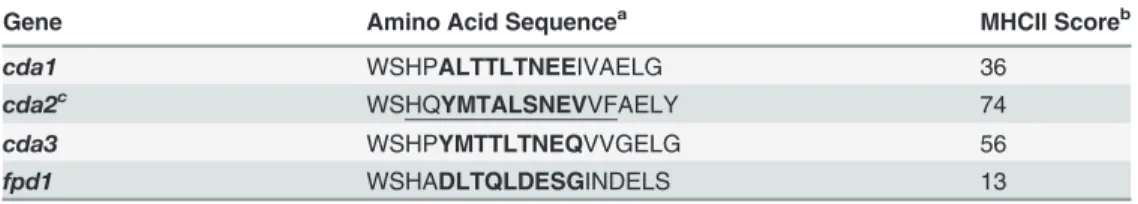
acid peptide from an immunodominant cryptococcal protein, chitin deacetylase 2 (Cda2)
(Table 1) [17]. The Cda2-MHCII tetramer labeled a population of antigen-experienced (i.e.
CD44+) Th cells, but it did not stain non-activated (i.e. CD44−) Th cells fromC. neoformans
infected mice or CD44+ Th cells from naive mice (Figs.1A,S1for flow cytometry gating). In
addition, mice infected with aC. neoformansmutant (cda2Δ) that lacks Cda2 protein
expres-sion [18] had marked reductions in Cda2-MHCII tetramer binding cells (Fig. 1A). Though
Cda2 contains a dominant CD4+ T cell epitope, cross-reactivity to other closely related crypto-coccal proteins likely account for the remaining tetramer binding Th cells generated during
in-fection withcda2Δ(Table 1). Taken together, these studies show the Cda2-MHCII tetramer
reliably identified antigen-specific CD4+ T cells produced in response toC. neoformans
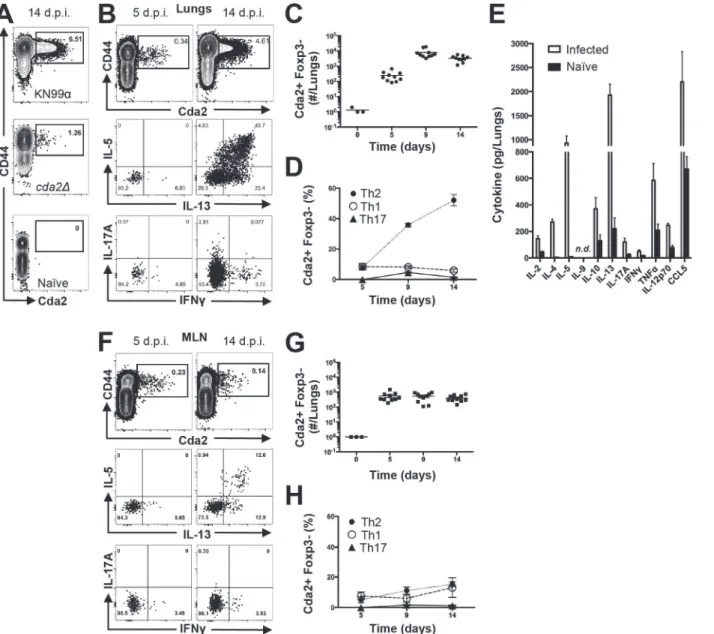
We characterized the immune response in the lung and lung-draining mediastinal lymph node (MLN) to determine the relative contributions of each site to CD4+ T cell subset differen-tiation. Pulmonary cryptococcal infection resulted in a progressive accumulation of Cda2-MHCII-specific T cells in the lungs that predominately expressed the Th2 cytokines IL-5 and/
or IL-13 (Fig. 1B-D). In addition, Th2 cytokines IL-5, IL-13, and CCL5 were among the most
abundant cytokines present in infected lung homogenates (Fig. 1E), and eosinophils, a
down-stream correlate of type-2 cytokines, represented an overwhelming majority of the bulk
leuko-cyte population in the lungs (S2 Fig.for flow cytometry gating,S3A Fig.). In contrast, the
Cda2-MHCII-specific Th2 cell response within the MLN (Fig. 1F-H) was significantly lower
than the response observed in the lungs, and eosinophils comprised an insubstantial
compo-nent of the lymph node resident leukocytes (S3 Fig.). These findings collectively suggest the
local inflammatory environment in the lung may shape the differentiation and/or promote the selective expansion of Th2 cells.
Interleukin-2 cytokine/antibody complexes enhance Th2 cell expansion
and disease
Due to the seemingly contradictory roles of Th2 cells in beneficial wound healing responses
and harmful allergic disease [19], it is not entirely clear whether Th2 cells simply correlate with
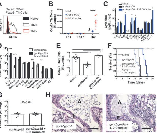
or cause disease associated with fungal infection. To test the causal relationship of Th2 cells with disease severity in this model, we augmented the endogenous Th2 cell response to fungal infection using IL-2 cytokine/antibody complex treatment. IL-2 can be targeted to the high af-finity IL-2 receptor to enhance Th cell proliferation by conjugating IL-2 cytokine with
anti-IL-2 antibody to form IL-anti-IL-2 cytokine/antibody complexes [20,21]. Since Th2 cells generated during
pulmonary cryptococcal infection expressed high levels of the alpha chain of the high affinity
IL-2 receptor (CD25) (Fig. 2A), we sought to use IL-2 complexes to boost the Th2 cell
response.
The wildtype strain ofC. neoformans, KN99α, induces an extremely aggressive infection
that leaves little room to increase the Th2 cell response. Consequently, we used an attenuated
strain ofC. neoformans, gpr4Δgpr5Δ(attenuation explained below/Fig. 5; deficient in
produc-tion of large, chitinous cells). Treatment with IL-2 complexes increased Th2 cell numbers
com-pared with similarly infected mice receiving antibody or cytokine alone (Fig. 2B). Th2 cells and
cytokines were also elevated in lung homogenates from infected mice treated with IL-2
com-plexes (Fig. 2C&D). In addition to Th2 cells, Regulatory T (Treg) cells can be expanded by IL-2
Table 1. CD4+ T cell Cda2-MHCII tetramer peptide sequence and putative cross reactive peptides from otherC. neoformanschitin deacetylases.
Gene Amino Acid Sequencea MHCII Scoreb
cda1 WSHPALTTLTNEEIVAELG 36
cda2c WSHQYMTALSNEVVFAELY 74
cda3 WSHPYMTTLTNEQVVGELG 56
fpd1 WSHADLTQLDESGINDELS 13
Theboldsequences indicate regions that align with the P1-P9 residues within the Cda2-MHCII tetramer core. The underlined sequences correspond to the total peptide included in the Cda2-MHCII tetramer. aHomologous amino acid sequences within the catalytic domain of chitin deacetylases.
bPeptide loading score on MHCII I-AB(74).
cCda2-MHCII tetramer made from this gene and amino acid sequence.
doi:10.1371/journal.ppat.1004701.t001
complex treatment [20,21] (S4A Fig.). This increase of Treg cells in mice treated with the IL-2 complex could theoretically suppress protective Th cell responses and allow Th2 cells to pre-dominate the response. However, IL-2 complex treatment did not affect Th1 or Th17 cell
numbers (Fig. 2B) and only minimal changes in IFNγcytokine (Fig. 2C) and monocyte
accu-mulation (Fig. 2D) were observed, showing IL-2 complex treatment did not eliminate the
effec-tor activity of protective Th1 cells (Fig. 2D). Instead, Schulze et al. [22] showed Treg cells
suppress Th2 cells during cryptococcal infection (S4B–S4C Fig.), suggesting the increase in
Treg cells due to IL-2 complex treatment would actually limit Th2 cell accumulation in this Fig 1. Type-2 Helper T Cells Accumulate in the Lungs of Mice Infected withC. neoformans.(A) Cda2-MHCII tetramer identifiesC. neoformans-specific helper T (Th) cells from mice 14 days post-infection with strain KN99α,cda2Δ, or age-matched, naïve mice. Flow cytometry plot (B) or graphs (C&D) from lung digests showing CD4+, Foxp3-, CD44+ Cda2+ Th cells expressing Th1 (IFNγ), Th2 (IL-5 & IL-13), or Th17 (IL-17A) cytokines. (E) Cytokines from lung homogenates 14 days post-infection with KN99αor age-matched, naïve mice. Flow cytometry plot (F) and graphs (G&H) from mediastinal lymph node suspensions of Th cells expressing Th1, Th2, or Th17 cytokines. Data are presented as mean +/- standard error with 2 independent experiments of at least 5 mice per group. Cda = Chitin deacetylase, CCL = chemokine ligand, IFN = interferon, IL = interleukin, MLN = mediastinal lymph node, TNF = tumor
necrosis factor.
system. Hence, IL-2 complexes can be used to augment the Th2 cell response during pulmo-nary fungal infection and assess the relationship between Th2 cells and fungal disease.
If Th2 cells promote disease, we hypothesized that increasing the Th2 response should
ac-celerate death during infection with the virulence-attenuated strain,gpr4Δgpr5Δ. IL-2 complex
treatment increased the Th2 cell response to levels even higher than the fully virulent KN99α
infection (Fig. 2E). Treatment with IL-2 complexes also greatly reduced the survival time of
in-fected mice (Fig. 2F) without affecting pulmonary fungal burden (Fig. 2G). Uninfected mice
treated with the same regimen of IL-2 complexes survived more than 30 days and remained
healthy (Fig. 2F), indicating the IL-2 complex treatment targeted detrimental cells that were
only present during infection. IL-2 treatment of infected mice also induced obvious lung Fig 2. IL-2 Complexes Augment Type-2 Helper T Cells and Enhance Fungal Disease.(A) CD25 expression by naïve (CD4+, CD44−), Th2- (CD4+, CD44+, IL-5-, IL-13−), and Th2+ (CD4+, CD44+, IL-5+, IL-13+) cells. (B) Cda+ Th cell numbers in the lungs ofgpr4Δgpr5Δinfected mice treated with 5μg IL-2, 25μg anti-IL-2 antibody (JES6-1A12), or both (IL-2 Complex). Th1, Th17, and Th2 cells were identified by production of IFNγ, IL-17A, and IL-5/13, respectively. (C) Cytokines from lung homogenates of mice infected withgpr4Δgpr5Δor age-matched naïve animals, with or without IL-2 complex treatment. (D) Pulmonary leukocytes from mice infected withgpr4Δgpr5Δ, with or without IL-2 complex treatment. (E) Cda2+ Th2 cells from lungs of mice infected with fully virulent KN99α, attenuatedgpr4Δgpr5Δ, or mice infected withgpr4Δgpr5Δand treated with IL-2 complex. (F) Survival of naïve mice or mice infected withgpr4Δgpr5Δeither with or without IL-2 complex treatment.P-value represents log-rank test comparing each survival curve with 10 mice per group to:
gpr4Δgpr5Δvs.gpr4Δgpr5Δ+ IL-2 complex,P<0.0005;gpr4Δgpr5Δvs. KN99α,P<0.0005;gpr4Δgpr5Δ+ IL-2 complex vs. KN99α,P= 0.23. (G) Fungal burden within lungs of mice with or without IL-2 complex 14 days post-infection withgpr4Δgpr5Δ. (H) Hematoxylin and eosin staining of lungs from mice infected withgpr4Δgpr5Δor similarly infected and treated with IL-2 complexes. A = brochial airway, E = epithelial cell layer. Data are presented as the mean +/-standard error with at least 2 independent experiments per group.*=P<0.05,**=P<0.005,***=P<0.0005 by Mann-WhitneyU. Cda = chitin deacetylase, CCL = chemokine ligand, DC = Dendritic Cells, IFN = interferon, IL = interleukin, ILC = Innate Lymphoid Cells, MΦ= Macrophages, NK = Natural Killer. TNF = tumor necrosis factor.
doi:10.1371/journal.ppat.1004701.g002
pathology consistent with increased Th2 activity, noted by increased metaplasia of the
bronchi-olar epithelium and mucous obstruction of the airways (Fig. 2H). En masse, these data indicate
that Th2 cells exacerbate pulmonary disease during fungal infection.
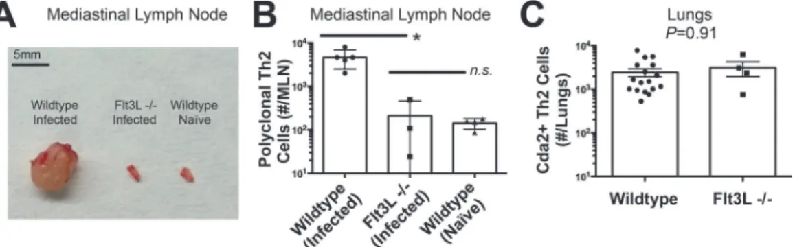
Th2 cells are primed at the site of infection by IRF4-dependent
conventional dendritic cells
The diminished Th2 response in the MLN compared to the lung led us to question whether lymphoid priming was required for Th2 cell induction during pulmonary fungal infection. Fms-like tyrosine kinase 3 ligand (Flt3L) is a differentiation factor for several hematopoietic cell subsets, and genetic deletion of Flt3L causes defects in antigen presenting cell traffic
be-tween the site of infection and secondary lymphoid organs [23]. Flt3L deficient mice infected
withC. neoformansneither experienced mediastinal lymphadenopathy (Fig. 3A) nor elicited a
polyclonal Th2 response in the MLN (Fig. 3B). Surprisingly, the Th2 cell response in the lungs
afterC. neoformansinfection was unaffected by Flt3L deficiency compared to wildtype animals
(Fig. 3C), indicating lymphoid priming is not required for pulmonary Th2 cell accumulation.
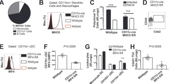
To determine the immune cell intermediate that primes Th2 cells in the lungs, we relied on the fact that Th cells are MHCII restricted. Three leukocyte subsets that express MHCII exist
in the lungs of mice infected withC. neoformans: monocytes, CD11c+ cells, and B cells
(Fig. 4A). Of these, CD11c+ cells are the most abundant in the lungs during cryptococcal
infec-tion (Fig. 4A). Consequently, we interrupted the specific interaction between CD11c+ cells and
Th cells by generating mice with conditional deletion of MHCII in cells that express CD11c
(CD11c-cre MHCII fl/fl) (Fig. 4B). Unlike NOD/SCID/Rag mice that fail to generate mature
Th cells, naïve CD11c-cre MHCII fl/fl mice produced an equivalent number of Th cells as
naïve wildtype mice (Fig. 4C), showing the peripheral Th cell compartment remained intact in
CD11c-cre MHII fl/fl mice. Thus, conditional deletion of MHCII on CD11c+ dendritic cells al-lowed specific disruption of the interaction between the dendritic cells and the Th cells in the periphery. Pulmonary Th cell expansion during cryptococcal infection was completely
abol-ished in CD11c-cre MHCII fl/fl mice (Fig. 4C-D). Consequently, MHCII-bearing CD11c+ cells
prime antigen-specific Th cells in the lungs of mice infected withC. neoformans.
CD11c+ cells are a heterogeneous group of macrophages and several dendritic cells (DC)
subsets in the lungs [24]. Therefore, we sought to discern the specific lineage of the CD11c+
an-tigen presenting cell that is responsible for pulmonary Th2 cell induction using an unbiased forward genetic screen of mouse lines genetically deficient in various CD11c+ subsets or their
ability to interact with Th cells via MHCII (S5A Fig.). Lysozyme M (LysM)-cre MHCII fl/fl
Fig 3. Lymphoid Priming is Dispensible for Pulmonary Th2 Cell Induction during Cryptococcal Infection.(A) Lymphadenopathy of mediastinal lymph nodes (MLN) after 14 days post-infection with strain KN99α. (B) Quantification of IL-5+ IL-13+ polyclonal Th2 cells from the MLN in wild-type and Flt3L−/− mice. (C) Quantification of IL-5+ IL-13+ antigen-specific Th2 cells contained in the lungs of wild-type and Flt3L−/−mice.
(macrophages and granulocytes [25]), BATF3−/−(CD103+ conventional dendritic cells [5]),
and CCR2−/−(monocytes and monocyte-derived dendritic cells [26]) mice generated robust
antigen-specific Th2 responses during cryptococcal infection (S5–S6Fig.). Only mice deficient
in CD11b+ conventional dendritic cells, abrogated using CD11c-cre IRF4 fl/fl mice,
experi-enced blunted Th2 cell accumulation with cryptococcal infection (Fig. 4E-H). Therefore, our
exhaustive search revealed lung-resident CDllc+ CDllb+ IRF4-dependent conventional DC (ferred to as CD11b+ conventional DC) are uniquely required for Th2 cell induction in re-sponse to pulmonary cryptococcal infection.
Fungal chitin correlates with Th2 cell induction and subsequent disease
Existing evidence and our data show CD11b+ conventional dendritic cells are capable ofinduc-ing both Th17 and Th2 cell responses to pulmonary fungal infection [27]; therefore, these DC
are not inherently programmed to specify a single Th cell lineage. Determination of Th2 cell fate by these lung resident DC must require higher order detection of specific features of the fungal infection. Many chitin-containing pathogens, as well as asthma/allergy models using
pu-rified chitin, evoke type-2 immunity [9,16,28]. Consequently, the striking Th2 cell response to
pulmonary fungal infection prompted us to explore the role of chitin as a Th2 cell adjuvant. Maintaining chitin homeostasis is critical for cell wall integrity and microbe vigor. Chitin synthases that regulate cell wall chitin deposition are often essential for fungal viability or are Fig 4. Interferon Regulatory Factor 4-Dependent Conventional Dendritic Cells Coordinate Th2 Cell Induction. (A)Antigen-presenting cells as a proportion of total MHCII+ cells in the lungs of infected mice.(B)MHCII expression by CD11b/c+ cells from mice 14 days post-infection with KN99α. Isotype refers to CD11b/c+ cells from wildtype mice stained with a rat IgG2b antibody of irrelevant specificity.(C)Quantification of pulmonary CD4+ Foxp3- CD44+ pulmonary Th cells.(D)Biexponential plot of CD4+ Foxp3- cells, indicating the absence of antigen-specific (CD44−, Cda2−) cells in the lungs of CD11c-cre MHCII mice infected with KN99α.(E)IRF4 expression by CD11b+ cDC from lungs of CD11cre IRF4 fl/fl or wildtype mice 14 days post-infection with KN99α. Isotype refers to CD11b+ cDC from wildtype mice stained with a rat IgG1 antibody of irrelevant specificity.(F)Geometric mean fluorescence intensity of CD11b+ cDC in from the lungs of infected CD11c-re IRF4 fl/fl mice.(G)Quantification of monocytes and dendritic cell subsets from the lungs of mutant or wildtype mice infected with KN99α.(H)Quantification of IL-5+ IL-13+ antigen-specific Th2 cells contained in the lungs of mutant or wildtype mice 14 days post-infection with KN99α. Data are presented as mean +/- standard error.*=P<0.05,***=P<0.0005, and n.s. = not significant by Mann-WhitneyU. Cda = chitin deacetylase, Flt3L = fms-like tyrosine kinase 3 ligand, MHC = major histocompatibility complex, cDC = conventional dendritic cells, IRF4 = interferon regulatory factor 4.
doi:10.1371/journal.ppat.1004701.g004
part of a redundant pathway [29]. As a result, studies that attempt to correlate loss-of-function mutations in chitin synthesis genes with modulation of the host response and attenuation of virulence would be challenging to interpret. Whether loss of Th2 cells and alterations in disease were due to a decrease in chitin, a loss of cryptococcal fitness/growth, or unmasking of other antigens due to modifications of the fungal cell wall or capsule would not be easily
distinguish-able [30].
In lieu of testing the requirement of chitin in Th2 cell induction, we exploited a natural
property ofC. neoformansto determine if increased fungal chitin was sufficient to expand Th2
cell formation. Approximately 20% of wildtype cryptococcal cells (KN99α) recovered from the
lungs of infected mice increase in diameter from<10μm to 15–100μm [31–33]. Previous
studies have shown these enlarged cells, known as titan cells, exhibit increased thickness of the
fungal cell wall [32]. Using the fluorescent dye calcofluor white to measure chitin content in
in-dividual cells by epifluorescence microscopy (Fig. 5A-B), or at the population level with flow
cytometry (S7A Fig.), we found the largeC. neoformanstitan cells contained more chitin, at a
higher density, than the typical sized cells.C. neoformansproduces several enzymes that
deace-tylate chitin to form chitosan [18,30]. Biochemical analyses additionally revealed that the
amount of chitosan produced by titan cells and typical size cells did not differ, whereas chitin
was significantly more abundant in the titan cells (Fig. 5C). Therefore, enhanced chitin content
accompanied cell size increases during formation of cryptococcal titan cells.
To control for the relative effects of cell size and chitin content on Th2-mediated disease, we
utilized several mutants ofC. neoformans. A strain with targeted deletions in G
protein-cou-pled receptor (gpr) 4 &gpr5 produces>95% typical sized cellsin vivo[34], and these cells
re-tain normal amounts of chitin (Fig. 5A). Deletion of the transcription factor Rim101
(rim101Δ) abolishes titan cell production [34,35], yet the typical sized cells have increased
ex-pression of chitin synthesis genes and elevated chitin content [36,37] (Fig. 5A). Using these
mutants, we were able to dissociate cryptococcal cell size from cell wall chitin as well as
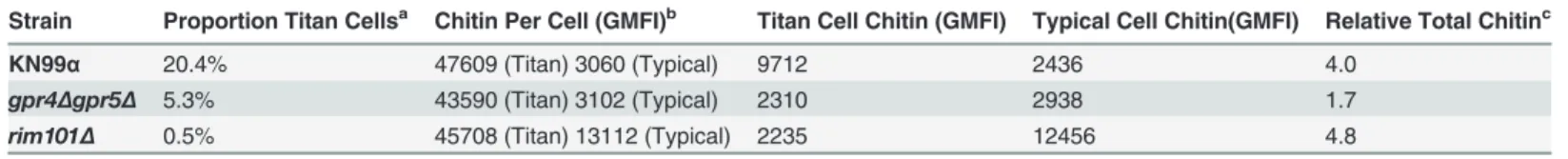
manip-ulate the total amount of chitin present during infection (Table 2).
We examined the impact of alterations in chitin content inC. neoformanson Th2 cell
accu-mulation in the lungs. Antigen-specific Th cell priming and Th2 cell differentiation were
re-duced in mice infected with the low chitingpr4Δgpr5Δstrain compared to infections with both
the high chitin KN99α(due to titan cell production) andrim101Δstrains (Fig. 5D-E). The
Cda2-independent polyclonal Th2 cell response togpr4Δgpr5Δinfection was also significantly
lower than the responses to KN99αandrim101Δinfections (S7B Fig.), indicating the defect in
antigen-specific Th2 cell accumulation togpr4Δgpr5Δinfection was not due to differential
ex-pression ofcda2between the strains. Finally, the secreted Th2 cytokines IL-5, IL-13, and CCL5
present in lung homogenates from mice infected withgpr4Δgpr5Δwere significantly reduced
compared to KN99αandrim101Δinfections (Fig. 5F). Of note, secreted Th1 and Th17
cyto-kines, interferon-γand IL-17A, did not concomitantly increase in response togpr4Δgpr5Δ
in-fection (Fig. 5F). Thus, Th cells not receiving strongly polarizing Th2 signals in thegpr4Δgpr5Δ
infection failed to acquire an alternate Th1 or Th17 cell differentiation fate. Since KN99αand
rim101Δstrains contribute more total chitin to the infection than thegpr4Δgpr5Δstrain
(Table 2,S7A Fig.), these data demonstrate that chitin abundance and not cell size positively
correlated with Th2 cell response intensity.
Uncontrolled factors affected by thegpr4Δgpr5Δorrim101Δmutations could correlate with
chitin levels and independently contribute to Th2 cell accumulation. To directly test if purified
chitin can increase Th2 cell numbers, mice were infected withgpr4Δgpr5Δ, and<10μm chitin
particles were co-administered into the lungs. Chitin treatments partially rescued the
antigen-specific cells, and therefore, chitin increased the potency of Th2 cell induction during C. neoformansinfection.
We examined the correlation between chitin, Th2 cell accumulation, and disease severity in our experimental model of cryptococcal infection. Infections with the high chitin strains, KN99αandrim101Δ, significantly hastened the time to death relative to mice infected with
gpr4Δgpr5Δ(Fig. 5H). Interestingly, all strains had equivalent pulmonary colony forming units
at 14-days post-infection, indicating the differences in disease were not simply due to a failure
to control the infection (Fig. 5I), but rather paralleled the total amount of chitin present during
infection (Table 2). In summary, both chitin production byC. neoformansand Th2 cell
accu-mulation directly correlated with exacerbation of lethal fungal disease.
Fig 5.neoformansChitin Correlates with Type-2 Helper T Cell Response and Subsequent Disease.(A&B) Cryptococcal cells isolated from lungs at 14 days post-infection and stained with Calcofluor White. Cells were either imaged with epifluorescence microscopy (A) or analyzed by flow cytometry with fluorescence intensity calculated per pixel to determine chitin density within the cell wall (B). (C) Total chitin and chitosan content normalized to dry weight of cryptococccal cells. (D) Flow cytometry plots of CD4+, Foxp3-, CD44+ Cda2+ Th cells expressing Th2 cytokines, IL-5 & IL-13, at 14 days post-infection and (E) the quantification of these plots. (F) Cytokines from lung homogenates of mice 14 days post-infection. (G) IL-5+ IL-13+ antigen-specific Th2 cells from lungs of mice 14 days post-infection without and with intranasal chitin particle treatment. (H)P-value represents log-rank test comparing each survival curve with 10 mice per group to:gpr4Δgpr5Δvs. KN99α, P<0.0005;gpr4Δgpr5Δvs.rim101Δ, P<0.0005; KN99αvs.rim101Δ, P<0.0005. (I) Fungal burden in the lungs at 14 days post-infection. Data are presented as the mean +/- standard error with at least 2 independent experiments per group.**=P<0.005,***=
P<0.0005 by Mann-WhitneyUor Kruskal Wallis ANOVA. Cda = chitin deacetylase, CCL = chemokine ligand, GlcNAc =N-acetylglucosamine, IFN = interferon, IL = interleukin.
doi:10.1371/journal.ppat.1004701.g005
Chitotriosidase deficiency confers resistance to Th2-mediated fungal
disease
We next investigated host intrinsic factors that could influence detrimental Th2 cell responses
to chitin—specifically the mammalian chitinases, AMCase and Chit1. pH-sensitive differences
in enzyme activity allow for an assessment of the contribution of each enzyme to chitin cleav-age. Consistent with published reports, recombinant AMCase cleaved 4-methlylumbelliferone chitotriose across a broad pH range, whereas recombinant Chit1 was only active at less acidic
pH (Fig. 6A) [38]. Chitinase activity in lung homogenates from infected mice was significantly
elevated compared with uninfected animals at pH 2 and pH 5 (Fig. 6A). Furthermore, lung
homogenates from infected mice genetically deficient in Chit1 or AMCase both showed
de-creased cleavage of the fluorescent chitin substrate (Fig. 6A). These data indicate both chitinase
enzymes are active during pulmonary fungal infection, and genetic abolishment of these en-zymes can be used to understand the effect of chitin degradation on Th2-mediated fungal pathogenesis.
To test the hypothesis that Th2 cell-associated disease depends on chitinases, we infected
wildtype, Chit1−/−, and AMCase−/−mice withC. neoformansand quantified the Th2 cell
re-sponse. Despite no differences in pulmonary fungal burden at 14-days post-infection (S8A
Fig.), Th2 cells were 10-fold less abundant in the lungs of infected Chit1−/−mice compared
with wildtype controls (Fig. 6B-C), and this trend was consistent with all the strains ofC.
neo-formanstested (S8B Fig.). Conversely, AMCase deficiency did not impact Th2 cell quantities
after cryptococcal infection (Fig. 6B-C). Furthermore, Chit1 deficiency and not AMCase
defi-ciency also significantly extended the survival of mice infected withC. neoformansrelative to
age matched, wildtype animals (Fig. 6D-E). The loss of Th2 cell accumulation with Chit1
defi-ciency was responsible for attenuation of disease, because the use of IL-2 complexes to boost
Th2 cell numbers (Fig. 6C) and associated cytokines (Fig. 6F) also hastened lethal disease in
Chit1−/−mice compared with infection-matched, untreated Chit1−/−controls (Fig. 6D).
Simi-lar to our previous studies using the IL-2 complex, Th1 cytokine production in the Chit1−/−
mice was only minimally affected by the IL-2 complex treatment (Fig. 6F). Thus, the presence
of Chit1, and not AMCase, positively influences Th2 induction and subsequent disease.
Digestion of chitin via chitotriosidase promotes Th2 cell accumulation
Chitin receptors in plants bind chitin oligomers [39], and chitin polymer size also influences
the mammalian immune response to chitin [40,41]. Since appropriately sized chitin fragments
could result from chitin digestion by Chit1, we used heterogeneous-length chitin and highly purified chitin heptamers to understand the effect of Chit1-associated degradation of chitin on Table 2. Differences in total chitin due to cell morphology.
Strain Proportion Titan Cellsa Chitin Per Cell (GMFI)b Titan Cell Chitin (GMFI) Typical Cell Chitin(GMFI) Relative Total Chitinc
KN99α 20.4% 47609 (Titan) 3060 (Typical) 9712 2436 4.0
gpr4Δgpr5Δ 5.3% 43590 (Titan) 3102 (Typical) 2310 2938 1.7
rim101Δ 0.5% 45708 (Titan) 13112 (Typical) 2235 12456 4.8
aProportion of cryptococcal titan cells in the lungs, as reported by Okagaki et al. 2011[34]. bMean
fluorescence intesity of calcofluor white in size-fractionated cryptococcal cells analyzed byflow cytometry. GMFI = geometric mean
fluorescence intensity
cEstimated total chitin calculated uising the folowing equation: Relative Total Chitin = [(Proportion Titan Cells × Titan GMFI) + (Proportion Typical Cells × Typical GMFI)]/KN99αTypical Cell GMFI
Th2 cell accumulation. Although treatment with heterogeneous-length chitin augmented Th2
cell induction in wildtype animals infected withgpr4Δgpr5Δcells (Fig. 5F), it did not increase
Th2 cell numbers in Chit1−/−mice (Fig. 6G). Conversely, inoculation with mass equivalent
amounts of chitin heptamers boosted the Th2 cell response inC. neoformans-infected animals
with Chit1 deficiency (Fig. 6G), revealing the requirement for Chit1 in chitin polymer
Fig 6. Chitotriosidase Promotes Chitin Recognition and Th2 Cell-mediated Disease.(A) Chitinase enzyme activity of recombinant enzymes or lung homogenates from 14 days post-infected or naïve mice. (B) Flow cytometry plots of CD4+, Foxp3-, CD44+ Cda2+ Th cells expressing Th2 cytokines, IL-5 & IL-13, at 14 days post-infection and (C) the quantification of these plots. (D&E) Survival of mutant or wildtype mice infected with KN99αeither with or without IL-2 complex treatment. Logrank test comparing each survival curve relative with 10 mice for each group to: Chit1−/−vs. wildtype,P<0.005; Chit1−/−vs. Chit1−/−+ IL-2 complex,P<0.0005; Chit1−/−+ IL-2 complex vs. wildtype,P= 0.07. (F) Cytokines from lung homogenates of naïve or wildtype and Chit1−/− mice 14 days post-infection with KN99αor age-matched, naïve Chit1
−/−mice. (G) IL-5+ IL-13+ antigen-specific Th2 cells from lungs of Chit1−/−mice 14 days post-infection and mice treated with intranasal chitin particles or chitin heptamer fragments (C7). (H) Chitinase enzyme activity of human plasma without (LEFT) or with (RIGHT) cryptococcal lysate antigen stimulation. Data are presented as the mean +/- standard error with at least 2 independent experiments per group.*=P<0.05,**=P<0.005,***=P<0.0005 by Mann-WhitneyUor Kruskal Wallis ANOVA. AMCase = Acidic Mammalian Chitinase, C7 = chitin heptamer, CCL = chemokine ligand, Cda = chitin deacetylase, Chit1 = Chitotriosidase, IFN = interferon, IL = interleukin, TNF = tumor necrosis factor.
doi:10.1371/journal.ppat.1004701.g006
recognition can be bypassed by providing exogenous chitin fragments. Therefore, our data demonstrate a role for Chit1 in the chitin cleavage pathway that leads to Th2
cell accumulation.
We next examined how chitin fragments influence the upstream pathway of Th2 cell induc-tion. To test the hypothesis that DC interact directly with chitin fragments, we cultured prima-ry pulmonaprima-ry leukocytes from infected mice in the presence of R-phycoerithrin-fluorophore conjugated chitin heptamers (GN7) or unbound REP-streptavidin (SA). While
RPE-GN7 labeled a subset of B cells that have been shown to bind chitin [9], RPE-GN7 did not
ad-here to conventional CD11b+ DC (S9A Fig.). Thus, we did not detect direct binding of chitin
heptamers to the conventional DC. Furthermore, conventional CD11b+ DC stimulatedex vivo
with PMA + ionomycin produced an important Th2 cell differentiation cytokine, IL-4, yet this
DC subset did not express IL-4 upon stimulation with chitin heptamers (GN7) (S9B Fig.).
Combined, these data suggest the CD11b+ conventional dendritic cells may not sense chitin levels directly.
Pulmonary epithelial cells respond to chitin [42] and secrete several Th2-inducing alarmins,
thymic-stromal lymphopoietin (TSLP), IL-25, and IL-33 [28,43,44]. As a result, alarmin
recep-tors on DC could potentially mediate the indirect recognition of chitin, leading to Th2 cell po-larization during pulmonary fungal infection. We found CD11b+ conventional DC from the lungs of fungal infected mice expressed high levels of TSLP receptor, but not receptors for
IL-25 or IL-33 (S9C–S9D Fig.), indicating this DC subset is capable of sensing TSLP generated by
epithelial cells. Taken together, our data suggest Th2 cell induction by conventional CD11b+ DC appears to involve an indirect recognition of chitin oligomers.
Chitotriosidase activity correlates with cryptococcal infection in humans
The importance of Chit1 in promoting Th2 cell-mediated disease in an experimental model of cryptococcosis prompted us to investigate the relevance of chitotriosidase activity in human fungal disease. Blood samples were collected from human donors: 46 Ugandan patients pre-senting at the hospital for the first time with AIDS and 38 similar AIDS patients experiencingacute cryptococcal disease (S1 Table). We analyzed chitotriosidase and AMCase enzymatic
ac-tivity in plasma from each group as described for the mouse lung homogenates. Chitin sub-strate cleavage at pH = 5 was significantly elevated in plasma from AIDS patients with cryptococcal infection when compared to AIDS patients without cryptococcal infection
(Fig. 6H). Comparatively low levels of enzymatic activity were detected at pH = 2 for each
group, indicating that chitotriosidase and not acidic mammalian chitinase is the predominate
chitinase produced by humans with cryptococcal infection (Fig. 6H).
To determine if the difference in chitotriosidase activity was due to an inherent propensity or deficiency in chitotriosidase expression, we used cryptococcal lysate antigens to stimulate chitinase production in whole blood samples from the human donors. Stimulation of whole blood from all patients induced robust chitotriosidase activity relative to unstimulated samples,
and chitinase activity did not differ between the groups (Fig. 6H), indicating all human donors
had equivalent capacity to produce chitotriosidase. Chitinase activity was not detectable in
pure cryptococcal culture supernatants or cryptococcal lysate antigens (S10 Fig.), and as a
re-sult, the chitinase activity detected in the assays with human samples was not due to
Discussion
An association between pulmonary fungal exposure and allergic Th2 inflammation is well
es-tablished [1,3]. Fungal proteases [45] and fungal chitin [46] impact the innate immune
re-sponses underlying allergic inflammation, but elements of Th2 cell induction are enigmatic. Using an experimental model of pulmonary cryptococcosis, we demonstrated that inhalation ofC. neoformansestablishes a robust pulmonary infection, and the potent antigen-specific Th2 cell accumulation required lung resident CD11b+ conventional DC. Since these CD11b+
con-ventional DC can stimulate Th2 or Th17 cell differentiation in response toC. neoformansand
Aspergillus fumigatusexposure respectively [27], these DC must interpret specific features of the infection to direct Th2 cell fate. To this end, we found cryptococcal chitin and exogenous administration of chitin particles correlated with increased Th2 cell accumulation. We further
showed that chitotriosidase activity was highest in mice and humans infected withC.
neofor-mans, and Chit1 was necessary for efficient Th2 cell induction and disease in our murine model of cryptococcosis. Taken together, these data indicate the host response to fungal chitin is an important factor that enhances Th2 cell production during pulmonary fungal infection.
Our findings narrow an important gap in the mechanism of pattern recognition of fungal
chitin in the lungs (Fig. 7). We have shown Chit1 functions as a“gatekeeper”in making chitin
fragments available to host surveillance, thereby promoting Th2 cell accumulation and disease. Fig 7. Model of Th2 Cell-mediated Disease during Pulmonary Fungal Infection. 1) Chitotriosidase is released by the host and degrades fungal chitin to generate small chitin fragments, such as chitin heptamers.
2) Chitin recognition occurs by an unknown mechanism that could involve either epithelial cell production of alarmins, such as thymic stromal lymphopoietin (TLSP) [9] or antibodies that bind chitin [9].3) These chitin-based signals are recognized by lung-resident CD11b+ conventional dendritic cells that are capable of producing IL-4, an essential Th2 cell differentiation factor.4) The adaptive Th2 cell response results in enhanced disease in the absence of significant changes in pulmonary fungal burden.
doi:10.1371/journal.ppat.1004701.g007
Additionally, the use of pharmaceutical grade chitin heptamers to augment Th2 cell responses establishes a new minimum component for chitin recognition in vertebrates. While we did not
detect direct interactions between the CD11b+ conventional DC and labeled chitinex vivo,
other systems have shown chitin binding to the mannose receptor and subsequent recognition
by TLR9 and/or NOD2 [47]. Alternatively, pulmonary allergy models demonstrate that lung
epithelial cells recognize chitin fragments and produce the necessary alarmins for Th2 cell
in-duction: TSLP, IL-25, and IL-33 [43,44,48]. In our model, CD11b+ conventional DC express
high levels of TSLPR. Finally, natural IgM has been shown to bind fungal carbohydrates,
in-cluding chitin, and facilitate the interaction of DC and fungal carbohydrates [9]. Signals, such
as alarmins or antibody-chitin complexes, could be received by CD11b+ conventional DC to direct Th2 cell differentiation via IL-4 or another novel pathway.
A major impediment in understanding Th cell responses to pulmonary fungal infection has been the lack of reagents to detect antigen-specific Th cells. Antigen-specific reagents are par-ticularly important when examining Th2 responses, because Th2 cells can be induced by the wounding that occurs during infection. Our ability to track endogenously derived,
antigen-spe-cific Th cells with pMHCII tetramers [49] allowed us to present for the first time that Th2 cells
produced in response to fungal pathogens are not part of a generalized wound healing response but are fungal-antigen specific. Unlike T cell receptor transgenic approaches, pMHCII tetra-mers permitted us to monitor the population of infection-specific Th cells in the polyclonal repertoire, while maintaining physiologic precursor frequency and clonal expansion. Thus, we are able to examine the Th cell response during the natural course of infection and keep all other variables constant. The availability of these pMHCII tetramers will undoubtedly empow-er cryptococcal researchempow-ers and accelempow-erate the field of fungal immunology.
The use of IL-2 complexes allowed us to conveniently and reliably augment the Th2 cell re-sponse to further understand unappreciated elements underlying Th2-mediated disease. This strategy is amenable to any host or microbial genetic model, which facilitates direct compari-sons. Also, this gain-of-function approach permitted us to test the sufficiency of Th2 cells to
ex-acerbate disease. These data, combined with loss-of-function studies by other groups [50,51],
alter the longstanding paradigm that susceptibility to lethal fungal disease is traditionally viewed as a breakdown in protective immunity. This paradigm is supported by the higher prev-alence of invasive fungal disease in immunosuppressed individuals, including people living with HIV/AIDS, cancer patients undergoing chemotherapy, and solid organ transplant recipi-ents. However, we propose that in addition to the lack of a protective response, an independent development of a harmful Th2 cell response further exacerbates disease. This is particularly im-portant in the the case of human cryptococcocosis were a compromised immune system not only lacks sufficient quantities of lymphocytes to resolve the fungal infection, but the residual
Th cell repertoire is plastic and detrimentally influenced by the microbe [52–55].
A subset of innate lymphoid cells (ILC) produce the Th2 cytokines, IL-5 and IL-13 [56], and
these so-called ILC2 have been shown to contribute to allergic airway disease [4]. While IL-2
complex treatment dramatically increases Th2 cell accumulation and enhances pulmonary dis-ease, ILC numbers are not affected by IL-2 complex treatment in our model. Likewise, CD25+
ILC2 exist in the lungs under homeostatic conditions [57], yet uninfected mice exhibit no ill
ef-fects of IL-2 complex treatment. However, the developmental relationship of ILC2 to lympho-cytes, combined with the lack of lineage markers expressed by ILC, make it challenging to separate the relative effects of Th2 cells and ILC2 in driving immunopathology. Thus, our con-clusion that Th2 cells are vital mediators of disease does not categorically exclude the participa-tion of ILC2 in this process.
CD11b+ conventional DC are an ontologically distinct mononuclear phagocyte subset that
functionally unique in programing specific Th cell differentiation, building evidence seems to indicate a plastic role of these cells in Th cell induction. CD11b+ conventional IRF4-dependent DC have been shown to coordinate Th2 cell priming following protease inoculation and worm
infection in the skin [58], as well as house dust mite extract installation in the lungs [59].
How-ever, several research groups have found the same DC subset, using identical host genetic
sys-tems, can control Th17 cell differentiation in the gut under homeostatic conditions [60] and
the lungs after fungal infection [27]. While these observations infer that CD11b+ conventional
DC are plastic, these cells may have different functions in the skin and gut compared to the lungs that could explain their role in priming Th17 and Th2 cell responses. Our findings
show-ing Th2 primshow-ing byC. neoformans, combined with work by Schlitzer et al. showing
IRF4-de-pendent DC are capable of priming Th17 cells in response toA. fumigatusto pulmonary
fungal infection, highlight a fatal flaw in the notion that DC subsets are inherently specialized to control Th cell lineage fate. As a result, we explored an alternative hypothesis that higher order signals (e.g. chitin recognition) are required for pulmonary CD11b+ conventional DC to promote Th2 cell priming to fungal infection.
Gene deletions in microbes can cause pleiotropic phenotypes, as seen in therim101Δstrain
(35) that can complicate interpretation of the effects of these mutations on host responses. However, the data obtained from the mutants utilized in this study support the conclusion that fungal chitin promotes Th2-mediated disease for several reasons. First, equivalent fungal bur-den in the lungs at 14 days post-infection indicates these mutations do not confer an inherent survival advantage or disadvantage for the fungus, and any effect the mutants have on leuko-cyte accumulation or disease is not simply driven by antigen load. Second, Rim101 transcrip-tionally controls many elements associated with cryptococcal virulence, including pH
responses, encapsulation, cell enlargement, and iron sequestration [61]. While individual
mu-tations in each of these pathways should result in a loss of virulence, therim101Δmutants
par-adoxically exhibit equivalent or accelerated disease [36]. Altered expression of cell wall
synthesis genes combined with unmasking of the cell wall has been posited [36,37], and in
par-ticular, our data implicate cell wall chitin in enhancing fungal pathogenesis associated with rim101deficiency. More importantly, we viewrim101Δas an essential control for the effect cryptococcal cell size has on Th2 cell formation more than an independent test of the
hypothe-sis that fungal chitin drives Th2 cell priming. Thus, we needed a strain ofC. neoformansthat
produces mostly small cells but retained elevated chitin density to offset the loss of elevated chi-tin density with titan cell deficiency. At a minimum, these data prove that large cryptococcal cell size is not driving robust Th2 cell accumulation. Finally, due to the structural similarities, as well as the interdependent synthetic pathways, it is extremely challenging to decouple how chitin and chitosan separately impact a complex biological system. Although we cannot rule
out immunomodulatory effects of chitosan in our experiments withC. neoformansmutants,
we have confirmed chitin alone, when provided as an adjuvant, is sufficient to augment Th2
cell accumulation. Interpretation of theCryptococcuschitin mutant data in the context of our
additional data regarding host recognition of exogenous chitin builds a compelling argument that fungal chitin promotes detrimental Th2 cell induction.
Acidic mammalian chitinase has been previously implicated in innate type-2 responses
[62]. However, a head-to-head comparison of the effect of both mammalian chitinases,
chito-triosidase and acidic mammalian chitinase, has never been performed in any model, much less in the context of Th2 cell responses to fungal infection. Surprisingly, chitotriosidase, and not acidic mammalian chitinase, influenced Th2 cell accumulation and disease during pulmonary fungal infection. While no direct explanation exists, the different patterns of expression of AMCase and Chit1 could explain our results. AMCase is produced by a number of cells includ-ing several leukocyte subsets and pulmonary epithelial cells, whereas Chit1 expression is
restricted to mononuclear phagocytes, including DC [38,63]. Since these DC are the main coor-dinators of antigen presentation to CD4+ T cells, the close proximity of chitin degradation and recognition likely allows the DC to efficiently influence the fate of CD4+ T cells during pulmo-nary fungal infection. Furthermore, the varying pH-dependent enzymatic activity of Chit1 and AMCase suggest these enzymes may function in disparate anatomical and subcellular compart-ments that may bias their participation in response to pulmonary fungal infection.
Considering Th2 cell responses likely evolved to resist parasitic infection [10], the
asymmet-ric global distribution ofCHIT1alleles [64] combined with the data presented herein offers a
unique perspective on why individuals from tropical regions with endemic parasites tend to
ex-perience frequent and severe mycosis [65–69]. Ethnic groups historically residing in regions
with highly prevalent parasites likeStrongyloidestend to maintain functionalCHIT1, whereas
populations from more temperate climates with lower endemic parasitic burdens frequently
possess enzymatically inactiveCHIT1alleles [64]. Perhaps continuous fungal exposure in the
absence of parasitic encounters provides sufficient negative selection pressure to eliminate
functionalCHIT1alleles from these populations (e.g. Europe). Conversely, ethnic groups
his-torically residing in tropical areas (e.g. Africa) that maintain functionalCHIT1alleles may have
enhanced protection against common parasitic exposures, while these same individuals
experi-ence exacerbation of Th2-mediated fungal disease [67–69].
In conclusion, we elucidated a novel mechanism of Th2 cell induction during fungal infec-tion. CD11b+ DC, and importantly recognition of chitin via Chit1, drive the generation of dele-terious Th2 cells responding to pulmonary fungal infection. Next generation anti-fungal treatments should not only block fungal growth, but should also target the host immune
re-sponse. A recent trial used IFNγin combination with traditional anti-fungal therapy to
pro-mote beneficial immune responses. This treatment improved cryptococcal clearance, yet it had
no significant impact on patient survival [70]. Our study suggests treatments that additionally
aim to suppress the pathologic Th2 response, perhaps through chitotriosidase inhibition, may be necessary to improve clinical outcomes. Ultimately, the coordinated efforts of microbiolo-gists, immunologists and infectious disease physicians will enable personalized medicine ap-proaches that effectively combat lethal fungal infections by inhibiting fungal growth, promoting beneficial host responses, and dampening pathologic inflammation.
Materials and Methods
Ethics statement
This study was approved by the institutional review boards of the University of Minnesota, Makerere University, and the Uganda National Council of Science and Technology. Written informed consent was obtained from all human participants prior to inclusion in the sutdy,
and all data were de-identified [71]. All animal experiments were done in concordance with
the Animal Welfare Act, U.S. federal law, and NIH guidelines. Mice were handled in accor-dance with guidelines defined by the University of Minnesota Institutional Animal Care and Use Committee (IACUC) protocol numbers 1010A91133 and 1207A17286 and University of Massachuesets IACUC protocol number A-1802.
Mice
All mice used in this study were derived from a C57BL/6 background. C57BL/6J, LysM-cre
(B6.129P2-Lyz2tm1(cre)Ifo/J), CD11c-cre (B6.Cg-Tg (Itgax-cre) 1-1Reiz/J), MHCIIloxP
(B6.129X1-H2-Ab1tm1Koni/J), Batf3−/−(B6.129S(C)-Batf3tm1Kmm/J), CCR2−/−
(B6.129(Cg)-Ccr2tm2.1Ifc/J), B6.129(Cg)-Foxp3tm3(DTR/GFP)Ayr/J) mice were purchased from Jackson
(Hudson, NY). Crosses were performed when necessary to generate the mouse strains used in
this study, as indicated inS2 Table. Chit1−/−[72] mice were infected and processed in the
lab-oratory of Kirsten Nielsen. AMCase−/−[73] mice were infected and processed in the
laborato-ry of Stuart Levitz per MTA stipulations. All mice were housed in specific pathogen–
free conditions.
Cryptococcus
Cryptococcus neoformansvar.grubiistrains were streaked on yeast peptone dextrose (YPD) agar plates and incubated for 2 days at 30°C. YPD broth was inoculated with colonies from the aforementioned plate and incubated for 16 hours at 30°C with gentle agitation. The inoculum was prepared by pelleting the culture, washing 3 times with phosphate buffered saline (PBS),
and resuspending in PBS at a concentration of 2x10⁶cells/mL. All strains used in this study
were on a KN99αgenetic background, and the complemented strains have wildtype
pheno-types [18,34,35].
Infection
A well established intranasal pulmonary aspiration model of cryptococcosis was used for this
study [74]. 6–8 week old, sex-matched mice were anesthetized with pentobarbitol or isoflurane.
5x10⁴cryptococcal cells in 25μL of PBS was placed on the nares of each mouse, and the mice
aspirated the inoculum into the lower respiratory tract. Finally, the mice were suspended by their incisors for 5 minutes and subsequently placed upright in their cage until regaining con-sciousness. For survival studies, ten mice per group were infected as described above. Animals were monitored for morbidity and sacrificed when endpoint criteria were reached. Endpoint criteria were defined as 20% total body weight loss, loss of 2 grams of weight in 2 days, or symp-toms of neurological disease.
Tetramer production
Nine amino acid peptides from Cda2 were selected using a MHCII loading algorithm [75].
pMHCII tetramers were produced as previously described [76]. In short, biotinylated pMHCII
monomers were expressed inDrosophila melanogasterS2 cells and isolated from culture
super-natants by affinity chromatography. Streptavidin-Phycoerythrin (Prozyme) was added to pMHCII monomers at a 4:1 molar ratio. Finally, tetramer formation was assessed by western blot analysis.
Pulmonary leukocyte preparation
Lung leukocytes were isolated as previously described [77]. Briefly, lungs were excised and
minced to generate approximately 1 mm3pieces. The lung mince was incubated in HBSS
(Invi-trogen, Grand Island, NY) + 1.3 mM EDTA solution for 30 min at 37°C with agitation, and then transferred to RPMI-1640 (Invitrogen) medium supplemented with 5% Fetal Bovine Serum (FBS) (Invitrogen) and 150 U/ml type I collagenase (Invitrogen) and incubated for 1 h
at 37°C with agitation. The cells were passed through a 70μm filter, pelleted, and resuspended
in 44% Percoll-RPMI medium (GE Life Sciences, Pittsburgh, PA). A Percoll density gradient
was created (44% top, 67% bottom), and the samples were centrifuged for 20 min at 650 xg.
The leukocytes at the interface were removed, washed 2 times with RPMI medium, and
resus-pended in PBS + FBS at a concentration of 107cells/ml. CD4+ T cells were enriched using a
Dynabeads CD4+ T Cell Negative Isolation Kit (Life Technologies, Grand Island, NY) per
manufacture’s instructions. After enrichment,*106cells were suspended in 200μL of
restimulation buffer (RPMI + 10% FBS + 1% Penicillin/Streptomycin + 5μg Brefeldin A) with
(stimulated) or without (unstimulated) 10 ng phorbol myristate acetate (PMA) and 50 ng iono-mycin. After 6 hours, the cells were washed and immediately prepared for flow cytometry.
IL-2 complexes
5μg of murine IL-2 cytokine (Biolegend) and 25μg of clone JES6-1A12 anti-IL-2 antibody
(Bio X Cell, West Lebanon, NH) were added to 100μL of PBS at room temperature. Each
mouse received intraperitoneal injections of IL-2 complexes every other day beginning at 5 days post-infection.
Flow cytometry
Samples were incubated for 5 minutes with CD16/32 antibody (Biolegend) to block the Fc re-ceptor and prevent nonspecific antibody binding. 25nM Cda2-tetramer was added to the sam-ple and incubated at 25°C for 1 hour in the dark. Samsam-ples were surface-stained at 4°C for 30
minutes with the following antibodies (see gatingS1 Fig.): CD4 (RM4-5, BV605, Biolegend),
CD8 (53-6.7, APC-eFluor 780, eBioscience, San Diego, CA), CD11b (M1/70, PE-Cy5,
eBioscience), CD11c (N418, PE-Cy5, eBioscience), B220 (RA3-6B2, PE-Cy5, eBioscience), and CD44 (IM7, Alexa Fluor 700, Biolegend). The cells were then incubated in Foxp3 Transcription Factor Buffer (eBioscience) at 4°C for 30 minutes. The cells were labeled with antibodes against the following intracellular antigens: Foxp3 (FJK-16s, FITC, eBioscience), IL-5 (TRFK5, APC, Biolegend), IL-13 (eBio13A, eFluor 450, eBioscience), IL-17A (TC11-18H10.1, BV650,
Biole-gend), and IFNγ(XMG1.2, PE-Cy7, eBioscience). 1:200 antibody concentrations were used for
surface staining, and 1:100 antibody concentrations were used for intracelluar staining. For
data acquisition, events from the entire sample (500,000–1,000,000) were collected on a BD
FACSCanto II flow cytometer (BD Biosciences, San Jose, CA), and the data were analyzed with FlowJo X (Tree Star Inc., Ashland, OR).
To account for cell loss during CD4+ T cell enrichment and mitogen restimulation, several calculations were performed. Total leukocyte numbers were determined by hemacytometer count after Percoll density gradient separation. 2.5% of the sample was stained with the follow-ing antibodies: Ly6G (RB6-8C5, APC-eFluor 780, eBioscience), Ly6C (HK1.4, eFluor 450, eBioscience), CD11b (M1/70, BV650, Biolegend), CD11c (N418, BV605, Biolegend), NK1.1 (PK136, AF700, Biolegend), CD3 (17A2, PE-Cy5, Biolegend), CD4 (RM4-5, FITC, Biolegend), CD19 (6D5, PE-Cy7, Biolegend), Sca1 (D7, APC, Biolegend), and Siglec F (E50-2440, PE, BD
Biosciences). Cells were identified as the following (see gatingS2 Fig.): Th cells =
Dendritic cell subsets were determined using the following antibodies and gating strategy. CD3 (17A2, PE-Cy5, Biolegend), CD19 (6D5, PE-Cy5, Biolegend), Siglec F (E50-2440, APC, BD Biosciences), CD64 (X54-5/7.1, PE, Biolegend), MHCII (M5/114.15.2, AF700, Biolegend), CD11c (N418, BV605, Biolegend), CD11b (M1/70, BV650, Biolegend), CD103 (2E7, e450,
eBioscience), FcεRI (MAR-1, PE-Cy7, Biolegend), TSLPR (PE, R&D Systems), IL-25R (9B10,
PE, Biolegend), IL-33Rα(DIH9, PE, Biolegend), IL-4 (BVD6-24G2), PE, Biolegend), and
GN7-PE [78]. See gatingS5B Fig.Dump = CD3+, CD19+, Siglec F+. Monocyte-derived DC =
Dump-, CD64+, CD11c+, MHCII+, CD11b+, CD103-, FcεRI+. CD103+ cDC = Dump-,
CD64-, CD11c+, MHCII+, CD103+, CD11b-. CD11b+ cDC = Dump-, CD64-, CD11c+,
MHCII+, CD11b+, CD103-. While CD103, CD11b, FcεRI, and CD64 surface markers allow
convenient detection of each DC subset, it is unclear whether other developmentally and/or functionally unrelated cells can express similar markers. Consequently, genetic blockade in the developmental pathways of each DC subset were assumed to be completely penetrant, notwith-standing the persistence of cells expressing surrogate markers for each ontological subset.
Lung cytokines
Lungs from mice 14-days post-infection were excised, snap frozen in liquid nitrogen, and ho-mogenized in 3 mL of T-PER (Thermo Fisher Scientific) with Complete Protease Inhibitor Cocktail (Roche, Indianapolis, IN). The lung homogenate was pelleted, and the supernatant was collected and stored at 80°C until analysis. Samples were diluted 1:4 in assay buffer imme-diately before processing. Cytokines were quantified using Luminex technology according to manufacturer instructions (Bio-Rad, Hercules, CA).
Lung histology
Lungs were removed from mice 14 days post-infection, perfused via the right ventricle with cold PBS, inflated with 10% formalin (Thermo Fisher Scientific, Rockford, IL), and placed in a container of 10% formalin. Tissues were dried with organic solvent, embedded in parafin, sec-tioned, and stained with hematoxylin and eosin, before images were captured.
Fungal chitin/chitosan quantification
Cryptococcal cells were isolated from the lungs after enzymatic digestion and density gradient separation, as described above. Cells were fixed with 3.7% formaldehyde and stored at 4°C
until analyzed. The cells were standardized to 1x10⁶cells/mL and stained for 2 minutes at 25°C
with 1μg/mL of Calcofluor White (Sigma Aldrich, St. Louis, MO) [79]. Cells were washed and
immediately processed with an epifluorescence microscope (Axio Imager M1, 40X/0.6 lens, Zeiss Filter Set 02, Axio Cam MRc5, Axiovision 4.8; Carl Zeiss, Inc., Munich, Germany).
Ima-geJ software (NIH.gov) was used to calculate fluorescence intesity per pixel. For flow
cytome-try, large and typical sized cells were first gated by forward scatter properties to distinguish size. Chitin/chitosan content was then determined by 405 nm laser excitation and fluorescence
detection at*450 nm.
Biochemical chitin/chitosan quantification was adapted from Banks et al. [30]. Purified
titan cell (>15μm) and typical sized cell (<15μm) samples collected from infected mice were
each divided into two aliquots: one treated with acetic anhydride to fully acetylate the chitin/
chitosan polymer and the other was left untreated. 5μl of purifiedStreptomyces griseus
chiti-nase (5 mg/ml in PBS) was added to hydrolyze chitin toN-acetylglucosamine (GlcNAc) and
samples were incubated for 3 days at 37°C. For colorimetric determination of GlcNAc, the Morgan-Elson method was adapted for microplate readers. Chitinase-treated samples were in-cubated with 0.27 M sodium borate (pH 9.0) and heated at 99.9°C for 10 minutes. Immediately
upon cooling to room temperature, freshly diluted 10X DMAB solution (10 g p-dimethylami-nobenzaldehyde in 12.5 ml concentrated HCl and 87.5 ml glacial acetic acid) was added, fol-lowed by incubation at 37°C for 20 minutes. Absorbance at 585 nm was recorded for each sample. Standard curves were prepared from stocks of 0.2 to 2.0 mM of GlcNAc (Sigma). The amount of GlcNAc was calculated as mol/g cells (dry weight). The acetylated samples con-tained chitin plus chitosan, and the untreated sample concon-tained chitin. The difference between the two measurements estimated the amount of chitosan.
Exogenous chitin
Chitin was prepared as previously published [42]. Chitin from shrimp shells (Sigma Aldrich)
was pulverized with a mortar and pestle. 12.5N HCl was added, and the slurry was incubated at 40°C for 30 minutes. Chilled 10N NaOH was added until a neutral pH was attained. The
sam-ple was centrifuged at 2x10⁴gfor 5 minutes, the supernatant was decanted, and the sample was
suspended in deionized water (dH2O). This step was repeated 3 times followed by a wash in
ethanol. The sample was pelleted, suspended in dH2O, and filtered through a 10μm membrane
(EMD Millipore, Billerica, MA). The solution containing<10μm chitin was dried with a
SpeedVac (Thermo Scientific, West Palm Beach, FL). The powder was weighed and resus-pended in PBS to make a concentration of 50 mg/mL (i.e. 10X). Endotoxin was measured by Limulus amebocyte lysate assay (Associates of Cape Cod, East Falmouth, MA) and was found to be less than 0.03 EU/mL. Chitin heptamers were purchased from Carbosynth (Berkshire,
UK). Mice were anesthetized and allowed to aspirate 125μg of chitin suspended in 25μL of
PBS into the lungs at 0, 5, and 10 d.p.i. Pulmonary leukocytes from wildtype mice 14 days
post-infection with KN99αwere cultured and stimulatedex vivofor 5 hours with PMA + ionomycin
(as previously described for Th cells) + 125μg of chitin heptamers + golgi stop, or golgi stop
alone (unstimulated) before processing by flow cytometry.
Chitinase activity
CycLex Acidic Mammalian Chitinase (AMCase) and Chitotriosidase (Chit1) Fluorometric As-says (MBL International, Woburn, MA) were used to detect chitinase activity. In brief, each of the following were added to pH 2 and pH 5 buffers containing 4-Methylumbelliferyl
Chito-triose: 25 ng of recombinant AMCase, 25 ng recombinant Chit1, 10μL of mouse lung
homoge-nate, 10μL of human plasma, 10μL of lysate antigen, or 10μL of culture supernatant of
KN99αgrown in YPD. The samples were incubated at 37°C in a Synergy H1 Microplate
Reader (Biotek, Winooski, VT) and 360 nm excitation/450 nm emission readings were ob-tained every 2 minutes. The relative fluorescent units (RFU) at 1 hour of incubation were com-pared to the RFU of serial dilutions of 4MU standard, and the molar concentration of cleaved chitin was calculated.
Antigen stimulation of whole blood
The assay was performed as previously described [55]. Cell wall antigens were prepared from
Cryptococcus neoformans, strain KN99α. The cells were flash frozen in liquid nitrogen, com-bined with glass beads, and vortexed vigorously for 2 hours at 4°C to disrupt the cells. The in-soluble fraction (i.e., cell wall) was analyzed for protein concentrations (bicinchoninic acid protein assay; Thermo Fisher Scientific, Rockford, IL). Endotoxin levels in all antigen
prepara-tions were undetectable (<0.06 U/ml) byLimulusamebocyte lysate assay (Associates of Cape
Cod, East Falmouth, MA). Whole-blood samples were obtained from AIDS patients at
screen-ing for theCryptococcusOptimal Timing of Anti-retroviral Therapy Trial in Sub-Saharan