Helper T Cell Epitope-Mapping Reveals MHC-Peptide
Binding Affinities That Correlate with T Helper Cell
Responses to Pneumococcal Surface Protein A
Rajesh Singh1, Shailesh Singh2, Praveen K. Sharma2, Udai P. Singh3, David E. Briles4, Susan K. Hollingshead4, James W. Lillard, Jr.1*
1Department of Microbiology, Biochemistry, and Immunology, Morehouse School of Medicine, Atlanta, Georgia, United States of America,2Department of Microbiology and Immunology, University of Louisville School of Medicine, Louisville, Kentucky, United States of America,3Department of Pathology, Microbiology and Immunology, University of South Carolina School of Medicine, Columbia, South Carolina, United States of America,4Department of Microbiology, University of Alabama at Birmingham School of Medicine, Birmingham, Alabama, United States of America
Abstract
Understanding the requirements for protection against pneumococcal carriage and pneumonia will greatly benefit efforts in controlling these diseases. Several proteins and polysaccharide capsule have recently been implicated in the virulence of and protective immunity againstStreptococcus pneumonia. Pneumococcal surface protein A (PspA) is highly conserved amongS. pneumoniastrains, inhibits complement activation, binds lactoferrin, elicits protective systemic immunity against pneumococcal infection, and is necessary for full pneumococcal virulence. Identification of PspA peptides that optimally bind human leukocyte antigen (HLA) would greatly contribute to global vaccine efforts, but this is hindered by the multitude of HLA polymorphisms. Here, we have used an experimental data set of 54 PspA peptides andin silicomethods to predict peptide binding to HLA and murine major histocompatibility complex (MHC) class II. We also characterized spleen-and cervical lymph node (CLN)-derived helper T lymphocyte (HTL) cytokine responses to these peptides afterS. pneumonia strain EF3030-challenge in mice. Individual, yet overlapping peptides, 15 amino acids in length revealed residues 199 to 246 of PspA (PspA199–246) consistently caused the greatest IFN-c, IL-2, IL-5 and proliferation as well as moderate IL-10 and IL-4
responses by ex vivo stimulated splenic and CLN CD4+ T cells isolated from S. pneumoniastrain EF3030-challeged F
1
(B66BALB/c) mice. IEDB, RANKPEP, SVMHC, MHCPred, and SYFPEITHIin silicoanalysis tools revealed peptides in PspA199–246
also interact with a broad range of HLA-DR, -DQ, and -DP allelles. These data suggest that predicted MHC class II-peptide binding affinities do not always correlate with T helper (Th) cytokine or proliferative responses to PspA peptides, but when used together within vivovalidation can be a useful tool to choose candidate pneumococcal HTL epitopes.
Citation:Singh R, Singh S, Sharma PK, Singh UP, Briles DE, et al. (2010) Helper T Cell Epitope-Mapping Reveals MHC-Peptide Binding Affinities That Correlate with T Helper Cell Responses to Pneumococcal Surface Protein A. PLoS ONE 5(2): e9432. doi:10.1371/journal.pone.0009432
Editor:Jo¨rg Hermann Fritz, University of Toronto, Canada
ReceivedSeptember 10, 2009;AcceptedFebruary 2, 2010;PublishedFebruary 25, 2010
Copyright:ß2010 Singh et al. This is an open-access article distributed under the terms of the Creative Commons Attribution License, which permits unrestricted use, distribution, and reproduction in any medium, provided the original author and source are credited.
Funding:This study was supported by funds from the National Institutes of Health Grants AI057808, GM09248, MD00525, and RR03034. The funders had no role in study design, data collection and analysis, decision to publish, or preparation of the manuscript.
Competing Interests:The authors have declared that no competing interests exist. * E-mail: [email protected]
Introduction
Pneumococcal pneumonia is the most common cause of childhood deaths in the developing world and among the top ten causes of death in aged populations worldwide; recently, antibiotic-resistant S. pneumonia strains have emerged [1,2,3,4]. Hence, vaccines against these strains are greatly needed. This study characterizes the HTL epitopes of a candidate pneumococ-cal vaccine antigen, PspA, which is a highly conserved, cell wall-associated surface protein that plays a major role in pneumococcal virulence by binding human lactoferrin and interferes with com-plement deposition on the bacterial surface [5]. During the course of invasive disease, antibodies against PspA peak during the convalescent phase, but CD4+
T cell help is required for optimal protective immune responses to PspA [6,7].
A central event in the adaptive immune response to invasive microorganisms is the specific recognition of processed antigens bound to the peptide-binding region of MHC class II molecules on the surface of antigen-presenting cells. These peptide antigens are
subsequently detected by the T cell receptor (TCR) of CD4+
T cells, which proliferate, secrete cytokines, and differentiate into antigen-specific Th effector cells. To induce protective immunity, HTL epitopes contained in synthetic peptide vaccines must: (i) match those naturally presented to the immune system during infection, (ii) be recognized by the majority of the human popu-lation, and (iii) induce an appropriate effector immune response to eliminate the pathogen of interest. Single epitope-based vaccines may, however, have drawbacks. For example, the mono-specificity of the induced immune response might miss the emergence of sequence mutants that would potentially escape the vaccine’s protective effect [8]. It is also unlikely that T cells from genetically distinct populations would recognize, and respond to a single peptide epitope.
These obstacles are secondary to the wide-ranging polymor-phisms of HLA molecules that present antigenic peptides to T cells. Indeed, a unique set of epitopes from a given protein antigen will be presented to T cells of an individual bearing hundreds of unique HLA molecules. Additionally, some HLA molecules may
not be able to bind to any of the peptides derived from a given protein [9,10]. The major challenge of peptide-based vaccines is the identification of one or more epitope(s) that bind to many HLA alleles and cover close to 100% of the genetically diverse human population [11]. Thus, the identification of peptides that bind to multiple HLA types, the so-called ‘‘promiscuous’’ or ‘‘universal’’ epitope(s), could lead to effective coverage of the human population using peptide-based vaccine.
Until recently, the search for immunodominant peptides relied on the direct testing of overlapping peptides or peptide libraries. Fortunately, the identification of MHC binding motifs allowed for the prediction of potential T cell epitopes [12,13]. To identify the immunodominant epitopes of PspA, we usedin silicoMHC affinity measurement methods using both affinity data from the Immune Epitope Database and Analysis Resource (IEDB) [14], eluted peptide data from the SYFPEITHI [12] database as well as RANKPEP [15], SVMHC [16], and MHCPred tools [17,18], which predicted the PspA peptides that bind HLA-DR, -DQ, and -DP alleles. To correlate these predictions with in vivo immuno-genicity, PspA-specific HTL proliferation and cytokine responses were measured and correlated with predicted peptide-MHC binding affinities. A novel human isolate of capsular group 19 pneumococci, which was passed in mice to yieldS. pneumoniastrain EF3030, which has a greater propensity to cause nasal or pulmonary infections than sepsis when given intranasally, was used to accomplish this objective [19]. Further, F1 (B66Balb/c) mice have reduced susceptibility toS. pneumoniastrain EF3030 and express functional I-Ab, I-Ad, I-Eb, and I-Ed. After these mice were nasally challenged with S. pneumonia strain EF3030, CLN- and spleen-derived CD4+
T cells were isolated andex vivo stimulated with PspA peptides. Together, thesein silico and in vivomethods revealed immunodominant PspA HTL epitopes that might serve as vaccine antigens.
Results
Peptide Selection, Binding Analysis, and Overview of PspA Predicted Secondary Structure
The aligned PspA amino acid sequence using 24 unrelatedS. pneumonia strains, was previously shown to contain helical and charged immunogenic domains (i.e., Regions A, A*, B, and C) [20]. As reported previously, the secretion signal peptide for PspA extends into the first 50 amino acids and has.50% amino acid identity among strains. Region A encodes the first 100 amino acids (,300 nucleotides) of PspA, beginning with the first amino acid of
the mature protein. PspA is less conserved over the second half of Region A, where sequences begin to diverge and fall into groups. The amino terminal end of Region A* is hypervariable, but the C-terminal end of Region A* and much of Region B are more conserved among strains. Region C is proline-rich.
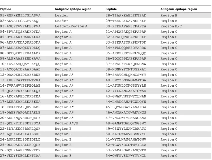
We created individual, yet overlapping peptides, that were 15 amino acids in length (Table 1). The entire sequence of PspA was used to predict the protein structure as well as b turn (t) using PSIPRED (http://bioinf.cs.ucl.ac.uk/psipred/) [21] and COUDES (http://bioserv.rpbs.jussieu.fr/Coudes/index.html) [22] methods. Coiled–coiled (C) as well as helical (H) structures were noted throughout PspA (Figure 1). There were nob turns or potential asparagine (N) endopeptidase sites in Regions A or A*.The majority of Region B is coiled with small helix (PspA242–246) and strand
(PspA286–293) domains. In contrast, Region C displays an array of
complex secondary structures as well as numerous potential N endopeptidase sites. The latter sites are typically found in bacterial cell wall-associated domains and known to enhance antigen-processing for MHC presentation [23].
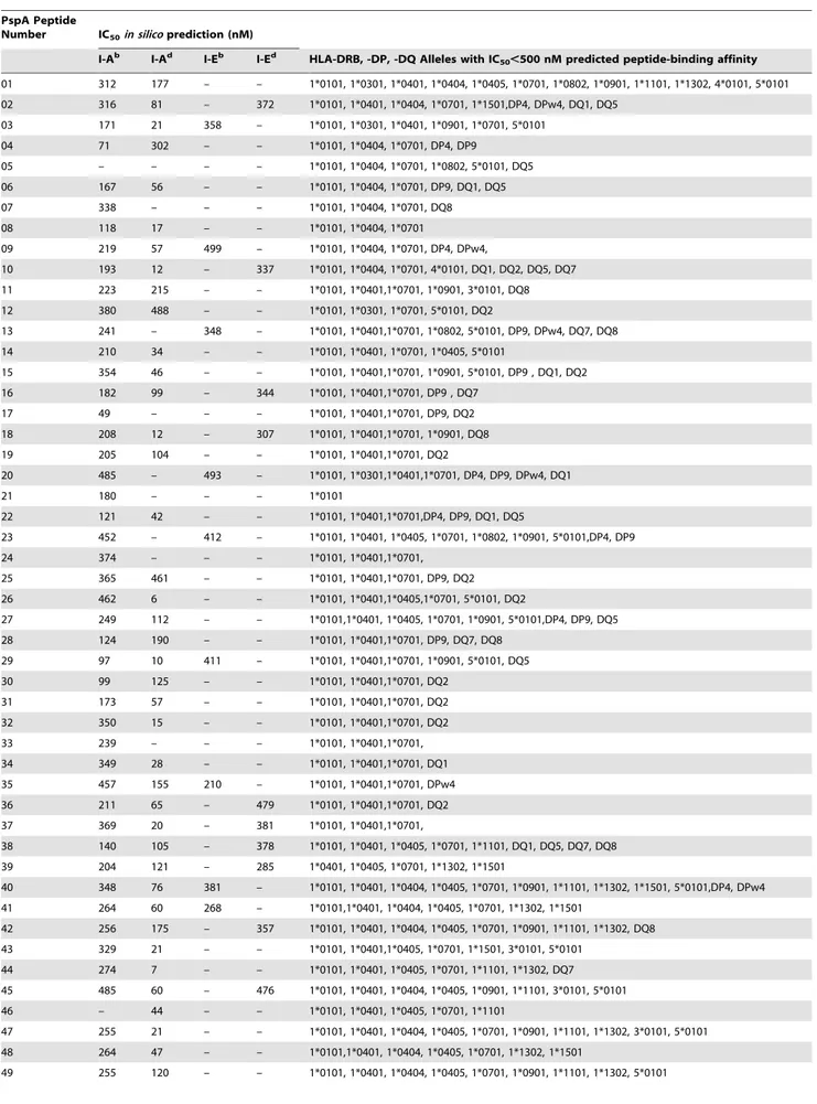
Next, the PspA peptide dataset was was used to determine MHC II binding affinities (Table 2). These data span a total of 16 human and 4 mouse MHC class II types. IEDB, MHCPred, RANKPEP, SVMHC and SYFPEITHI MHC class II epitope databases scanned the entire sequence of PspA. In brief, PspA peptides were compared with archived peptide datasets of previously measured peptide-MHC class II affinities. Peptides were classified into binders (IC50,500 nM) and non-binders
(IC50$500 nM) based onin silico-derived binding affinities. This
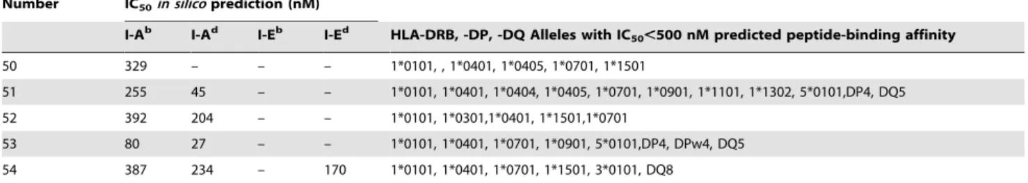
analysis revealed that nearly all PspA peptides could potentially bind a variety of mouse and human MHC class II molecules. Finally, the amino acid sequence comprising PspA peptides 19 to 22 (or PspA199–246) was aligned with sequences from nearly
100 clinically relevant family 1 S. pneumonia strains (Table 3). PspA199–246 is highly conserved among S. pneumonia strains and
contains the C-terminal end of Region A* and the beginning of Region B [20].
PspA Peptide-Specific Systemic and Mucosal CD4+T Cell
Proliferation Responses
To better determine whether predicted PspA peptide-MHC class II binding affinities corresponded with HTL proliferation, PspA peptide-specific CD4+
T cell responses were characterized 28 days after S. pneumoniae strain EF3030 or mock (naı¨ve) challenge. PspA peptide-specific proliferative responses by naı¨ve CD4+
T cells were relatively low (Figure 2). However, spleen- or CLN-derived CD4+
T cell from S. pneumonia strain EF3030-challenged mice showed selective yet significant proliferation indexes to PspA peptides. Spleen-derived HTLs fromS. pneumonia
strain EF3030-challenged mice significantly proliferated in re-sponse to PspA peptides 21, 22, and 23 than compared to naı¨ve controls. CLN CD4+
T cell PspA peptide-specific proliferation responses were moderately higher than similar cells isolated from the spleen of S. pneumonia strain EF3030-challenged mice, with comparatively higher responses to PspA peptides 21 and 23.
PspA Peptide-Specific T Helper Cytokine Profiles
In general, pneumococcal infection resulted in significantly higher HTL cytokine secretion byex vivoPspA peptide-stimulated CD4+
T cells from the spleen as well as CLNs ofS. pneumoniastrain EF3030-challenged mice, than compared to naı¨ve mice (Figures 3 and 4). In contrast to proliferation responses, spleen-derived CD4+
T cells fromS. pneumoniastrain EF3030-challenged mice secreted higher levels of IFN-c and IL-2 after PspA peptide ex vivo
stimulation than did similar cells from CLNs. HTLs from CLNs of pneumococcal-challenged mice, significantly responded to PspA peptides 20 and 21. CD4+
T cells isolated from the spleen and CLNs also significantly secreted Th2 cytokines after ex vivo
stimulation of PspA peptides, than compared to naı¨ve mice. Similar to proliferation responses, CLN CD4+
T cells from S. pneumoniastrain EF3030-challenged mice significantly secreted IL-10 following PspA peptide restimulation, with comparatively higher responses to PspA peptides 19, 20, and 21. Splenic HTLs selectively secreted significant levels of IL-10 in response to PspA peptides 13, 19, and 21 than compared to naı¨ve mice (Figure 5). While cells from naı¨ve mice did not significantly respond to PspA peptides, CD4+
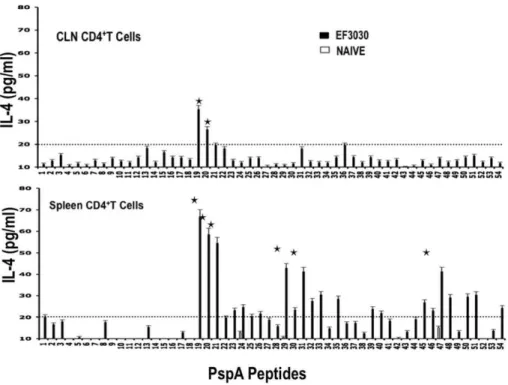
T lymphocytes fromS. pneumoniastrain EF3030-challenged mice also significantly secreted IL-4 and IL-5 after PspA peptideex vivo stimulation (Figures 6 and 7). In particular, there were comparatively higher responses to PspA peptides 19, 20, and 21 by splenic HTLs. Similar CLN Th2 cells secreted IL-4 in response to peptides 19 and 20 whereas heightened IL-5 secretion was noted in response to peptides 19 to 22 as well as 29 and 35, than compared to naive mice.
PspA HTL Epitopes
In summary, CD4+
T cells from S. pneumonia strain EF3030-challenged mice consistently mounted significant yet select proliferation and IL-10 responses (CLN..spleen), IFN-c, IL-2 and IL-4 secretion (spleen..CLN) and IL-5 expression (spleen#CLN) largely in response to PspA peptides 19, 20, 21, and 22. Moreover, PspA peptides 21 and 22 mounted compar-atively high proliferation responses, 20 and 21 induced consistently high IFN-cand IL-2 responses, and 19, 20, and 21 caused IL-10, IL-4 and IL-5 responses by HTLs isolated from Pneumococci-exposed mice.
Predicted PspA Peptide-MHC Class II Alleles Binding Affinities and Correlation with Proliferation and Cytokine Secretion Responses
PspA peptides 19, 20, 21, and 22 mounted significant HTL responses, and displayed strong predictive binding affinities to numerous HLA-DR, -DQ, and -DP as well as I-Ab and I-Ed haplotypes. This is best illustrated by viewing a 3-dimensional plot of the proliferation index as well as IFN-c, 10, 2, 4, and/or IL-5 responses compared with MHC allele binding affinities (Figures 8 and 9). PspA peptide-specific T cell proliferation and IFN-c, IL-10, IL-2, IL-4 and IL-5 secretion by CLN and splenic CD4+
T cells from
S. pneumoniastrain EF3030-challenged mice was higher than the naı¨ve
group. In general, CLN HTLs from mice previously challenged with
S. pneumoniastrain EF3030) secreted high levels of IFN-c, IL-2, IL-4, IL-5 and IL-10 as well as enhanced proliferation in response to PspA peptides (19, 20.21, 22) stimulation.
PspA peptides 19, 20, 21, and 22 were predicted to bind I-Ab /I-Ad, I-Ab/I-Eb, I-Ab and I-Ab/I-Ad, respectively, with IC50,500 nM. From these, PspA peptide 20 was predicted to
have marginal binding affinities to I-Aband I-Ebwith IC50= 485
and 493 nM, respectively. This also corresponded with relatively high IL-10 responsiveness. Spleen-derived CD4+
T cells secreted significant amounts of IFN-c, IL-2, IL-4 and IL-5 as well as proliferated in response to PspA peptides 19, 20, 21, and 22 (i.e., PspA199–246) stimulation from mice previously challenged withS.
pneumoniastrain EF3030. Peptide 20 or 23 stimulation of splenic HTLs resulted in comparatively high secretion of IL-10. Similar to PspA peptide 20, peptide 23 was predicted to have moderate I-Ab and I-Ebbinding affinity i.e., IC50= 452 and 412 nM, respectively.
Peptides that induced spleen-derived CD4+
T cells to secrete high levels of Th1 (IFN-c/IL-2) and Th2 (IL-4/IL-5) cytokines also correlated with relatively high MHC binding affinities. It is important to note that several PspA peptides predicted to tightly bind I-A and/or I-E alleles did not always correspond with elevated cytokine secretion (e.g., peptides 6, 18, 30, and 53). Table 1.Overlapping PspA peptides and antigenic region description.
Peptide Antigenic epitope region Peptide Antigenic epitope region
01-MNKKKMILTSLASVA Leader 28-TIAAKKAELEKTEAD Region B
02-ASVAILGAGFVASQP Leader 29-TEADLKKAVNEPEKP Region B
03-ASQPTVVRAEESPVA Leader/Region A 30-PEKPAPAPETPAPEA Region B/C
04-SPVASQSKAEKDYDA Region A 31-APEAPAEQPKPAPAP Region C
05-DYDAAKKDAKNAKKA Region A 32-APAPQPAPAPKPEKP Region C
06-AKKAVEDAQKALDDA Region A 33-PEKPAEQPKPEKTDD Region C
07-LDDAKAAQKKYDEDQ Region A 34-KTDDQQAEEDYARRS Region C
08-DEDQKKTEEKAALEK Region A 35-ARRSEEEYNRLTQQQ Region C
09-ALEKAASEEMDKAVA Region A 36-TQQQPPKAEKPAPAP Region C
10-KAVAAVQQAYLAYQQ Region A 37-APAPKTGWKQENGMW Region C
11-AYQQATDKAAKDAAD Region A 38-NGMWYFYNTDGSMAT Region C
12-DAADKMIDEAKKREE Region A* 39-SMATGWLQNNGSWYY Region C
13-KREEEAKTKFNTVRA Region A* 40-SWYYLNSNGAMATGW Region C
14-TVRAMVVPEPEQLAE Region A* 41-ATGWLQYNGSWYYLN Region C
15-QLAETKKKSEEAKQK Region A* 42-YYLNANGAMATGWAK Region C
16-AKQKAPELTKKLEEA Region A* 43-GWAKVNGSWYYLNAN Region C
17-LEEAKAKLEEAEKKA Region A* 44-LNANGAMATGWLQYN Region C
18-EKKATEAKQKVDAEE Region A* 45-LQYNGSWYYLNANGA Region C
19-DAEEVAPQAKIAELE Region A* 46-ANGAMATGWAKVNGS Region C
20-AELENQVHRLEQELK Region A* 47-VNGSWYYLNANGAMA Region C
21-QELKEIDESESEDYA Region A*/B 48-GAMATGWLQYNGSWY Region C
22-EDYAKEGFRAPLQSK Region B 49-GSWYYLNANGAMATG Region C
23-LQSKLDAKKAKLSKL Region B 50-MATGWAKVNGSWYYL Region C
24-LSKLEELSDKIDELD Region B 51-WYYLNANGAMATGWV Region C
25-DELDAEIAKLEDQLK Region B 52-TGWVKDGDTWYYLEA Region C
26-DQLKAAEENNNVEDY Region B 53-YLEASGAMKASQWFK Region C
27-VEDYFKEGLEKTIAA Region B 54-QWFKVSDKWYYVNGL Region C
Individual, yet overlapping,Streptococcus pneumoniastrain R6 PspA peptides, 15 amino acids in length were used inex vivoandin silicoassays. The antigenic epitope regions based on homologous alignment of PspA amino acid sequences from other strains were previously described as leader, A, A*, B, and C regions [20]. doi:10.1371/journal.pone.0009432.t001
PspA HTL Epitopes
Figure 1. Modular PspA amino acid sequence showing regions of predicted immunogenicity and secondary structure.Major domains of PspA are indicated. The aligned amino acid sequence shows the previously defined PspA windows A, A*, B and C. The PspA amino acid (AA) sequence was used to predict helical (H), coiled (C),astrand (E),bturns (t), and asparagine endopeptidase sites (N).
doi:10.1371/journal.pone.0009432.g001
PspA HTL Epitopes
Table 2.Overview of PspA peptide predicted binding affinities to MHC class II alleles.
PspA Peptide
Number IC50in silicoprediction (nM)
I-Ab I-Ad I-Eb I-Ed HLA-DRB, -DP, -DQ Alleles with IC
50,500 nM predicted peptide-binding affinity
01 312 177 – – 1*0101, 1*0301, 1*0401, 1*0404, 1*0405, 1*0701, 1*0802, 1*0901, 1*1101, 1*1302, 4*0101, 5*0101
02 316 81 – 372 1*0101, 1*0401, 1*0404, 1*0701, 1*1501,DP4, DPw4, DQ1, DQ5
03 171 21 358 – 1*0101, 1*0301, 1*0401, 1*0901, 1*0701, 5*0101
04 71 302 – – 1*0101, 1*0404, 1*0701, DP4, DP9
05 – – – – 1*0101, 1*0404, 1*0701, 1*0802, 5*0101, DQ5
06 167 56 – – 1*0101, 1*0404, 1*0701, DP9, DQ1, DQ5
07 338 – – – 1*0101, 1*0404, 1*0701, DQ8
08 118 17 – – 1*0101, 1*0404, 1*0701
09 219 57 499 – 1*0101, 1*0404, 1*0701, DP4, DPw4,
10 193 12 – 337 1*0101, 1*0404, 1*0701, 4*0101, DQ1, DQ2, DQ5, DQ7
11 223 215 – – 1*0101, 1*0401,1*0701, 1*0901, 3*0101, DQ8
12 380 488 – – 1*0101, 1*0301, 1*0701, 5*0101, DQ2
13 241 – 348 – 1*0101, 1*0401,1*0701, 1*0802, 5*0101, DP9, DPw4, DQ7, DQ8
14 210 34 – – 1*0101, 1*0401, 1*0701, 1*0405, 5*0101
15 354 46 – – 1*0101, 1*0401,1*0701, 1*0901, 5*0101, DP9 , DQ1, DQ2
16 182 99 – 344 1*0101, 1*0401,1*0701, DP9 , DQ7
17 49 – – – 1*0101, 1*0401,1*0701, DP9, DQ2
18 208 12 – 307 1*0101, 1*0401,1*0701, 1*0901, DQ8
19 205 104 – – 1*0101, 1*0401,1*0701, DQ2
20 485 – 493 – 1*0101, 1*0301,1*0401,1*0701, DP4, DP9, DPw4, DQ1
21 180 – – – 1*0101
22 121 42 – – 1*0101, 1*0401,1*0701,DP4, DP9, DQ1, DQ5
23 452 – 412 – 1*0101, 1*0401, 1*0405, 1*0701, 1*0802, 1*0901, 5*0101,DP4, DP9
24 374 – – – 1*0101, 1*0401,1*0701,
25 365 461 – – 1*0101, 1*0401,1*0701, DP9, DQ2
26 462 6 – – 1*0101, 1*0401,1*0405,1*0701, 5*0101, DQ2
27 249 112 – – 1*0101,1*0401, 1*0405, 1*0701, 1*0901, 5*0101,DP4, DP9, DQ5
28 124 190 – – 1*0101, 1*0401,1*0701, DP9, DQ7, DQ8
29 97 10 411 – 1*0101, 1*0401,1*0701, 1*0901, 5*0101, DQ5
30 99 125 – – 1*0101, 1*0401,1*0701, DQ2
31 173 57 – – 1*0101, 1*0401,1*0701, DQ2
32 350 15 – – 1*0101, 1*0401,1*0701, DQ2
33 239 – – – 1*0101, 1*0401,1*0701,
34 349 28 – – 1*0101, 1*0401,1*0701, DQ1
35 457 155 210 – 1*0101, 1*0401,1*0701, DPw4
36 211 65 – 479 1*0101, 1*0401,1*0701, DQ2
37 369 20 – 381 1*0101, 1*0401,1*0701,
38 140 105 – 378 1*0101, 1*0401, 1*0405, 1*0701, 1*1101, DQ1, DQ5, DQ7, DQ8
39 204 121 – 285 1*0401, 1*0405, 1*0701, 1*1302, 1*1501
40 348 76 381 – 1*0101, 1*0401, 1*0404, 1*0405, 1*0701, 1*0901, 1*1101, 1*1302, 1*1501, 5*0101,DP4, DPw4
41 264 60 268 – 1*0101,1*0401, 1*0404, 1*0405, 1*0701, 1*1302, 1*1501
42 256 175 – 357 1*0101, 1*0401, 1*0404, 1*0405, 1*0701, 1*0901, 1*1101, 1*1302, DQ8
43 329 21 – – 1*0101, 1*0401,1*0405, 1*0701, 1*1501, 3*0101, 5*0101
44 274 7 – – 1*0101, 1*0401, 1*0405, 1*0701, 1*1101, 1*1302, DQ7
45 485 60 – 476 1*0101, 1*0401, 1*0404, 1*0405, 1*0901, 1*1101, 3*0101, 5*0101
46 – 44 – – 1*0101, 1*0401, 1*0405, 1*0701, 1*1101
47 255 21 – – 1*0101, 1*0401, 1*0404, 1*0405, 1*0701, 1*0901, 1*1101, 1*1302, 3*0101, 5*0101
48 264 47 – – 1*0101,1*0401, 1*0404, 1*0405, 1*0701, 1*1302, 1*1501
49 255 120 – – 1*0101, 1*0401, 1*0404, 1*0405, 1*0701, 1*0901, 1*1101, 1*1302, 5*0101
PspA HTL Epitopes
Discussion
The immune system is remarkably robust in responding to a multitude of foreign antigens. T cells are crucial for generating an efficient immune response following recognition of foreign antigen in the context of MHC. The polymorphism of MHC genes leads to differences in immune responsiveness. While peptide vaccines potentially circumvent the problem of using whole antigen or attenuated pathogens as vaccines, this approach is impeded by the exhaustive MHC repertoire [24]. Hence, the identification of optimal or common HTL epitopes is imperative in mounting a protective immune response. To this end, MHC a chains have limited variability compared to MHCbchains suggesting that the binding affinity of MHC b chains dictates antigenic specificity [25,26]. This restricts the utility of peptides as vaccines. The discovery of ‘‘promiscuous’’ or ‘‘universal’’ peptides that can bind multiple HLA (bchain) allele would solve many of these problems. While HLA-transgenic mice have been used to map HTL epitopes [27], the limited number of HLA transgenic mice are not representative of all populations. Hence, the current study is the first of many to map clinically relevant HTL pneumococcal epitopes. We have utilizedin silicomethods for predicting class II-restricted peptides and evaluated immunogenicity by ex vivo
peptide-restimulation.
Protein secondary structure consists of regular elements such as a-helices andb-sheets, and irregular elements such as b-bulges, random coils, and tight turns. Tight turns are generally classified asd-,c-,b-,a-, andp-turns according to the number of residues involved [28]. b-turns have important biological tasks [29]. We predicted b-turns in PspA using a new and highly accurate secondary structure prediction software, PSIPRED, which incor-porates two feed forward neural networks that perform an analyses on PSI-BLAST position-specific-iterated- BLAST peptide se-quence [30]. b-turns were abundant in PspA Region C, which did not have immunodominant HTL epitopes. While b-sheet structures were not detected, analysis revealed PspA hasa-helical secondary structure content and is predominantly a coiled-coil structure. These structural properties correlate with PspA function and anti-complement activity [31]. In general, PspA peptides with continuous helix or strand predicted secondary structures were not considered immunodominant; instead, PspA peptides 19, 20, 21, and 22 (or PspA199–246) were estimated to predominantely have a
coiled secondary structure.
In addition to protein secondary structure, proteases and MHC class II co-mingle in the antigen-processing compartment and compete for peptides that satisfy requirements for protease or MHC recognition, respectively. Indeed, several proteases are
implicated in processing antigen and the MHC class II-bound invariant chain [32,33,34]. The proteolytic separation of MHC class II-bound epitopes was found to be a rate-limiting step in the presentation of T cell epitopes [35]. The level and activity of N endopeptidases can directly control the proteolysis and presenta-tion of T cell epitopes [36]. In contrast to other proteases, N endopeptidase is required for both antigen and invariant chain (Ii) processing [37,38,39,40]. Hence, N endopeptidase can have both positive and negative effects on the outcome of antigen processing [23,41,42]. Future studies will be required to verify whether the candidate HTL peptides are able to induce protective immunity against to pneumococcal infection.
PspA is highly immunogenic and is considered a promising vaccine candidate for combating pneumococcal infection [43,44,45]. In our model,S. pneumonia strain EF3030 promoted substantial PspA peptide-specific HTL responses. We show that PspA199–246 (i.e., PspA peptides 19, 20, 21, and 22) is highly
immunogenic and likely encompasses HLA class-binding epitopes to support pneumococcal immunity. Further, PspA 199–246 is
highly conserved among 100 different family 1S. pneumoniastrains (Table 3). In confirmation, Region B lies within PspA199–246and
was found to be important in eliciting protective pneumococcal immunity [46]. Taken together, our findings support the rationale for additional studies to explore the utility of PspA199–246-based
vaccines.
S. pneumonia has co-evolved with man and no doubt has numerous immune evasion mechanisms to avoid detection by T cells. From the pathogen’s perspective, it would be critical to maintain PspA function, while reducing detection of a T cell immuno-dominant epitope (i.e., peptide 21). PspA peptide 21 restimulation of pneumococcal-infected mice induced significant cytokine production and proliferation, yet was predicted to be poorly recognized by mouse and human MHC class II alleles. In contrast, other immunodominant regions exist within peptides 38 to 41 and might be protective since they invoked CD4+
T cell proliferation as well as T helper cytokine responses. However, these peptides reside in Region C, which has several potential, N endopeptidase sites along withb turn secondary structures that would optimally expose these sites for cleavage. In particular, peptide 40 has a highly conserved N endopeptidase site (i.e., NxN) that lies in the middle of a pronouncedbturn secondary structure (ttttt). While this intact peptide would potentially bind several MHC class II alleles, it is also likely that it would be cleaved before or after MHC-binding by N endopeptidases.
The Th1-associated cytokine, IL-2 promotes T cell prolifera-tion. Our data show PspA199–246peptides mounted comparatively
high IL-2 and proliferation HTL recall responses in mice
PspA Peptide
Number IC50in silicoprediction (nM)
I-Ab I-Ad I-Eb I-Ed HLA-DRB, -DP, -DQ Alleles with IC
50,500 nM predicted peptide-binding affinity 50 329 – – – 1*0101, , 1*0401, 1*0405, 1*0701, 1*1501
51 255 45 – – 1*0101, 1*0401, 1*0404, 1*0405, 1*0701, 1*0901, 1*1101, 1*1302, 5*0101,DP4, DQ5
52 392 204 – – 1*0101, 1*0301,1*0401, 1*1501,1*0701
53 80 27 – – 1*0101, 1*0401, 1*0701, 1*0901, 5*0101,DP4, DPw4, DQ5
54 387 234 – 170 1*0101, 1*0401, 1*0701, 1*1501, 3*0101, DQ8
*Dashes (–) represent the predicted affinity of peptides that poorly (i.e., IC50.500 nM) bind mouse I-Ab, I-Eb, I-Ad, or I-Edalleles. Similarly, absent HLA alleles are those that poorly (i.e., IC50.500 nM) bind the corresponding peptide.
doi:10.1371/journal.pone.0009432.t002 Table 2.Cont.
PspA HTL Epitopes
Table 3.Alignment of PspA199–256amino acid sequences from family 1 Pneumococci strains.
Strain NCBI Accession Number *Conserved amino acid sequence
D39/R6 NP_357715 DAEEVAPQAKIAELENQVHRLEQELKEIDESESEDYAKEGFRAPLQSK
WU2 AAF27710 EVAPQAKIAELENQVHRLEQELKEIDESESEDYAKEGFRAPLQSK
195 AAF68105 EEVAPQAKIAELENQVHRLEQELKEIDESDSEDYIKEGFRAPLQSE
SP19 AAF68093 AEEVAPQAKIAELENQVHKLEQKLKEIDESDSEDYVKEGFRAPLQSE
CGSP14, R41 YP_001834837, ABY67182 EVAPQAKIAELENQVHRLEQDLKDINESDSEDYVKEGLRAPLQSE
RHG79, OVP-41721 ABY67197, ACR50702 HAEEVAPQVKIAELENQVHKLEQKLKEIDESDSEDYVKEGLRAPLQSE
EF3030 to be determined .. EVALQAKIAELENQVHRLETELKEIDESDSEDYVKEGLRVPLQSE
c2, OVP-43533, OVP-42723, OVP-43431, R24729, DBL5, HUB-6893, St 371/00
ACM45238, ACR50689, ACR50693, ACR50694, ABY67184, AAF27706, ACR50678, ABR53733
HAKEVAPQAKIAELENQVHRLEQDLKDINESDSEDYVKEGLRAPLQSE
L81905, RH5, BG9739, MC-247 AAF27705, ABV60383, AAF27700, ACR50682 RAKEVVLQAKIAELENEVHKLEQKLKEIDESDSEDYVKEGFRAPLQSE
70585 YP_002739507 RAKEVALQAKIAELENEVHRLETKLKEIDESDSEDYVKEGLRAPLQSE
AC94 AAF27698 RAKEVALQAKIAELENEVHRLETELKEIDESDSEDYVKEGLRVPLQSE
SP6-BS73, EF6796, BG9163, RHG63 ZP_01819322, AAF27709, AAF27711,
ABY67195
EVALQAKIAELEYEVQRLEKELEEINESDSEDYAKEGFRAPLQSK
SP18-BS74 ZP_01829602 HAEEVVPQAKIAELENEVQKLEKDLKEIDESDSEDYVKEGLRAPLQSE
SP200, MC-332, SP221 AAF67354, ACR50683, AAF68099 RAKEVALQAKIAELENQVHRLETELKEIDESDSEDYVKEGLRVPLQSE
BG8838, R30318 AAF27703, ABW07806 HAEEVVPQAKIAELENEVQKLEKDLKEIDESDSEDYVKEGLRAPLQSE
R30397, R171, BG6692 ABV60382, ACH72677, AAF27704 HAEEVVPQAKIAELENEVQKLEKDLKEIDESDSEDYVKEGLRAPLQSE
HUB-7682 ACR50697 RAKEVALQAKIAELENEVHRLETKLKEIDESDSEDYVKEGLRAPLQSE
130 AAF68103 HAEEVVPQAKIAELENEVQKLEKDLKEIDESASEDYVKEGLRAPLQSE
R30318 ABW07807 RAKEVALQAKIAELENEVHRLETKLKETDESDSEDYVKEGLRAPLQSE
OVI-2328 ACR50701 HAKEVVPQAKIAELENEVQKLEKDLKEIDESDSEDYVKEGLRAPLQSE
*CDC1873-00, ST858, *SP6-BS73, *EF6796, ST860, *SRF10, SP23-BS72, *g5, E134, BG9163
ZP_02709307{, ABN71686, ZP_01820249{,
AAD00184{, ABN71687, AAF73809{,
ZP_01835080, AAF73801{, AAF13457,
AAF13460
DAEEYALEAKIAELEYEVQRLEKELKEIDESDSEDYLKEGLRAPLQSK
232 AAF68104 HAEEVVPQAKIAELENEVQKLEKDLKEIDESASEDYVKEGLRAPLQSE
P1031, R30087 YP_002737416, ABY67187 RAKEVALQAKIAELENEVHRLETKLKETDESDSEDYVKEGLRAPLQSE
CDC3059-06 ZP_02717970 HAEEVAPQAKIAELEHEVQKLEKALKEIGESDSEDYVKEGLRAPLQSE
OVP-42725 ACR50703 LFLQAKIAELENEVHKLEQKLKEIDESDSEDYVKEGFRAPLQSE
PN124 AAN37735 AKIAELENQVHRLEQDLKDINESDSEDYVKEGFRAPLQSE
DBL6A AAF27701 RAKEVVLQAQIAELENEVHKLEPKLKEIDESDSEDYVKEGFRAPLQSE
St 435/96 AAL92492 HAEEVAPQAKIAELEHEVQKLEKALKEIDESDSEDYVKEGLRAPLQFE
EF10197 AAF27708 RAKEVVLHAKLAELENEVHKLDQKLKEIDESDSEDYVKEGFRAPLQSE
R402 ABY67181 HAEEVAPQAKIAELEHEVQKLEKALKEIDESDSEDYVKEGLRAPLQFE
DBL1 AAF27702 RAKEVALQAKIAELENEVYRLETELKGIDESDSEDYVKEGLRAPLQSE
HUB-4197, 237 ACR50680, AAF68102 HAEEVAPQAKIAELEHEVQKLEKALKEIDESDSEDYVKEGLRAPLQFE
PspA
HTL
Epitopes
PLoS
ONE
|
www.plos
one.org
7
February
2010
|
Volume
5
|
Issue
2
|
Strain NCBI Accession Number *Conserved amino acid sequence
c1, SP194, RHG95, HUB-2371, PC4, RH21, RH12
ACM45237, AAF68092, ABV60384, ACR50685, ABV30914, ABY67192, ABW07810
RAKEVALQAKIAELENEVYRLETELKGIDESDSEDYVKEGLRAPLQSE
SP23-BS72, SP196, URSP2, 233, 152, 164, BG8743, 183, HUB-6892, 90, 177, 137, 39, RH9
ZP_01834257, AAF67355, AAR20918, AAF70097, AAF70096, AAF70094, AAF27699, AAF70095, ACR50684, AAF70093, AAF70091, AAF70090, AAF70092, ABW07809
KYALEAKIAELEYEVQGLEKELKEIDESDSEDYIKEGLRAPLQSK
R23661, R30360, OVP-40742 ABV30913, ABY67189, KYALEAKIAELEYEVQRLEKEIKEIDESDSEDYLKEGLRAPLQSE
R11561 ACH72679 EVAPQAKIAELENQVHRLEQDL---SDSEGYVKEGLRAPLQSE
E134 AAF27707 KYALEAKISELEYEVQGLGKELKEIDESDSEDYXKEGLRAPLQSK
SP356 AAN37734 IAELENEVYRLETELKGIDESDSEDYVKEGLRAPLQSE
R83 ACH72676 KYALEAKIAELEYEVQRVEKEIK--DESDSEDYLKEGLRAPLQSE
P105 ABE67219 LEKEIKEIDESDSEDYLKEGLRAPLQSE
P755, P13 ABE67236, ABE67218 LKEIDESDSEDYVKEGFRAPLQSE
P1151 ABE67224 LKEIDESDSEDYIKEGVRAPLQSK
P308 ABE67222 LKEIDESDSEDYIKEGLRAPLQSK
P176, 371/00, P1161 ABE67232, AAL92493, ABE67225 LKEIDESDSEDYVKEGLRAPLQSE
*Alignment of conservedStreptococcus pneumoniaPspA199–256amino acid sequences appear as white text in black boxes.
{NCBI accession number of PspC that align with PspA199–246. doi:10.1371/journal.pone.0009432.t003
Table 3.Cont.
PspA
HTL
Epitopes
PLoS
ONE
|
www.plos
one.org
8
February
2010
|
Volume
5
|
Issue
2
|
Figure 2. Proliferation responses of PspA peptide-specific systemic and mucosal CD4+T cells during pneumococcal carriage.Spleen
and cervical lymph node (CLN) lymphocytes were isolated from F1 (B66Balb/c) mice, 28 days after intranasal challenge withStreptococcus pneumonia
strain EF3030 (&) and naı¨ve (%). CD4+T cells were incubated with 1
mM of PspA peptide (15 amino acid peptides that overlapped every 11 residues)
plus mitomycin C-treated naı¨ve syngeneic feeder cells, for 3 days, at a ratio of 5:16106cells. Proliferation was measured by BrdU incorporation, which was measured by ELISA. The data presented are the mean OD450. Experimental groups consisted of 10 mice. The results were expressed as the mean 6the standard error mean (SEM) of the response from 3 replicate determinations of three independent experiments.
doi:10.1371/journal.pone.0009432.g002
Figure 3. PspA peptide-specific IFN-csecretion by CD4+T cell following pneumococcal challenge.Groups of 10 F1 (B66Balb/c) mice were
intranasally challenged with 107CFUs ofS. pneumoniastrain EF3030 in a 15ml volume of Ringer’s solution. Spleen and cervical lymph node (CLN) lymphocytes
were isolated from mice, 28 days after intranasal challenge withStreptococcus pneumoniastrain EF3030 (&) and naı¨ve (%). CD4+
T cells were incubated with 1mM of PspA peptide (15 amino acid peptides that overlapped every 11 residues) plus mitomycin C-treated naı¨ve syngeneic feeder cells, for 3 days, at a ratio of
5:16106cells. The results were expressed as the mean
6the standard error mean (SEM) of IFN-csupernatant levels from 3 replicate determinations of three independent experiments. IFN-cproduction of cultured supernatants was determined by Luminex capable of detecting.2 pg/ml of IFN-c.
doi:10.1371/journal.pone.0009432.g003
PspA HTL Epitopes
previously challenged with S. pneumonia strain EF3030. Another Th1 cytokine, IFN-c, is required for protective pneumococcal immunity [47]. CD4+
T cells from S. pneumonia strain EF3030-challenged mice secreted significant amounts of IFN-cfollowingex vivoPspA peptide re-stimulation. IFN-cblockade accelerated the death of animals during pneumococcal infection [48], whereas treatment of mice with IFN-cenhanced the survival of mice [49]. However, confounding studies suggest that too much IFN-cand too little IL-10 can inhibit pneumococcal clearance during S. pneumonia infection that is secondary to influenza virus infection [50].
IL-10 has been suggested to be both deleterious and important for pneumococcal immunity. On one hand, administration of anti-IL-10 antibody was shown to enhance pneumococcal immunity [51], while others showed this Th2-associated cytokine is critical for MARCO-1 expression and subsequent pneumococcal clear-ance [50]. We show that PspA199–246 stimulates pneumococcal
strain EF3030-primed CD4+
T cells to secrete IL-10. Interestingly, HTLs from CLN mounted IL-10 responses to more peptides, than similar cells isolated from the spleen. Perhaps this contributes to establishing pneumococcal carriage by supporting selective pneumococcal clearance by CLN..spleen antigen-presenting cells after stimulation with CD4+
T cell-derived IL-10, whereas IFN-c-secreting HTLs might support spleen..CLN macrophag-es activation and/or internalization ofS. pneumonia.
In the absence of IL-10, a marked increase in pro-inflammatory cytokines is induced during pneumococcal infection [52]. To this end, IL-10 plays an indispensable role in mucous cell metaplasia and hyperplasia. IL-10 attenuates the proinflammatory cytokine response and its absence hampers effective clearance of the
infection, and reduces survival of pneumococcal infection [53]. We have shown that CCL5 inhibition resulted in lower IFN-c -secreting CD4+
T cells and significantly more PspA-specific IL-10-producing CD4+
T cells, which corresponded with the transition from pneumococcal carriage to lethal pneumonia [45,54]. Thus, the precise contribution of IL-10 in pneumococcal immunity remains uncertain, but the preponderance of the evidence suggests excessive IL-10 responses play a deleterious role in pneumococcal immunity, but moderate levels of this cytokine are required for optimal adaptive (humoral) immune responses toS. pneumoniaand reduced mucosal hyperplasia.
An effective intranasal conjugate pneumococcal vaccine using interleukin-12 (IL-12) as a mucosal adjuvant induced protection and increased expression of lung and splenic IFN-c and IL-10 mRNAs and protected mice from lethal challenge [55]. Thus, interplay and requirement of the HTL-derived IFN-cand IL-10 in pneumococcal carriage and pneumonia will require further study. In addition, the adjuvants or cytokines, e.g., IL-12, required by antigen presenting cells to promote IFN-cand IL-10 secreting, PspA-specific T cells will be addressed in the future.
Some studies suggest that Th2 cytokines do not support optimal pneumococcal immunity. Mice primed to mount Th2 cell responses followed by pneumococcal infection showed an increase in the number of Pneumococci and an increase in sinus inflammation than compared to naive or Th1 -primed groups [56]. IL-4 plays a central role in directing the development of the Th2 phenotype and IL-4 responses in lung have been associated with an increased risk to pneumococcal infection [57]. While IL-4 does not stimulate T cell proliferation, it induces the growth of lymphoblasts [58]. IL-5 was originally defined as a Th2 cell-derived cytokine that triggers B cell
Figure 4. PspA peptide-specific IL-2 secretion by CD4+T cell following pneumococcal challenge.
Groups of 10 F1 (B66Balb/c) mice were intranasally challenged with 107CFUs ofS. pneumoniastrain EF3030 in a 15
ml volume of Ringer’s solution. Spleen and Cervical lymph node (CLN)
lymphocytes were isolated from mice, 28 days after intranasal challenge withStreptococcus pneumoniaestrain EF3030 (&) and naı¨ve (%). CD4+
T cells were incubated with 1mM of PspA peptide (15 amino acid peptides that overlapped every 11 residues) plus mitomycin C-treated naı¨ve syngeneic
feeder cells, for 3 days, at a ratio of 5:16106cells. The results were expressed as the mean6the standard error mean (SEM) of IL-2 supernatant levels from 3 replicate determinations of three independent experiments. IL-2 production of cultured supernatants was determined by Luminex capable of detecting.2 pg/ml of IL-2.
doi:10.1371/journal.pone.0009432.g004
PspA HTL Epitopes
activation and differentiation into plasma cells [59]. PspA199–249
-specific HTLs from S. pneumonia strain EF3030-challenged mice secreted significant amounts of IL-4 (spleen..CLN) and IL-5 (spleen#CLN) largely in response to PspA peptides 19, 20, 21, and 22. However, the uncertain role of IL-4 and IL-5 in pneumococcal cellular immunity makes correlations of these cytokines with protective immunity difficult.
The role of Th17 cells in pneumococcal immunity has not been extensively studied. However, recent reports suggest that IL-17A supports antibody responses to pneumococcal capsular polysac-charides [60]. Mice lacking the IL-17A receptor or mice with neutrophil depletion are more susceptible to pneumococci [61]. Additional studies on the role of HTL-derived IL-17 would greatly contribute to the field and will be required to understand how secretion of this cytokine correlates with pneumococcal immunity. While the precise role of peptide MHC class II interactions that determine protective pneumococcal immunity are not known, this study addresses important questions that are relevant to MHC polymorphisms and antigen responsiveness. A number of studies have definitively proven a cause and effect relationship between human MHC genes and resistance to infection [62,63] as well as autoimmune diseases [64]. I-A, which is highly homologous to HLA-DQ [65], typically restricts antigen-specific CD4+
T cells in mice, whereas I-E (homologous to HLA-DR) [66,67,68] has been reported to control non-responsiveness through antigen-specific suppressor cells [69]. Further studies will be required to determine whether I-E or I-A as well as DQ or DR molecules might be involved in pneumococcal antigen non-responsiveness or cytokine secretion in mouse or man, respectively. To this end, many of the PspA peptides were predicted to bind I-A, while relatively few
were predicted to bind I-E. These studies support the use ofin silico
and in vivo methods to validate T cell responsiveness to PspA peptide-based vaccines.
Materials and Methods
Animals
Female F1 (B66Balb/c) mice, aged 8 to 12 weeks, contain MHC class II haplotype and corresponding TCR diversity that approaches those seen in man [70,71] and were purchased from Jackson Laboratories. All mice were housed in horizontal laminar flow cabinets free of microbial pathogens. Routine antibody screening for a large panel of pathogens and routine histological analysis of organs and tissues were performed to insure that mice were pathogen free.
S. pneumoniaStrain EF3030 Growth and Challenge S. pneumonia capsular strain EF3030 was among the human isolates of capsular group 19 that were previously examined and found to be relatively non-invasive in mice [72]. Pneumococci were grown in Todd Hewitt broth and stored frozen in aliquots at
280uC, in 20% glycerol, in sterile lactated Ringer’s solution (Ringer’s) (Abbott Labs, North Chicago, IL) [73,74]. To establish nasal carriage, Pneumococci were introduced into groups of mice (8 to 12 week old) by nasal administration. The animals were anesthetized with ketamine (100 mg/ml) and xylazine (20 mg/ml), mixed at a 4:1 (vol/vol) ratio. The anesthesia mixture was injected intramuscularly into the right hamstring muscle at a dose of 100 mg of ketamine per kg of body weight. After anesthesia was established, the mice were inoculated with approximately 107
Figure 5. PspA peptide-specific IL-10 secretion by CD4+T cell following pneumococcal challenge.Groups of 10 F1 (B66Balb/c) mice
were intranasally challenged with 107CFUs ofS. pneumoniastrain EF3030 in a 15ml volume of Ringer’s solution. Spleen and Cervical lymph node
(CLN) lymphocytes were isolated from mice, 28 days after intranasal challenge withStreptococcus pneumoniastrain EF3030 (&) and naı¨ve (%). CD4+T
cells were incubated with 1mM of PspA peptide (15 amino acid peptides that overlapped every 11 residues) plus mitomycin C-treated naı¨ve
syngeneic feeder cells, for 3 days, at a ratio of 5:16106cells. The results were expressed as the mean
6the standard error mean (SEM) of IL-10 supernatant levels from 3 replicate determinations of three independent experiments. IL-10 production of cultured supernatants was determined by Luminex capable of detecting.2 pg/ml of IL-10.
doi:10.1371/journal.pone.0009432.g005
PspA HTL Epitopes
Figure 6. PspA peptide-specific IL-4 secretion by CD4+T cell following pneumococcal challenge.Groups of 10 F1 (B66Balb/c) mice were
intranasally challenged with 107CFUs ofS. pneumoniastrain EF3030 in a 15
ml volume of Ringer’s solution. Spleen and Cervical lymph node (CLN) lymphocytes
were isolated from mice, 28 days after intranasal challenge withStreptococcus pneumoniastrain EF3030 (&) and naı¨ve (%). CD4+T cells were incubated with
1mM of PspA peptide (15 amino acid peptides that overlapped every 11 residues) plus mitomycin C-treated naı¨ve syngeneic feeder cells, for 3 days, at a ratio of
5:16106cells. The results were expressed as the mean
6the standard error mean (SEM) of IL-4 supernatant levels from 3 replicate determinations of three independent experiments. IL-4 production of cultured supernatants was determined by Luminex capable of detecting.2 pg/ml of IL-4.
doi:10.1371/journal.pone.0009432.g006
Figure 7. PspA peptide-specific IL-5 secretion by CD4+T cell following pneumococcal challenge.
Groups of 10 F1 (B66Balb/c) mice were intranasally challenged with 107CFUs ofS. pneumoniastrain EF3030 in a 15
ml volume of Ringer’s solution. Spleen and Cervical lymph node (CLN) lymphocytes
were isolated from mice, 28 days after intranasal challenge withStreptococcus pneumoniastrain EF3030 (&) and naı¨ve (%). CD4+
T cells were incubated with 1mM of PspA peptide (15 amino acid peptides that overlapped every 11 residues) plus mitomycin C-treated naı¨ve syngeneic feeder cells, for 3 days, at a ratio of
5:16106cells. The results were expressed as the mean6the standard error mean (SEM) of IL-5 supernatant levels from 3 replicate determinations of three independent experiments. IL-5 production of cultured supernatants was determined by Luminex capable of detecting.2 pg/ml of IL-5.
doi:10.1371/journal.pone.0009432.g007
PspA HTL Epitopes
colony forming units (CFU) ofS. pneumoniastrain EF3030 in 15ml of Ringer’s solution using a 25-gauge ball-tipped gavage needle [75]. Experimental groups consisted of 10 mice and studies were repeated 3 times. The guidelines proposed by the committee for the Care of Laboratory Animal Resources Commission of Life Sciences - National Research Council were followed to minimize animal pain and distress. All procedures involving mice were approved by the Morehouse School of Medicine Committees (IACUC).
Pneumococcal Antigens
54 overlapping peptides, spanning the entire length of S. pneumoniastrain D39/R6 PspA protein sequence (NCBI Accession # NP_357715), starting with the first 15 residues at the N-terminus, was synthesized by the multipin synthesis method by Chiron Mimotopes Peptide Systems. Peptides overlapped by four amino acids (Table 1) and were acetylated at the N- terminus and ended with a COOH-terminal. Purity of these peptides was approximately 95%. The peptides were dissolved in a mixture (v/ v) of 75% dimethyl sulfoxide and 25% water, to a concentration of 70 mM, divided into small aliquots and stored frozen at280uC.
Tissue Collection and Cell Isolation
Mice were sacrificed by CO2inhalation to collect spleen and
CLNs for single cell isolation of lymphocytes 28 days followingS. pneumonia strain EF3030 challenge. Individual single cell
suspen-sions of spleen and CLNs were collected and prepared by aseptically removing tissues and passage through a sterile wire screen. Unpooled CD4+ T cells were further separated by
OctoMACSTM (Miltenyi Biotec) using negative selection. Re-maining (non-CD4+
) cells, were used as accessory feeder cells for antigen peptide-specific stimulation assays after mitomycin C (Sigma-Aldrich) treatment.
Cytokine Quantitation by LuminexTMAnalysis
Purified CD4+
T cells and mitomycin C-treated feeder cells were cultured at a density of 56106 and 106 cells per ml, respectively, in complete medium containing 1mM of each PspA peptide at 37uC in 5% CO2. For the assessment of cytokine
production, 100mL of culture supernatants from 96-well flat bottom plates (Corning Glass Works) were harvested 3 days after
ex vivoPspA peptide stimulation to determine the levels of IL-10 and IFN-c secreted by CD4+
T cells. phorbol-12-myristate-13-acetate (PMA) 1mg/ml was used as a positive control , ovalbumin (1mg/ml) and medium only is used as negative control to reduce the background reading. Supernatant cytokine levels were determined by the BeadlyteTM mouse multi-cytokine detection (Bio-Rad). Briefly, filter bottom ELISA plates were rinsed with 100mL of Bio-plex assay buffer and liquid was removed using a MilliporeTMMultiscreen Separation Vacuum Manifold System set at 5 mm Hg. Analyte beads in assay buffer were added to the wells followed by 50mL of serum or standard solution. The plates were
Figure 8. 3D plot of Th1/Th2 cytokine secretion relative to proliferation or I-A/I-E predicted peptide-binding by cervical lymph node-derived CD4+T cells.The panels summarize
IFN-c, IL-10, IL-2, IL-4, IL-5 and proliferation responses of PspA peptide-specific CD4+T cells
isolated from cervical lymph nodes of F1 (B66Balb/c) mice, 28 days afterS. pneumoniastrain EF3030- challenge and predicted I-A or I-E binding affinities. Y-axis and X-axis indicate the concentration (ng/ml) of IFN-cand IL-10, IL-2, IL-4, IL-5 respectively, secreted by PspA peptide-stimulated CD4+
T cells. The Z- axis represents the predicted I-A or I-E binding affinities (Kd). PspA peptides 19, 20, 21 and 22 appear as white circles, while remaining peptides are open circles.
doi:10.1371/journal.pone.0009432.g008
PspA HTL Epitopes
incubated for 30 minutes at room temperature with continuous shaking (at setting#3) using a Lab-LineTMInstrument Titer Plate Shaker. The filter bottom plates were washed, as before, and centrifuged at 3006g for 30 seconds. Subsequently, 50mL of anti-mouse IL-10 or IFN-cantibody-biotin reporter solution was added in each well, after which the plates were incubated with continuous shaking for 30 min followed by centrifugation and washing. Next, 50mL streptavidin-phycoerythrin (PE) solution was added, and the plates were incubated with continuous shaking for 10 min at RT. 125mL of Bio-plex assay buffer was added, and BeadlyteTM readings were measured using a LuminexTM System and calculated using Bio-plexTM software (Bio-Rad). The cytokine BeadlyteTMassays were capable of detecting.5 pg/mL for each analyte.
Cell Proliferation
Lymphocyte proliferation was measured by a 5-Bromo-29 -deoxy uridine (BrdU) absorption and detection (Roche Diagnos-tics). In brief, purified CD4+
T cells were cultured at a density of 56106cells/mL, with 106mitomycin C-treated feeder cells/mL in complete medium containing 1mM of PspA peptide at 37uC in 5% CO2. After 2 days of ex vivoantigen stimulation, cells were
transferred to polystyrene 96 well plates (Corning Glass Work). 10mL of BrdU labeling solution (10mM final concentration per well) were added and incubated for 18 hours at 37uC with 5% CO2. The cells were then fixed and incubated with 100mL of
nuclease in each well for 30 minute at 37uC. The cells were washed with complete media and incubated with BrdU-POD solution for 30 minute at 37uC. BrdU incorporation was developed with an 2,29–azino-bis 3- ethylbenzthiazoline-6-sulfonic acid (ABTS) solution and optical density (OD) was read at 450 nm. The proliferation index (PI) was calculated as follows. Antigen-specific CD4+ T cell proliferation was obtained by
measuring 5-Bromo-29-deoxy uridine (BrdU) incorporation, ac-cording to manufacturer’s instructions (Roche Diagnostics). BrdU absorption or optical density at 450 nm (OD450) was detected
using a scanning multi-well SpectraMax 250 spectrophotometer (Molecular Devices). PI = OD450in peptide stimulated cell/OD450
in un-stimulated cells6100. The results were expressed as mean6
the standard error mean (SEM) of the response of 3 replicate determinations from three independent experiments. Statistical significance was assessed by student’s t test.
MHC Class II Epitope Prediction Using External Tools
IEDB (http://www.immuneepitope.org/), SYFPEITHI (http:// www.syfpeithi.de/), SVMHC (http://www.bs.informatik.unituebingen. de/SVMHC/), RANKPEP (http://bio.dfci.harvard.edu/RANKPEP/), and MHCPred (http://www.jenner.ac.uk/MHCPred) external software(s) were used to predict peptide binding affinities to mouse I-A and I-E as well as HLA-DR, -DP and -DQ. In brief, for average relative binding (ARB) evaluation, 10-fold cross validation results stored at IEDB were used to estimate performance. Because the
Figure 9. 3D plot of Th1/Th2 cytokine secretion relative to proliferation or I-A/I-E predicted peptide-binding by spleen- derived CD4+T cells.
The panels summarize IFN-c, IL-10, IL-2, IL-4, IL-5 and proliferation responses of PspA peptide-specific CD4+T cells isolated from spleen
of F1 (B66Balb/c) mice, 28 days afterS. pneumoniastrain EF3030-challenge and predicted I-A or I-E binding affinities. Y-axis and X-axis indicate the concentration (ng/ml) of IFN-cand IL-10, IL-2, IL-4,IL-5 respectively, secreted by PspA peptide-stimulated CD4+T cells. The Z-axis represents the
predicted I-A or I-E binding affinities (Kd). PspA peptides 19, 20, 21 and 22 appear as white circles, while remaining peptides are open circles. doi:10.1371/journal.pone.0009432.g009
PspA HTL Epitopes
binding of peptides to MHC class II molecules is not dependent on exact size, derivation of MHC class II ARB matrices followed an iterative procedure. For the first iterative step, a matrix was generated from a set of nine-residue core sequences randomly obtained from each peptide sequence in the training set. For subsequent cycles, nine-residue core sequences were used to generate a matrix. The overall binding affinity of a peptide was predicted using the highest scoring nine-residue core sequence. For the SYFPEITHI prediction, we patched each testing peptide with three glycine residues at both ends before evaluation for prediction. This was recommended by the creators of SYFPEITHI method to ensure that all potential binders were correctly presented to the prediction algorithm. For all other methods, the original tested peptides were submitted directly for prediction. Peptide sequences were sent to web servers one at a time and predictions were extracted from the server’s response. To assign a single prediction for peptides longer than nine amino acids in the context of tools predicting the affinity of 9 core-binding regions, we took the highest affinity prediction of all possible 9-mers within the longer peptide as the prediction result. For each MHC class II molecule whose binding can be predicted by three or more algorithms, the top three methods were selected that gave the best performance. For each method, peptides were tested and ranked by their scores with higher ranks for better binders. For each tested peptide, three ranks from
different methods were taken and the median rank was taken as the consensus score. Peptides were classified into binders (IC50,500 nM) and nonbinders (IC50$500 nM), as practical
cutoffs.
Statistics
Data are expressed as the mean6SEM and compared using a two-tailed student’st-test or an unpaired Mann Whitney U test. The results were analyzed using Microsoft Excel for Macintosh computers and were considered statistically significant ifpvalues were less than 0.01. When cytokine or antibody levels were below the detection limit (BD), they were recorded as one-half the lower detection limit for statistical analysis.
Acknowledgments
The content of this manuscript benefited from many fruitful conversations with members of the Morehouse School of Medicine and the University of Alabama at Birmingham
Author Contributions
Conceived and designed the experiments: JL. Performed the experiments: RS SS PKS UPS. Analyzed the data: RS SH JL. Contributed reagents/ materials/analysis tools: DB SH JL. Wrote the paper: RS JL.
References
1. Nasrin D, Collignon PJ, Wilson EJ, Pilotto LS, Douglas RM (1999) Antibiotic resistance inStreptococcus pneumoniaeisolated from children. Journal of Paediatrics & Child Health 35: 558–561.
2. Hooper DC (2001) Emerging mechanisms of fluoroquinolone resistance. Emerging Infectious Diseases 7: 337–341.
3. Morita JY, Zell ER, Danila R, Farley MM, Hadler J, et al. (2002) Association between antimicrobial resistance among pneumococcal isolates and burden of invasive pneumococcal disease in the community. Clinical Infectious Diseases 35: 420–427.
4. Richter SS, Heilmann KP, Coffman SL, Huynh HK, Brueggemann AB, et al. (2002) The molecular epidemiology of penicillin-resistantStreptococcus pneumoniae
in the United States, 1994–2000. Clinical Infectious Diseases 34: 330–339. 5. Bignell GR, Huang J, Greshock J, Watt S, Butler A, et al. (2004) High-resolution
analysis of DNA copy number using oligonucleotide microarrays. Genome Research 14: 287–295.
6. Costantino HR, Firouzabadian L, Wu C, Carrasquillo KG, Griebenow K, et al. (2002) Protein spray freeze drying. 2. Effect of formulation variables on particle size and stability. Journal of Pharmaceutical Sciences 91: 388–395.
7. Alrutz MA, Srivastava A, Wong KW, D’Souza-Schorey C, Tang M, et al. (2001) Efficient uptake ofYersinia pseudotuberculosisvia integrin receptors involves a Rac1-Arp 2/3 pathway that bypasses N-WASP function. Molecular Microbiology 42: 689–703.
8. Ertl HC, Xiang Z (1996) Commentary: Novel vaccine approaches. J Immunol 156: 3579–3582.
9. Brooks-Walter A, Briles DE, Hollingshead SK (1999) The pspC gene of
Streptococcus pneumoniaeencodes a polymorphic protein, PspC, which elicits cross-reactive antibodies to PspA and provides immunity to pneumococcal bacteremia. Infection & Immunity 67: 6533–6542.
10. Sturniolo T, Bono E, Ding J, Raddrizzani L, Tuereci O, et al. (1999) Generation of tissue-specific and promiscuous HLA ligand databases using DNA micro-arrays and virtual HLA class II matrices.[see comment]. Nature Biotechnology 17: 555–561.
11. Cunha-Neto E (1999) MHC-restricted antigen presentation and recognition: constraints on gene, recombinant and peptide vaccines in humans. Brazilian Journal of Medical & Biological Research 32: 199–205.
12. Rammensee HG (1995) Chemistry of peptides associated with MHC class I and class II molecules. Current Opinion in Immunology 7: 85–96.
13. Meister GE, Roberts CG, Berzofsky JA, De Groot AS (1995) Two novel T cell epitope prediction algorithms based on MHC-binding motifs; comparison of predicted and published epitopes fromMycobacterium tuberculosisand HIV protein sequences. Vaccine 13: 581–591.
14. Kang H, Mansel RE, Jiang WG (2005) Genetic manipulation of stromal cell-derived factor-1 attests the pivotal role of the autocrine SDF-1-CXCR4 pathway in the aggressiveness of breast cancer cells. International Journal of Oncology 26: 1429–1434.
15. Reche PA, Glutting JP, Zhang H, Reinherz EL (2004) Enhancement to the RANKPEP resource for the prediction of peptide binding to MHC molecules using profiles. Immunogenetics 56: 405–419.
16. Donnes P, Kohlbacher O (2006) SVMHC: a server for prediction of MHC-binding peptides. Nucleic Acids Research 34: W194–197.
17. Gu Y, Filippi MD, Cancelas JA, Siefring JE, Williams EP, et al. (2003) Hematopoietic cell regulation by Rac1 and Rac2 guanosine triphosphatases. Science 302: 445–449.
18. Parry CS (2008) Flanking p10 contribution and sequence bias in matrix based epitope prediction: revisiting the assumption of independent binding pockets. BMC Struct Biol 8: 44.
19. Briles DE, Hollingshead SK, Paton JC, Ades EW, Novak L, et al. (2003) Immunizations with Pneumococcal Surface Protein A and Pneumolysin Are Protective against Pneumonia in a Murine Model of Pulmonary Infection with
Streptococcus pneumoniae. Journal of Infectious Diseases 188: 339–348.
20. Hollingshead SK, Becker R, Briles DE (2000) Diversity of PspA: mosaic genes and evidence for past recombination inStreptococcus pneumoniae. Infect Immun 68: 5889–5900.
21. McGuffin LJ, Bryson K, Jones DT (2000) The PSIPRED protein structure prediction server. Bioinformatics 16: 404–405.
22. Fuchs PF, Alix AJ (2005) High accuracy prediction of beta-turns and their types using propensities and multiple alignments. Proteins 59: 828–839.
23. Manoury B, Hewitt EW, Morrice N, Dando PM, Barrett AJ, et al. (1998) An asparaginyl endopeptidase processes a microbial antigen for class II MHC presentation. Nature 396: 695–699.
24. Reche PA, Reinherz EL (2003) Sequence variability analysis of human class I and class II MHC molecules: functional and structural correlates of amino acid polymorphisms. Journal of Molecular Biology 331: 623–641.
25. Corte G, Damiani G, Calabi F, Fabbi M, Bargellesi A (1981) Analysis of HLA-DR polymorphism by two-dimensional peptide mapping. Proceedings of the National Academy of Sciences of the United States of America 78: 534–538. 26. Shackelford DA, Strominger JL (1980) Demonstration of structural
polymor-phism among HLA-DR light chains by two-dimensional gel electrophoresis. Journal of Experimental Medicine 151: 144–165.
27. Yu JJ, Goluguri T, Guentzel MN, Chambers JP, Murthy AK, et al. (2009) Francisella tularensis T Cell Antigen Identification Using Humanized HLA-DR4 Transgenic Mice. Clin Vaccine Immunol.
28. Chou KC (2000) Prediction of tight turns and their types in proteins. Anal Biochem 286: 1–16.
29. Kaur H, Raghava GP (2002) An evaluation of beta-turn prediction methods. Bioinformatics 18: 1508–1514.
30. Altschul SF, Madden TL, Schaffer AA, Zhang J, Zhang Z, et al. (1997) Gapped BLAST and PSI-BLAST: a new generation of protein database search programs. Nucleic Acids Res 25: 3389–3402.
31. Jedrzejas MJ, Lamani E, Becker RS (2001) Characterization of selected strains of pneumococcal surface protein A. J Biol Chem 276: 33121–33128.
32. Riese RJ, Wolf PR, Bromme D, Natkin LR, Villadangos JA, et al. (1996) Essential role for cathepsin S in MHC class II-associated invariant chain processing and peptide loading. Immunity 4: 357–366.
33. Pluger EB, Boes M, Alfonso C, Schroter CJ, Kalbacher H, et al. (2002) Specific role for cathepsin S in the generation of antigenic peptides in vivo. Eur J Immunol 32: 467–476.
PspA HTL Epitopes
34. Villadangos JA, Riese RJ, Peters C, Chapman HA, Ploegh HL (1997) Degradation of mouse invariant chain: roles of cathepsins S and D and the influence of major histocompatibility complex polymorphism. J Exp Med 186: 549–560.
35. Castellino F, Zappacosta F, Coligan JE, Germain RN (1998) Large protein fragments as substrates for endocytic antigen capture by MHC class II molecules. J Immunol 161: 4048–4057.
36. Manoury B, Mazzeo D, Fugger L, Viner N, Ponsford M, et al. (2002) Destructive processing by asparagine endopeptidase limits presentation of a dominant T cell epitope in MBP. Nat Immunol 3: 169–174.
37. Chen JM, Rawlings ND, Stevens RA, Barrett AJ (1998) Identification of the active site of legumain links it to caspases, clostripain and gingipains in a new clan of cysteine endopeptidases. FEBS Lett 441: 361–365.
38. Chen JM, Dando PM, Rawlings ND, Brown MA, Young NE, et al. (1997) Cloning, isolation, and characterization of mammalian legumain, an asparaginyl endopeptidase. J Biol Chem 272: 8090–8098.
39. Blum JS, Cresswell P (1988) Role for intracellular proteases in the processing and transport of class II HLA antigens. Proc Natl Acad Sci U S A 85: 3975–3979. 40. Amigorena S, Webster P, Drake J, Newcomb J, Cresswell P, et al. (1995) Invariant chain cleavage and peptide loading in major histocompatibility complex class II vesicles. J Exp Med 181: 1729–1741.
41. Antoniou AN, Blackwood SL, Mazzeo D, Watts C (2000) Control of antigen presentation by a single protease cleavage site. Immunity 12: 391–398. 42. Watts C (2001) Antigen processing in the endocytic compartment. Curr Opin
Immunol 13: 26–31.
43. McDaniel LS, Yother J, Vijayakumar M, McGarry L, Guild WR, et al. (1987) Use of insertional inactivation to facilitate studies of biological properties of pneumococcal surface protein A (PspA). Journal of Experimental Medicine 165: 381–394.
44. Berry AM, Yother J, Briles DE, Hansman D, Paton JC (1989) Reduced virulence of a defined pneumolysin-negative mutant ofStreptococcus pneumoniae. Infection & Immunity 57: 2037–2042.
45. Palaniappan R, Singh S, Singh UP, Sakthivel SK, Ades EW, et al. (2005) Differential PsaA-, PspA-, PspC-, and PdB-specific immune responses in a mouse model of pneumococcal carriage. Infection & Immunity 73: 1006–1013. 46. McDaniel LS, Ralph BA, McDaniel DO, Briles DE (1994) Localization of protection-eliciting epitopes on PspA ofStreptococcus pneumoniaebetween amino acid residues 192 and 260. Microbial Pathogenesis 17: 323–337.
47. Pomeroy C, Rubins JB (1997) Role of gamma interferon in the pathogenesis of bacteremicpneumococcal pneumonia. Infection & Immunity 65: 2975–2977. 48. Weigent DA, Huff TL, Peterson JW, Stanton GJ, Baron S (1986) Role of
interferon in streptococcal infection in the mouse. Microb Pathog 1: 399–407. 49. Lynch JM, Briles DE, Metzger DW (2003) Increased protection against
pneumococcal disease by mucosal administration of conjugate vaccine plus interleukin-12. Infect Immun 71: 4780–4788.
50. Sun K, Metzger DW (2008) Inhibition of pulmonary antibacterial defense by interferon-gamma during recovery from influenza infection. Nat Med 14: 558–564.
51. Menahem S, Rahayoe AU, Brawn WJ, Mee RB (1992) Interrupted aortic arch in infancy: a 10-year experience. Pediatr Cardiol 13: 214–221.
52. Tsuchiya K, Komori M, Zheng QY, Ferrieri P, Lin J (2008) Interleukin-10 is an essential modulator of mucoid metaplasia in a mouse otitis media model. Ann Otol Rhinol Laryngol 117: 630–636.
53. van der Poll T, Marchant A, Keogh CV, Goldman M, Lowry SF (1996) Interleukin-10 impairs host defense in murinepneumococcal pneumonia. J Infect Dis 174: 994–1000.
54. Palaniappan R, Singh S, Singh UP, Singh R, Ades EW, et al. (2006) CCL5 modulates pneumococcal immunity and carriage. Journal of Immunology 176: 2346–2356.
55. Khan AQ, Shen Y, Wu Z-Q, Wynn TA, Snapper CM (2002) Endogenous pro-and anti-inflammatory cytokines differentially regulate an in vivo humoral response toStreptococcus pneumoniae. Infection & Immunity 70: 749–761.
56. Yu X, Sperling A, Blair C, Thompson K, Naclerio R (2004) Antigen stimulation of TH2 cells augments acute bacterial sinusitis in mice. J Allergy Clin Immunol 114: 328–334.
57. Kang CI, Rouse MS, Patel R, Kita H, Juhn YJ (2009) Allergic airway inflammation and susceptibility topneumococcal pneumoniain a murine model with real-time in vivo evaluation. Clin Exp Immunol 156: 552–561.
58. Murtaugh MP, Johnson CR, Xiao Z, Scamurra RW, Zhou Y (2009) Species specialization in cytokine biology: is interleukin-4 central to the T(H)1-T(H)2 paradigm in swine? Dev Comp Immunol 33: 344–352.
59. Kouro T, Takatsu K (2009) IL-5- and eosinophil-mediated inflammation: from discovery to therapy. Int Immunol 21: 1303–1309.
60. Malley R, Srivastava A, Lipsitch M, Thompson CM, Watkins C, et al. (2006) Antibody-independent, interleukin-17A-mediated, cross-serotype immunity to pneumococci in mice immunized intranasally with the cell wall polysaccharide. Infect Immun 74: 2187–2195.
61. Lu YJ, Gross J, Bogaert D, Finn A, Bagrade L, et al. (2008) Interleukin-17A mediates acquired immunity to pneumococcal colonization. PLoS Pathog 4: e1000159.
62. Fuller-Espie SL, Murphy GA, Brett SJ, Lechler RI (1997) Quantitative but not qualitative variation in MHC class II alters CD4 interaction and influences T cell repertoire formation. Cellular Immunology 177: 49–61.
63. Mack DG, Johnson JJ, Roberts F, Roberts CW, Estes RG, et al. (1999) HLA-class II genes modify outcome of Toxoplasma gondii infection. International Journal for Parasitology 29: 1351–1358.
64. Rosloniec EF, Brand DD, Myers LK, Esaki Y, Whittington KB, et al. (1998) Induction of autoimmune arthritis in HLA-DR4 (DRB1*0401) transgenic mice by immunization with human and bovine type II collagen. Journal of Immunology 160: 2573–2578.
65. Bono MR, Strominger JL (1982) Direct evidence of homology between human DC-1 antigen and murine I-A molecules. Nature 299: 836–840.
66. Walker LE, Ferrone S, Pellegrino MA, Reisfeld RA (1980) Structural polymorphism of the beta chain of human HLA-DR antigens. Molecular Immunology 17: 1443–1448.
67. Silver J, Ferrone S (1979) Structural organization of human DR antigens and their murine analog, I-E antigens: serologic, genetic, and functional implications. Transplantation Proceedings 11: 1743–1744.
68. Hurley CK, Nunez G, Winchester R, Finn OJ, Levy R, et al. (1982) The human HLA-DR antigens are encoded by multiple beta-chain loci. Journal of Immunology 129: 2103–2108.
69. Ottenhoff TH, Walford C, Nishimura Y, Reddy NB, Sasazuki T (1990) HLA-DQ molecules and the control of Mycobacterium leprae-specific T cell nonresponsiveness in lepromatous leprosy patients. European Journal of Immunology 20: 2347–2350.
70. Ridgway WM, Fathman CG (1999) MHC structure and autoimmune T cell repertoire development. Curr Opin Immunol 11: 638–642.
71. Gregersen PK (1989) HLA class II polymorphism: implications for genetic susceptibility to autoimmune disease. Lab Invest 61: 5–19.
72. Briles DE, Crain MJ, Gray BM, Forman C, Yother J (1992) Strong association between capsular type and virulence for mice among human isolates of
Streptococcus pneumoniae. Infection & Immunity 60: 111–116.
73. Aaberge IS, Eng J, Lermark G, Lovik M (1995) Virulence ofStreptococcus pneumoniaein mice: a standardized method for preparation and frozen storage of the experimental bacterial inoculum. Microbial Pathogenesis 18: 141–152. 74. Briles DE, Ades E, Paton JC, Sampson JS, Carlone GM, et al. (2000) Intranasal
immunization of mice with a mixture of the pneumococcal proteins PsaA and PspA is highly protective against nasopharyngeal carriage of Streptococcus pneumoniae. Infection & Immunity 68: 796–800.
75. Kadioglu A, Gingles NA, Grattan K, Kerr A, Mitchell TJ, et al. (2000) Host cellular immune response to pneumococcal lung infection in mice.[erratum appears in Infect Immunol 2000 Apr;68(4):2390]. Infection & Immunity 68: 492–501.
PspA HTL Epitopes