D
12,14
-Prostaglandin J
2
in Neuronal Plasma Membrane
Yasuhiro Yamamoto1, Kenkichi Takase1, Junji Kishino2, Megumi Fujita3, Noboru Okamura2,3, Toshiyuki Sakaeda2,4, Masafumi Fujimoto2,5, Tatsurou Yagami1,2*
1Division of Physiology, Department of Pharmaceutical Health Care, Faculty of Pharmaceutical Sciences, Himeji Dokkyo University, Himeji, Japan,2Discovery Research Laboratories, Shionogi and Co., Ltd., Osaka, Japan,3Department of Clinical Pharmacy, School of Pharmaceutical Sciences, Mukogawa Women’s University, Nishinomiya, Japan,4Center for Integrative Education of Pharmacy Frontier, Kyoto University Graduate School of Pharmaceutical Sciences, Kyoto, Japan,5Laboratory of Applied Pharmacology, Faculty of Pharmacy, Chiba Institute of Science, Choshi, Japan
Abstract
15-deoxy-D12,14-prostaglandin J2(15d-PGJ2) is one of factors contributed to the neurotoxicity of amyloidb(Ab), a causative protein of Alzheimer’s disease. Type 2 receptor for prostaglandin D2(DP2) and peroxysome-proliferator activated receptorc (PPARc) are identified as the membrane receptor and the nuclear receptor for 15d-PGJ2, respectively. Previously, we reported that the cytotoxicity of 15d-PGJ2 was independent of DP2 and PPARc, and suggested that 15d-PGJ2 induced apoptosis through the novel specific binding sites of 15d-PGJ2different from DP2 and PPARc. To relate the cytotoxicity of 15d-PGJ2 to amyloidoses, we performed binding assay [3H]15d-PGJ2and specified targets for 15d-PGJ2associated with cytotoxicity. In the various cell lines, there was a close correlation between the susceptibilities to 15d-PGJ2and fibrillar Ab. Specific binding sites of [3H]15d-PGJ
2 were detected in rat cortical neurons and human bronchial smooth muscle cells. When the binding assay was performed in subcellular fractions of neurons, the specific binding sites of [3H]15d-PGJ
2were detected in plasma membrane, nuclear and cytosol, but not in microsome. A proteomic approach was used to identify protein targets for 15d-PGJ2in the plasma membrane. By using biotinylated 15d-PGJ2, eleven proteins were identified as biotin-positive spots and classified into three different functional proteins: glycolytic enzymes (Enolase2, pyruvate kinase M1 (PKM1) and glyceraldehyde 3-phosphate dehydrogenase (GAPDH)), molecular chaperones (heat shock protein 8 and T-complex protein 1 subunita), cytoskeletal proteins (Actinb, F-actin-capping protein, Tubulinband Internexina). GAPDH, PKM1 and Tubulinbare Ab-interacting proteins. Thus, the present study suggested that 15d-PGJ2plays an important role in amyloidoses not only in the central nervous system but also in the peripheral tissues.
Citation:Yamamoto Y, Takase K, Kishino J, Fujita M, Okamura N, et al. (2011) Proteomic Identification of Protein Targets for 15-Deoxy-D12,14-Prostaglandin J 2in
Neuronal Plasma Membrane. PLoS ONE 6(3): e17552. doi:10.1371/journal.pone.0017552
Editor:Howard Gendelman, University of Nebraska, United States of America
ReceivedOctober 24, 2010;AcceptedFebruary 4, 2011;PublishedMarch 18, 2011
Copyright:ß2011 Yamamoto et al. This is an open-access article distributed under the terms of the Creative Commons Attribution License, which permits unrestricted use, distribution, and reproduction in any medium, provided the original author and source are credited.
Funding:The authors acknowledge the following sources of support: Grant-in-Aid 20500346 from the Ministry of Education, Culture, Sports, Science, and Technology of Japan. This funder had no role in study design, data collection and analysis, decision to publish, or preparation of the manuscript. JK, NO, TS, MF and TY are employed by Shionogi and Co., Ltd. The present study includes data provided by JK, NO, TS, MF and TY when at Shiogi and Co., Ltd. Since the data were not applied to pharmaceutical development, Shionogi approved the submission of the present study to contribute to the development of academic study.
Competing Interests:JK, NO, TS, MF and TY are employed by Shionogi and Co., Ltd. There are no patents, products in development or marketed products to declare. This does not alter the authors’ adherence to all the PLoS ONE policies on sharing data and materials.
* E-mail: [email protected]
Introduction
Eicosanoids are divided into two groups, according to their mechanism of action: the conventional eicosanoids,e.g., prostaglan-din D2(PGD2) and the cyclopentenone-type PGs,e.g., 15-deoxy-D12,14-PGJ2(15d-PGJ2). PGD2 has been considered to be a
pro-inflammatory mediator in pro-inflammatory diseases such as Alzhei-mer’s disease (AD) and Asthma. In AD, PGD2formation increased
in the frontal cortex of the patients when compared with those of the healthy subjects [1]. AD is characterized pathologically by cortical atrophy, neurodegeneration and deposits of amyloid protein in the various regions of brain such as cerebral cortex [2]. Amyloidb(Ab) generated PGD2 from cortical neurons before inflammation [3].
However, the toxicity of PGD2via its GTP-binding protein-coupled
PGD2receptors does not occur. First, the PGD2receptor blocker
did not inhibit PGD2-induced neuronal cell death [4]. Second, little
mRNA of the PGD2receptor is observed in the rat [5] and human
[6] cerebral cortex. Third, few binding sites of [3H]PGD2 were
detected in the plasma membranes from rat cortices [4]. Fourth, the extent of specific [3H]PGD2in total biding is much lower (30–40%)
than that of [3H]15d-PGJ2(.80%), although binding sites of PGD2
have been reported in synaptosomes of rat [7] and human brains [6]. Fifth, the LD50value (8.2mM) of PGD2is much higher than the
affinity for PGD2 receptor (dissociation constant = 14 nM) [5].
Finally, PGD2required a latent time to exert toxicity. PGD2was
non-enzymatically metabolized to prostaglandin J2 (PGJ2), D12
-PGJ2 and 15d-PGJ2 [4]. Among PGD2 metabolites, 15d-PGJ2
exhibited most potent inflammatory effects [4]. Taken together, PGD2 appeared to mediate inflammation via 15d-PGJ2 in the
amyloidoses.
The surface receptors specific for 15d-PGJ2 have not been
identified, and 15d-PGJ2is believed to be actively transported into
cells. It possesses an a, b-unsaturated carbonyl group in the cyclopentane ring that can form covalent adducts with free thiols in proteins by Michael addition. 15d-PGJ2 covalently binds to
activated receptorc (PPARc) [9], [10]. Recently, 15d-PGJ2 has
been implicated in the antiproliferation independently from PPARc[11]. Moreover, 15d-PGJ2inhibits the NF-kB–dependent
gene expression through the covalent modification at Cys179 in IkB kinase [12]. Previously, we have found the novel binding sites of 15d-PGJ2 on the cell surface [4]. [3H]15d-PGJ2 bound
specifically to plasma membranes of cortical neurons. Among PGD2metabolites, 15d-PGJ2exhibited the highest affinity for the
specific binding sites. Other eicosanoids and PPAR agonists did not affect the specific binding sites. 15d-PGJ2 regulated cell
numbers in primary cultures of rat cortical neurons. The neurotoxicity of 15d-PGJ2 was the most potent among PGD2
and its metabolites, whereas little effect of other eicosanoids and PPAR agonists was detected. In peripheral tissues, 15d-PGJ2also
exhibited toxicity independently of PPARc. In response to basic fibroblast growth factor, bronchial smooth muscle cells (BSMC) proliferate and remodel airway in asthma [13]. 15d-PGJ2inhibits
proliferation in a PPARc-independent manner[14]. Thus, the identification of cell surface targets for 15dPGJ2is required to clear
how 15d-PGJ2induces cell toxicity and involves in amyloidoses.
In the present study, we identified cell surface targets for 15d-PGJ2in cortical neurons. In general, glycolytic enzymes, molecular
chaperones and cytoskeletone identified as membrane targets for 15d-PGJ2are known to localize in the cytosol, but their roles on
the cell surface have not been elucidated sufficiently. Here, we propose hypothetical role of membrane targets for 15d-PGJ2on
the cell toxicity and amyloidoses.
Materials and Methods
Materials
Dulbecco’s modified Eagle’s medium, Leibovitz’s L-15 medium, Roswell Park Memorial Institute 1640 medium, MCDB, CS-C, trypsin, deoxyribonuclease I, fetal bovine serum (FBS), horse serum (HS), penicillin, and streptomycin were obtained from Invitrogen (Carlsbad, CA). Ab (25–35) was purchased from Bachem AG (Bubendorf, Switzerland). [3H]PGD2(115 Ci/mmol)
and human hepatocytes was purchased from Perkin Elmer Life Science Products (Boston, MA). Human BSMC and human dermal fibroblasts were purchased from Lonza (Basel, Switzer-land). PGD2, PGJ2, D12-PGJ2, 15d-PGJ2 and biotinylated
15d-PGJ2 were obtained from Cayman Chemicals (Ann Arbor, MI;
Cabru, Milan, Italy). ImmobilineTM DryStrip Gels (pH 3–10), Amersham ECL PlusTM Western Blotting Detection Reagents, were obtained from GE Healthcare Bio-Sciences Corp. (Piscat-away, NJ). Iodoacetamide, dithiothreitol (DTT), ethyleneglycol bis tetraacetic acid (EGTA) and ATP (disodium salt) were from Sigma-Aldrich (Milan, Italy). Sequence grade modified trypsin was purchased from Promega (Madison, WI; Milan, Italy), andN -(1-pyrenyl) iodoacetamide was from Molecular Probes (Eugene, OR). Horseradish peroxidase-linked antibody against biotine was obtained from Cell Signaling Technology (Boston, MA). The protein concentration was measured using the BCA protein assay reagent obtained from Thermo Fisher Scientific. (Rockford, IL). All other chemicals were of reagent grade.
Tissue cultures
All procedures were conducted in accordance with NIH guidelines concerning the Care and Use of Laboratory Animals and with the approval of the Animal Care Committee of the Himeji Dokkyo University. Rat cortical neurons, human BSMC, human hepatocytes and human dermal fibroblasts were cultured as previously reported [15]. Cerebral cortices from the cerebral cortex of day-19 Sprague-Dawley rat embryos were dissociated in isotonic
buffer with 4 mg/ml trypsin and 0.4 mg/ml deoxyribonuclease I. Cells were plated at a density of 2.56105cells/cm2on poly-L-lysine-coated dishes in conditioning medium, Leibovitz’s L-15 medium supplemented with 5% FBS and 5% horse serum at 37uC in 5% CO2and 9% O2. On day 1 after plating, cultures were treated with
0.1mM arabinosylcytosine C. On day 4, cortical cultures were immunostained with anti-MAP2 specific for neurons, anti-GFAP for astrocytes, and anti-microglial antigen (OX-42). Cultures prepared by this method, consisted of approximately 95% neurons. Human BSMC were cultured at a density of 3.56103cells/cm2on
48-well plates in Molecular, Developmental, and Cellular Biology medium supplemented with 5% FBS, 50mg/ml gentamicin, 50 ng/ ml amphotericin. Human hepatocytes were cultured at a density of 56104cells/cm2on 48-well plates in CS-C medium (Applied Cell Biology Research Institute) supplemented with 10% FBS. Human dermal fibroblasts were cultured at a density of 56104cells/cm2on
48-well plates in DMEM supplemented with 10% FBS, 50 units/ml penicillin, and 50mg/ml streptomycin.
Aggregation assessment of fAb
A stock solution of fibrillar Ab(25–35) (fAb) was prepared by dissolving Abat 1 mM in deionized water and incubating Abat 37uC for 2–5 days to aggregate the peptide and stored at220uC until use [16]. The aggregation state of fAb was assessed in two ways. First, light microscopy was used to identify the presence of precipitated peptides both in stock solutions and after their addition to tissue culture wells; the observations were confirmed by three observers. Second, the aggregation state of fAbwas assessed by migration patterns after sodium dodecyl sulfate-polyacrylamide gel electrophoresis (SDS-PAGE). Samples of fAb stock solutions were added to reducing buffer, heated at 100uC for 3 min, and electrophoresed on 15% SDS–PAGE at 70 V.
Cell viability
Two different methods were employed for assessment of cell viability as previously reported [15]. First, the 3-(4,5-dimethylthia-zol-2-yl)-2,5-diphenyl tetrazolium bromide dye (MTT) reduction assay reflecting mitochondrial succinate dehydrogenase activity was employed. Second, residual cells were counted according to morphologic criteria; neurons with intact neurites and a smooth, round soma were considered viable, whereas those with degen-erated neurites and an irregular soma were considered nonviable. BSMC with extended cell bodies and their bright phase-contrast appearance were considered viable, whereas those with shrank and round cell bodies were considered nonviable.
Cell fractionation
Cell fractionation was performed as previously reported [17]. Cerebral cortices from rat brains were homogenized in 3 volumes of ice-cold STEA solution (0.25 M sucrose, 5 mM Tris-HCl (pH 7.5), 1 mM EGTA and 50 karikllein units/ml aprotinin). The homog-enate was filtered through three meshes and centrifuged at 7006g
for 10 min. Fractionations of nuclear and plasma membrane; The pellet was resuspended in 120 ml of STEA solution by gentle homogenization, and the resuspension was dispersed in 1080 ml of isosmotic Percoll solution (15.7% Percoll, 0.25 M sucrose, 1 mM EGTA, 50 karikllein units/ml aprotinin and 10 mM Tris HCI (pH 7.5)). The mixture was centrifuged at 35,0006g for 30 min. The resulting pellet was suspended in HEA solution (50 mM Hepes-NaOH (pH 7.4), 1 mM EGTA and 50 kIU/ml aprotinin) as the nuclear fraction. On the other hand, the second band from the surface in the supernatant was collected, washed by dilution with 2– 3 volumes of HEA solution and centrifuged at 10,0006g for 30 min.
fraction and stored in liquid nitrogen until used [18]. Fractionations of cytosol and microsome: The supernatant was centrifuged at 7,0006g for 10 min. The resulting supernatant was recentrifuged at
100,0006g for 1 h. The pellet was used as the microsomal fraction.
The supernatant was used as the cytosolic fraction.
Binding assay of [3H]15d-PGJ2
Binding assay of [3H]15d-PGJ2 were performed as previously
reported [18]. The standard reaction mixture of 10 nM [3 H]15d-PGJ2contained 50 mM Tris-HCl buffer (pH 8.0), 100 mM NaCl
and plasma membranes (10mg) in a total volume of 100ml. Incubation was initiated by addition of the reaction mixture to plasma membranes, and was carried out at 4uC for 24 h. We determined non-specific binding by performing incubations with [3H]15d-PGJ2 in the presence of 100mM unlabeled 15d-PGJ2.
The specific binding was calculated by subtraction of the non-specific binding from the total binding. Data are expressed as means6standard error of the mean (S.E.M.) values (n = 4).
Protein separation by two-dimensional electrophoresis
Membrane preparation and binding assay of biotinylated 15d-PGJ2were conformed to ‘‘Binding assay of [3H]15d-PGJ2’’. The
standard reaction mixture of 1mM biotinylated 15d-PGJ2contained
50 mM Tris-HCl buffer (pH 8.0), 100 mM NaCl and plasma membranes (400mg) in a total volume of 4 ml. Incubation was initiated by addition of the reaction mixture to plasma membranes, and was carried out at 4uC for 24 h in the presence or absence of unlabeled 15d-PGJ2. We determined non-specific binding by
performing incubations with biotinylated 15d-PGJ2in the presence
of 100mM unlabeled 15d-PGJ2. According to the method of Toda
and Kimura [19], two-dimensional electrophoresis was performed with the CoolPhoreStar Horizontal Gel Electrophoresis Unit IPG-IEF (Anatech: Tokyo, JP). The samples containing 400mg of membrane lysates were dissolved in a rehydration buffer (5 M urea, 2 M thiourea, 2%(w/v) CHAPS, 2%(w/v) SB3-10, 2% Pharmalytes and 65 mM DTT) for the first dimensional isoelectric focusing (IEF). The pH range of the IEF was 3–10. Before IEF was performed, the gel strips were incubated with a swelling buffer (6 M urea, 2 M thiourea, 2%(w/v) TritonX-100, 2%(w/v) SB3-10, 2% Pharmalytes, 2.5 mM acetic acid, 0.0025% BPB and 13 mM DTT). After IEF was performed, the gel strips were incubated with an SDS buffer (6 M urea, 32 mM DTT, 2%(w/v) SDS, 0.0025% BPB, 30%(v/v) glycerol, and 25 mM Tris-HCl pH 6.8) for 10 min, and then with
Figure 2. Effect of 15d-PGJ2 on the cell viability. (A) Cortical neurons, BSMC, hepatocytes and dermal fibroblasts were treated with 10mM 15d-PGJ2or vehicle (0.1% ethanol). (B) Cortical neurons (circles) and BSMC (triangles) were treated with 15d-PGJ2 at the indicated concentrations. MTT-reducing activities of cortical neurons and other cells were determined 24 h or 48 h later, respectively. Data are expressed as means6 S.E.M. (n = 4). *P,0.05, **P,0.01, compared with control by ANOVA followed by Dunnett’s test.
doi:10.1371/journal.pone.0017552.g002
an alkylation buffer (6 M urea, 243 mM iodoacetamide, 2%(w/v) SDS, 0.0025% BPB, 30%(v/v) glycerol, and 25 mM Tris-HCl pH 6.8) for 10 min. For the second dimensional electrophoresis, polyacrylamide gel (12% acrylamide, 0.4% bis-acrylamide, 10.6% glycerol, 0.1% SDS, 1.2% APS, 0.1% (v/v) TEMED and 369 mM Tris-HCl pH 8.8) was used. All procedures followed the manufac-turer’s protocol. Separated proteins were then fixed in the gel using 1) 40% methanol and 10% acetic acid, 2) 10% methanol and 7% acetic acid, and 3) 10% methanol and 8% acetic acid. Then, they were stained with SYPRO Ruby protein gel stain, and scanned using the FluoroPhoreStarH3000 (Anatech: Tokyo, JP). The protein spots were visualized by Progenesis Same Spots (Nonliner Dynamics Ltd: Newcastle upon Tyne, UK). For immunoblotting, gels were transferred to polyvinylidene fluoride membranes (Millipore Co., Bedford, USA). The membranes were incubated with phosphate-buffered saline containing 0.1% Tween20 (PBS/Tween) and 5% skim milk for blocking and washed with PBS/Tween. This procedure was followed by the addition of horseradish peroxidase-conjugated anti-biotin antibody and ECL reagents (GE Healthcare Bio-Sciences). The spots were visualized by LAS-3000 (Aisin Seiki Co., Ltd., Aichi, Japan).
Identification of 15d-PGJ2-targeted proteins
Gel pieces were washed in 50 mM ammonium bicarbonic acid containing 50% acetonitrile for 10 min, twice. Then, they were
dried in block incubator Bl-516S (ASTEC Co., ltd.; Tokyo, JP) at 95uC for 10 min. Each sample was proteolyzed with 10ml 1 mM ammonium bicarbonic acid containing 200 ng trypsin overnight at 37uC. The peptide in each gel was extracted with 50% acetonitrile containing 0.1% TFA followed by sonication for 15 min. The supernatant was collected, and peptides were further extracted with 75% acetonitrile containing 0.1% TFA followed by sonication for 15 min. Peptide extracts were concentrated to
,10ml using Speedvac concentrator. Then, they were desalted with Ziptip (Millipore Co.) and mixed with an equal volume of 5 mg a-cyano-4hydroxycinnamic acid (Shimadzu GLC ltd.; Tokyo, JP) dissolved in 0.5 ml 50% acetonitrile containing 0.1% TFA. One micro liter samples were spotted onto a matrix assisted laser desorption/ionization (MALDI) plate. After air drying, spots were identified by MALDI time of flight mass spectrometry (MALDI-TOF MS: Shimazu, AXIMA TOF2TM). MS spectra were collected over m/z 500–3500. The acquisition parameters were Tunig mode: Reflectron, Mass range: 1–3500, Max Laser Rep Rate: 10.0, CID: off, Power: 75, Profiles: 200, Shots: 5, Ion
Figure 3. 15d-PGJ2 induced morphorogical degeneration in
cortical neurons and BSMC.Cortical neurons (A and B) and BSMC (C and D) were treated with vehicle (A and C) or 10mM 15d-PGJ2(B and D). Vehicle was 0.1% ethanol. Cortical neurons and BSMC were examined by phase-contrast microscopy 24 h and 48 h later, respectively. doi:10.1371/journal.pone.0017552.g003
Figure 4. 15d-PGJ2downregulated cortical neurons and BSMC. (A) Cortical neurons and BSMC were treated with vehicle (control) or 10mM 15d-PGJ2. Vehicle was 0.1% ethanol. Cell densities (open columns) and MTT-reducing activities (closed columns) in cortical neurons and BSMC were determined 24 h or 48 h later, respectively. (B) Cortical neurons and BSMC were treated with vehicle (control), PGD2, PGJ2,D12-PGJ2or 15d-PGJ2at 10mM. Vehicle was 0.1% ethanol. MTT-reducing activities in cortical neurons (open columns) and BSMC (closed columns) were determined 8 h or 48 h later, respectively. Data are expressed as means6S.E.M. (n = 4). **P,0.01, compared with control by ANOVA followed by Dunnett’s test.
gate: Blank 900, P. Ext: 2500, Scenario: Advanced, Profile average: All profiles, Peak width: 2 chans, Smoothing method: Gaussian, Smoothing filter width: 2 chans, Baseline filter width: 16 chans, Peak detection method: Thresh hold Apex, Thresh hold offset 0.500 mV, Use monoisotopic peak picking, Minimum mass 500, Maximam mass: 3500, Resolution of the MS analyzer was 1,000 (0–1k Da), 5,000 (1 kDa–2 kDa) and 10,000 (.2 kDa).Mi-nimum isotope: 1, Maximum intensity variation: 90 and Overlapping distributions Minimum peak percent: 10. Proteins were identified with the MASCOT (Matrix Science, London) searching algorithms using the Swiss-plot database. Probability-based MOWSE scores were estimated by comparison of search results against estimated random match population and were reported as-10* log 10(p), where p is the absolute probability. Scores greater than 50 were considered significant, meaning that for scores higher than 50 the probability that the match is a
random event is lower than 0.05. The sequence version of the Swiss-Prot were heat shock cognate 71 kDa protein (Hspa8): 1, Internexin a: 2, Tubulin b2b: 1, glial fibrillary acidic protein (GFAP): 2, keratin, type I cytoskeletal 20 (CK20): 2, T-complex protein 1 subunita(TCP1a): 1, pyruvate kinase M1 (PKM1): 3, Enolase 1: 4, Enolase 2: 2, Actinb: 1, F-actin-capping protein subunit a-2 (CapZa2): 1 and glyceraldehyde-3-phosphate dehy-drogenase (GAPDH): 3. The interrogation parameters were Type of search: Peptide Mass Fingerprint, Enzyme: Trypsin, Fixed modifications: Carbamidomethyl (C), Variable modifications:
Gln-.pyro-Glu (N-term Q), Glu-.pyro-Glu (N-term E), Oxidation (M), Mass values: Monoisotopic, Protein Mass: Unrestricted, Peptide Mass Tolerance:60.5 Da, Peptide Charge State: 1+and Max Missed Cleavages: 1. Angiotensin II and ACTH were used as an internal standard. All protein identifications were in the expected size and PI range based on position in the gel.
Western blotting
The standard reaction mixture of 1mM biotinylated 15d-PGJ2
contained 50 mM Tris-HCl buffer (pH 8.0), 100 mM NaCl and plasma membranes (400mg) in a total volume of 4 ml. Incubation was initiated by addition of the reaction mixture to plasma membranes, and was carried out at 4uC for 24 h. Membrane lysates were incubated with Streptavidin Agarose beads (Invitro-gen, Carlsbad, CA) at room temperature for 30 min. The beads were rinsed three times with lysis buffer. The proteins were eluted by boiling the beads in Laemmli sample buffer and analysed by SDS-PAGE followed by immunodetection with antibodies to GAPDH (rabbit polyclonal, abcam [ab9485], Cambridge, UK), PKM1 (goat polyclonal, abcam [ab6191]), Enolase 2 (goat polyclonal, Santa Cruz [sc-31859], Santa Cruz, CA), Tubulinb
(rabbit polyclonal, Santa Cruz [sc-9104]), TCP1a (mouse monoclonal, Enzo Life Sciences [ADI-CTA-191-D], New York, NY), Internexin a (mouse monoclonal, Millipore [MAB5224], Billerica, MA) and Actinb(mouse monoclonal, abcam [ab8226]). This procedure was followed by the addition of horseradish peroxidase-conjugated secondary antibody and ECL reagents.
Statistical analysis
Data are given as means 6 S.E.M. (n = number of observations). Data were analyzed statistically by use of Student’s non-pairedttest for comparison with the control group, and data on various inhibitors and blocker groups were analyzed statistically by use of two-way ANOVA followed by Dunnett’s test for comparison with the PG group (15d-PGJ2,D
12
-PGJ2, PGJ2, PGD2
and 15d-PGD2). The half maximal inhibitory concentration
(IC50), the half maximal lethal dose (LD50) and the half maximal
lethal time (LT50) were calculated by Microsoft Excel Fit.
Results
Susceptibilities of various cell lines to amyloid protein
Sensitivities of various cell lines to amyloid protein were examined in the central nervous system and peripheral tissues. Cortical neurons, BSMC, hepatocytes and dermal fibroblasts were exposed to fAb or vehicle (ionized water) for 48 h, and their viability was quantified by the MTT-reducing activity. In comparison with vehicle, fAb significantly reduced the viability of cortical neurons and BSMC at 10mM. On the other hand, fAb
did not significantly affect the viability of hepatocytes and dermal fibroblasts (Figure 1A). In neuronal cells and BSMC among tested cell lines, amyloid protein inhibited the cell viability in a concentration-dependent manner (Figure 1B).
Figure 5. Binding assay of [3H]15d-PGJ2 to subcellular
frac-tions.(A) Proteins fractionated to plasma membranes, nuclear, cytosol or microsome from cortical neurons were incubated with 10 nM [3H]15d-PGJ
2 at 25uC for 1 h in the absence or presence of 10mM unlabeled 15d-PGJ2. Total binding, nonspecific binding and specific binding were hatched columns, open columns and closed columns, respectively. Data are expressed as means (n = 2). (B) Effects of 15d-PGJ2 and its precursors on the binding of 10 nM [3H]15d-PG J2to cortical neurons (open columns) and BSMC (closed columns). Plasma mem-branes (10mg/protein) were incubated with [3H]15d-PGJ2at 4uC for 24 h in the presence of unlabeled PGD2, PGJ2,D12-PGJ2or 15d-PGJ2at the indicated concentrations. The control value of [3H]15d-PGJ2binding in cortical neurons and BSMC were 2523 cpm and 1309 cpm, respectively. Data are expressed as means 6S.E.M. (n = 4). *P,0.05, **P,0.01, compared with control by ANOVA followed by Dunnett’s test.
Figure 6. Biotinylated 15d-PGJ2induced neuronal cell death.(A) Cortical neurons were treated with 15d-PGJ2(open circles) or biotinylated 15d-PGJ2(closed circles) at the indicated concentrations in the serum-free medium. MTT-reducing activities were determined 18 h later. Data are expressed as means6S.E.M. (n = 4). **P,0.01, compared with control by ANOVA followed by Dunnett’s test. (B) Cortical neurons were treated with vehicle (control), 3mM 15d-PGJ2or 3mM biotinylated 15d-PGJ2 in the serum-free medium. Vehicle was 0.1% ethanol. Cortical neurons were photographed by phase-contrast microscopy 18 h later.
doi:10.1371/journal.pone.0017552.g006
Table 1.Comparison of the specific binding sites for [3H]15d-PGJ
2in plasma membranes to authentic receptors, DP1 and DP2.
Ligand Apoptosis Apoptosis SBJ DP1 DP2
LD50(mM) LT50(h) IC50(mM) Ki (nM) Ki (nM)
15d-PGJ2 1 4 1.6 280 3.2
D12-PGJ
2 1 6 7 100 6.8
PGJ2 2 10 11 0.9 6.6
PGA2 5 .10 67 n.d. 23000
PGD2 .10 .10 .100 1.7 2.4
15d-PGD2 .10 .10 n.d. 6374 2.9
Apoptotic effects of 15d-PGJ2–related compounds were correlated to their affinities for DP1, DP2 and the specific binding sites for [3H]15d-PGJ2(SBJ). LD50: The
concentration of 15d-PGJ2–related compounds required to induce apoptosis in the half of neurons which were cultured for 24h in the absence of serum. LT50: The time
of 10mM 15d-PGJ2–related compounds required to induce apoptosis in the half of neurons which were cultured in the absence of serum. IC50: The concentration of
15d-PGJ2-related compounds required to inhibit half of the specific binding of [3H]15d-PGJ2to SBJ. LD50, LT50and IC50were calculated from Yagami et al.[4]. These data
on Ki: The Ki values of 15d-PGJ2-related compounds to DP1 and DP2 were referred from Sawyer et al[53].
Sensitivities of various cell lines to 15d-PGJ2
We examined susceptibilities to 15d-PGJ2in cortical neurons,
BSMC, hepatocytes and dermal fibroblasts. These cell lines were exposed to 15d-PGJ2or vehicle (0.1% ethanol), and their viability
was quantified by the MTT-reducing activity. In comparison with vehicle, 15d-PGJ2 significantly reduced the viability of cortical
neurons and BSMC at 10mM. On the other hand, 15d-PGJ2did
not significantly affect cell viability of hepatocytes and dermal fibroblasts (Figure 2A). As well as amyloid protein, 15d-PGJ2also
reduced the cell viability of neuronal cells and BSMC, but neither hepatocytes nor dermal fibroblasts in a concentration-dependent manner (Figure 2B).
In control cultures, neurons had extended neurites and smooth, round cell bodies (Figure 3A). On the other hand, some cell bodies shrank and lost their bright phase-contrast appearance in 15d-PGJ2
-treated cultures. By 24 h, there were markedly fewer cells, and extensive debris was seen attached to the substratum (Figure 3B). In control cultures, BSMC extended cell bodies and exhibited their bright phase-contrast appearance (Figure 3C). When BSMC were cultured, we confirmed that the cell density was increased (data not shown). This increment was significantly prevented by 10mM 15d-PGJ2(Figure 3B and 3D). In 15d-PGJ2-treated cultures, some cell
bodies shrank and became round (Figure 3D). Thus, there was a close correlation between susceptibilities to 15d-PGJ2and amyloid
protein, suggesting an involvement of 15d-PGJ2 in the amyloid
protein-induced inflammation.
Effects of PGD2and Its metabolites on the viability of
cortical neurons and BSMC
MTT assay is a colorimetric assay for measuring the activity of enzymes that reduce MTT or close dyes to formazan dyes. These reductions take place only when reductase enzymes in mitochon-dria are active, and therefore conversion is often used as a measure of viable (living) cells. Previously, we have reported that there was a linear relationship between cell density and MTT-reducing activities in cortical neurons [15]. As well as the MTT-reducing activity, the cell density was reduced by 10mM 15d-PGJ2 in
cortical neurons and BSMC (Figure 4A). MTT-reduction assay is also established for various cell types other than neurons to enable accurate, straightforward quantification of changes in their cell densities.
In most experiments, the neurotoxicity of 15d-PGJ2 was
evaluated at 10mM for 24 h in the presence of serum. Since PGD2can be non-enzymatically metabolized to PGJ2, D12-PGJ2
and 15d-PGJ2in the present culture medium [4], it is very difficult
to compare their neurotoxic potencies. When serum was deprived from culture medium to decelerate the metabolism of PGD2, we
have succeeded in detecting their neurotoxic hierarchy by the treatment with each PG at 10mM for 8 h. We observed that serum-deprivation did not induced neuronal cell death within 8 h. The growth-inhibitory effect of PGD2 and its metabolites at
10mM was 15d-PGJ2.D 12
-PGJ2.PGJ2&PGD2in sequence
(Figure 4B). On the other hand, 15-deoxy-D12,14-PGD2
(15d-PGD2) did not affect MTT-reducing activity of neuronal cells. In
BSMC, 15d-PGJ2 significantly decreased MTT-reducing
activi-ties. Although D12
-PGJ2 showed a tendency to decrease
MTT-reducing activity, the inhibitory effect was significantly detected in neither 15d-PGD2,D
12
-PGJ2, PGJ2nor PGD2.
Specific binding sites of 15d-PGJ2in the plasma
membranes of cortical neurons and BSMC
Cortical neurons were fractionated into nuclear, plasma membrane, cytosol and microsome. Binding assay of [3
H]15d-PGJ2 was performed at room temperature for 1 h. The ratio of
specific binding of [3H]15d-PGJ2to total binding were 78%, 66%,
45% and 4% in the fraction of plasma membrane, nuclear, cytosol and microsome, respectively (Figure 5A). Previously, we have reported the binding assay of [3H]15d-PGJ2 in the plasma
membrane under optimal conditions at 4uC for 24 h [4]. The ratio of specific binding of [3H]15d-PGJ2to total binding was more than
80% in the cortical neuron. The inhibitory effect of 15d-PGJ2
-related compounds at 100mM was 15d-PGJ2.D12-PGJ2.PGJ2 &PGD2in sequence (Figure 5B). 15d-PGJ2displaced the specific
binding of [3H]15d-PGJ2 in a concentration-dependent manner
(Figure 5B). In BSMC, 15d-PGJ2also inhibited the specific binding
of [3H]15d-PGJ2in a concentration-dependent manner (Figure 5B).
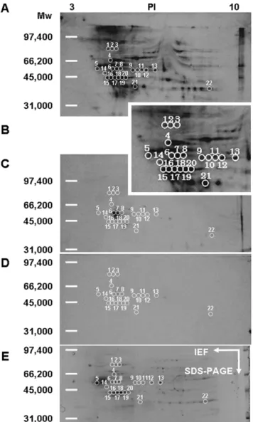
Figure 7. Identification of biotinylated 15d-PGJ2-modified
proteins in the plasma membrane of cortical neurons.Western blotting: Membrane proteins (400mg) were incubated with 1mM biotinylated 15d-PGJ2at 4uC for 24 h in the presence of vehicle (A), Magnified photograph including spot 1-21 (B), 10mM unlabeled 15d-PGJ2(C) and 100mM unlabeled 15d-PGJ2(D). SYPRO Ruby: Membrane proteins (400mg) were incubated with 1mM biotinylated 15d-PGJ2at 4uC for 24 h in the presence of vehicle (E). The proteins were separated by isoelectrofocusing (pH 3-10) and then by SDS-PAGE. The white circles denote spots excised for subsequent identification by MALDI-TOF analysis, as described under Expreimental Procedures.
TheIC50value of 15d-PGJ2to the specific binding of [ 3
H]15d-PGJ2
in BSMC was 31mM, and 20-fold higher than that (1.6mM) in neuronal cells. The binding sites of 15d-PGJ2in cortical neurons
could also be recognized byD12-PGJ2and PGJ2, whereas those in
BSMC could be specifically done by 15d-PGJ2(Figure 5B). In the
two cells, the MTT-reducing activities of 15d-PGJ2 and its
Figure 8. MALDI-TOF mass spectrum of the tryptic digest of spot 8.Spot 8 from Figure 7D was digested in gel with trypsin, and the resulting peptides were analyzed by MALDI-TOF MS as detailed in the experimental section. (A) Typical mass spectrum from a representative experiment. (B) Probability based Mowse Score. (C) Positions of matched peptides in the sequence of GFAP.
doi:10.1371/journal.pone.0017552.g008
Figure 9. Peptide matches of spot 8 with GFAP.List of the monoisotopic masses of some of the peptides identified showing their position in the sequence of GFAP.
precursors were paralleled to the affinities of these ligands for the membrane specific binding sites of 15d-PGJ2.
Comparison of the specific binding sites for [3H]15d-PGJ2
in plasma membranes to authentic receptors, DP1 and DP2
In peripheral tissues including nerves, chemoattractant receptor-homologous molecule expressed on Th2 cells has been identified as a type 2 receptor for PGD2(DP2), and reported to be also a membrane
receptor for 15d-PGJ2[20]. We compared characterization of specific
binding sites for [3H]15d-PGJ2(SBJ) and DP2. According to LD50
and LT50, the apoptotic effect of 15d-PGJ2-related compounds was
15d-PGJ2.D12-PGJ2.PGJ2.PGA2&PGD2.15d-PGD2in
sequence (Table 1). In the view of IC50, the affinity of 15d-PGJ2–
related compounds for SBJ was 15d-PGJ2.D12-PGJ2.PGJ2.
PGA2&PGD2.15d-PGD2in sequence (Table 1). On the other
hand, the affinity of 15d-PGJ2–related compounds for DP2 was
PGD2.15d-PGD2.15d-PGJ2.PGJ2.D12-PGJ2&PGA2in
sequence (Table 1). In addition, the affinity of 15d-PGJ2–related
compounds for DP1 was PGJ2 .PGD2 & D12-PGJ2 .
15d-PGJ2.15d-PGD2 . PGA2 in sequence (Table 1). Thus, the
apoptotic effect of 15d-PGJ2-related compounds was correlated to
their affinities for SBJ, but not to those for DP1 or DP2.
Isolation of Targets for 15d-PGJ2
To identify target proteins for 15d-PGJ2, membrane proteins
were labeled with biotinylated 15d-PGJ2 under the serum-free
condition to reduce non-specific binding. Under this condition, biotinylated 15d-PGJ2induced neuronal cell death in a
concentra-tion- dependent manner as well as 15d-PGJ2. Their LD50 values
were almost 1mM (Figure 6A). Biotinylated 15d-PGJ2suppressed
the extension of neurites and shrank cell bodies in a similar fashion to 15d-PGJ2(Figure 6B). Next, neuronal plasma membranes were
incubated with 1mM biotinylated 15d-PGJ2 in the absence or
presence of 15d-PGJ2 at the indicated concentrations. Then,
membrane proteins modified with biotinylated 15d-PGJ2 were
separated by two-dimensional gel electrophoresis. The patterns that were given by western blot analysis probed with anti-biotin antibody-HRP and SYPRO Ruby fluorescence staining are shown in Figure 7. Several biotinylated 15d-PGJ2-protein conjugates were
detected as biotin-positive spots (Figure 7A and 7B). 15d-PGJ2
inhibited the modification of proteins with the biotinylated 15d-PGJ2in a concentration-dependent manner (Figure 7C and 7D). At
100mM, 15d-PGJ2eliminated almost completely the biotin-positive
spots (Figure 7D). After superimposition of both patterns, the SYPRO Ruby-stained proteins that coincided with the biotin-positive spots were excised from two-dimensional gels (Figure 7E), subjected to trypsin digestion, and then successfully analyzed by MALDI-TOF MS fingerprint analysis (Figure 8A).
Identification of Targets for 15d-PGJ2
Spot 8 corresponding to a 50 kDa 15d-PGJ2-protein conjugate
was one of the targets of the modification by biotinylated 15d-PGJ2, as seen in Figure 8. Using MASCOT, the probability based
MOWSE score was 267 for GFAP (p,0.05) (Figure 8B), with 28
Table 2.Membrane proteins targeted for 15d-PGJ2.
No Protein Swiss-plot MW PI Score Matches Coverage
1 Hspa8 P63018 71055 5.37 72 25/48 49
2 Hspa8 P63018 71055 5.37 100 13/20 23
3 Hspa8 P63018 71055 5.37 114 14/20 24
4 Internexina P23565 56224 5.20 89 11/20 18
5 Tubulinb2b Q3KRE8 49931 4.78 99 11/20 21
6 GFAP P47819 49984 5.35 97 14/40 40
7 GFAP P47819 49984 5.35 292 31/40 51
8 GFAP P47819 49984 5.35 267 28/39 56
9 CK20 P25030 49414 5.28 52 12/19 31
10 TCP1a P28480 60835 5.86 51 10/33 22
11 PKM1 P11980 58331 6.63 52 9/29 26
12 Enolase1 P04764 47098 6.16 73 9/20 22
13 Enolase1 P04764 47098 6.16 62 8/20 23
14 Enolase 2 P07323 47111 5.03 50 7/20 19
15 Actinb P60711 42052 5.29 73 7/20 52
16 Actinb P60711 42052 5.29 65 9/37 28
17 Actinb P60711 42052 5.29 79 8/19 23
18 Actinb P60711 42052 5.29 58 9/38 27
19 Actinb P60711 42052 5.29 48 8/34 23
20 Actinb P60711 42052 5.29 82 9/24 26
21 CapZa2 Q3T1K5 33118 5.57 62 8/30 34
22 GAPDH P04797 36090 8.14 73 10/36 27
Spots that were excised from the gel show in Figure 7E were identified by tryptic digestion and MALDI-TOF MS. Shown are the spot number, name of the identified protein, the accession number in the SwissProt database, the theoretical molecular mass and isoelectric point, the probability based MOWSE score, the number of peptides matched according to the Mascot database, the percentage of the protein sequence that is covered by the identified peptides.
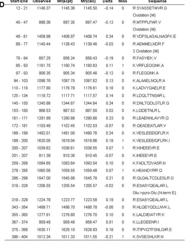
peptide matches (error60.02%) (Figure 9), which represents 56% sequence coverage (Figure 8C). Table 2 lists the identity of 22 protein spots, which could be identified in three independent experiments. The multiple gel spots for a single identification could be ascribed to posttranslational modification, such as phosphorylation. For example, spot 6 could contain 3 phosphor-ylation sites (T129, T130 and Y283), which represented the probability based MOWSE score59, 16 peptide matches, 32% sequence coverage. Spot 7 could contain 1 phosphorylation site (Y283), which represented the probability based MOWSE score 188, 31 peptide matches, 51% sequence coverage. On the other hand, the phosphorylation site of spot 8 was not detected. The identified proteins fall into several different functional classes, including glycolytic enzymes (Enolase 1, Enolase 2, GAPDH and PKM1), molecular chaperones (Hsp8a and TCP1a) and cytoskel-tones (Tubulinb2b, Actinb, Internexina, GFAP and CapZa2).
Next, we attempted to detect the 15d-PGJ2-target adducts in the
plasma membranes exposed to the biotinylated 15d-PGJ2. by
streptavidin agarose pull-down assays. Western blot revealed that 15d-PGJ2interacted with Actinb, Enolase 2, GAPDH, Internexin a, PKM1, TCP1a and Tubulin b2b (Figure 10). Since plasma membranes were prepared from adult cerebral cortices including neurons and astrocytes, non-neuronal enolase1 and GFAP appeared to be derived from astrocytes.
Regions homologous to the binding site of 15d-PGJ2in
targeted proteins
Several lines of evidences indicate the covalent binding sites of 15d-PGJ2 in previous target proteins. To ascertain whether the
cysteine residue in the present target proteins responded to the covalent binding sites of 15d-PGJ2in previous target proteins or
not, homologous regions were searched (Table 3). As query sequences, we used the amino acid sequences of the previous target proteins, in which the covalent binding sites of 15d-PGJ2are
identified: Cys374 of Actin b (P60711) [21], Cys269 of c-Jun (NP_068607) [22], Cys184of H-ras (NP_001091711) [23], Cys179 of IkB-kinaseb(Q9QY78)8, Cys285of PPARc(NP_619725) [8], Cys35 and Cys69 of thioredoxin (NP_446252) [24]. Hspa8 contained Cys603responded to the Cys179of IkB-kinase b. The amino sequence of Hspa8 from Lys597to Leu610was homologous to that of IkB-kinase b from Lys171 to Leu186. Based on the comparison between the two sequences, the initial score, the optimal score and the identity were 15, 29 and 31%, respectively. In a similar fashion, Internexina, Tubulin b2b, GFAP, CK20, TCP1a, PKM1, Enolase 1, Enolase 2, Actin b, CapZa2 and GAPDH contained the cysteine residue responded to Cys69 of thioredoxin, Cys184 of H-ras, Cys269 of c-Jun, Cys69 of thioredoxin, Cys35 of thioredoxin, Cys35 of thioredoxin, Cys184 of H-ras, Cys184of H-ras, Cys374of actinb, Cys179of IkB-kinase b8, Cys35 of thioredoxin, respectively. Thus, the present target proteins also contained the cysteine residue responded to the previous covalent binding site of 15d-PGJ2, and exhibited
homologous sequences around the specific binding site.
Discussion
Cortical neurons and BSMC sensitive to amyloid protein were susceptible to 15d-PGJ2. [
3
H]15d-PGJ2bound specifically to the
two cells, suggesting that 15d-PGJ2 played an important role in
amyloidoses not only in the central nervous system but also in the peripheral tissues. The specific binding sites of [3H]15d-PGJ2were
detected in the neuronal subcellular fractions of nuclear, cytosol and plasma membrane, but not in the microsomal fraction. 15d-PGJ2binds to the nuclear receptor, PPARc[9] and the cytosolic
protein, Ras [23]. In peripheral tissues including nerves, chemoattractant receptor-homologous molecule expressed on Th2 cells has been identified as a type 2 receptor for PGD2
(DP2), and reported to be also a membrane receptor for 15d-PGJ2
[20]. Contrary to its mRNA, little protein of DP2 has yet been detected in the central nerve. Furthermore, we ruled out the possibility that the specific binding site of 15d-PGJ2in the plasma
membrane of cortical neurons was DP2. First, few binding sites of [3H]PGD2 are detected in plasma membranes from rat cortices
[4]. Although binding sites of [3H]?12-PGJ2and [3H]PGJ2are also
detected in plasma membranes, those are displaced most potently by 15d-PGJ2 among PGD2 metabolites [4]. Second, a DP2
selective agonist, 15d-PGD2 do not affect the cell number of
neuronal cells and BSMC (Figure 3B and Table 1). Third, the LD50value (.10mM) of PGD2is much higher than the affinity for
PGD2receptor (dissociation constant = 8.8 nM) [20].
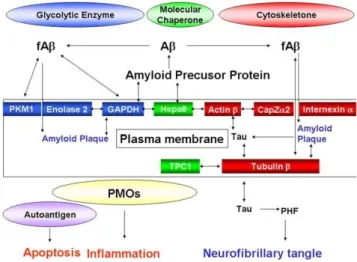
In the present study, we identified membrane proteins targeted for 15d-PGJ2including glycolytic enzymes, molecular chaperones
and cytoskeletons (Table 2 and Figure 10). GAPDH, Enolase 1, Enolase 2 and PKM1 were previously believed to perform exclusively ‘house-keeping’ glycolysis. GAPDH is not only found in the cytoplasm, but also closely associated with the plasma membrane [25]. GAPDH catalyses the conversion of glyceralde-hyde 3-phosphate to D-glycerate 1,3-bisphosphate. Reduction in glycolysis precedes cognitive dysfunction and is therefore believed to be an important early event in AD development [26]. Apart from its glycolytic role, overexpression of the particular mem-brane-associated GAPDH has a direct role in neuronal apoptosis [27] (Figure 11). GAPDH is located in amyloid plaques [28], Figure 10. Interactions of 15d-PGJ2 with targets. Membrane
proteins (400mg) were incubated with 1mM biotinylated 15d-PGJ2at 4uC for 24 h. Membrane lysates were incubated with Streptavidin Agarose. The presence of targeted proteins was detected by immunoblot analysis, and the incorporation of biotinylated 15d-PGJ2 into immunoprecipitates was detected with ECL.
interacts with the C-terminal region of amyloid precursor protein (APP) [29], and co-precipitates with fAb[30]. Furthermore, GAPDH associates tightly with Enolase 2 and Hspa8, and makes
uptrans-plasma-membrane oxidoreductases (PMOs), the
extracel-lular redox sensor for signaling external oxidative stress to the cell [31].
Enolase 1 and Enolase 2 belong to a superfamily of abundantly expressed carbon-oxygen lyases known for the catalysis of 2-phosphoglycerate to phosphoenolpyruvate. Ubiquitous enolase1 and neuron specific enolase 2 exist as monomers and also as dimmers on the neuronal membrane surface [32]. Recent studies have demonstrated that enolases possess different regulatory properties from glycolysis in the brain [33]. Enolase1 is one of the most consistently up-regulated and oxidatively modified proteins in brain of subjects of early-onset AD [34]. Enolase1 and enolase 2 are autoantigen targets in post-streptococcal autoimmune disease of central nervous system (Figure 11). The anti-enolase antibodies induce neuronal apoptosis [35]. Enolase 2 is part of neuronal PMOs, and the anti-enolase2 antibody can inhibit PMO activity on the plasma membrane [31].
Pyruvate kinase transfers a phophate from phosphoenolpyr-uvate to ADP. Pyrphosphoenolpyr-uvate kinase is also defined as the autoantigen, and its antibodies induce neuronal apoptosis [35] (Figure 10). The significant increase in pyruvate kinase activity is found in frontal and temporal cortex of AD brains [36]. Pyruvate kinase is elevated in the cortical neurons undergoing Ab-mediated apoptosis [37]. Pyruvate kinase is co-precipitated with fAb[30]. Biotinylated 15d-PGJ2 binds to PKM1 in mesangial cells [38], supporting our
results.
Hsp8a is dnaK-type molecular chaperone heat shock protein 72-ps1 in the PMO complex [31]. It is located in the cytoplasm [39], but nuclear localization and accumulation near or at the plasma membrane in stressed cells and in synaptosomal mem-branes has been observed [40]. Hsp8a binds to the cytoplasmic domain near the post-transmembrane region of APP (Figure 11). TCP1ais a selective molecular chaperone in tubulin biogenesis, by that nascent tubulin subunits are bound to TCP1aand released in assembly competent forms. Cytoskeletal proteins are deficient and aggregated in AD. When TCP1ais related to its natural and Figure 11. Hypothetical roles of targets for 15d-PGJ2 on
amyloidoses.Membrane target proteins for 15d-PGJ2were glycolytic enzymes (Enolase 2, PKM1 and GAPDH), molecular chaperones (Hsp8a and TCP1a), and cytoskeletal proteins (Actinb, CapZa2, Tubulinband Internexin a). These proteins were factors associated with the two remarks of AD, the amyloid plaque and the neurofibrillary tangle. Beyond classical roles as glycolytic enzymes and molecular chaperones, GAPDH, Enolase2 and Hsp8a appear to form the complex of PMOs and contribute to the generation of reacting oxygen species by 15d-PGJ2. doi:10.1371/journal.pone.0017552.g011
Table 3.Regions homologous to the binding site of 15d-PGJ2 in targeted proteins.
Initial Score
Protein Sequences Optimal Score
Identity
IkB-kinaseb Query 171 KELDQGSLCTSFVGTL 186 15
| | | . . . | . . . | 29
Hspa8 Sbjct 597 KELEK– –VCNPI I T KL 610 5/16 (31%)
Thioredoxin Query 60 DDCQDVAADCE 70 16
. . | . | | | 24
Internexina Sbjct 173 EEV QRLR ARCE 183 4/11 (36%)
H-ras Query 183 SCKCV 187 14
| | . | . 28
Tubulinb2b Sbjct 126 SCDCL 130 3/5 (60%)
c-Jun Query 261 RNRI AAS KCRKRKL 274 16
| . . . . | . | . | 21
GFAP Sbjct 284 RRQLQALTC DLE S L 297 4/14 (28%)
Thioredoxin Query 59 VDDCQDVAADCEVK 72 8
. . | | . | | . . 20
CK20 Sbjct 136 I KDAQ I ENARCVLQ 149 4/14 (28%)
Thioredoxin Query 34 PCKMI KPFFH 43 18
. | | . . . . 23
TCP1a Sbjct 124 ACKEAVRYIN 133 2/10 (20%)
Thioredoxin Query 35 CKMI 38 17
| | . . 23
PKM1 Sbjct 165 CKVV 168 2/4 (50%)
H-ras Query 178 GPGCMSCKCV L 188 17
. . | . | | . | . | 37
Enolase 1 Sbjct 331 AAGE KSCNCL L 341 5/11 (45%)
H-ras Query 183 SCKCVL 188 17
. | . | . | 34
Enolase 2 Sbjct 336 ACNCLL 341 3/6 (50%)
Actinb Query 356 WISKQEYDESGPSIVHRKCF 375 117
| | | | | | | | | | | | | | | | | | | | 117
Actinb Sbjct 356 WISKQEYDESGPSIVHRKCF 375 20/20 (100%)
IkB-kinaseb Query 169 YAKELDQGSL CT SFVGTLQ 187 21
| . | | . . . . | | . . . . 38
CapZa2 Sbjct 131 YVKEHYPNGVCTVYGKKVD 149 5/19 (26%)
Thioredoxin Query 35 CKMIKP 40 11
| . . | | 28
GAPDH Sbjct 245 CRLEKP 250 3/6 (50%)
Homologies were determined with Lipman-Pearson searching algorithms using the Swiss-plot database. As query sequences, we used the amino acid sequences of the previous target proteins, in which the covalent binding sites of 15d-PGJ2are identified: Cys374of Actinb(P60711) [50], Cys269of c-Jun
(NP_068607) [51], Cys184of H-ras (NP_001091711) [20], Cys179of I
kB-kinaseb
(Q9QY78) [12], Cys285of PPARc(NP_619725) [8], Cys35and Cys69of thioredoxin
(NP_446252) [52]. As subject sequences, we used the amino acid sequences of our target proteins. The listed sequences exhibited the highest score in the initial score, the optimal score and the identity.
specific substrate tubulinb, the ratio is significantly decreased in the temporal, frontal, parietal cortex and in thalamus of AD patients [41]. Relatively decreased molecular chaperoning of tubulin b by TCP1a is suggested to lead to misfolded tubulin aggregating and accumulating in plaques and tangles, a hallmark of AD (Figure 11).
Tubulin has been identified as a membrane component of synaptosomes and various plasma membranes. Both tubulinaand
b have been shown to associate with the amyloid deposits of familial amyloidosis [42] and to bind to the Absequence of APP [43]. Moreover, tubulinbis retained by a monomeric Abcolumn [44], and co-precipitated with fAb [30] (Figure 11). The tau protein interacts with tubulin to stabilize microtubules and promote tubulin assembly into microtubules. PGJ2 induces
caspase-mediated cleavage of tau, generatingDtau, an aggregation prone form known to seed tau aggregation prior to neurofibrillary tangle formation [45]. Hyperphosphorylation of the tau protein (tau inclusions) can result in the self-assembly of tangles of paired helical filaments and straight filaments, which are involved in the pathogenesis of AD [46]. Biotinylated 15d-PGJ2binds to tubulinb
in mesangial cells [38], supporting our results.
AD-linked human Ab synergistically enhances the ability of wild-type tau to promote alterations in the actin cytoskeleton (Figure 11) and neurodegeneration [47]. The ability of globular actin to rapidly assemble and disassemble into filaments is critical to many cell behaviors. F-actin-capping protein subunit a-2 (CapZa2) regulates growth of the actin filament by capping the barbed end of growing actin filaments (Figure 11). Members of the actin-depolymerizing factor (ADF)/cofilin family are important regulators of actin dynamics. ADF and cofilin’s ability to increase actin filament dynamics is inhibited by their phosphorylation on Ser3 by LIM kinase 1 and other kinases [48] Ab dystrophy requires LIM kinase 1-mediated phosphorylation of ADF/cofilin and the remodeling of the actin cytoskeleton [49]. Biotinylated 15d-PGJ2 covalently binds to actin b in various cells [38] other
than neurons, supporting our results in neurons.
Internexin ais classified as a type IV neuronal intermediate filament. Internexina also co-assembles with the neurofilament (NF) triplet proteins [50]. The protein is expressed by most, if not all, neurons as they commence differentiation and precedes the expression of the NF triplet proteins [51]. Although the interaction of internexinawith amyloid proteins has not yet been reported, Internexina, and not NF triplet, ring-like reactive neurites are present in end-stage AD cases, indicating the relatively late involvement of neurons that selectively contain Internexin a
(Figure 11). Another intermediate filament protein, GFAP is expressed exclusively in astrocytes. Abincreased the total number of activated astrocytes, and elevated the expression of GFAP by Ab-induced spontaneous calcium transients [52]. 15d-PGJ2
suppresses inflammatory response by inhibiting NF-kB signaling at multiple steps as well as by inhibiting the PI3K/Akt pathway independent of PPARcin primary astrocytes [53].
In conclusion, membrane target proteins for 15d-PGJ2 were
factors associated with the two remarks of AD, the amyloid plaque and the neurofibrillary tangle. Beyond classical roles as glycolytic enzymes and molecular chaperones, GAPDH, enolase 2 and Hsp8a can form the antioxidant complex of PMOs responded to the extracellular oxidative stress. 15d-PGJ2 might regulate the
activity of PMOs during inflammation and degeneration. Apart from glycolysis, pyruvate kinase and enolase might be involved in the 15d-PGJ2–induced apoptosis as autoantigens. Thus, the
present study sheds light on the ecto-enzymes targeted for 15d-PGJ2as a prelude to the death receptor stimulated by 15d-PGJ2or
the antioxidant complex regulated by 15d-PGJ2.
Author Contributions
Conceived and designed the experiments: TY. Performed the experiments: YY KT JK M. Fujita NO TS. Analyzed the data: YY KT TY. Contributed reagents/materials/analysis tools: M. Fujimoto. Wrote the paper: YY TY.
References
1. Iwamoto N, Kobayashi K, Kosaka K (1989) The formation of prostaglandins in the postmortem cerebral cortex of Alzheimer-type dementia patients. J Neurol 236: 80–84.
2. Selkoe DJ (1991) The molecular pathology of Alzheimer’s disease. Neuron 6: 487–498.
3. Yagami T, Ueda K, Asakura K, Sakaeda T, Kuroda T, et al. (2001) Effects of S-2474, a novel nonsteroidal anti-inflammatory drug, on amyloid beta protein-induced neuronal cell death. Br J Pharmacol 134: 673–681.
4. Yagami T, Ueda K, Asakura K, Takasu N, Sakaeda T, et al. (2003) Novel binding sites of 15-deoxy-delta12,14-prostaglandin J2 in plasma membranes from primary rat cortical neurons. Exp Cell Res 291: 212–227.
5. Wright DH, Nantel F, Metters KM, Ford-Hutchinson AW (1999) A novel biological role for prostaglandin D2 is suggested by distribution studies of the rat DP prostanoid receptor. Eur J Pharmacol 377: 101–115.
6. Boie Y, Sawyer N, Slipetz DM, Metters KM, Abramovitz M (1995) Molecular cloning and characterization of the human prostanoid DP receptor. J Biol Chem 270: 18910–18916.
7. Sasaguri T, Masuda J, Shimokado K, Yokota T, Kosaka C, et al. (1992) Prostaglandins A and J arrest the cell cycle of cultured vascular smooth muscle cells without suppression of c-myc expression. Exp Cell Res 200: 351– 357.
8. Shiraki T, Kamiya N, Shiki S, Kodama TS, Kakizuka A, et al. (2005) Alpha, beta-unsaturated ketone is a core moiety of natural ligands for covalent binding to peroxisome proliferator-activated receptor gamma. J Biol Chem 280: 14145–14153.
9. Forman BM, Tontonoz P, Chen J, Brun RP, Spiegelman BM, et al. (1995) 15-Deoxy-delta 12, 14-prostaglandin J2 is a ligand for the adipocyte determination factor PPAR gamma. Cell 83: 803–812.
10. Kliewer SA, Lenhard JM, Willson TM, Patel I, Morris DC, et al. (1995) A prostaglandin J2 metabolite binds peroxisome proliferator-activated receptor gamma and promotes adipocyte differentiation. Cell 83: 813–819.
11. Ward JE, Gould H, Harris T, Bonacci JV, Stewart AG (2004) PPAR gamma ligands, 15-deoxy-delta12,14-prostaglandin J2 and rosiglitazone regulate human
cultured airway smooth muscle proliferation through different mechanisms. Br J Pharmacol 141: 517–525.
12. Rossi A, Kapahi P, Natoli G, Takahashi T, Chen Y, et al. (2000) Anti-inflammatory cyclopentenone prostaglandins are direct inhibitors of IkappaB kinase. Nature 403: 103–108.
13. Gabazza EC, Taguchi O, Tamaki S, Takeya H, Kobayashi H, et al. (1999) Thrombin in the airways of asthmatic patients. Lung 177: 253–262. 14. Patel HJ, Belvisi MG, Bishop-Bailey D, Yacoub MH, Mitchell JA (2003)
Activation of peroxisome proliferator-activated receptors in human airway smooth muscle cells has a superior anti-inflammatory profile to corticosteroids: relevance for chronic obstructive pulmonary disease therapy. J Immunol 170: 2663–2669.
15. Yagami T, Ueda K, Asakura K, Hata S, Kuroda T, et al. (2002) Human group IIA secretory phospholipase A2 induces neuronal cell death via apoptosis. Mol Pharmacol 61: 114–126.
16. Yagami T, Takahara Y, Ishibashi C, Sakaguchi G, Itoh N, et al. (2004) Amyloid beta protein impairs motor function via thromboxane A2 in the rat striatum. Neurobiol Dis 16: 481–489.
17. Yagami T, Tohkin M, Matsubara T (1990) Sex difference in adrenergic receptor-mediated glycogenolysis in rat livers. Jpn J Pharmacol 54: 365–374. 18. Yagami T (1995) Differential coupling of glucagon and beta-adrenergic
receptors with the small and large forms of the stimulatory G protein. Mol Pharmacol 48: 849–854.
19. Toda T, Kimura N (1997) Standardization of protocol for immobiline 2-D PAGE and construction of 2-D PAGE protein database on World Wide Web home page. Jpn J Electroph 41: 13–19.
20. Hata AN, Zent R, Breyer MD, Breyer RM (2003) Expression and molecular pharmacology of the mouse CRTH2 receptor. J Pharmacol Exp Ther 306: 463–470.
22. Perez-Sala D, Cernuda-Morollon E, Canada FJ (2003) Molecular basis for the direct inhibition of AP-1 DNA binding by 15-deoxy-delta 12,14-prostaglandin J2. J Biol Chem 278: 51251–51260.
23. Oliva JL, Perez-Sala D, Castrillo A, Martinez N, Canada FJ, et al. (2003) The cyclopentenone 15-deoxy-delta 12,14-prostaglandin J2 binds to and activates H-Ras. Proc Natl Acad Sci U S A 100: 4772–4777.
24. Shibata T, Yamada T, Ishii T, Kumazawa S, Nakamura H, et al. (2003) Thioredoxin as a molecular target of cyclopentenone prostaglandins. J Biol Chem 278: 26046–26054.
25. Rogalski AA, Steck TL, Waseem A (1989) Association of glyceraldehyde-3-phosphate dehydrogenase with the plasma membrane of the intact human red blood cell. J Biol Chem 264: 6438–6446.
26. Arias C, Montiel T, Quiroz-Baez R, Massieu L (2002) beta-Amyloid neurotoxicity is exacerbated during glycolysis inhibition and mitochondrial impairment in the rat hippocampus in vivo and in isolated nerve terminals: implications for Alzheimer’s disease. Exp Neurol 176: 163–174.
27. Ishitani R, Sunaga K, Hirano A, Saunders P, Katsube N, et al. (1996) Evidence that glyceraldehyde-3-phosphate dehydrogenase is involved in age-induced apoptosis in mature cerebellar neurons in culture. J Neurochem 66: 928–935. 28. Tamaoka A, Endoh R, Shoji S, Takahashi H, Hirokawa K, et al. (1996)
Antibodies to amyloid beta protein (A beta) crossreact with glyceraldehyde-3-phosphate dehydrogenase (GAPDH). Neurobiol Aging 17: 405–414. 29. Schulze H, Schuler A, Stuber D, Dobeli H, Langen H, et al. (1993) Rat brain
glyceraldehyde-3-phosphate dehydrogenase interacts with the recombinant cytoplasmic domain of Alzheimer’s beta-amyloid precursor protein. J Neurochem 60: 1915–1922.
30. Verdier Y, Huszar E, Penke B, Penke Z, Woffendin G, et al. (2005) Identification of synaptic plasma membrane proteins co-precipitated with fibrillar beta-amyloid peptide. J Neurochem 94: 617–628.
31. Bulliard C, Zurbriggen R, Tornare J, Faty M, Dastoor Z, et al. (1997) Purification of a dichlorophenol-indophenol oxidoreductase from rat and bovine synaptic membranes: tight complex association of a glyceraldehyde-3-phosphate dehydrogenase isoform, TOAD64, enolase-gamma and aldolase C. Biochem J 324(Pt 2): 555–563.
32. Ueta H, Nagasawa H, Oyabu-Manabe Y, Toida K, Ishimura K, et al. (2004) Localization of enolase in synaptic plasma membrane as an alphagamma heterodimer in rat brain. Neurosci Res 48: 379–386.
33. Butterfield DA, Lange ML (2009) Multifunctional roles of enolase in Alzheimer’s disease brain: beyond altered glucose metabolism. J Neurochem 111: 915–933. 34. Butterfield DA, Reed T, Newman SF, Sultana R (2007) Roles of amyloid beta-peptide-associated oxidative stress and brain protein modifications in the pathogenesis of Alzheimer’s disease and mild cognitive impairment. Free Radic Biol Med 43: 658–677.
35. Dale RC, Candler PM, Church AJ, Wait R, Pocock JM, et al. (2006) Neuronal surface glycolytic enzymes are autoantigen targets in post-streptococcal autoimmune CNS disease. J Neuroimmunol 172: 187–197.
36. Bigl M, Bruckner MK, Arendt T, Bigl V, Eschrich K (1999) Activities of key glycolytic enzymes in the brains of patients with Alzheimer’s disease. J Neural Transm 106: 499–511.
37. Lovell MA, Xiong S, Markesbery WR, Lynn BC (2005) Quantitative proteomic analysis of mitochondria from primary neuron cultures treated with amyloid beta peptide. Neurochem Res 30: 113–122.
38. Stamatakis K, Sanchez-Gomez FJ, Perez-Sala D (2006) Identification of novel protein targets for modification by 15-deoxy-delta12,14-prostaglandin J2 in
mesangial cells reveals multiple interactions with the cytoskeleton. J Am Soc Nephrol 17: 89–98.
39. Pelham HR (1984) Hsp70 accelerates the recovery of nucleolar morphology after heat shock. Embo J 3: 3095–3100.
40. Whatley SA, Leung T, Hall C, Lim L (1986) The brain 68-kilodalton microtubule-associated protein is a cognate form of the 70-kilodalton mammalian heat-shock protein and is present as a specific isoform in synaptosomal membranes. J Neurochem 47: 1576–1583.
41. Schuller E, Gulesserian T, Seidl R, Cairns N, Lube G (2001) Brain t-complex polypeptide 1 (TCP- 1) related to its natural substrate beta1 tubulin is decreased in Alzheimer’s disease. Life Sci 69: 263–270.
42. Baumann MH, Wisniewski T, Levy E, Plant GT, Ghiso J (1996) C-terminal fragments of alpha- and beta-tubulin form amyloid fibrils in vitro and associate with amyloid deposits of familial cerebral amyloid angiopathy, British type. Biochem Biophys Res Commun 219: 238–242.
43. Islam K, Levy E (1997) Carboxyl-terminal fragments of beta-amyloid precursor protein bind to microtubules and the associated protein tau. Am J Pathol 151: 265–271.
44. Oyama R, Yamamoto H, Titani K (2000) Glutamine synthetase, hemoglobin alpha-chain, and macrophage migration inhibitory factor binding to amyloid beta-protein: their identification in rat brain by a novel affinity chromatography and in Alzheimer’s disease brain by immunoprecipitation. Biochim Biophys Acta 1479: 91–102.
45. Arnaud LT, Myeku N, Figueiredo-Pereira ME (2009) Proteasome-caspase-cathepsin sequence leading to tau pathology induced by prostaglandin J2 in neuronal cells. J Neurochem 110: 328–342.
46. Alonso A, Zaidi T, Novak M, Grundke-Iqbal I, Iqbal K (2001) Hyperpho-sphorylation induces self-assembly of tau into tangles of paired helical filaments/ straight filaments. Proc Natl Acad Sci U S A 98: 6923–6928.
47. Fulga TA, Elson-Schwab I, Khurana V, Steinhilb ML, Spires TL, et al. (2007) Abnormal bundling and accumulation of F-actin mediates tau-induced neuronal degeneration in vivo. Nat Cell Biol 9: 139–148.
48. Morgan TE, Lockerbie RO, Minamide LS, Browning MD, Bamburg JR (1993) Isolation and characterization of a regulated form of actin depolymerizing factor. J Cell Biol 122: 623–633.
49. Bamburg JR, Bloom GS (2009) Cytoskeletal pathologies of Alzheimer disease. Cell Motil Cytoskeleton 66: 635–649.
50. Ching GY, Liem RK (1998) Roles of head and tail domains in alpha-internexin’s self-assembly and coassembly with the neurofilament triplet proteins. J Cell Sci 111(Pt 3): 321–333.
51. Kaplan MP, Chin SS, Fliegner KH, Liem RK (1990) Alpha-internexin, a novel neuronal intermediate filament protein, precedes the low molecular weight neurofilament protein (NF-L) in the developing rat brain. J Neurosci 10: 2735–2748.
52. Chow SK, Yu D, Macdonald CL, Buibas M, Silva GA (2010) Amyloid-beta directly induces spontaneous calcium transients, delayed intercellular calcium waves, and gliosis in rat cortical astrocytes. ASN Neuro 2: 15–23.


![Figure 5. Binding assay of [ 3 H]15d-PGJ 2 to subcellular frac- frac-tions. (A) Proteins fractionated to plasma membranes, nuclear, cytosol or microsome from cortical neurons were incubated with 10 nM [ 3 H]15d-PGJ 2 at 25 u C for 1 h in the absence or pre](https://thumb-eu.123doks.com/thumbv2/123dok_br/18377920.356064/5.918.93.440.378.852/binding-subcellular-proteins-fractionated-membranes-microsome-cortical-incubated.webp)
![Table 1. Comparison of the specific binding sites for [ 3 H]15d-PGJ 2 in plasma membranes to authentic receptors, DP1 and DP2.](https://thumb-eu.123doks.com/thumbv2/123dok_br/18377920.356064/6.918.94.572.397.1005/table-comparison-specific-binding-plasma-membranes-authentic-receptors.webp)