45
EFFECT OF NATURAL PLANT EXTRACTS ON PORCINE OVARIAN FUNCTIONS
Attila Kádasi*
1, Aneta Štochmaľová
3,
Nora Maruniaková
1, Adriana Kolesárová
1, Roland Grossman
4, Alexander V. Sirotkin
2,3Address(es):
1Slovak University of Agriculture in Nitra, Faculty of Biotechnology and Food Sciences, Department of Animal Physiology, Department of Animal Physiology Tr. A.
Hlinku2, 949 76 Nitra, Slovak Republic;
2Institute for Genetics and Reproduction of Farm Animals, Animal Production Research Centre Nitra, Lužianky, Slovak Republic; 3Constantine the Philosopher University in Nitra, Faculty of Natural Sciences, Department of Zoology and Anthropology, 4
Department of Functional Genomics and Bioregulation, Institute of Animal Science, Mariensee, Neustadt, Germany
*Corresponding author: attila.kadasi@gmail.com
ABSTRACT
Keywords: Natural plant, proliferation, apoptosis, hormone secretion, ovarian cells
INTRODUCTION
Nowadays, the study of natural plant and their substances with pharmacological activity has become an emerging trend in nutritional and pharmacologic research. Natural plant extracts represent a rich group used as remedies for human and animal diseases and for regulation of particular physiological processes (Hammer et al., 1999; Nostro et al., 2000). These plant extracts are used due to their preventive, antibacterial and therapeutic effects, anticancer and apoptosis inducing-properties (Blanko et al., 2003). Ginkgo biloba extract displays free radical scavenging and antioxidant actions (Marcocci
et al., 1994). In vitro experiments showed that Ginkgo extract and its components have significant anti-proliferative effects in ovarian cancer cells (Ye et al., 2007). It is known that Rooibos tea contains abundant flavonoids (Shimoi et al., 1996), aspalathin, chrysoeriol, orientin, isoorientin, vitexin, isovitexin, quercetin, isoquercitrin and rutin (Duke et al., 2002). Rooibos tea is commonly used for treating cardiac arrhythmias, colic, diarrhea (Duke et al., 2002), asthma (Brown,
1995) and hypertension (Nakano, 1997). Flaxseed is a rich source of 3
components with demonstrated cardioprotective effects (Chantal et al., 2009), can inhibit arrhythmogenesis during ischemia-reperfusion (Ander et al., 2004), inhibit atherogenesis (Prasad, 2005), and protect against vascular dysfunction during hypercholesterolemic conditions (Dupasquier et al., 2006). RSV was chemically 3, 4', 5 – trihydroxystilbene, is one of the natural phytoalexins (Hain
et al., 1990)and is found in grapevines, in soft fruits and hazelnut (Frémont,
2000). CURC is Curcuma longa L. extract and chemicaly is 1,7-bis(4-hydroxy-3-methoxyphenyl)-1,6-eptadiene-3,5-dione (Nadkarmi, 1976). Among effects of CURC extract is reduction of P secretion in mature follicle of swine GCs
(Nurcahyo and Kadarsih, 2003). Green tea is obtained from the leaves and the
leaf buds of the plant Camellia sinensis. GTPP may induced reductions in the levels of sex steroids hormones as testosterone and estradiol and possible negative effects on reproductive performance (Kao et al., 2000) on granulosa cell functions (Basini et al., 2005) and in vitro fertilization in swine (Spinaci et al.,
2006). The main components of green tea are polyphenols. 50-80 % of
polyphenols are represented by special flavonoids - catechins, especially epigallocatechin-3-gallate (EGCG) (Fukai et.al., 1991; Khan et al., 2006). Long term consumption of green tea may influence the incidence of obesity, diabetes, and cardiovascular disease (Kao et al., 2000). Direct effect of these natural plant and plant molecules on healthy ovarian cells functions remaines unknow. The aim of our studies was to analyze the effects of selected plants, GB, RB, FL, and
plant substances GTPP, RSV and CURC on markers of proliferation, apoptosis and secretory activity of porcine ovarian granulosa cells.
MATERIAL AND METHODS
Granulosa cells were aspirated from the ovaries of Slovakian white gilts after slaughter at a local abattoir. The subsequet procedures followed standard protocols according to Sirotkin, et al.(2008). After formed confluent monolayer and medium replacement experimental cells were cultured in the presence of GB (Shangai TECH Chemical Indutry Testing Co., Ltd), RB
(Clanwilliam, South Africa), FL (MEDU s.r.o., Čenkovce, Slovakia), RSV,
CURC, GTPP and EGCG (all from Changsha Sunfull Bio-tech. Co, Hunan China) alone at concentrations of 0; 1; 10 and 100 µg.ml-1.
After removing the medium from chamber slides, cell were washed in ice-cold PBS (pH 7.5), fixed in paraformaldehyde (4% in PBS, pH 7.2-7.4; 60 min) and
held at 4°C to await immunocytochemistry. The medium from the plate wells was gently aspirated and frozen at -24 °C to await EIA. Concentrations of P4, T
and L were determined in 25 µl aliquots of incubation medium by EIA,
previously validated for use in culture medium, by using antisera against steroids (produced in the Institute of Animal Science, Neustadt, Germany) as previously described and characterised (Sirotkin et al., 2008). Significant differences
among the experiments were evaluated using Student´s T-test and one/two-way ANOVA folowed by paired Wilcoxon-Mann Whitney test, by using Sigma Plot 11.0 software (Systat Software, GmbH, Erkhart, Germany). Differences against control at P<0.05 were considered as significant.
RESULTS AND DISCUSSION Immunocytochemistry
In our study of plants extracts (GB, RB, FL, RSV and EGCG) significantly inhibited the percentage of cells containing PCNA at all doses added. Proliferation of GCs was diminished also after addition of CURC (at dose 10 µg.ml-1) and GTPP (at doses 10 and 100 µg.ml-1). These datas comfirmed antiproliferative activity on cancer cells (Salganik, 2001; Kim et al., 2005; Chen
et al., 2002; Xu et al., 2003; Wei et al., 2007; Zhang et al., 2008; Demark-Wahnefried et al., 2001; Thompson et al., 2005; Zhou et al., 2009), on interstitial theca cells (Wong et al., 2010), in rat ovarian GCs (Ortega et al.,
2012), on non-ovarian and swine ovarian cells (Nurcahyo and Kadarsih, 2003;
Huh et al., 2004; Spinella et al., 2006; Basini et al., 2005a,b). Our results are This report provides information about the impact of chosen natural plant extracts on basic ovarian functions. This article summarizes our results concerning the effect of selected plant extracts on proliferation, apoptosis and hormone secretion – release of progesterone (P4), testosterone (T) and leptin (L) on porcine granulosa cells (GC), We analyzed effects ofginkgo (GB), rooibos (RB), flaxseed (FL), green tea polyphenols (GTPP), green tea - epigallocatechin-3-gallate (EGCG), resveratrol (RSV) and curcumin (CURC) (0; 1; 10 and 100 μg.ml-1) on markers of proliferation, apoptosis and secretory activity of porcine ovarian granulosa cells by using immunocytochemistry and EIA. It was demonstrated, that all these natural plants and plant molecules inhibited the accumulation of proliferation-related peptide (PCNA) and apoptosis-associated peptide (Bax) in cultured. Furthermore, it was observed that natural plant extracts altered progesterone, testosterone and leptin release in porcine ovarian cells.It is concluded, thatGB, RB, FL, RSV, CURC, GTPP and EGCG can directly affect ovarian cells and thereforethey could potentially influence ovarian functions.
ARTICLE INFO
Received 2. 12. 2014 Revised 7. 1. 2015 Accepted 20. 1. 2015 Published 2. 2. 2015
Regular article
J Microbiol Biotech Food Sci / Kádasi et al. 2015 : 4 (special issue 2) 45-48
46 not inline with Pantsi et al. (2001) reports of the cardio-protective properties of aqueous rooibos extracts via the inhibition of apoptosis. These observation is the first observation on healthy ovarian cells , that these plants and their molecules
can directly suppress ovarian cell proliferation and therefore potentially inhibit ovarian follicle growth and development.
Table 1. The percentage of cells containing marker of proliferation PCNA in cultured ovarian granulosa cells cultured with and without (control) GB, RB, FL, RSV,
CURC, GTPP and EGCG
Supplement
Doses of supplement added (µg.ml-1)
0 (control) 1 10 100
GB 45.21±1.07
(1900)
24.00±0.99*
(500)
20.50±1.12*
(400)
22.55±1.35*
(550)
RB 45.21±1.07
(1900)
28.75±2.03*
(400)
26.20±1.44*
(500)
22.20±0.76*
(500)
FL 45.21±1.07(1900) 23.40±0.67*
(500)
21.75±1.10*
(400)
17.40±0.85*
(500)
RSV 51.00±1.43
(1404)
36.30±1.20*
(356)
39.50±1.50*
(273)
34.00±10.00*
(320)
CURC 51.00±1.43(1404) 50.5±3.43
(867)
42.83±1.49*
(743)
44.71±2.56
(823)
GTPP 49.86±1.3
(3311)
45.13±2.17
(903)
34.6±3.98*
(597)
41.0±3.53*
(658)
EGCG 49.86±1.3
(3311)
40.42±2.99*
(844)
38.88±3.46*
(907)
36.88±2.53*
(909)
All the values represent % of cells containing particular antigen, means ± SEM, *- significant (P<0.05) differences with control (cells not treated with plant molecules). In the brackets is a number of counted cells.
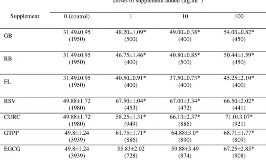
In our study plant extracts (GB, RB, FL, RSV, CURC and GTPP) significantly stimulated the number of cells containing Bax at all used doses (except doses 1 and 10 µg.ml-1of EGCG). Pro-apoptotic effect is confirmed on cancer lines (Salganik, 2001; Chen et al., 2002; Xu et al., 2003; Wei et al.,
2007; Zhang et al., 2008; Demark-Wahnefried et al., 2001; Thompson et al.,
2005; Zhou et al., 2009; Chen and Huang, 1998;Zheng et al. 2004), in theca
cells (Wong et al., 2010), on rat ovarian GCs (Ortega et al., 2012) via activation of apoptotic peptide caspase 3/7. Our results not correspondent with Wei et al.
(2000); Ni et al. (1996); Fan et al. (2006) who demonstated antiapoptotic effect of GB. These observation is the first demonstration, that these plants and their molecules can directly promote ovarian cell apoptosis and therefore potentially stimulated ovarian cell death and ovarian follicular atresia.
Table 2 The percentage of cells containing Bax The percentage of cells containing marker of apoptosis Bax in cultured ovarian granulosa cells cultured with and
without (control) GB, RB, FL, RSV, CURC, GTPP and EGCG
Supplement
Doses of supplement added (µg.ml-1)
0 (control) 1 10 100
GB 31.49±0.95(1950) 48.20±1.09*(500) 49.00±0.38*(400) 54.00±0.82*(450)
RB 31.49±0.95
(1950)
46.75±1.46*
(400)
40.80±0.85*
(500)
50.44±1.59*
(450)
FL 31.49±0.95
(1950)
40.50±0.91*
(400)
37.50±0.73*
(400)
45.25±2.10*
(400)
RSV 49.88±1.72
(1980)
67.50±1.04*
(453)
67.00±3.34*
(472)
66.50±2.02*
(441)
CURC 49.88±1.72
(1980)
58.25±1.31*
(949)
66.13±2.37*
(886)
71.0±3.07*
(921)
GTPP 49.8±1.24
(3939)
61.75±1.71*
(886)
64.88±3.0*
(890)
68.71±1.77*
(809)
EGCG 49.8±1.24
(3939)
55.83±2.02
(728)
59.88±3.49
(874)
67.25±2.85*
(908)
Legends as in Table 1.
Release of hormones
In our experiment, GB and RSV addition significantly decreased respectively CURC, GTPP and EGCG increased P4 release. This secretion was not affect after RB treatment. This is the first evidence for an involvement of GB in the control of ovarian hormone secretion. Stimulated P4 release found
Kolesarova et al. (2012) after treatment of RSV alone and RSV in combination
with mycotoxin – deoxynivalenol (DON). Inhibited P4 secretion was detected by
Ortega et al. (2012) after RSV output on rat GCs, by Basini et al. (2010) in porcine GCs treatment of polymethoxystilben 2 – analogue of RSV, by
Nurcahyo and Kadarsih (2003) after CURC treatment on porcine GC from
J Microbiol Biotech Food Sci / Kádasi et al. 2015 : 4 (special issue 2) 45-48
47
Table 3 The secretion of P4 (ng.10-6 cells-1.day-1) in cultured ovarian granulosa cells cultured with and without (control) GB, RB, FL, RSV, CURC, GTPP and EGCG
(EIA).
Supplement
Doses of supplement added (µg.ml-1)
0 (control) 1 10 100
GB 2.32 ± 0.35 0.18 ± 0.05* 2.77 ± 0.83 -
RB 2.00 ± 0.22 2.34 ± 0.10 1.29 ± 0.43 -
RSV 78.10±5.75 52.00±2.26* 50.80±3.03* 41.40±5.90*
CURC 145.00±6.88 250.00±5.00* 250.00±5.00* 62.10±6.90
GTPP 81.20±6.28 172.00±28.40 250.00±5.00* 146.70±47.30
EGCG 81.20±6.28 63.70±6.47 230.00±5.00* 101.00±17.80
All the values represent P4 release, means ± SEM, *- significant (P<0.05) differences with control (cells not treated with plant molecules).
In our study, T release was stimulated after RSV, CURC and GTPP respectively inhibited after GTPP and EGCG administration on porcine GCs. This is the first evidence about impact of these plant molecules on T release by swine ovaries. It might be hypothesised, that reduction in P4 outpout might indicate, that plant extracts can reduce ovarian cell luteinisation, which is
characterised by promotion of P4 production and reduction in P4 derivates – androgens and estrogens. Both P4 and T have antiproliferative and proapoptotic properties, therefore they can suppress growth of ovarian follicles (Sirotkin, 2014), whilst plant extracts can affect these processes.
Table 4 The secretion of T in pg.10-6 cells-1.day-1in cultured ovarian granulosa cells cultured with and without (control) GB, RB, FL, RSV, CURC, GTPP and EGCG
(EIA).
Supplement
Doses of supplement added (µg.ml-1)
0 (control) 1 10 100
RSV 420.70±54.90 496.70±27.50 777.00±15.0* 1932.00±41.9*
CURC 787.00±82,90 596.00±91,80 549.00±88.40 1203.28±47.70*
GTPP 344.46±79.20 965.00±29.90* 154.00±20.50* 156.00±15.20*
EGCG 344.46±79.20 274.40±12.10* 270.60±45.90 297.00±50.80
Legends as in Table 3
In our study, all used plant decreased L release. Since leptin is considered as a hormonal stimulator of ovarian functions and fecundity (Spicer,
2001; Ogunwobi and Beales, 2007; Sirotkin, 2014), it may be proposed, that the analysed plants can suppress ovarian functions via inhibition of leptin output.
Table 5. The secretion of L in ng.10-6 cells-1.day-1 in cultured ovarian granulosa cells cultured with and without (control) GB, RB, FL, RSV, CURC, GTPP and EGCG
(EIA).
Supplement
Doses of supplement added (µg.ml-1)
0 (control) 1 10 100
GB 1.61 ± 0.83 0.76 ± 0.54 0.18 ± 0.03* 1,67 ± 0.04
RB 1.87 ± 0.00 1.06 ± 0.27* 1.14 ± 0.33* 1.62 ± 0.11*
FL 4.17 ± 0.38 4.68 ± 0.52 2.82 ± 0.33* 4.3 ± 0.52
Legends as in Table 3
CONCLUSION
The present review suggest a possible inhibitory impact of GB, RB, FL, RSV, CURC, GTPP and EGCG on proliferation (accumulation of PCNA) and stimulatory influence on apoptosis (accumulation of Bax) in porcine granulosa cells. Also addition of these natural plant and natural molecules affect the release of P4,T and L. Our results suggest a direct effect of these plant extracts on proliferation, apoptosis and hormone release in porcine ovaries. Taken together, these data suggest that GB, RB, FL, RSV, CURC, GTPP and EGCG can suppress porcine reproductive (ovarian) function –inhibit ovarian cell proliferation, promote their apoptosis and alter release of hormones. These direct inhibitory action of medical and food plants on ovarian cells functions observed in our experiments should be validated by further in vivo experiments. If this action will be confirmed, the potential anti-reproductive action of these plant extracts should be taken into account by their consumption by humans and farm animals.
Acknowledgments: This work was financially supported by the the Ministry of
Education, Science, Research and Sport of the Slovak Republic projects no. 1/0022/13, APVV no. 0137-10, 0854-11 and APVV-0304-12, and no. 740531-OPVaV-2011/2.2/07-SORO. This publication was written during realization of
the project "ZDRAVIE“ no. 26220220176 supported by the Operational
Programme Research and Development funded from the European Regional Development Fund.
REFERENCES
ANDER, B.P., WEBER, A.R., RAMPERSAD, P.P, GILCHRIST, J.S.C., PIERCE, G.N., LUKAS, A. 2004. Dietary flaxseed protects against ventricular fibrillation induced by ischemia- reperfusion in normal and hypercholesterolemic Rabbits. Nutrition, 134(12), 3250–3256.
J Microbiol Biotech Food Sci / Kádasi et al. 2015 : 4 (special issue 2) 45-48
48 BASINI, G., BIANCO, F., GRASSELLI, F. 2005b. EGCG, a major component of green tea, inhibits VEGF production by swine granulosa cells. BioFactors, 23, 25–33. http://dx.doi.org/10.1002/biof.5520230104
BASINI, G., TRINGALI, C., BAIONI, L., BUSSOLATI, S., SPATAFORA, C., GRASSELLI, F. 2010. Biological effects on granulosa cells of hydroxylated and methylated resveratrol analogues. Molecular Nutrition & Food Research, 54, 2, 236-43. http://dx.doi.org/10.1002/mnfr.200900320
BLANCO, A. R., LA TERRA, M. S., BABINI, G., GARBISA, S., ENEA, V., RUSCIANO, D. 2003. Epigallocatechin-3-gallate inhibits gelatinase activity of some bacterial isolates from ocular infection, and limits their invasion through gelatine. Biochimica et Biophysica Acta., 1620, 273–81.
http://dx.doi.org/10.1016/s0304-4165(03)00007-2
BROWN, D. 1995. Encyclopaedia of herbs and their uses. Dorling Kindersley, London, 244. ISBN 0-7513-020-31.
DEMARK-WAHNEFRIED, W., PRICE, D.T., POLASCIK, T.J., ROBERTSON, C.N., ANDERSON, E.E., PAULSON, D.F., WALTHER, P.J., GANNON, M., VOLLMER, R.T. 2001. Pilot study of dietary fat restriction and flaxseed supplementation in men with prostate cancer before surgery: exploring the effects on hormonal levels, prostate-specific antigen, and histopathologic features. Urology, 58(1), 47–52. http://dx.doi.org/10.1016/s0090-4295(01)01014-7 DUKE, J.A., BOGENSCHUTZ-GODWIN, M. J., DU CELLIAR, J., DUKE, P. A. K. 2003. Hand book of medicinal herbs. Journal of Natural Products, 66(5), 612-613. http://dx.doi.org/10.1021/np030701k
DUPASQUIER, C.M., WEBER, A.M., ANDER, B.P., RAMPERSAD, P.P., STEIGERWALD, S., WIGLE, J.T., MITCHELL, R.W., KROEGER, E.A., GILCHRIST, J.S.C., MOGHADASIAN, M.M., LUKAS, A., PIERCEET, G.N. 2006. Effects of dietary flaxseed on vascular contractile function and atherosclerosis during prolonged hypercholesterolemia in rabbits. AJP: Heart and Circulatory Physiology, 291(6), H2987–H2996.
http://dx.doi.org/10.1152/ajpheart.01179.2005
FAN, L.H., WANG, K.Z, CHENG, B. 2006. Effects of Ginkgo biloba extract on lipid peroxidation and apoptosis after spinal cord ischemia/reperfusion in rabbits. Chinese Journal of Traumatology, 9(2), 77-81.
HAMMER, K.A., CARSON, C.F., RILEY, T.V. 1999. Antimicrobial activity of essential oils and other plant extracts. Journal of Applied Microbiology, 86(6), 985-990. http://dx.doi.org/10.1046/j.1365-2672.1999.00780.x
HUH, S. W., BAE, S. M., KIM, Y. W., LEE, J. M., NAMKOONG, S. E., LEE, I. P., KIM, S. H., KIM, C. K., AHNA, W. S.2004. Anticancer effects of (_)-epigallocatechin-3-gallate on ovarian carcinoma cell lines. Gynecologic Oncology, 94, 760–768. http://dx.doi.org/10.1016/j.ygyno.2004.05.031
CHANTAL, M.C. RODRIGUEZ-LEYVA, D., PIERCE, G.N. 2009. Experimental and clinical research findings on the cardiovascular benefits of consuming flaxseed. Nutrition and Metabolism, 34(5), 965-974. http://dx.doi.org/10.1139/h09-087
CHEN, H. W. and HUANG, H. C. 1998. Effect of Curcumin on Cell Cycle Progression and Apoptosis in Vascular Smooth Muscle Cells. British Journal of Pharmacology, 124, 6, 1029-40. http://dx.doi.org/10.1038/sj.bjp.0701914
CHEN, Q., YANG, G.W., AN, L.G. 2002. Apoptosis of hepatoma cells SMMC-7721 induced by Ginkgo biloba seed polysacharide. World Journal of Gastroenterology, 8(5), 832-836.
KIM, K., RHEE, K.H., YOO, J.H., LEE, J.G., LEE, J.H., YOO, J.B. 2005. Ginkgo biloba extract (EGb 761) induces apoptosis by the activation of caspase-3 in oral cavity cancer cells. Oral Oncology, 41(4), 383-389. http://dx.doi.org/10.1016/j.oraloncology.2004.09.013
KOLESAROVA, A., CAPCAROVA, M., MARUNIAKOVA, N., LUKAC, N., CIERESZKO, R. E., SIROTKIN, A.V. 2012. Resveratrol inhibits reproductive toxicity induced by deoxynivalenol. Journal of Environmental Science and Health, part A, 47 (9), 329-34. http://dx.doi.org/10.1080/10934529.2012.672144 Letters in Applied Microbiology, 30(5), 379–384.
http://dx.doi.org/10.1046/j.1472-765x.2000.00731.x
MARCOCCI, L., MAGUIRE, J.J., DROY-LEFAIX, M. T., PACKER, L. 1994. The nitric oxide-scavenging properties of Ginkgo biloba extract EGb 761. Biochemical and Biophysical Research Communications, 201(2), 748-755. http://dx.doi.org/10.1006/bbrc.1994.1764
NAKANO, M., NAKASHIMA, H., ITOH, Y. 1997. Anti-human immunodeficiency virus activity of oligosaccharides from rooibos tea (Aspalathus linearis) extracts in vitro. Leukemia, 11(3), 128-130.
NI, Y., ZHAO, B., HOU, J., XIN, W. 1996. Preventive effect of Ginkgo biloba extract on apoptosis in rat cerebellar neuronal cells induced by hydroxyl radicals. Neuroscience Letters, 214(2-3), 115–118. http://dx.doi.org/10.1016/0304-3940(96)12897-4
NOSTRO, A., GERMANO, M.P., ANGELO, V.D., MARINO, A., CANNATELLI. 2000. Extraction methods and bioautography for evaluation of medicinal plant antimicrobial activity
NURCAHYO, H. and KADARSIH, S. S. 2003. The Effects of Curcumin and Pentagamavunon-0 (PGV-0) on the Steroidogenesis, Proliferative Activity, and Apoptosis in Cultured Porcine Granulosa Cells at Varying Stages of Follicular Growth. Deskripsi Fisik, 28, 261.
OGUNWOBI, O.O., BEALES, L.P. 2007. The anti-apoptotic and growth stimulatory actions of leptin in human colon cancer cells involves activation of
JNK mitogen activated protein kinase, JAK2 and PI3 kinase/Akt. International Journal of Colorectal Disease, 22(4), 401-409. http://dx.doi.org/10.1007/s00384-006-0181-y
ORTEGA, I., WONG, D. H., VILLANUEVA, J. A., CRESS, A.
B., SOKALSKA, A., STANLEY, S. D., DULEBA, A. J. 2012. Effects of
resveratrol on growth and function of rat ovarian granulosa cells. Fertility and Sterility., 98, 6, 1563-73. http://dx.doi.org/10.1016/j.fertnstert.2012.08.004
PANTSI, J.L., MARNEWICJ, J.L., ESTERHUYSE, A.J., RAUTENBACH, F., ROOYEN, J. 2011. Rooibos (Aspalathus linearis) offers cardiac protection against ischaemia/reperfusion in the isolated perfused rat heart. Phytomedicine, 18(14), 1220–1228.
PRASAD, K. 2005. Hypocholesterolemic and antiatherosclerotic effect of flax lignan complex isolated from flaxseed. Atherosclerosis, 179(2), 269–275. http://dx.doi.org/10.1016/j.atherosclerosis.2004.11.012
SALGANIK, R.I. 2001. The benefits and hazards of antioxidants: controlling apoptosis and other protective mechanisms in cancer patients and the human population. Journal of the American College of Nutrition, 20 (5), 464-472. http://dx.doi.org/10.1080/07315724.2001.10719185
SHIMOI, K., MASUDA, S., SHEN, B., FURUGORI, M., KINAE, N. 1996. Radioprotective effects of antioxidative plant flavonoids in mice. Mutation Research/Fundamental and Molecular Mechanisms of Mutagenesis, 350(1), 153-161. http://dx.doi.org/10.1016/0027-5107(95)00116-6
SIROTKIN, A. V. 2014. Regulators of ovarian functions. New York: Nova Science Publishers Inc., 194 p. ISBN 978-1-62948-574-4.
SIROTKIN, A., MLYNCEK, M., MAKAREVICH, A. V., FLORKOVICOVA, I. 2008. Leptin affects proliferation-, apoprosis-, and protein kinase A-related peptides in human ovarian granulosa cells. Physiological Research, 57, 3, 437-42.
SPICER, L.J. 2001. Leptin: a possible metabolic signal affecting reproduction. Domestic Animal Endocrinology, 21(4), 251-270. http://dx.doi.org/10.1016/s0739-7240(01)00120-5
SPINELLA, F., ROSANÒ, L., DI CASTRO, V. et al. 2006. Green tea polyphenol epigallocatechin-3-gallate inhibits the ovarian carcinoma endothelin axis and downstream signaling pathways in ovarian carcinoma. Molecular Cancer Therapeutics, 5, 1483-1492. http://dx.doi.org/10.1158/1535-7163.mct-06-0053
THOMPSON, L.U. 2005. Dietary Flaxseed Alters Tumor Biological Markers in Postmenopausal Breast Cancer. Clinical Cancer Research, 11(10), 3828-.3835. http://dx.doi.org/10.1158/1078-0432.ccr-04-2326
WEI, T. 2000. Hydrogen peroxide-induced oxidative damage and apoptosis in cerebllar granule cells: protection by Ginkgo biloba extract. Pharmacological Research, 41(4), 427-433. http://dx.doi.org/10.1006/phrs.1999.0604
WONG, D. H., VILLANUEVA, J. A., CRESS, A. B., DULEBA, A. J. 2010.
Effects of resveratrol on proliferation and apoptosis in rat ovarian theca-interstitial cells. Molecular Human Reproduction, 16, 4, 251–259.
http://dx.doi.org/10.1093/molehr/gaq002
XU, A.H., CHEN, H.S., SUN, B.C., XIANG, X.R., CHU, Y.F., ZHAI, F., JIA, L.CH. 2003. Therapeutic mechanism of ginkgo biloba exocarp polysaccharides on gastric cancer. World Journal og Gastroenteroly, 9(11), 2424-2427.
YE, B., APONTE, M., DAI, Y., LI, L., HO, M-CH.D., VITONIS, A., EDWARDS, D., HUANG, T-N., CRAMER, D.W. 2007. Ginkgo biloba and ovarian cancer prevention: Epidemiological and biological evidence. Cancer Letters, 251(1), 43-52. http://dx.doi.org/10.1016/j.canlet.2006.10.025
ZHANG, F., CUI, Y., CAO, P. 2008. Effect of quercetin on proliferation and apoptosis of human nasopharyngeal carcinoma HEN1 cells. Journal of Huazhong University of Science and Technology, 28(3), 369-372. http://dx.doi.org/10.1007/s11596-008-0333-0
ZHENG, L., TONG, Q., WU, C. 2004. Growth-inhibitory effects of curcumin on ovary cancer cells and its mechanisms. Journal of Huazhong University of Science and Technology [Medical Sciences], 24, 1, 55-8.
http://dx.doi.org/10.1007/bf02830706
ZHOU, Y.J., LIU, Y.E., CAO, J., ZENG, G., SHEN, C., LI, Y., ZHOU, M., CHEN, Y., PU, W., POTTERS, L., SHI, Y.E. 2009. Vitexins, Nature-Derived Lignan Compounds, Induce Apoptosis and Suppress Tumor Growth. Clinical
Cancer Research, 15(16), 5161-5169.