Article
ISSN 0102-695X doi: 10.1590/S0102-695X2011005000100 Received 22 Jul 2010 Accepted 14 Nov 2010 Available online 10 Jun 2010
and induction apoptosis in PC-3 cells of
extract from
Patrinia heterophylla
Bo Yang, Na Li, Yiqi Wang, Jin Chen, Rusong Zhang
*College of Pharmaceutical Sciences, Zhejiang Chinese Medical University, People’s Republic of China
Abstract: Patrinia heterophylla Bunge, Caprifoliaceae, is a traditional Chinese medicine that has been used for cancer therapy. In our study, a panel of human cancer cells was treated with extract of Patrinia heterophylla Bunge. (PHEB), MTT study showed that PC-3 Human prostate adenocarcinoma was the most
responsive (IC50 9.21±0.32 μg/mL) one to cell growth inhibition, the further study
also demonstrated that PHEB could inhibit the proliferation of PC-3 based on a concentration- and time-dependent manner. The transplanted model of sarcoma 180 (S180) and hepatoma 22 (H22) was established in mice, the study demonstrated that i.p. administration of 20, 40, 60 mg/kg PHEB exhibited a significant inhibitory effect on the growth of transplantation tumor, with inhibition rate 23.9, 48.4 and 53.6% on S180 and 21.0, 46.3 and 57.2% on H22, respectively. To investigate the molecular mechanism of PHEB in PC-3, the morphological changes of apoptosis were observed by fluorescent microscopy, apoptosis rate was analyzed by flow cytometry (FCM). Morphological characterizations such as apoptotic bodies and membrane blebs were shown by microscopy. The increase of an early apoptotic population was observed in a dose-dependent manner. These results suggest that PHEB has anti-tumor effects and its mechanism is attributed partially to apoptosis induced.
Keywords:
antitumor effect apoptosis MTT
Patrinia heterophylla
PC-3 PHEB
Introduction
Chinese herbal Mutouhui is the dry root and rhizome of Patrinia heterophylla Bunge. which belong to family Caprifoliaceae. As a traditional Chinese medicine, it functions dissipating heat and detoxification, activating blood flow, drying dampness and curing leukorrhagia, and it is usually be used in folk medicine for treating intestinal carbuncle, dysentery, cervical cancer and gastric cancer. Modern pharmacological studies show that plants of Patrinia have significant antitumor effects (Chiu et al., 2006;
Lu et al., 2009; Zhang et al., 2008a). However, there
is still no systematic study on antitumor effect of P. heterophylla.
PHEB is a fraction that has been extracted and isolated based on bioactivity-guided fractionation from the root and rhizome of P. heterophylla by the authors. The results of Tilden reaction were positive, and it indicated that PHEB is of the unsaturated terpenoids. We have established the preparation method for it, and chemical components studies show that there existed iridoids in PHEB. Seven iridoids have been isolated and identified from PHEB (the chemical components studies will be reported in the future). Iridoid is a
wide class of the main components existed in Patrinia
(Liu et al., 2006; Yang et al., 2004). Reports showed
that iridoids have the effect of sedative and hypnotic (Cometa et al., 2009; Chen et al., 2007). However, there is almost no other report about the antitumor effect of iridoids. In this study, we report the first evaluation in vitro and in vivo of the antitumor effects of PHEB in which the main chemical components are iridoids.
Apoptosis is a form of cell death triggered under a variety of physiological conditions and is tightly regulated by a number of genes that promote or block cell death at different stages. Apoptosis has been suggested as a novel target for cancer chemoprevention and chemotherapy (Makin & Dive, 2001; Sun et al., 2004). In this study, PHEB was also evaluated whether it has the activity of induction apoptosis in PC-3.
Material and Methods
Plant material and PHEB preparation
Patrinia heterophylla Bunge, Caprifoliaceae, was purchased from Hangzhou Chinese medicine herbal tablets factory and authenticated by Prof. RuSong
Chinese Medical University. Voucher specimens (No. 080928) were deposited at the Chinese Herb Resources
and Engineering Laboratory of the university.
Dried root and rhizome of P. heterophylla (5 kg) were smashed into crude powder, then, it was extracted with twenty times 80% ethanol by percolation method at room temperature. The ethanol extract was partitioned between water and ethyl acetate. The extract of ethyl acetate (PHE) was then isolated by silica gel column chromatography, and was gradient eluted by dichlormethane-ethyl acetate. According to the different polarity, PHE was isolated to A, B, C, D four fractions, the fraction B is PHEB.
Reagents
RPMI-1640 and DMEM medium was purchased
from Invitrogen Life Technologies Corporation (USA). Bovine serum was purchased from Hangzhou Sijiqing
Biotechnology Co. (Hangzhou, China). Trypsin, Acridine Orange (AO) and 3-(4,5-dimethylthiazol-2-yl)-2, 5-diphenyltetrazolium bromide (MTT) was obtained from Boehringer Mannheim (Mannheim, Germany). Penicillin/ streptomycin solution was purchased from Sigma (St.
Louis, MO). Cyclophosphamide was obtained from Jiangsu Hengrui Medicine Co., LTD.
Cell cultures and animals
PC-3 Human prostate adenocarcinoma,
COLO-205 colorectal carcinoma, MCF-7 breast carcinoma, gastric SGC-7901 cancer, NB4 leukemia cells, HepG2 hepatocellular carcinoma were provided by the Cell Bank of Shanghai Institute of Cell Biology, Chinese Academy of Sciences (Shanghai, China). MCF-7, SGC-7901, NB4 and HepG2 were cultured in RPMI 1640
medium, PC-3 and COLO-205 in DMEM medium,
all supplemented with 0.25% sodium bicarbonate, 10% heat-inactivated fetal bovine serum (FBS), and 2% penicillin-streptomycin. All the cell cultures were incubated at 95% relative humidity, 5% CO2, and 37
oC, and passaged two times a week. Ascitic sarcoma
180 and ascitic hepacarcinoma 22 tumor cells were
provided by Zhejiang Academy of Medical Science.
Healthy male ICR mice which weigh 20±2 g were provided by Animal Experimental Center of
Zhejiang Chinese Medical University. Certificate of the mice is syxk (Zhe) 2008-003. All experiments involving
mice were approved by the Institutional Animal Care and Use Committee.
Antiproliferative effect of PHEB on different cancer cells
Antiproliferative effect was measured by
microculture tetrazolium (MTT) assay (Mosmann, 1983). Six cancer cells, namely PC-3 human prostate
adenocarcinoma, COLO-205 colorectal carcinoma,
MCF-7 breast carcinoma, gastric SGC-7901 cancer, NB4 leukemia cells, HepG2 hepatocellular carcinoma, were seeded in 96-well flat-bottom plates at cell density of 2.0×10³ per well for 24 h. PHEB with various
concentrations, 2.5-100 μg/mL were then added, for 72 h. After incubation, followed by adding 20 μL MTT solution (5 mg/mL, w/v, in phosphate-buffered saline
(PBS) pH 7.4) in each well and being left for 3 h. The
blue formazan precipitate was dissolved using 150 μL
DMSO, absorbance was read on a Microplate Reader (EIx800, Bio-TEK instruments, Inc, USA) at 570 nm after shaking plates for 5 min.
Concentration- and time-dependent effects of PHEB on the viability of PC-3 cells
Above results of MTT study showed that PC-3 was the most responsive one to cell growth inhibition. The relations of PC-3 cell growth inhibition and PHEB concentration, exposure time was then be studied by MTT assay. Briefly, PC-3 cells were seeded within 96-well culture plates (1.5×10³ cells/well). After overnight
adherence, the cells were treated with PHEB 5, 10, 20
μg/mL or without PHEB (normal control, 0.1% DMSO)
respectively for 0, 24, 48, 72, 96 h, respectively. Each concentration group included four wells. The medium was replaced at two-day intervals. At the end of treatment, the medium was discarded and the cells were
washed twice with PBS. 20 μL MTT at a concentration of 5 mg/mL in PBS was added to each well. The cells were incubated for another 4 h and then 150 μL DMSO
was added to each well. Absorbance at a test wavelength of 570 nm was measured using a Microplate Reader. All experiments were conducted three times.
Tumor transplantation in vivo
According to protocols of mouse tumor transplants models (Wand, 1997). Under sterile circumstance, ascites of bearing sarcoma 180 mice and hepatoma 22 mice were drawed and diluted with normal saline to final concentrations of 1×10⁷ cells/
mL. ICR mice were inoculated with S180 or H22 cells
subcutaneously into right axillary fossa, each mouse in
a volume of 0.2 mL. After 24 h, mice were randomly
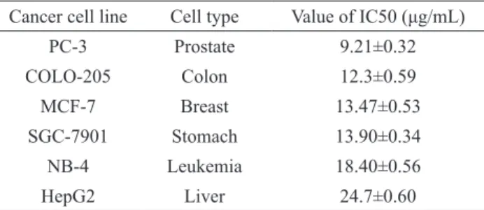
while PC-3 was the most sensitive (IC50 9.21±0.32 μg/ mL) cancer cell to the PHEB-induced growth inhibition
(Table 1). Therefore, mechanistic actions on the growth inhibition of PHEB were further investigated in the PC-3 human prostate adenocarcinoma cells.
Table 1. Values of IC50 for PHEB on growth of various cancer cells (mean±SD, n=3).
Cancer cell line Cell type Value of IC50 (μg/mL)
PC-3 Prostate 9.21±0.32
COLO-205 Colon 12.3±0.59
MCF-7 Breast 13.47±0.53
SGC-7901 Stomach 13.90±0.34
NB-4 Leukemia 18.40±0.56
HepG2 Liver 24.7±0.60
Concentration- and time-dependent effects of PHEB on the viability of PC-3 cells
Using MTT assay, the growth inhibitory effect of different concentrations of PHEB on PC-3 cells is shown in Figure 1. The data indicated that growth inhibitory effect of PHEB on the tested cell line increased in a concentration- and time-dependent manner, particularly between 48 and 96 h of treatment. The graph clearly demonstrates that the
IC50 of PHEB on PC-3 is about 10 μg/mL at 72 h.
0 24 48 72 96
0.2 0.4 0.6 0.8 1.0 1.2 1.4 1.6
a
b
so
rb
a
n
ce
(5
7
0
n
m)
t(h)
Figure 1. Concentration- and time-dependent effects of PHEB on
the growth inhibition of PC-3 cells. ■, DMSO 0.1%; ▼, PHEB 5 μg/mL; ▲, PHEB 10 μg/mL; ●, PHEB 20 μg/mL.
Tumor transplantation in vivo
The anti-tumor effect of PHEB in vivo was evaluated by the inhibition rate of tumor mass. Data (Table 2 and 3) showed that i.p. PHEB 40 and 60 mg/kg had a
signiicant antitumor effect on the growth of S180 with
inhibition rate 48.4% and 53.6%, and 46.3% and 57.2% on H22, respectively.
immediately weighed.
Morphological study of apoptosis by acridine orange staining assay
Acridine orange staining assay (Renvoize et al., 1998) was used to observe the morphological changes of apoptotic cells treated by PHEB. PC-3 human prostate adenocarcinoma cells were seeded within 6-well culture plates (1×10⁵ cells/well). After
incubation for 24 h, PHEB of different concentrations
(final dose is 5, 10, 20 μg/mL), DMSO (normal control,
0.1%) was added to the wells. Having been treated with PHEB for 48 h, the medium were discarded and the cells were washed twice with D-Hanks, it was then
stained with 5 μL acridine orange for 10 min at room
temperature in the dark. Processed cells were observed with a fluorescence microscope (Olympus, Japan).
Cell apoptosis analysis by flow cytometry
AnnexinV-FITC/PI staining assay was used to analysis cell apoptosis (Cao et al., 2006). PC-3 human prostate adenocarcinoma cells were seeded in T25 culture flask for 24 h. The cells were then
treated with 5, 10, 20 μg/mL PHEB, DMSO (0.1%)
for 24 h, respectively. After incubation, the cells were trypsinized, washed with PBS, and fixed overnight in ice-cold 70% ethanol. After fixation, 1×10⁶ cells were
washed twice with PBS, resuspended in 1 mL of DNA binding buffer solution, then, 10 μL propidium iodide (PI) and 5 μL AnnexinV-FITC was added, and analyzed
with FACSCalibur flow cytometer (BD Biosciences, USA). Proportion of apoptotic cells was measured using the control software of the flow cytometer.
Statistics
All data are expressed as mean±SD. The divergence between the treated and the controlled was analyzed by t-test. A probability of p<0.05 was considered significant.
Results
Antiproliferative effects of PHEB on different cancer cells
The PC-3 human prostate adenocarcinoma,
COLO-205 colorectal carcinoma, MCF-7 breast
carcinoma, gastric SGC-7901 cancer, NB4 leukemia cells, HepG2 hepatocellular carcinoma cells were treated respectively with various concentrations of PHEB, for 72 h. Among those cancer cells tested,
Morphological study of apoptosis by acridine orange staining assay
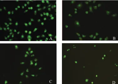
After treatment of PC-3 cells with PHEB at different concentrations for 48 h, compared with vehicle treated control, marked morphological changes of cell apoptosis such as fragmentation and condensation of chromatin, nuclear fragmentations, membrane blebs and apoptotic bodies were found clearly in the cells treated with
10, 20 μg/mL PHEB, using acridine orange (AO) staining
(Figure 2). These suggest that apoptosis occurred in these cells, and these effects were increased by increasing in concentration of PHEB.
Cell apoptosis analysis by low cytometry
In view of the morphological changes of cell apoptosis, we were interested in determining whether PHEB also induced apoptosis in PC-3 in the early stage of the treatment, we harvest cells at 24 h, rather than 48 h as many other studies did. The cells were treated with
0.1% DMSO alone or 5, 10, 20 μg/mL PHEB for 24 h,
representative results for the PC-3 cells were shown in Figure 3. In PC-3 cells, the percentage of apoptotic cells was 3.0, 13.0, 24.1, and 34.1% respectively. Thus, PHEB
has signiicant apoptosis-inducing effect on PC-3 cells in vitro.
Table 2. Inhibitory effects of PHEB on the growth of S180 in tumor-bearing mice (mean±SD, n=3).
Groups Dose (mg/kg) n Tumor weight (g) weight gain/g Inhibitory rate
(%)
Beginning End
Control - 10/10 2.453±0.421 20.6±1.34 28.7±1.47
-PHEB
20 10/10 1.867±0.485 20.3±1.28 27.4±1.41 23.9
40 10/10 1.266±0.575** 20.3±1.39 22.1±1.37 48.4
60 10/10 1.138±0.493** 20.4±1.27 21.3±1.29 53.6
CP 25 10/10 1.065±0.312** 20.2±1.43 23.9±1.52 56.6
Table 3. Inhibitory effects of PHEB on the growth of H22 in tumor-bearing mice (mean±SD, n=3).
Groups Dose (mg/kg) n Tumor weight (g) weight gain/g Inhibitory rate
(%)
Beginning End
Control - 10/10 2.659±0.364 20.4±1.18 29.4±1.51
-PHEB
20 10/10 2.100±0.392 20.3±1.30 26.3±1.46 21.0
40 10/10 1.427±0.416** 20.5±1.28 21.5±1.34 46.3
60 10/10 1.138±0.295** 20.2±1.25 20.4±1.38 57.2
CP 25 10/10 1.254±0.254** 20.3±1.42 23.6±1.19 52.8
Note: Control: 0.9% NaCl, CP: cyclophosphamide; Inhibition rate (IR %) = (1 - Tumor weight Treated / Tumor weight Controlled)×100%. * p<0.05, **
p<0.01 vs. control.
Figure 2. Morphology studies of PHEB-treated PC-3 cells for 48 h with luorescent staining AO. The cells were treated with 0.1%
DMSO, various concentrations of PHEB, for 48 h. Fixed cells were stained with acridine orange (5 μL) and examined by luorescence microcopy (A: DMSO 0.1%, B: PHEB 5 μg/mL, C: PHEB 10 μg/mL, D: PHEB 20 μg/mL). The arrow indicates the apoptotic cell.
A B
Discussion
Patrinia exhibits diverse pharmacological effects. Volatiles from Patrinia villosa (Thunb.) Juss. have the antioxidant activity (Xie et al., 2008). Pretreatment with Patrinia scabiosifolia has an anti-inflammatory effect on cholecystokinin (CCK) octapeptide-induced acute pancreatitis (AP) (Seo et al., 2006). Chemical constitutes extracted from P. villosa has the effect of
anti-inflammation (Li et al., 2008). P. heterophylla has been used in traditionally Chinese medicine for treating intestinal carbuncle, dysentery; it has also been suggested for treating gastric cancer and cervical cancer. The main components contained in Patrinia are triterpenoid saponin, flavonoid, volatile oil etc. There are some iridoids reported recent years. So far studies
have indicated that the material bases of Patrinia
antitumor effect are triterpenoid saponin (Zhang et al.,
2008b; Jung et al., 2004). Our studies clearly indicated that PHEB in which the main components are iridoids has significant antiproliferative effect on a panel of human cancer cells in vitro, it could effectively inhibit the growth of sarcoma 180 mice and hepatoma 22 mice in a concentration-dependent manner, simultaneously. This will supply the scientific evidences to reasonably use P. heterophylla.
PHEB also induced typical apoptotic features. It is known that apoptosis is important cellular events that can account for the cancer preventive effects of selenium (Kamesaki, 1998). Apoptosis is considered as an active suicidal response to various physiological or pathological stimuli including anticancer agents. It is
Figure 3. The apoptosis-inducing effect of PHEB on PC-3 cells. PC-3 cells were treated with 0.1% DMSO, various concentrations of
not only a genetically controlled mechanism essential for development, maintenance of tissue homeostasis and elimination of unwanted or damaged cells such as tumor cells (Chiu et al., 2006), but also a commonly accepted mechanism of anti-tumor effect of chemotherapeutic drugs. Ethyl acetate extract of Patrinia scabiosifolia was found to induce apoptosis in MCF-7 cells, involving
downregulated Bcl-2/Bcl-XL expression. Interestingly,
the ethyl acetate extract of Patrinia scabiosifolia induced apoptosis could not be inhibited by the caspase-9 inhibitor (Chiu et al., 2006). PHEB was isolated from ethyl acetate extract of P. heterophylla. The components of PHEB may have some similarity with ethyl acetate extract of Patrinia scabiosaifolia. Furthermore, the antiproliferative effect of PHEB is better than EAE-PS. So, the mechanism of PHEB-induced apoptosis in PC-3 should be studied in the future.
In our study, PHEB could effectively inhibit the growth of S180 mice and H22 mice in a concentration-dependent manner. Antitumor activity of PHEB in vivo is almost the same as cyclophosphamide, this is pleasantly surprised us. For PHEB is still crude extract, after isolation
and puriication, it may exhibit stronger antitumor effect
in vivo. Sarcoma 180 and hepatoma 22 are tumor in mice, in further study, a human prostate adenocarcinoma cancer implant model using nude mice will be established to evaluate the antitumor effect in vivo.
Acknowledgements
We thank Dr. Xiaoying Zhang of South Dakota
State University for critically reading the manuscript.
References
Cao Y, Adhikari S, Ang AD, Clement MV, Wallig M, Bhatia
M 2006. Crambene induces pancreatic acinar cell apoptosis via the activation of mitochondrial pathway.
Am J Physiol Gastrointest Liver Physiol 291: G95-101.
Chen YF, Zhang H, Zhang QY, Wu JZ, Li N, Rahman K, Zheng HC, Qin LP 2007. Antinociceptive activity of n-butanol
fraction from MeOH extracts of Paederia scandens in mice. Pharmazie 62: 943-948.
Chiu LC, Ho TS, Wong EY, Ooi VE 2006. Ethyl acetate extract
of Patrinia scabiosaefolia downregulates anti-apoptotic
Bcl-2/Bcl-X(L) expression, and induces apoptosis in
human breast carcinoma MCF-7 cells independent of caspase-9 activation. J Ethnopharmacol 105: 263-268.
Cometa MF, Parisi L, Palmery M, Meneguz A, Tomassini L 2009.
In vitro relaxant and spasmolytic effects of constituents from Viburnum prunifolium and HPLC quantiication of
the bioactive isolated iridoids. J Ethnopharmacol 123: 201-207.
Jung HJ, Lee CO, Lee KT, Choi J, Park HJ 2004.
Structure-activity relationship of oleanane disaccharides isolated from Akebia quinata versus cytotoxicity against cancer cells and NO inhibition. Biol Pharm Bull 27: 744-747. Kamesaki H 1998. Mechanisms involved in
chemotherapy-induced apoptosis and their implications in cancer chemotherapy. Int J Hematol 68: 29-43.
Li N, Zhao B, Yu YF, Dong XP 2008. Studies on anti-inlammation chemical constitutes of Patrinia villosa. Zhong Yao Cai
31: 51-53.
Liu RH, Zhang WD, Gu ZB, Zhang C, Su J, Xu XK 2006. Two
new iridoids from roots of Patrinia scabra Bunge. Nat Prod Res 20: 866-870.
Lu WZ, Geng GX, Li QW, Li J, Liu FZ, Han ZS, Gao DW, Yan X, Yang XL 2009. Anti-tumor activity of polysaccharides
isolated from Patrinia scabra Bunge on U14 cervical carcinoma bearing mice. Am J Chin Med 37: 933-944. Makin G, Dive C 2001. Apoptosis and cancer chemotherapy.
Trends Cell Biol 11: S22-S26.
Mosmann T 1983. Rapid colorimetric assay for cellular growth and survival: application to proliferation and cytotoxicity assays. J Immunol Methods 65: 55-63.
Renvoize C, Biola A, Pallardy M, Breard J 1998. Apoptosis:
identiication of dying cells. Cell Biol Toxicol 14: 111-120.
Seo SW, Park CS, Hong SH, Kwon KB, Moon HC, Song BK,
Kim KY, Park YM, Song HJ, Kim HM, Park SJ 2006.
Inhibitory effect of Patrinia scabiosaefolia on acute pancreatitis. World J Gastroentero 12: 1110-1114.
Sun SY, Hail Jr, N, Lotan R 2004. Apoptosis is a novel target for
cancer chemoprevention. J Natl Cancer I 96: 662-672. Wand WR 1997. In vivo methods. In Teicher, B.A. (ed.)
Anticancer drug development guide, preclinical screening, clinical trials, and approval. Totowa: Humana Press Inc, p 59-213.
Xie Y, Peng J, Fan G, Wu Y 2008. Chemical composition and
antioxidant activity of volatiles from Patrinia villosa Juss obtained by optimized supercritical luid extraction. J Pharm Biomed Anal 48: 796-801.
Yang GJ, Gu ZB, Liu WY, Qiu Y, Li TZ, Zhang WD 2004. Two
new iridoids from Patrinia scabra. J Asian Nat Prod Res 6: 277-280.
Zhang T, Li Q, Li K, Li Y, Li J, Wang G, Zhou S. 2008a. Antitumor
effects of saponin extract from Patrinia villosa (Thunb.) Juss on mice bearing U14 cervical cancer. Phytother Res 22: 640-645.
Zhang T, Li Q, Li K, Li Y, Li J, Wang G, Zhou S 2008b. Antitumor
effects of saponin extract from Patrinia villosa (Thunb.) Juss. on mice bearing U14 cervical cancer. Phytother Res 22: 640-645.
*Correspondence
Rusong Zhang
College of Pharmaceutical Sciences, Zhejiang Chinese Medical
University
548 Binwen Road, Hangzhou 310053, People’s Republic of China