Pro-Apoptotic Function in Ovarian Granulosa Cells
Xiao-Ming Liu, Fei-Fei Yang, Yi-Feng Yuan, Rui Zhai, Li-Jun Huo*
Key Laboratory of Agricultural Animal Genetics, Breeding and Reproduction, Education Ministry of China, College of Animal Science and Technology, Huazhong Agricultural University, Wuhan, People’s Republic of China
Abstract
Follicular atresia is a process of spontaneous degradation of follicles, hindering growth and development in the mammalian ovary. Previous studies showed that follicular atresia was caused by apoptosis of granulosa cells, for which a number of apoptosis-related genes have already been identified. The roles of p53 in apoptosis of mouse granulosa cells and its post-translational modification are still unclear. The main objective of this study was to explore the roles of p53 in mouse granulosa cells. We found that mouse p53b, but not p53a, could be SUMOylated by SUMO-1 at lysine 375, which was essential for the protein stability of p53b in a dose-dependent manner. Immunofluorescent staining showed that wild p53b was located in the nucleus of granulosa cells, while its mutation of SUMOylated site (K375R) was localized in both nucleus and cytoplasm, implying that SUMOylation was necessary for the nuclear localization of p53b in granulosa cells. Overexpression of wild-type p53b, but not the mutation of SUMOylation site (K375R), significantly induced the expression of apoptosis-related gene, Bax, and increased the level of apoptosis in granulosa cells. This suggested that SUMO-1
modification of p53b was essential for inducing apoptosis in granulosa cells. Our results provide strong evidences that modification of p53b by SUMO-1 at lysine 375 was necessary for its activity to induce apoptosis in mouse granulosa cells, and it was involved in the regulation of p53b protein stability and nuclear localization. This implies that modification of p53b by SUMO-1 might regulate follicular atresia by inducing the apoptosis of ovarian granulosa cells in mice.
Citation:Liu X-M, Yang F-F, Yuan Y-F, Zhai R, Huo L-J (2013) SUMOylation of Mouse p53b by SUMO-1 Promotes Its Pro-Apoptotic Function in Ovarian Granulosa Cells. PLoS ONE 8(5): e63680. doi:10.1371/journal.pone.0063680
Editor:Qing-Yuan Sun, Institute of Zoology, Chinese Academy of Sciences, China
ReceivedNovember 21, 2012;AcceptedApril 5, 2013;PublishedMay 16, 2013
Copyright:ß2013 Liu et al. This is an open-access article distributed under the terms of the Creative Commons Attribution License, which permits unrestricted use, distribution, and reproduction in any medium, provided the original author and source are credited.
Funding:This study was supported by National Natural Science Foundation of China (Grant No.31071273), the Specialized Research Fund for the Doctoral Program of Higher Education of China (Grant No. 20100146110011), and the Fok Ying-Tong Education Foundation, China (Grant No. 121029). The funders had no role in study design, data collection and analysis, decision to publish, or preparation of the manuscript.
Competing Interests:The authors have declared that no competing interests exist.
* E-mail: [email protected]
Introduction
During the process of follicular atresia, follicles spontaneously degrade in the mammalian ovary, hindering growth and development. More than 99% of follicles disappear, primarily due to apoptosis of granulosa cells [1–6]. Although atresia can occur at any time during follicular development, the majority of follicles become atretic during the early antral stage of develop-ment [7]. The transition from preantral to antral follicles occurs after the granulosa cells are exposed to gonadotropin. The gonadotropin initiates differentiation of the granulosa cells making them susceptible to apoptosis.
Apoptosis in granulosa cells is characterized by nucleosome DNA fragmentation, cell shrinkage, membrane blistering, and the formation of apoptotic bodies [8]. Many apoptosis-related factors have been implicated in follicular atresia, including death ligands and receptors, intracellular pro- and anti-apoptotic molecules, cytokines, growth factors, and a number of apoptosis-related genes, including the p53 gene [9–11].
The p53 protein is an antiproliferative transcription factor that increases the rate of transcription of various genes involved in mitosis and apoptosis [9,12–15]. It plays a critical role in cell cycle regulation (G1/S transition), DNA repair, and induction of apoptosis [14,16–18]. In granulosa cells, p53 content is correlated with contents of Fas and FasL, death ligands that are involved in
the induction of apoptosis and are regulated by gonadotropins [19]. When gonadotropin induced, p53 becomes markedly elevated, suggesting that induction of atresia is p53-dependent [20]. Furthermore, overexpression of p53 resulted in increased Fas content and apoptosis [16]. However, mouse p53 gene contains only a promoter, which differs from the selective promoter of the human p53 gene; the alternatively spliced RNA species of tumor-suppressor gene p53 (which contains an additional 96 bases derived from intron 10) is present at approximately 25 to 30% the level of regularly spliced p53 RNA in both normal epidermal cells, carcinoma cells, and mouse liver and testis cells [21]. Precisely because of this alternative splicing, there are two types of p53 protein: p53a encodes 390 amino acids and p53b encodes 381 amino acids. The main difference between p53a and p53b is the C-terminal sequence. However, the biological properties of different types of the p53 protein in mice have not yet been determined.
processes, including cell proliferation, differentiation, and apopto-sis [25,27]. Studies have reported that human p53 can be modified by SUMO-1 and the SUMOylation site is lysine386 [28–30]. Conjugation of SUMO-1 to wild-type p53 results in an increased transactivation ability of p53 [28,30]. However, SUMOylation has no effect on mutant p53 transcriptional activity [29,31]. In addition to comparing wild-type and SUMOylation-deficient p53 for transactivation, studies analyzed potential differences in localization and growth inhibition or apoptosis. Mutating the p53 SUMO-acceptor site lysine386 to arginine had no obvious effect on p53 localization [29], but one study generated p53-SUMO-1 fusion protein as a model for the effect of SUMO modification on the localization and function of p53, showing that p53-SUMO-1 fusion protein significantly redistributed from the nucleus to the cytoplasm when the SUMOylation site lysine386 was destroyed [32]. Studies showed the SUMOylation of p53 enhanced the apoptosis in Saos-2 cells [33]. In addition, SUMO modification of Drosophila p53 is required for its pro-apoptotic activity [34].
While it is not very clear whether p53 is involved in regulating follicular atresia and granulosa cells apoptosis, and its regulatory mechanism is also unclear. Furthermore, there are two types of p53 protein (p53a and p53b) in mice, and the roles of these p53 forms in mouse granulosa cells and whether they can be SUMOylated have not been reported. In this study, the main objective is to explore the roles of p53 in mouse granulosa cells and the effects of SUMOylation.
Materials and Methods
Experimental Animals
We obtained immature 21 to 23 d-old Kunming White female mice from the Centre of Laboratory Animals of Hubei Province (Wuhan, PR China). All animal treatment procedures were approved by the Ethical Committee of the Hubei Research Center of Experimental Animals (Approval ID: SCXK (Hubei) 2008-0005). Mice were housed under controlled temperature (24uC) and lighting (12 h light/12 h darkness) with food and water ad libitum. Follicle development was primed by injection of each mouse with 10 IU pregnant mare serum gonadotropin (PMSG; SanSheng, Ningbo), and mice were killed by cervical dislocation 44–48 h later.
Plasmid Construction
A 1310-bp mouse p53a and a 1213-bp mouse p53b cDNA sequence were amplified using polymerase chain reaction (PCR) from mouse ovary cDNA using the following primers: p53a-Forward 59-CGGGATCCGGCAGGGTGTCACGCTTCT-39; p53a- Reverse 59-CGGAATTCCG AGGGACCGGGAG-GATTGT-39; p53b- Forward 59 -CGGGATCCGG-CAGGGTGTCACGCTTCT-39; and p53b- Reverse 59 -GCGAATTCGGAGGGATGAAGTGATGGGA-39. Both in-cluded BamHI and EcoRI restriction sites. We subcloned into pCMV-N-HA to generate a HA-tagged p53a and p53b cDNA.
To generate the p53b lysine 375 arginine mutant, two pairs of primers were used for PCR: Pair 1- Forward 59 -CGGGATCCGGCAGGGTGTCACGCTTCT-39; Pair 1- Re-verse 59- GGGCTTTCCTCCCTGATCAAGGCTTGG -39; Pair 2- Forward 59- CGGGATCCGGCAGGGTGT-CACGCTTCTCCGAAGACTGGATT-39; and Pair 2- Reverse 59 -GCGAATTCGGAGGGATGAAGTGATGGGAGCTAG-CAGTTTGGGCTTTCCTCCCTG -39. Amino acid of p53b was mutated from lysine to arginine using the p53b cDNA sequence as
the template for the first reaction and the product as the template for the second reaction. The results were confirmed by sequencing. HA-tagged sumo-1 or ubc9, or Flag-tagged sumo-1 was preserved in our laboratory.
In vitro Culture of Granulosa Cells and DNA Transfection
Granulosa cells from pre-ovulatory follicles (pre-GCs) were obtained from ovaries of 21 to 23 d-old Kunming White female mice injected with 10 IU PMSG (SanSheng, Ningbo) 44–48 h. Granulosa cells were cultured in 6-well culture plates in Dulbecco’s Modified Eagle’s Medium/Nutrient F-12 (DMEM/ F12; Gibco) medium with 10% fetal bovine serum (FBS; Invitrogen), 60 mg/mL penicillin, and 50 mg/mL streptomycin. All cultures were maintained in DMEM/F12 medium at 37uC in a humidified atmosphere of 5% CO2. After being cultured 24 h, granulosa cells were washed by phosphate-buffered saline (PBS), and cultured for 24 h in fresh serum-free DMEM/F12 medium before DNA transfections.
For DNA transfection or co-transfection, granulosa cells were plated and then transfected with 4mg of the desired plasmids for 24 h with Lipofectamine LTX (Invitrogen) according to manu-facturer’s instructions. Desired plasmids and LTX were diluted in Opti-MEM (Gibco) medium and incubated for 5 min. This solution was then mixed with granulosa cells at a proportion of 1:1.5 for 30 min; 24 h after transfection, granulosa cells were collected for protein extraction or apoptosis analysis.
To study the effect of SUMO-1 mediated modification of p53b on its protein level in granulosa cells, different dosages of pCMV-Flag-sumo-1 plasmids (2mg, 3mg, 4mg, and 6mg) were
co-transfected with a fixed proportion of HA-p53b or pCMV-HA-p53bK375R(2mg) plasmids into granulosa cell.
Protein Extraction, Immunoprecipitation and Western Blot Analysis
Subconfluent cells seeded on 6-well culture plates were transfected with the expression vectors indicated. At 24 h after transfection, cells were washed twice with ice-cold PBS and harvested in 80mL of ice-cold RIPA buffer (Santa Cruz),
containing 10mM phenylmethylsulfonyl fluoride (PMSF;
Ding-Guo, Beijing) and 10 mg/mL protease inhibitors cocktail (Santa Cruz). Protein lysis was performed on ice for 20 min. Then, the lysates were centrifuged at 12000 rpm for 5 min, and the supernatant was collected and stored at280uC.
The concentration of total protein was determined by bicinchoninic acid (BCA) assay (Pierce, Rockford, USA), and 20mg of total protein was subjected to gel electrophoresis. The
cell lysates mixed with 26SDS gel-loading buffer were loaded on 4% stacking gel and 10% separating gel, and were then transferred to 0.2mm polyvinylidene fluoride (PVDF) membrane
modifi-cation of p53 protein, membranes were probed with the HA antibody or p53 antibody, respectively, while the membrane was probed with b-actin antibody for normalization. The band intensities were measured with Gel-Pro analyzer 4.0 (Media Cybernetics, USA).
Immunoprecipitation (IP) was conducted to detect if p53 could be modified by SUMO-1 in granulosa cells in vivo. Briefly, after transfection with pCMV-Flag-sumo-1 plasmid or pCMV-Flag plasmid (as a negative control), granulosa cells (1062107) were harvested in 1 mL lysis buffer (50 mM Tris HCl, pH 7.4, 150 mM NaCl, 1 mM EDTA, 1% Triton X-100) containing 10mM PMSF
(DingGuo, Beijing) and 10 mg/mL protease inhibitors cocktail (Santa Cruz). The extract was centrifuged at 12,000 g for 15 min at 4uC, and the supernatant were collected. 800 ul of the supernatant were incubated with ANTI-FLAG M2 Affinity beads (Sigma-Aldrich) overnight at 4uC with continual shaking. After recovery by centrifugation at 8,000 g for 30 s, the beads were washed four times with ice cold TBS (50 mM Tris HCl, 150 mM NaCl, pH 7.4). Then Flag-fusion proteins were eluted by 100mL
36FLAG peptide (Sigma-Aldrich) at 4uC with gentle shaking for 30 min. After elution, the beads were centrifuged at 8,000 g for 30 s and the supernatant was collected. For Western blot, the supernatant was mixed with equal volume of 26SDS loading buffer and heated for 5 min at 100uC before being loaded on a SDS-PAGE gel. Polyclonal rabbit anti-p53 IgG was used as the primary antibody.
In order to confirm which type of p53 (p53a or p53b) could be modified by SUMO-1, granulosa cells were co-transfected with pCMV-Flag-sumo-1 plasmid and pCMV-HA-p53a plasmid or pCMV-HA-p53b plasmid. After transfection, Flag tagged SUMO-1 were pulled down by Flag antibody and then HA antibody were used to detect the expression of p53a or p53b by Western blot.
Immunofluorescence Cytochemistry
In order to detect the subcellular localization pattern of p53b and p53bK375R, pre-GCs were plated on coverslips and transfected with pCMV-N-HA-p53b or pCMV-N-HA-p53bK375R vector respectively. After 24 h transfection, cells were washed twice with PBS and were fixed in 4% paraformaldehyde (Santa Cruz) for 15 min. Next, the cells were washed in PBS three times and then permeabilized in PBS containing 0.5% Triton-100 (Santa Cruz) at room temperature for 20 min. After blocking in PBS containing 5% bovine serum albumin (BSA) (Santa Cruz), cells were washed in PBS three times. After that, cells were incubated with anti-HA primary antibody (1:100 dilution, CWBIO, Beijing) at 37uC for 2 h, followed by FITC-conjugated secondary antibody (1:100 dilution, Boster, Wuhan) in the dark. The nucleus of cells was stained in 10mg/mL propidium iodide (PI; Santa Cruz) for 5 min.
Slides were mounted in DABCO (Sigma-Aldrich,) and viewed under a Zeiss- LSM 510 Meta confocal microscope.
For co-localization study of p53b or p53bK375Rwith SUMO-1 in granulosa cells, pre-GCs were co-transfected with pCMV-Flag-sumo-1 plasmid and p53b plasmid or pCMV-HA-p53bK375Rplasmid. After 24 h transfection, SUMO-1 and p53b or p53bK375R were detected by polyclonal rabbit anti-SUMO-1 antibody (1:200 dilution; Santa Cruz) or monoclonal mouse anti-HA antibody (1:100 dilution, CWBIO, Beijing), respectively. Cy3-conjugated anti-rabbit IgG antibody (1:100 dilution, Boster, Wuhan) or FITC-conjugated anti-mouse IgG antibody (1:100 dilution, Boster, Wuhan) were used as the secondary antibody, respectively. The nucleus was stained in 10mg/mL 49,69
,-diamidino-2-phenylindole (DAPI; Santa Cruz).
Cell Apoptosis Assay
Pre-GCs were transfected with HA-p53b, pCMV-N-HA-p53bK375R, or negative control pCMV-N-HA for 24 h. Cells were washed twice in PBS, then trypsinized and collected for apoptosis assay. Apoptosis was performed by using the AnnexinV kit (AntGene, Wuhan) according to manufacturer’s instructions. Cells were incubated in AnnexinV–FITC and PI solution at room temperature in the dark for 15 min, then 300mL of 16binding buffer was added to each sample. Flow cytometric analysis was conducted using a BD FACSCalibur (Becton, Dickinson and Company, USA; Ex, 488 nm and Em, 530 nm).
Real-time RT-PCR Analysis
To determine genes regulated by SUMOylation of p53b, at 24 h after transfection, total RNA was prepared using the RNAiso Plus (TaKaRa, Dalian), and in vitro transcription was carried out using PrimeScript RT reagent Kit With gDNA Eraser (TaKaRa, Dalian). Real-time PCR was then conducted to quantify the steady-state mRNA levels of the tested genes using SsoFastTM EvaGreenH Supermix (Bio-Rad, USA) on the Bio-Rad CFX96 Real-time PCR System. The threshold cycle (Ct) was used to determine the relative expression level of each gene by normal-izing to the Ct ofb-actinmRNA. The method of 2–ddCt was used to calculate the relative fold change of each gene. To ensure that only target-gene sequence-specific, non-genomic products were amplified by real-time PCR, careful design and validation of each primer pair, as well as cautious manipulation of RNA, were undertaken to quantify the steady-state mRNA levels ofBaxand housekeeping gene b-actin (internal control). The primers used were Bax- forward: 5-CCAGGATGCGTCCACCAA-3; Bax-reverse: 5-CAAAGTAGAAGAGGGCAACCAC-3; b-actin- for-ward: 5- CCCATCTACGAGGGCTAT-3; and b-actin- reverse: 5-TGTCACGCACGATTTCC-3. Calculation of the relative fold change of Bax was done using the method of 22DDCt. In each experiment, levels of Bax mRNA were presented as relative changes to a specific group (control), in which its expression level was set at 1.
Data Analysis and Statistics
All experiments were performed independently at least three times, and data are presented as mean6SD. Differences between groups were analyzed by one-way ANOVA followed by Tukey’s Honesty Significant Difference (HSD) test using SPSS (Version 17.0; SPSS, Chicago, IL, USA). P,0.05 was considered signifi-cantly different, andP,0.01 was extremely significantly different.
Results
SUMOylation Increased the Expression Level of Mouse p53 Protein
SUMO-1 Conjugation to p53b in vivo Requires Lysine 375
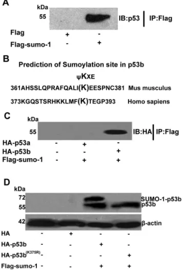
Based on the results above, we speculated that the mouse p53 protein could also be modified by SUMO-1. Immunoprecipitation (IP) study was designed to confirm whether mouse p53 could be SUMO-1 modified or not in granulosa cells. Pre-GCs were transfected with pCMV-Flag-sumo-1 plasmid or pCMV-Flag plasmid (as a negative control) for 24 h. Flag-tagged protein were pulled down by anti-Flag affinity beads and then analyzed by Western blot using anti-p53 antibody. A visible band of p53 was observed in the sample of pCMV-Flag-sumo-1 transfected granulosa cells, but not in the control sample (Fig. 2A). This study clearly showed that mouse p53 protein could be SUMO-1 modified in granulosa cells in vivo. However, there are two types of p53 in mouse, and which type of p53 that could be modified was unknown. Previous studies showed lys386 of human p53 was required for SUMO-1 modification in Saos-2 cells [29,35,36]. To identify which type of mouse p53 could be modified by SUMO-1 in mouse, we aligned the protein sequence of mouse p53a and p53b to human p53 around the SUMOylation site at lys386 (Fig. 2B). The lysine 375 of 374IKEE377 in mouse p53b was aligned to the lysine within the consensus SUMOylation site of human p53, 385FKTE388 (Fig. 2B). The region 374IKEE377 of mouse p53b was also confirmed as a high probability SUMOyla-tion site, with a score of 2.943 predicted by SUMOsp2.0.4 software. However, there are no conserved amino acids in mouse p53a with mouse p53b at lys375 or human p53 at lys386, and there are no SUMOylated sites in mouse p53a predicted by SUMOsp2.0.4 software. In order to further confirm if only p53b, but not p53a, could be SUMO-1 modified in granulosa cells, pre-GCs were co-transfected with Flag-tagged sumo-1 plasmid and HA-tagged p53a or p53b plasmid. After transfection, the Flag-tagged fusion protein were pulled down by Flag antibody and then detected by western blot using HA antibody. The results showed that only p53b, but not p53a, could be co-immunoprecipitated by Flag-tagged sumo-1 (Fig. 2C).
Although Lys375 of p53b were predicted by SUMOsp2.0.4 software as a putative SUMO-1 modified site, it is still unknown whether this Lys375 is necessary for p53b’s modification by SUMO-1. Therefore, we mutated lysine 375 of mouse p53b to arginine and generated a mutant, HA-tagged p53b (K375R) expression plasmid. Wild-type p53b plasmid or mutant p53b plasmid, with sumo-1 plasmid, were co-transfected into mouse
Figure 1. SUMOylation increased the expression level of mouse p53 protein. Pre-GCs were seeded in 6-well culture plates and transfected with 2mg of HA-p53a, HA-p53b, HA-sumo-1, HA-ubc9, or
empty HA expression plasmids. 20mg total extracts were prepared and
resolved by SDS–PAGE and analyzed by Western blot using anti-HA (the top group) or anti-p53 (the middle group) specific antibody. Positions of molecular weight markers, free HA-p53 or putative p53/HA–SUMO-1 and p53/HA-UBC9 conjugates are indicated.b-actin is a loading control. doi:10.1371/journal.pone.0063680.g001
Figure 2. Identification of SUMOylation of p53b in vivo at Lys 375.(A) Immunoprecipitation analysis of SUMOylation of p53 in vivo. Pre-GCs were transfected with Flag-sumo-1 plasmid or pCMV-Flag plasmid. pCMV-Flag-tagged protein were pulled down by anti-pCMV-Flag affinity beads and then analyzed by Western blot using anti-p53 antibody. A visible band of p53 was detected in the sample of pCMV-Flag-sumo-1 transfected granulosa cells, but not in the control sample. (B) SUMOylation consensus site aligned in sequences of p53 from human and mouse. The SUMOylation consensus site isyKXE with a hydrophobic residue, such as Phenylalanine (F), Isoleucine (I), or Leucine (L). Sequences of human p53 and mouse p53b were aligned. Lys386 in human p53 is SUMO-1 modified and the mouse p53b has a highly homologous sequence to this SUMOylation site. (C) Co-immunoprecip-itation of SUMO-1 with p53a or p53b. Pre-GCs were co-transfected with Flag-tagged sumo-1 plasmid and HA-tagged p53a or HA-tagged p53b plasmid. Flag-tagged proteins were pulled down by anti-Flag affinity beads and then analyzed by Western blot using anti-HA antibody. A band of p53 (p53b) was detected in the sample of Flag-sumo-1 plasmid and p53b plasmid co-transfected granulosa cells, but not in the HA-p53a plasmid and Flag-sumo-1 plasmid co-transfection group or control sample. (D) p53b is SUMOylated at lys375. Pre-GCs were co-transfected with 2mg of wild type HA-p53b or mutant p53b (K375R) plasmid and
2mg of Flag-sumo-1 plasmid. 20mg total extracts were prepared and
resolved by SDS–PAGE and analyzed by Western blot using the anti-HA-specific antibody. Positions of molecular weight markers, free p53 or p53-SUMO-1 conjugates are indicated.b-actin is a loading control. After transfection, two bands could be detected in the group with co-transfection of wild type p53b and sumo-1, the lower molecular weight below was considered as free p53b and the higher molecular weight above was p53b-SUMO-1 as the SUMOylated p53b, but only one band (mutant p53b) could be observed in the group with co-transfection of mutant p53b and sumo-1 plasmids, while the p53b (K375R)-SUMO-1-band was not observed.
pre-GCs and analyzed by Western blot to examine the expression and SUMOylation. When cells were co-transfected with wild-type p53b plasmid and sumo-1 plasmid, two bands were detected (Fig. 2D, lane 3), but only one band could be observed when co-transfected with mutated p53b (K375R) plasmid and sumo-1 plasmid (Fig. 2D, lane 4). The lower band (55 kDa) was considered p53b protein and the band above was considered SUMO-1 covalently conjugated p53b protein, SUMOylated p53b. These results supported our hypothesis that mouse p53b could be SUMO-1 modified, and lysine 375 of p53b was necessary for SUMO-1 modification. The SUMOylation of mouse p53b at lys375 by SUMO-1 raised another interesting question: what regulatory roles of SUMOylation of p53b plays on the function of p53b in mouse ovarian granulosa cells. Further experiments were conducted to explore the effects of SUMOylation modification of p53b on its protein stability, subcellular localization, and biological activity in mouse granulosa cells.
SUMO-1 Modification Enhanced the Protein Stability of p53b in a Dose-dependent Manner
Our results above showed that transfection with sumo-1 or ubc9 plasmid could enhance the expression level of p53 in granulosa cells. To further explore the patterns of SUMO-1 modification to enhance the stability of p53b, pre-GCs were co-transfected with HA-wild-type p53b or HA-p53b (K375R) plasmid and different amounts of Flag–sumo-1 plasmids. Total proteins were extracted and subjected to Western blot analysis with HA antibody (Fig. 3). As predicted, increasing the amount of Flag-sumo-1 plasmid for transfection resulted in a significant increase of both SUMOylated p53b and free p53b in a dose-dependent manner (Fig. 3A and B, compare lanes 3, 4, 5, and 6), but did not increase p53b (K375R) (Fig. 3C). These results demonstrated that SUMO-1 modification was responsible for increasing the stability of p53b in a dose-dependent manner, and further confirmed that Lys 375 was required for SUMOylation of p53b by SUMO-1.
SUMO-1 Modification was Required for the Nuclear Accumulation of p53b in Granulosa Cells
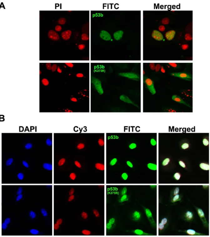
In order to study whether SUMO-1 modification of p53b at Lys375 is involved in the regulation of p53b’s subcellular localization in granulosa cells, pre-GCs were transfected with HA-p53b plasmid or mutant HA-p53b (K375R) plasmid, immu-nofluorescence cytochemistry was used to detect the subcellular localization of p53b protein by HA antibody. Wild type p53b proteins were accumulated only in the nucleus of pre-GCs, while mutant p53b (K375R) were localized in both nucleus and cytoplasm of pre-GCs (Fig. 4A). Meanwhile, the co-localization of SUMO-1 with p53b was observed in the nucleus of granulosa cells after co-transfection with sumo-1 plasmid and p53b plasmid. Mutant p53b (K375R) was still localized in both nucleus and cytoplasm of granulosa cells after co-transfection with sumo-1 plasmid and p53b (K375R) plasmid. Overall, the results indicated that SUMO-1 modification of p53b at Lys 375 was required for its nuclear accumulation in mouse granulosa cells.
p53b-induced Cell Apoptosis was Enhanced by SUMO-1 Modification
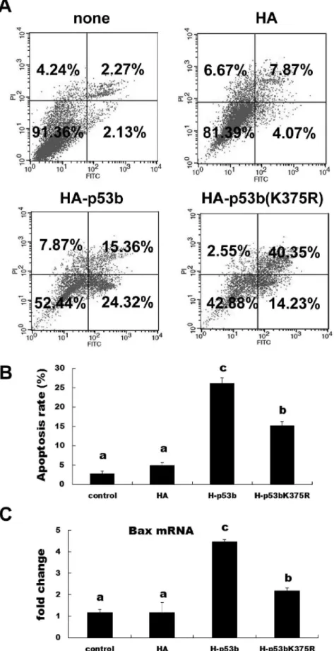
As p53 is a cellular gatekeeper [14,37], one of its roles is to survey cellular stress and induce apoptosis, if necessary. Therefore, we investigated the roles of mouse p53b in inducing apoptosis of pre-GCs. Cells were transfected with wild-type p53b or mutant p53b (K375R) for 24 h and then collected for apoptosis analysis by flow cytometer. The apoptosis rate was significantly increased by
transfection with wild-type p53b compared with the control group and mutant p53b (K375R; Fig. 5A and 5B;P,0.05). The mutant p53b (K375R) transfected cells also showed significant increases in apoptosis compared with the control group (p,0.05), but were significantly lower in apoptosis compared with wild p53b transfected group (p,0.05). In addition, RT-PCR was used to detect the expression of a marker of apoptosis (Bax). Overexpres-sion of either wild type p53b or mutant p53b (K375R) could significantly increase the expression ofBaxmRNA, compared with control group. However, overexpression of wild type p53b could induce much higher expression level ofBax mRNA, compared with mutant p53b (Fig. 5C). These results were consistent with our other findings related to the apoptosis rate of granulosa cells. All these results implied that SUMO-1 modification of p53b enhanced the ability of p53b to induce apoptosis in pre-GCs. Mutating the SUMOylation site of p53b could significantly weaken the activity of p53b to induce apoptosis and apoptosis-related gene,Bax. On the other hand, in addition to inducing apoptosis, the cell death ratio was increased by mutation of p53b (K375R), but not by wild-type p53b (Fig. 5A).
Discussion
In normal cells, p53 is maintained at a low level, which is partly due to the short half-life of the protein [38]. However, in response to a variety of stress signals, p53 is stabilized, causing protein to accumulate and activating p53-dependent transcription. Related factors, including lower levels of the MDM2-mediated poly-ubiquitination of p53 and others, improved post-translational modifications [22–24].
Although human p53 had been reported to be modified by SUMO-1 [11,28,39], there are two kinds of p53 (p53a and p53b) in mouse; whether mouse p53 could be SUMOylated was unclear. In this study, overexpression of sumo-1 or ubc9 could increase the protein level of p53 in granulosa cells, and a specific migrating band was also observed by using HA antibody, implying that SUMOylation is involved in the stability of p53 protein by post-translational modification. Based on the prediction results of SUMOsp2.0.4 software, there is a conserved SUMOylated site at lys375 of p53b, but not in p53a. The results of IP confirmed that mouse p53b, but not p53a, could be SUMO-1 modified at lys375. Further experiments showed that lys375 mutation of p53b could result in its nucleus-cytoplasm translocation and decrease its ability to induce apoptosis, confirming that p53b modified at lys375 by SUMO-1 plays crucial roles for p53 functions in granulosa cells.
ubiquitination), SUMOylation of p53b may inhibit its degradation by competing for the same lysine residue that was required for p53 ubiquitination or by interfering with conjugation of ubiquitin molecules to neighboring sites [46]. However, further studies will be required to address this issue.
Another issue to be addressed is the role of the SUMO-1 modification pathway in the subcellular distribution of p53b. In normal cell circumstances, p53 protein is present in the nucleus, and p53 nuclear export is critical to determine their degradation; we can speculate that the distribution of p53 in the nucleus and cytoplasm is a key factor in determining its stability. Interestingly, it has been reported that p53-SUMO-1 fusion protein localize to PML bodies. While damages the use of the fusion proteins by
mutating the C-terminal glycines in the SUMO-1 portion (DGG) to inhibit the formation of isopeptide bonds between SUMO-1 and target lysines, the localization of p53-SUMO-1DGG is predominantly at the nuclear envelope with some cytoplasmic staining [32]. Similarly, SUMOylation helped recruit Drosophila p53 to nuclear dot-like structures that could be marked by human PML and theDrosophilahomologue of Daxx [34]. In our study, we also found that the localization of p53b was in the nucleus; after mutation, the nucleus localization was lost compared to wild p53b and part of the p53b was localized in the cytoplasm (Fig. 4 A and B). So our results directly demonstrated that SUMOylation was required for the nuclear localization of p53b, which provided a
Figure 3. SUMO-1 modification enhanced the stability of p53b in a dose-dependent manner.(A) Pre-GCs were co-transfected with 2mg
HA-p53b plasmid and different amounts (2mg, 3mg, 4mg, and 6mg) of Flag-sumo-1 plasmids. 20mg total proteins were prepared and resolved by
SDS–PAGE and analyzed by Western blot using anti-HA-specific antibody. Positions of molecular weight markers, free p53b and p53b-SUMO-1 conjugates are indicated.b-actin is a loading control. (B) Relative expression quantity of p53b and SUMO-1-p53b were determined by densitometric scans. The total amount ofb-actin present in the lower set of lanes was used to standardize the amount of p53b and SUMO-1-p53b present in the upper set of lanes. The value expressed by each bar represents the mean6SD (n = 3). Different letters indicated statistical difference (P,0.05). (C)
Pre-GCs were co-transfected with 2mg HA-p53b (K375R) and different amounts (2mg, 3mg, 4mg, and 6mg) of Flag-sumo-1 plasmids. HA antibody
were used to detect the expression of p53b (K375R) after transfection. The results showed that elevating the amount of transfected Flag-sumo-1 resulted in a simultaneous increase in the level of SUMOylated p53b and free p53b, but not mutant p53b (K375R).
novel platform for explaining why the SUMOylation of p53b increased its stabilization.
Research has firmly established that p53, is the cellular gatekeeper for growth, division, apoptosis, tumor suppression, and reproductive regulation [14,38,47]. Loss of the p53 gene in female mice leads to a significant decrease of fertility. The p53
gene product regulates maternal reproduction at the implantation stage of the embryo [48]. In addition, p53 is required for the induction of apoptosis [49]. Apoptosis of granulosa cells is an essential component of ovarian follicular atresia [50], and it has been reported that p53 protein is mainly expressed in the apoptotic granulosa cells of atretic follicles in the ovary [51,52]
Figure 4. SUMO-1 modification of p53b at Lys 375 was required for its nuclear accumulation in granulosa cells.(A) Pre-GCs were transfected with HA-tagged p53b or mutant p53b (K375R) plasmid, respectively. Then immunofluorescence cytochemistry was used to detect the subcellular localization of p53b or p53b (K375R) by HA antibody. The immunostaining signal of p53b were observed in nucleus of granulosa cells, but p53b (K375R) were seen in both nucleus and cytoplasm of granulosa cells (green). The nucleus was stained by PI (red). (B) Pre-GCs were co-transfected with HA-tagged p53b or mutant p53b (K375R) plasmid with Flag-tagged sumo-1 plasmid. Wild type p53b and mutant p53b (K375R) were detected by HA antibody with FITC-conjugated secondary antibody (green) and SUMO-1 was detected by SUMO-1 antibody with Cy3-conjugated secondary antibody (red), and the nucleus was stained by DAPI (blue). Co-localization of SUMO-1 and p53b in the nucleus were observed, but p53b (K375R) were still localized in both nucleus and cytoplasm of granulosa cells.
Figure 5. SUMOylation of p53b induced apoptosis in granulosa cells.(A) Representative flow cytometric analysis of apoptotic cells stained with Annexin V-FITC and PI after transfection with 2mg wild type p53b, mutant p53b (K375R) plasmid, control plasmid or non-transfection control
and p53 decreases the expression of the apoptosis-suppressing gene bcl-2 while simultaneously increased the expression ofBax, a gene that encodes a dominant inhibitor of bcl-2 protein [53,54]. Previous studies had shown that human p53 could be SUMO-1 modified, and when mutated the SUMOylation site lys386, the ability of p53 to induce apoptosis was weakened or even disappeared [33]. Mutation of both SUMOylation sites of Drosophilap53 dramatically reduced the transcriptional activity of p53 and its ability to induce apoptosis in transgenic flies [34]. In our study, SUMOylation of p53b enhanced the apoptosis in pre-GCs; mutating the SUMOylation site of p53b could significantly weaken the activity of p53b in inducing cells apoptosis (Fig. 5A and 5B). Actually, we also tested the effect of sumo-1 or p53/ sumo-1 co-transfection on apoptosis of granulosa cells, the results showed that sumo-1 transfection could also induce apoptosis of granulosa cells, but co-transfection with both p53 and sumo-1 has much more significant effect of inducing apoptosis of granulosa cells (data not shown). Meanwhile, overexpression of p53b significantly increased the expression of Bax (Fig. 5C). Interest-ingly, destruction of the SUMOylation site simultaneously decreased the expression ofBax(Fig. 5C). To some extent, p53b was regulated by SUMO-1 modification at the transcriptional level. However, further studies will be required to address whether SUMO-1 modification could enhance mouse p53b-dependent transactivation. Moreover, FSH is a potent survival factor of granulosa cells during follicular development. The future study
about how FSH affects the SUMOylation of p53b-induced apoptosis during folliculogenesis will help us in better understand-ing of follicular development.
In conclusion, our data demonstrated that mouse p53b, but not p53a, can be SUMO-1 modified, and the SUMOylation site is lys375. The SUMOylation modification is clearly important for the functions of mouse p53b, regulating its stabilization, nucleus-cytoplasm translocation, and pro-apoptosis ability. These results provide important information about the roles and regulatory pathways of p53 in follicular granulosa cell apoptosis, follicular atresia, and ovarian cancer.
Acknowledgments
This work was conducted in the Key Laboratory of Agricultural Animal Genetics, Breeding and Reproduction, Education Ministry of China, College of Animal Science and Technology, Huazhong Agricultural University. We are also grateful to Yao Hang (State Key Laboratory of Agricultural Microbiology, Huazhong Agricultural University) for provid-ing technical assistances for confocal microscopy.
Author Contributions
Conceived and designed the experiments: XML LJH. Performed the experiments: XML FFY YFY RZ. Analyzed the data: XML LJH. Wrote the paper: XML LJH.
References
1. Hughes FM, Jr., Gorospe WC (1991) Biochemical identification of apoptosis (programmed cell death) in granulosa cells: evidence for a potential mechanism underlying follicular atresia. Endocrinology 129: 2415–2422.
2. Gougeon A (1996) Regulation of ovarian follicular development in primates: facts and hypotheses. Endocr Rev 17: 121–155.
3. Jiang JY, Cheung C, Wang Y, Tsang BK (2003) Regulation of cell death and cell survival gene expression during ovarian follicular development and atresia. Front Biosci 8: 222–237.
4. Kaipia A, Hsueh AJ (1997) Regulation of ovarian follicle atresia. Annu Rev Physiol 59: 349–363.
5. Perez GI, Robles R, Knudson CM, Flaws JA, Korsmeyer SJ, et al. (1999) Prolongation of ovarian lifespan into advanced chronological age by Bax-deficiency. Nature genetics 21: 200–203.
6. Tilly JL, Kowalski KI, Johnson AL, Hsueh AJ (1991) Involvement of apoptosis in ovarian follicular atresia and postovulatory regression. Endocrinology 129: 2799–2801.
7. Hirshfield AN (1991) Development of follicles in the mammalian ovary. Int Rev Cytol 124: 43–101.
8. Boone DL, Carnegie JA, Rippstein PU, Tsang BK (1997) Induction of apoptosis in equine chorionic gonadotropin (eCG)-primed rat ovaries by anti-eCG antibody. Biol Reprod 57: 420–427.
9. Crook T, Marston NJ, Sara EA, Vousden KH (1994) Transcriptional activation by p53 correlates with suppression of growth but not transformation. Cell 79: 817–827.
10. MATSUDA-MINEHATA F, INOUE N, GOTO Y, MANABE N (2006) The regulation of ovarian granulosa cell death by pro-and anti-apoptotic molecules. Journal of Reproduction and Development 52: 695–705.
11. MATSUDA-MINEHATA F, Maeda A, Cheng Y, Sai T, Gonda H, et al. (2008) Regulation of granulosa cell apoptosis by death ligand–receptor signaling. Animal Science Journal 79: 1–10.
12. Bargonetti J, Manfredi JJ (2002) Multiple roles of the tumor suppressor p53. Curr Opin Oncol 14: 86–91.
13. Fridman JS, Lowe SW (2003) Control of apoptosis by p53. Oncogene 22: 9030– 9040.
14. Levine AJ (1997) p53, the cellular gatekeeper review for growth and division. Cell 88: 323–331.
15. Vousden KH, Lu X (2002) Live or let die: the cell’s response to p53. Nat Rev Cancer 2: 594–604.
16. Kim JM, Yoon YD, Tsang BK (1999) Involvement of the Fas/Fas ligand system in p53-mediated granulosa cell apoptosis during follicular development and atresia. Endocrinology 140: 2307–2317.
17. Ko LJ, Prives C (1996) p53: puzzle and paradigm. Genes & development 10: 1054.
18. Ruaro EM, Collavin L, Del Sal G, Haffner R, Oren M, et al. (1997) A proline-rich motif in p53 is required for transactivation-independent growth arrest as induced by Gas1. Proceedings of the National Academy of Sciences 94: 4675. 19. Asselin E, Xiao CW, Wang YF, Tsang BK (2000) Mammalian follicular
development and atresia: role of apoptosis. Biol Signals Recept 9: 87–95. 20. Jiang JY, Cheung CK, Wang Y, Tsang BK (2003) Regulation of cell death and
cell survival gene expression during ovarian follicular development and atresia. Front Biosci 8: d222–237.
21. Han KA, Kulesz-Martin MF (1992) Alternatively spliced p53 RNA in transformed and normal cells of different tissue types. Nucleic acids research 20: 1979–1981.
22. Bode AM, Dong Z (2004) Targeting signal transduction pathways by chemopreventive agents. Mutat Res 555: 33–51.
23. Toledo F, Wahl GM (2006) Regulating the p53 pathway: in vitro hypotheses, in vivo veritas. Nat Rev Cancer 6: 909–923.
24. Olsson A, Manzl C, Strasser A, Villunger A (2007) How important are post-translational modifications in p53 for selectivity in target-gene transcription and tumour suppression? Cell Death & Differentiation 14: 1561–1575.
25. Melchior F (2000) SUMO-nonclassical ubiquitin. Annual review of cell and developmental biology 16: 591–626.
26. Meluh PB, Koshland D (1995) Evidence that the MIF2 gene of Saccharomyces cerevisiae encodes a centromere protein with homology to the mammalian centromere protein CENP-C. Molecular biology of the cell 6: 793.
27. Mu¨ller S, Hoege C, Pyrowolakis G, Jentsch S (2001) SUMO, ubiquitin’s mysterious cousin. Nature reviews Molecular cell biology 2: 202.
28. Gostissa M, Hengstermann A, Fogal V, Sandy P, Schwarz SE, et al. (1999) Activation of p53 by conjugation to the ubiquitin-like protein SUMO-1. The EMBO journal 18: 6462–6471.
29. Kwek SS, Derry J, Tyner AL, Shen Z, Gudkov AV (2001) Functional analysis and intracellular localization of p53 modified by SUMO-1. Oncogene 20: 2587– 2599.
30. Rodriguez MS, Desterro JMP, Lain S, Midgley CA, Lane DP, et al. (1999) SUMO-1 modification activates the transcriptional response of p53. Science’s STKE 18: 6455.
Expression level ofBaxmRNA. 24 h after transfection with 2mg wild type p53b, mutant p53b (K375R), control plasmid or non-transfection control
group, expression level ofBaxmRNA was detected by quantitative real-time PCR. Fold changes were calculated fromb-actinnormalized Ct values.
The value expressed by each bar represents the mean6SD (n = 3). Different superscripts denote statistical difference at aP,0.05.
31. Schmidt D, Muller S (2002) Members of the PIAS family act as SUMO ligases for c-Jun and p53 and repress p53 activity. Proc Natl Acad Sci U S A 99: 2872– 2877.
32. Carter S, Vousden KH (2008) p53-Ubl fusions as models of ubiquitination, sumoylation and neddylation of p53. Cell Cycle 7: 2519–2528.
33. Mu¨ller S, Berger M, Lehembre F, Seeler JS, Haupt Y, et al. (2000) c-Jun and p53 activity is modulated by SUMO-1 modification. Journal of Biological Chemistry 275: 13321–13329.
34. Mauri F, McNamee LM, Lunardi A, Chiacchiera F, Del Sal G, et al. (2008) Modification of Drosophila p53 by SUMO modulates its transactivation and pro-apoptotic functions. Journal of Biological Chemistry 283: 20848–20856. 35. Gostissa M, Hengstermann A, Fogal V, Sandy P, Schwarz SE, et al. (1999)
Activation of p53 by conjugation to the ubiquitin-like protein SUMO-1. EMBO J 18: 6462–6471.
36. Rodriguez MS, Desterro JM, Lain S, Midgley CA, Lane DP, et al. (1999) SUMO-1 modification activates the transcriptional response of p53. EMBO J 18: 6455–6461.
37. Kinzler KW, Vogelstein B (1997) Cancer-susceptibility genes. Gatekeepers and caretakers. Nature 386: 761–763.
38. Hofseth LJ, Hussain SP, Harris CC (2004) p53: 25 years after its discovery. Trends Pharmacol Sci 25: 177–181.
39. Hay RT (2005) SUMO: a history of modification. Mol Cell 18: 1–12. 40. Perry JJ, Tainer JA, Boddy MN (2008) A SIM-ultaneous role for SUMO and
ubiquitin. Trends Biochem Sci 33: 201–208.
41. Weger S, Hammer E, Heilbronn R (2005) Topors acts as a SUMO-1 E3 ligase for p53 in vitro and in vivo. FEBS letters 579: 5007–5012.
42. Yang X, Li H, Zhou Z, Wang WH, Deng A, et al. (2009) Plk1-mediated phosphorylation of Topors regulates p53 stability. J Biol Chem 284: 18588– 18592.
43. Stehmeier P, Muller S (2009) Regulation of p53 family members by the ubiquitin-like SUMO system. DNA repair 8: 491–498.
44. Bakkers J, Camacho-Carvajal M, Nowak M, Kramer C, Danger B, et al. (2005) Report Destabilization ofDNp63a by Nedd4-Mediated Ubiquitination and Ubc9-Mediated Sumoylation, and Its Implications on Dorsoventral Patterning of the Zebrafish Embryo. Cell Cycle 4: 790–800.
45. Ghioni P, D’Alessandra Y, Mansueto G, Jaffray E, Hay RT, et al. (2005) The protein stability and transcriptional activity of p63alpha are regulated by SUMO-1 conjugation. Cell Cycle 4: 183–190.
46. Ulrich HD (2005) Mutual interactions between the SUMO and ubiquitin systems: a plea of no contest. Trends in cell biology 15: 525–532.
47. Hu W (2009) The role of p53 gene family in reproduction. Cold Spring Harbor perspectives in biology 1.
48. Hu W, Feng Z, Teresky AK, Levine AJ (2007) p53 regulates maternal reproduction through LIF. Nature 450: 721–724.
49. Meikrantz W, Schlegel R (1995) Apoptosis and the cell cycle. Journal of cellular biochemistry 58: 160–174.
50. Hussein MR (2005) Apoptosis in the ovary: molecular mechanisms. Human reproduction update 11: 162–178.
51. Miyashita T, Krajewski S, Krajewska M, Wang HG, Lin H, et al. (1994) Tumor suppressor p53 is a regulator of bcl-2 and bax gene expression in vitro and in vivo. Oncogene 9: 1799.
52. Tilly J, Tilly K, Kenton M, Johnson A (1995) Expression of members of the bcl-2 gene family in the immature rat ovary: equine chorionic gonadotropin-mediated inhibition of granulosa cell apoptosis is associated with decreased bax and constitutive bcl-2 and bcl-xlong messenger ribonucleic acid levels. Endocrinol-ogy 136: 232–241.
53. Cory S, Adams JM (2002) The Bcl2 family: regulators of the cellular life-or-death switch. Nature Reviews Cancer 2: 647–656.