Hybridoma Cell by Eukaryotic Ribosome Display
Yonghua Qi1,2, Congming Wu1, Suxia Zhang1, Zhanhui Wang1, Siyang Huang1, Lei Dai1, Shaochen Wang1, Lining Xia1,3, Kai Wen1, Xingyuan Cao1, Yongning Wu4, Jianzhong Shen1*
1National Center for Veterinary Drug Safety Evaluation, College of Veterinary Medicine, China Agricultural University, Beijing, China,2College of Animal Science, Henan Institute of Science and Technology, Xixiang, China,3College of Veterinary Medicine, Xinjiang Agricultural University, Urmuqi, China,4Institute for Nutrition and Food Safety, Chinese Center for Disease Control and Prevention, Beijing, China
Abstract
Background:Ribosome display technology has provided an alternative platform technology for the development of novel low-cost antibody based on evaluating antibiotics derived residues in food matrixes.
Methodology/Principal Findings:In our current studies, the single chain variable fragments (scFvs) were selected from hybridoma cell lines against sulfadimidine (SM2) by using a ribosome library technology. A DNA library of scFv antibody
fragments was constructed for ribosome display, and then mRNA–ribosome–antibody (MRA) complexes were produced by a rabbit reticulocyte lysate system. The synthetic sulfadimidine-ovalbumin (SM2-OVA) was used as an antigen to pan MRA
complexes and putative scFv-encoding genes were recovered by RT-PCRin situfollowing each panning. After four rounds of ribosome display, the expression vector pCANTAB5E containing the selected specific scFv DNA was constructed and transformed intoEscherichia coliHB2151. Three positive clones (SAS14, SAS68 and SAS71) were screened from 100 clones and had higher antibody activity and specificity to SM2by indirect ELISA. The three specific soluble scFvs were identified to
be the same molecular weight (approximately 30 kDa) by Western-blotting analysis using anti-E tag antibodies, but they had different amino acids sequence by sequence analysis.
Conclusions/Significance:The selection of anti-SM2 specific scFv byin vitroribosome display technology will have an
important significance for the development of novel immunodetection strategies for residual veterinary drugs.
Citation:Qi Y, Wu C, Zhang S, Wang Z, Huang S, et al. (2009) Selection of Anti-Sulfadimidine Specific ScFvs from a Hybridoma Cell by Eukaryotic Ribosome Display. PLoS ONE 4(7): e6427. doi:10.1371/journal.pone.0006427
Editor:Cameron Neylon, University of Southampron, United Kingdom
ReceivedApril 19, 2009;AcceptedJune 19, 2009;PublishedJuly 29, 2009
Copyright:ß2009 Qi et al. This is an open-access article distributed under the terms of the Creative Commons Attribution License, which permits unrestricted use, distribution, and reproduction in any medium, provided the original author and source are credited.
Funding:This work was supported by the Program for Cheung Kong Scholars and Innovative Research Team in University, National Natural Science Fund of Key Program and ‘‘863’’ Fund (Nos.IRT0866, Nos.30830082 and 2008AA10Z423) of China. The funders had no role in study design, data collection and analysis, decision to publish, or preparation of the manuscript.
Competing Interests:The authors have declared that no competing interests exist.
* E-mail: [email protected]
Introduction
Sulfadimidine, derivatives of r-aminobenzenesulfonamide, is
widely used in veterinary and human medicine for prophylactic and therapeutic purposes. It is also used as additive of animal feed due to their growth promotion properties. However, the proper withdrawal periods need to be done before slaughtering or milking in the medicated animals. Otherwise the meat and milk from these animals may be contaminated with residual SM2, leading to
adverse effects (toxic action and resistance) in human. In the USA, European Union and Canada, the maximum residue limit (MRL) of total sulfonamides in edible tissues is 100mg/kg, and 20mg/kg in Japan [1–3].
The monitoring programs, especially immunochemical screen-ing methods have been widely used to evaluate antibiotics derived residues in food matrixes. Current conventional methods for the analysis of sulfonamides derived residue are microbiological tests and analytical methods, such as thin-layer chromatography or high-performance liquid chromatography. However, these meth-ods require well equipped laboratory, trained personnels, high capital expenditure and time-consuming sample preparation steps.
Immunochemical assays such as enzyme linked-immunosorbent assay (ELISA) are simple, rapid, sensitive, specific, and generally cost-effective for large sample loads[4]. A number of immuno-chemical assays have been developed to screen sulfonamide [5–7]. However, Current sulfonamides immunochemical assays use conventional polyclonal (PAb) and monoclonal antibodies (MAb). PAbs are the easiest and quickest to produce, but they are not single molecular entities and sometimes cause nonspecific reactivity. MAbs are single molecular entities, and multiple clones are available for selection in the development process, but the preparation of MAb is more complex, and expensive cell culturing facilities are required for large scale production [8].
ScFv antibodies can be generated by phage display or ribosome display technologies. Although phage display represents a considerable progress compared to hybridoma technology, it is still not a perfect technique. First, the necessary transformation step limits the library size. Secondly, the selection in the context of the host environment cannot be avoided and their growth disadvantage or toxicity forEscherichia coli possibly lead to a loss of potential candidates. Furthermore, difficulties in eluting phages carrying antibodies with very high affinity may be encountered [13,14]. Ribosome display, created by Mattheakis et al and modified by Hanes and Plu¨ckthun as well as He and Taussig, is a robust tool for the isolation of specifically binding antibody fragments and non-immunoglobulin scaffolds [15–21]. It is based on the formation of a mRNA-Ribosome-Antibody(MRA) ternary complexs duringin vitroexpression. In the ribosome display, those of the limitations of phage display are circumvented by utilizing a cell-free transcription, translation and panning system. A larger capacity and further diversity of libraries will be built up and the random mutations can be introduced by PCR. It has exceptional strength in molecular evolution and affinity maturation. By using this novel technology, it is currently possible to select and evolve the high-affinity antibodies [20,22,23].
In this study, we hypothesize that scFvs specific for anti-sulfadimidine from a hybridoma cell can be produced and the affinity-matured efficiently using ribosome display technology and envisage that these unique scFvs will be valuable diagnostics in agriculture and the food industry. We hope that this study would provide a pathway for the development of a novel immunoassay on residual SM2detection by using recombinant antibody.
Results and Discussion
Antibody library construction
VH and VL fragments were amplified by RT-PCR from
hybridoma cell lines secreting anti-SM2MAb and assembled into
full-length scFvs library with the (Gly4Ser)3-linker sequence. The
amplified VH and VL fragments were the expected size (about
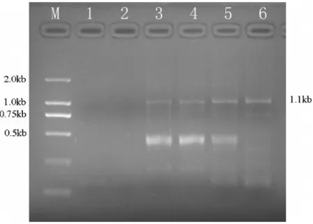
340 bp and 325 bp).(Fig. 1). The assembled approximate 0.8 kb full-length scFv fragments were used for the construction of templates of ribosome display.
Ribosome display and in situ RT-PCR recovery scFv
The gel-purified scFv fragments were digested and ligated into the vector pRDV. The ligation product was directly used as a template for the amplification of initial ribosome display library. The original scFv library was subjected toin vitrotranscription and translation usingTNTT7 Quick for PCR DNA kit to generate ternary MRA complexes. The target antigens were immobilized on microtiter plates. The selected specific scFv fragment based on mRNA of retained MRA complexes bound to SM2-OVA at the
plate wells was recovered after several washing with increasing stringency during the individual rounds of selection by in situ RT-PCR and SP-RT-PCR. The obtained products were applied to the affinity maturation or next round of panning. Each cycle of ribosome display was performed under the same conditions including the concentration of target antigens and the spanning washing time. As a whole, four cycles of selection on SM2-OVA, as
well as one round of affinity maturation were performed. The panning progress was monitored by examining the intensity of SP-PCR products (approximately 1.1 kb) on agarose gel–electropho-resis. The quantity of SP-PCR products continually increased during the next rounds of panning. Based on the result of SP-PCR, enrichment of specific scFvs was clearly confirmed. Meanwhile, no band was observed PBS-coated wells or when the untranslated mixture was used.(Fig. 2).
Here, we performed eukaryotic ribosome display by using anin vitro coupled transcription/translation system. These processes, based on theE.coli.S30and rabbit reticulocyte systems, had been described in previous studies [19,24–29]. We carried out the coupled transcription/translation steps, which were conducted by
Figure 1. Agarose gel electrophoresis of amplified VHchain, VLchain and assembled scFv fragments.Lane M: 100 bp plus DNA marker,
lane 1: VHfragments, lane 2: VLfragments, lane3: about 800 bp assembled scFv fragments.
doi:10.1371/journal.pone.0006427.g001
using a rabbit reticulocyte system. Briefly, after the transcription process, the translation was directly completed in tandem, potentially resulting in greater functional expression of scFvs antibodies [25]. Meanwhile, the rabbit reticulocyte system has lower RNase activity than anE.coli.S30 ribosome display system, leading to a less complicated selection condition [26]. Moreover, eukaryotic conditions may improve the translation or folding efficiency of some proteins. Then in our work, the eukaryotic ribosome display with coupled transcription/translation was used for selection and evolution of specific antibody.
In addition, the principal distinction between theE.coli.S30 and rabbit reticulocyte systems is in the DNA recovery step. In the
E.coli.S30 system, a chemical disruption procedure (EDTA) is introduced to dissociate the ribosome and release the mRNA, which is then purified before RT-PCR. Although this procedure efficiently isolates mRNA fromE.coliribosome complexes, it gives a relatively poor recovery from those generated by rabbit reticulocyte lysate [25,27], so that it cannot be applied directly to eukaryotic MRA complexes. We introduced the in situ RT-PCR procedure in which an internal primer is used for performing reverse transcription directly on the eukaryotic ribosome com-plexes without their disruption or mRNA isolation [28,29]. Not only does this simplify the recovery process, but it also avoids losses incurred in complex disruption. This procedure has been proved to be efficient and reliable in cDNA recovery in other studies [25,28,30]. The ineffectiveness, in the in situ procedure, of a primer that recognizes the 39 end of the mRNA, compared with the efficient use of one hybridizing upstream, is consistent with the interpretation that the ribosome is stalled at the end of the mRNA, which is consequently unavailable at the initiation of reverse transcription. The possibility of the 39 end of the mRNA being degraded, as has been speculated, has been excluded by showing that mRNA released from the ribosome complexes by EDTA, although low in yield, could be recovered and amplified by the 39
terminal primer with similar efficiency to the internal primer. The single-primer RT-PCR procedure presented here is a refinement
of the in situ DNA recovery method. It is based on the finding that single-primer PCR technology is capable of amplifying efficiently individual molecules of dsDNA fragments carrying identical flanking sequences at each end [31]. We have adapted this concept to in situ RT-PCR recovery by producing single-stranded cDNAs with complementary flanking 59 and 39 terminal sequences, so that PCR can be performed using a single consensus primer (KZ) to amplify the resultant cDNA templates. The method involves a new reverse transcription primer (RT-kz), the 39
end of which hybridizes,60 nt upstream of the 39terminus of the
mRNA, and the 59 end includes a consensus sequence from the transcription start to the translation initiation site. The in situ recovery method, using RT-PCR on an mRNA template that has not been prereleased from the ribosome, provides high sensitivity in a simple procedure and avoids sample loss.
The Characterization of soluble scFv
The selected specific scFv fragment after the fourth round was ligated with the expression vectorpCANTAB5Efor soluble scFvs expression. After electrotransformation, around 100 colonies from the selected library were isolated and soluble proteins of these clones were expressed using the nonsuppressor strain E.coli
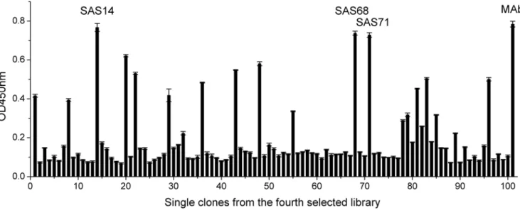
HB2151. The periplasmic extracts from individual clones were tested by indirect ELISA. The result showed that few clones showed positive to SM2in ribosome-ELISA before panning (Fig. S1), however, several clones from the fourth selected library had a good conjugation activity to specific SM2-OVA. Among these
clones, the three clones (SAS14, SAS68, SAS71) exhibiting the highest ELISA signals to SM2-OVA were not significantly
different to the anti-SM2MAb (Fig. 3). The result suggested that
the conjugation activity of scFvs was similar to that of parent MAb. The three specific soluble scFvs were confirmed by SDS-PAGE and Western-blotting analysis using anti-E tag antibodies. Approximately 30 kDa scFv protein was expressed from each of the three selected clones in the periplasmic extract sample compared to negative control (Fig. 4).
Figure 2. Selection and amplification of anti-SM2scFv gene over four rounds of ribosome display.After selection, the selected product
was amplified by SP-PCR and the SP-PCR products were analyzed by agarose gel electrophoresis. lane M: DL2000 DNA marker, lane 1: preliminary translation mixture selected on a PBS-coated well, lane 2: translation mixture selected on a OVA-coated well, lane 3: recovered band from the first ribosome display, lane 4: recovered band from the second round, lane 5: recovered band from the third cycle, lane 6: recovered band from the fourth selected library.
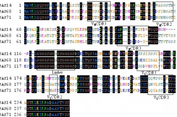
The deduced amino acid sequences of the above-mentioned three scFvs and complementary determining regions (CDR) were shown in Fig. 5. By using the DNA sequences, the VHand VL
gene families of the scFvs were designated based on Werner Mu¨ller’s database (DNAPLOT software). The heavy chains of scFvs SAS68 and SAS71 belong to the VH1 gene family and that
of SAS14 belongs to the VH2 gene family. The light chains of
clones SAS68 and SAS71 belong to the Vk IGKV12/13 subgroup and that of SAS14 belongs to the Vk IGKV4/5 subgroup. Sequencing alignment by the Vector NTI software program showed that the VH of the clones SAS68 and SAS71 have very
similar sequences (90.44% homology) and the VL of SAS14,
SAS68 and SAS71 shared 82.81% homology. Although the linking sequence of scFv SAS71 showed some mutations, these did not influence protein structure obviously. This indicated that a random mutation was induced by the second ribosome display,
but mutation affected scFv structures were selected. Sequencing data indicated that we succeeded in achieving our desired goals of library generation with the affinity maturation method.
In vitroselection cycles enable the introduction of further diversity every iteration, providing a means of protein evolution. Thus, ribosome display and similar methods are routes to increased diversity, and improve selection and a range of potentially novel molecules [32]. We carried out the affinity maturation by using error-prone PCR (EP-PCR) and staggered extension process (StEP) shuffling in tandem during the second round of ribosome display. The enrichment of specific scFvs was confirmed by the SP-PCR recovery band from each ribosome display cycle and the binding activity of scFvs from the fourth selected antibody library, respectively. After four rounds of ribosome display, three scFvs with high specificity and affinity were successfully selected. It showed that the slightly modified ribosome display technology was feasible for selection of specific scFvs against SM2.
Conclusions
In this report, we demonstrated the generation of anti-SM2
specific scFvs from hybridoma cell lines by eukaryotic ribosome display. Since ribosome display technology was first reported in 1994 [18], some early studies suggested antibody selection from a library using ribosome display[25,28,29,32,33]. However, there have been few reported about selection of scFvs against SM2.
Therefore, our work is significant to be an example for selecting specific scFvs against veterinary drugs by eukaryotic ribosome display. As an alternative to antiserum or MAb, the selected specific scFvs with high affinity can be used as detection reagent of SM2in foodstuffs in the future. In addition, this work provides a
novel pathway for the development of a rapid, sensitive and multi-residue immunoassay analysis technology for veterinary drugs detection with using recombinant antibody.
Materials and Methods
Plasmids, Strains and Reagents
All reagents used in the study were commercially available and were of reagent grade or better. All restriction enzymes and DNA modification enzymes were of molecular biology grade. DH5aand
Figure 4. Detection of soluble scFvs in periplasmatic extracts by Western Blot.M: low molecular weight protein marker, lanes1–3: Periplasmatic extracts of scFvs SAS14, SAS68, and SAS71.
doi:10.1371/journal.pone.0006427.g004
Figure 3. Clones were isolated from the fourth selected antibody library.Each clone was expressed for producing soluble scFv, and the binding activity of each scFv and the anti-SM2 MAb (positive control) was determined by ELISA (in triplicate). The error bars represent the standard deviation. doi:10.1371/journal.pone.0006427.g003
pGEM-T easy cloning vectors were purchased from TaKaRa; The ribosome display vector (pRDV) was obtained as a gift from the lab of Prof. Andreas Plu¨ckthun (Biochemisches Institut, Universita¨t Zu¨rich, Switzerland) [16]; The phagemid pCANTAB5E was from Amer-sham Biosciences; The bacterial host used for cloning and expression was the non-suppressor strain -E. coliHB2151, which was a gift from the lab of Prof. Yuanming Sui (Food Quality and Safety Research Institute, South China Agriculture University, P. R. China,); TNT T7 Quick for PCR DNA kit (rabbit reticulocyte cell free extract) was from Promega; Goat anti-E-Tag Antibody Affinity Purified HRP conjugated was purchased from Bethl Laboratories Inc.
Construction of scFv library
Total RNA was extracted from 107 Hybridoma cells produced previously by He et al [34] that secreted monoclonal antibody against SM2 by using SV Total RNA Isolation System (Promega, USA).
About 0.5mg of RNA was reverse transcribed by Oligo dT-Adaptor primers of the first-strand cDNA synthesis kit (TaKaRa, JAPAN). cDNA encoding the mouse variable heavy (VH) and light (VL) chains
were amplified by RT-PCR with degenerated immunoglobulin PCR primers (35 cycles at 94uC for 30 s, 55uC for 30 s and 72uC for 1 min). A complementary (Gly4Ser)3-linker was added by a
re-amplification. The full-length scFvs were assembled by using splicing overlap extension PCR. The purified products were cloned into the vector pRDV to add a 59-T7-promotor and ribosome binding site and 39-spacer region(Fig. 6). The templates of ribosome display were amplified directly from the ligation mixture [35]. The primers used in the PCR amplification were shown inTable 1.
In vitro transcription and translation
Eukaryotic ribosome display was conducted by a modified method, which was reported previously [24,26,36]. Briefly, 50ml
Figure 5. Alignment of the amino acid sequence of selected specific anti-SM2scFvs.CDRs and the (Gly4Ser)3linker are boxed. The regions
of CDR1-CDR3 were deduced according to Kabat database. doi:10.1371/journal.pone.0006427.g005
Figure 6. Construction of the template of ribosome display.In this generalized construct for ribosome display, the gene of interest is flanked by a 59sequence with the T7 promoter (T7) and eukaryotic translation initiation (Kozak) sequence, and a tolA domain is attached at the 39end as a spacer. The stop codon is removed to ensure stalling of the ribosome at the end of translation. X indicates removal of the stop codon.
of transcription/translation mixture, containing 40ml TNT T7 quick for PCR mix, 0.02 mM methionine and 1 mM Mg acetate, was set up in a siliconized tube with 100–700 ng purified PCR porducts. After incubation at 30uC for 60 min, the following reagents were added: 60 units DnaseI (TaKaRa, JAPAN), 7ml 106DNase I digestion buffer(400 mM Tris/HCl, pH 7.5, 60 mM
MgCl2, 100 mM NaCl) and adjusted the volume to 70ml with
dH2O. The incubation continued for another 20 min and then the
digested product was diluted with 210ml of cold PBS, containing 5 mM magnesium acetate [28,29].
Affinity selection and in situ RT-PCR Recovery
Microtiter plate was coated at 4uC overnight with 100ml of Avidin solution (0.066mM in PBS), and then washed with PBS for three times and blocked with blocking buffer PBSB (PBS with 1% (w/v) BSA) for 2 h at room temperature (RT). After another PBS washing for three times, the plate was coated with 100ml of Biotin-N-hydroxy-succinimide ester-sulfadimidine-ovalbumin (BNHS-SM2-OVA) at 4uC overnight. The coated plate was subsequently
washed by ice-cold washing buffer (PBS with 0.05 % (v/v) Tween 20 and 5 mM magnesium acetate) for three times, and then placed on ice for at least 10 min. 100ml the prepared TNT translation mixture containing the MRA ternary complexes was added to an antigen-coated well and incubated at 4uC for 2 h with gentle vibration. Followed three times wash with cold washing buffer and two times quick wash with ice-cold RNase-free water, in situ Single-Primer RT-PCR Recovery was performed in the plate wells carrying selected MRA complexes using PrimeScriptTM Reverse Transcriptase (TaKaRa). In brief, 12ml of solution A mixture, containing 1ml Primer RT-kz (10mM), 2ml dNTP (10 mM) and 9ml RNase-free water, was added into each MRA-bound well. After incubating at 48uC for 5 min and at least 30 s on ice, the following reagents were added into each well: 200 units Prime-ScriptTMReverse Transcriptase, 20 units Rnase Inhibitor, 5 mM DTT, 4ml 56PrimeScriptTMbuffer (250 mM Tris/HCl, pH 8.3,
15 mM MgCl2, 375 mM KCl) and adjusted the volume to 20ml
with RNase-free water. The plate was incubated at 42uC for 45 min followed by 5 min at 85uC. The mixture cooled to room temperature was then transfered to a fresh tube for subsequent
single-primer PCR (SP-PCR) using primer KZ. A further PCR step was introduced to regenerate the full-length construct avoiding shortening of the DNA fragment, compared to the original fragment [29]. The primers used in the PCR amplification were shown inTable 1. The purified PCR products were used for the next round of ribosome display or cloned intoE. coliHB2151 for expression.
Affinity maturation
The selected scFvs of first cycle were subjected to PCR-based random mutagenesis by using the error-prone PCR and staggered extension process (StEP) shuffling in tandem, according to the protocols described by Cadwell [37] and Zhao[38] to generate the initial mutant ribosome library of anti-Sulfanilamides scFvs. EP-PCR reactions were carried out under the following conditions, hereafter referred to as standard: 10 ng of DNA template, 106PCR buffer (Mg2+
Free, TaKaRa), 10 mM of each dNTP (dATP : dTTP = dGTP : dCTP = 1 : 5), 10mM of both primers (VHForNcoIand VLRevHindIII), 25 mM MgCl2and 2.5 U Taq
DNA polymerase (TaKaRa) in 50ml volume. PCR reactions were performed in an ABI thermocycler (Applied Biosystems Inc.) for 30 cycles: 1 min at 94uC, 1 min at 45uC and 2 min at 72uC, followed by 7 min extension at 72uC. Then the StEP shuffling was performed under the following conditions (5 min at 95uC, 80 cycles of 30 s at 94uC, and 5 s at 55uC, 5 min at 72uC), the StEP shuffling reactions contained (50ml final volume): 10 ng of the DNA products of EP-PCR, 106PCR buffer (Mg2+Plus, TaKaRa),
10 mM of dNTP mixtures, 10mM of both primers (VHForNcoI
and VLRevHindIII), 2.5 U Taq DNA polymerase (TaKaRa) and
dH2O. The purified StEP shuffling products were used for the
third round of ribosome display. The mutant repertoire was panned against target antigen as described above. One cycle of affinity maturation was performed.
Cloning and expression of scFv
After four selections, the obtained scFv fragment was amplified with forward primer scFvForSfiI with the SfiI restriction site (underlined) and reverse primer scFvRevNotI with the NotI
restriction site (underlined). The primers used in the PCR
Table 1.Synthesized oligonucleotides for the construction of scFv and ribosome display library.
Primer name sequence
VHFor AGATCTAGAGAATTCTGAGGAGACGGTGACCGTGGTCCCTTGGCCCCAG
VHRev AGATCTAGAAAGCTTAGGTCAAGCTGCAGCAGTCAGG
VLFor GGATACAGTTGGTGCAGCATC
VLRev GACATCCAGCTGACTCAGTCT
VHForNcoI CATGCCATGGATGGCCGTCAAGCTGCAGCAGTCAGGA
VHLinkRev ACCACCGGATCCGCCTCCGCCTAGATCTAGAGATTCTGAGGAGA
VLLinkFor GGAGGCGGATCCGGTGGTGGCGGATCTGGAGGTGGCGGAAGCGACATCCAGCTGACTCAGTC
VLRevHindIII CCCAAGCTTGGATACAGTTGGTGCAGCATC
T7B-kz GCAGCTAATACGACTCACTATAGGAACAGACCACCATGGCCGTCAAGCTGCAGCAG
tolAk CCGCACACCAGTAAGGTGTGCGGTTTCAGTTGCCGCTTTCTTTCT
RT-kz GAACAGACCACCATGCTGCTTCTGCCGCTTCC
KZ GAACAGACCACCATG
scFvForSfiI GTCCTCGCAACTGCGGCCCAGCCGGCCATGGCCGTCAAGCTGCAGCAGTCAGGA
scFvRevNotI GAGTCATTCTGCGGCCGCGGATACAGTTGGTGCAGCATC
The underlined sequence indicates theNcoI,HindIII,SfiIandNotIrestriction site for cloning. doi:10.1371/journal.pone.0006427.t001
amplification were shown inTable 1. The amplified product were digested with SfiI and NotI, then ligated with the vector pCANTAB5E by using a T4 DNA ligase (Promega, USA). The ligated products were transformed into E.coli HB2151 and the soluble scFv protein was expressed from each clone[39]. In brief, single colonies were grown in 5 ml of 26YT medium with
ampicillin (100mg/ml) and glucose (0.1 % (w/v)) to an OD600= 0.6 at 30uC/250 r.p.m, and induced by the addition of
IPTG (final concentration 1 mM) overnight at 30uC/180 r.p.m. The cells were pelleted at 4uC/4000 r.p.m for 10 min, and re-suspended in 0.5 ml ice-cold 16TES buffer (0.2 M Tris/HCl (pH 8.0), 0.5 mM EDTA, 0.5 M sucrose) and 0.75 ml ice-cold 1/ 56TES buffer. After incubation on ice for 40 min, the cells were
pelleted at 4uC/12000 r.p.m for 20 min, and the supernatant was retained as periplasmic extracts with the soluble scFvs. The expressed soluble scFvs were detected by using anti-E tag monoclonal antibody, since the pCANTAB5E vector contains an additional sequence encoding the E-tag.
ELISA assays
To screen anti-SM2specific scFvs, the expressed soluble scFvs
were analyzed by indirect ELISA. Microtiter plates were coated with 100ml of sulfadimidine-ovalbumin (SM2-OVA, 10mg/ml in
PBS) overnight at 4uC. Plates were washed with washing buffer and the blocking buffer (4 % (w/v) BSA in PBS, pH 7.4) was subsequently added at 37uC for 1 h with gentle shaking. Blocked plates were washed and 100ml of periplasmic extracts diluted 1:1 with PBSB were titrated followed by 1 h incubation at 37uC. Detection was performed with anHRP-labelled goat anti-E-tag antibody (1:10000 dilution with blocking buffer). Plates were developed with TMB-detection-solution and read at OD450 nm.
Immunoblot analysis of scFv expression
Periplasmic extracts from selected anti-sulfadimidine producing clones were subjected to SDS-PAGE on a 12 % polyacrylamide
gel and Western blot. After SDS-PAGE, the gel was transferred onto a nitrocellulose membrane. The transblotted membrane was blocked for 1 h with a blocking buffer (4 % (w/v) BSA in PBS, pH 7.4) and then incubated with anti-E-tag antibody HRP conjugated (1:2000) for 2 h at RT. 4-CN (4-chloro-1-naphthol, Sigma) was used as a peroxidase substrate to visualize the immunoreactivity.
Sequence analysis
Plasmid DNA from anti-sulfadimidine producing clones was isolated fromE. coliHB2151 using the QIAEX II gel extraction kit (QIAGEN). The scFv DNA was sequenced on both strands with the pCANTAB5E sequence primer set using an ABI Perkin Elmer 373A automated DNA sequencer.
Supporting Information
Figure S1 Clones were isolated from the unselected antibody library. Each clone was expressed for producing soluble scFv, and the binding activity of each scFv and the anti-SM2 MAb (positive control) was determined by ELISA (in triplicate). The error bars represent the standard deviation.
Found at: doi:10.1371/journal.pone.0006427.s001 (0.46 MB TIF)
Acknowledgments
We wish to thank Professors Andreas Plu¨ckthun and Yuan-Ming Sui for providing the test material used in this work.
Author Contributions
Conceived and designed the experiments: YQ SZ JS. Performed the experiments: YQ CW SH LD KW. Analyzed the data: YQ ZW SW LX. Contributed reagents/materials/analysis tools: XC YW. Wrote the paper: YQ.
References
1. McEvoy JD, Mayne CS, Higgins HC, Kennedy DG (1999) Transfer of sulphamethazine from contaminated dairy feed to cows’ milk. Vet Rec 144: 470–475.
2. Muldoon MT, Buckley SA, Deshpande SS, Holtzapple CK, Beier RC, et al. (2000) Development of a monoclonal antibody-based cELISA for the analysis of sulfadimethoxine. 2. Evaluation of rapid extraction methods and implications for the analysis of incurred residues in chicken liver tissue. J Agric Food Chem 48: 545–550.
3. Bruhlmann F, Chen W (1999) Tuning biphenyl dioxygenase for extended substrate specificity. Biotechnol Bioeng 63: 544–551.
4. Cliquet P, Cox E, Haasnoot W, Schacht E, Goddeeris BM (2003) Generation of group-specific antibodies against sulfonamides. J Agric Food Chem 51: 5835–5842.
5. Heering W, Usleber E, Dietrich R, Martlbauer E (1998) Immunochemical screening for antimicrobial drug residues in commercial honey. Analyst 123: 2759–2762.
6. Ko E, Song H, Park JH (2000) Direct competitive enzyme-linked immunosor-bent assay for sulfamethazine. J Vet Med Sci 62: 1121–1123.
7. Shelver WL, Shappell NW, Franek M, Rubio FR (2008) ELISA for sulfonamides and its application for screening in water contamination. J Agric Food Chem 56: 6609–6615.
8. Bashiardes S, Veile R, Wise CA, Szappanos L, Lovett M (2002) Positional cloning strategies for idiopathic scoliosis. Stud Health Technol Inform 91: 86–89. 9. Hoogenboom HR (2005) Selecting and screening recombinant antibody
libraries. Nat Biotechnol 23: 1105–1116.
10. Rothe A, Nathanielsz A, Hosse RJ, Oberhauser F, Strandmann EP, et al. (2007) Selection of human anti-CD28 scFvs from a T-NHL related scFv library using ribosome display. J Biotechnol 130: 448–454.
11. Deng P, Zhang HJ, Li Y, Liu W, Wang QP, et al. (2004) Evaluation of anti-HCV detection kits using recombinant antigens derived from various anti-HCV regions. Zhonghua Shi Yan He Lin Chuang Bing Du Xue Za Zhi 18: 354–355. 12. Korpimaki T, Rosenberg J, Virtanen P, Lamminmaki U, Tuomola M, et al. (2003) Further improvement of broad specificity hapten recognition with protein engineering. Protein Eng 16: 37–46.
13. Chen G, Yin YP, Yuan Q, Xia YX, Wang ZK (2007) High efficient expression and bio-activity assay of recombinant antibody for citrus bacterial canker disease. Wei Sheng Wu Xue Bao 47: 1066–1069.
14. Schier R, Marks JD (1996) Efficient in vitro affinity maturation of phage antibodies using BIAcore guided selections. Hum Antibodies Hybridomas 7: 97–105.
15. He M, Khan F (2005) Ribosome display: next-generation display technologies for production of antibodies in vitro. Expert Rev Proteomics 2: 421–430. 16. Binz HK, Amstutz P, Kohl A, Stumpp MT, Briand C, et al. (2004) High-affinity
binders selected from designed ankyrin repeat protein libraries. Nat Biotechnol 22: 575–582.
17. Kimura T, Iwase M, Kondo G, Watanabe H, Ohashi M, et al. (2003) Suppressive effect of selective cyclooxygenase-2 inhibitor on cytokine release in human neutrophils. Int Immunopharmacol 3: 1519–1528.
18. Mattheakis LC, Bhatt RR, Dower WJ (1994) An in vitro polysome display system for identifying ligands from very large peptide libraries. Proc Natl Acad Sci U S A 91: 9022–9026.
19. Hanes J, Pluckthun A (1997) In vitro selection and evolution of functional proteins by using ribosome display. Proc Natl Acad Sci U S A 94: 4937–4942. 20. MacPherson LJ, Bayburt EK, Capparelli MP, Carroll BJ, Goldstein R, et al. (1997) Discovery of CGS 27023A, a non-peptidic, potent, and orally active stromelysin inhibitor that blocks cartilage degradation in rabbits. J Med Chem 40: 2525–2532.
21. Yang LM, Wang JL, Kang L, Gao S, Liu YH, et al. (2008) Construction and analysis of high-complexity ribosome display random peptide libraries. PLoS ONE 3: e2092.
22. Hanes J, Jermutus L, Weber-Bornhauser S, Bosshard HR, Pluckthun A (1998) Ribosome display efficiently selects and evolves high-affinity antibodies in vitro from immune libraries. Proc Natl Acad Sci U S A 95: 14130–14135. 23. Hanes J, Jermutus L, Pluckthun A (2000) Selecting and evolving functional
proteins in vitro by ribosome display. Methods Enzymol 328: 404–430. 24. He M, Taussig MJ (1997) Antibody-ribosome-mRNA (ARM) complexes as
25. He M, Taussig MJ (2005) Ribosome display of antibodies: expression, specificity and recovery in a eukaryotic system. J Immunol Methods 297: 73–82. 26. Hanes J, Jermutus L, Schaffitzel C, Pluckthun A (1999) Comparison of
Escherichia coli and rabbit reticulocyte ribosome display systems. FEBS Lett 450: 105–110.
27. Douthwaite JA, Groves MA, Dufner P, Jermutus L (2006) An improved method for an efficient and easily accessible eukaryotic ribosome display technology. Protein Eng Des Sel 19: 85–90.
28. He M, Taussig MJ (2007) Eukaryotic ribosome display with in situ DNA recovery. Nat Methods 4: 281–288.
29. Rothe A, Nathanielsz A, Oberhauser F, von Pogge SE, Engert A, et al. (2008) Ribosome display and selection of human anti-cD22 scFvs derived from an acute lymphocytic leukemia patient. Biol Chem 389: 433–439.
30. Irving RA, Coia G, Roberts A, Nuttall SD, Hudson PJ (2001) Ribosome display and affinity maturation: from antibodies to single V-domains and steps towards cancer therapeutics. J Immunol Methods 248: 31–45.
31. Rungpragayphan S, Kawarasaki Y, Imaeda T, Kohda K, Nakano H, et al. (2002) High-throughput, cloning-independent protein library construction by combining single-molecule DNA amplification with in vitro expression. J Mol Biol 318: 395–405.
32. He M, Taussig MJ (2002) Ribosome display: cell-free protein display technology. Brief Funct Genomic Proteomic 1: 204–212.
33. He M, Menges M, Groves MA, Corps E, Liu H, et al. (1999) Selection of a human anti-progesterone antibody fragment from a transgenic mouse library by ARM ribosome display. J Immunol Methods 231: 105–117.
34. He JH, Shen JZ, Suo X, Jiang HY, Hou XL (2005) Development of a monoclonal antibody-based ELISA for detection of sulfamethazine and N4-acetyl sulfamethazine in chicken breast muscle tissue. J Food Sci 70: C113–C117.
35. Zahnd C, Amstutz P, Pluckthun A (2007) Ribosome display: selecting and evolving proteins in vitro that specifically bind to a target. Nat Methods 4: 269–279.
36. He M, Cooley N, Jackson A, Taussig MJ (2004) Production of human single-chain antibodies by ribosome display. Methods Mol Biol 248: 177–189. 37. Cadwell RC, Joyce GF (1992) Randomization of genes by PCR mutagenesis.
PCR Methods Appl 2: 28–33.
38. Zhao H, Giver L, Shao Z, Affholter JA, Arnold FH (1998) Molecular evolution by staggered extension process (StEP) in vitro recombination. Nat Biotechnol 16: 258–261.
39. Lee MS, Kwon MH, Kim KH, Shin HJ, Park S, et al. (2004) Selection of scFvs specific for HBV DNA polymerase using ribosome display. J Immunol Methods 284: 147–157.