P2Y2 Receptor and EGFR Cooperate to
Promote Prostate Cancer Cell Invasion via
ERK1/2 Pathway
Wei-Hua Li1,2, Ying Qiu1,2, Hong-Quan Zhang1,3, Xin-Xia Tian1,2*, Wei-Gang Fang1,2*
1Key Laboratory of Carcinogenesis and Translational Research, Ministry of Education, Peking University Health Science Center, Beijing, 100191, China,2Department of Pathology, Peking University Health Science Center, Beijing, 100191, China,3Department of Anatomy, Histology and Embryology, Peking University Health Science Center, Beijing, 100191, China
*wgfang@bjmu.edu.cn(W-GF); tianxinxia@163.com(X-XT)
Abstract
As one member of G protein-coupled P2Y receptors, P2Y2 receptor can be equally acti-vated by extracellular ATP and UTP. Our previous studies have proved that activation of P2Y2 receptor by extracellular ATP could promote prostate cancer cell invasion and metas-tasisin vitroandin vivovia regulating the expressions of some epithelial-mesenchymal
tran-sition/invasion-related genes (including IL-8, E-cadherin, Snail and Claudin-1), and the most significant change in expression of IL-8 was observed after P2Y2 receptor activation. However, the signaling pathway downstream of P2Y2 receptor and the role of IL-8 in P2Y2-mediated prostate cancer cell invasion remain unclear. Here, we found that extracellular ATP/UTP induced activation of EGFR and ERK1/2. After knockdown of P2Y2 receptor, the ATP -stimulated phosphorylation of EGFR and ERK1/2 was significantly suppressed. Fur-ther experiments showed that inactivation of EGFR and ERK1/2 attenuated ATP-induced invasion and migration, and suppressed ATP-mediated IL-8 production. In addition, knock-down of IL-8 inhibited ATP-mediated invasion and migration of prostate cancer cells. These findings suggest that P2Y2 receptor and EGFR cooperate to upregulate IL-8 production via ERK1/2 pathway, thereby promoting prostate cancer cell invasion and migration. Thus blocking of the P2Y2-EGFR-ERK1/2 pathway may provide effective therapeutic interven-tions for prostate cancer.
Introduction
Prostate cancer is one of the most common malignancies in human male population [1]. Most deaths related to prostate cancer are due to invasion and metastasis. Cell invasion and metasta-sis are complex processes that are regulated by multiple signaling pathways such as MAPK, Wnt and Notch pathways. Activation of these pathways is mainly dependent on interactions between receptors and extracellular signaling molecules [2].
OPEN ACCESS
Citation:Li W-H, Qiu Y, Zhang H-Q, Tian X-X, Fang W-G (2015) P2Y2 Receptor and EGFR Cooperate to Promote Prostate Cancer Cell Invasion via ERK1/2 Pathway. PLoS ONE 10(7): e0133165. doi:10.1371/ journal.pone.0133165
Editor:Karl X Chai, University of Central Florida, UNITED STATES
Received:March 21, 2015
Accepted:June 23, 2015
Published:July 16, 2015
Copyright:© 2015 Li et al. This is an open access article distributed under the terms of theCreative Commons Attribution License, which permits unrestricted use, distribution, and reproduction in any medium, provided the original author and source are credited.
Data Availability Statement:All relevant data are within the paper.
Funding:This work was supported by grants to WGF from National Natural Science Foundation of China (81321003) and 973 Program
(2010CB529402) from the Ministry of Science and Technology of China.
Extracellular adenosine 5’-triphosphate (ATP) is an important signaling molecule in tissue microenvironment, which mediates various biological functions via activation of P2 receptors [3]. Two subfamilies of P2 receptors have beαin mammalian cells. One is P2X family of
ligand-gated ion channel receptors (P2X1-7), and the other is P2Y family of G protein-coupled recep-tors (P2Y1, 2, 4, 6, 11, 12, 13, 14) [4]. Our previous study demonstrated that activation of P2Y receptors by ATP enhanced prostate cancer cell invasion [5]. We further found that P2Y2, a preferred receptor for ATP and UTP, contributed to the invasion and metastasis of prostate cancer cells [6]. However, the signaling pathway(s) downstream of P2Y2 receptor, especially in prostate cancer progression, is still not clear.
As a member of the CXC chemokine family, IL-8 expression is low in normal tissue and can be induced by a variety of stimuli such as growth factors and inflammatory cytokines in patho-logic conditions [7]. The expression of IL-8 is often elevated in human tumor cells and tissues [8]. It is reported that IL-8 functions as a significant regulatory factor in tumor microenviron-ment, and plays a crucial role in tumor invasion and metastasis [9]. We previously found that activation of P2Y2 receptor upregulated the expression and secretion of IL-8 [6]. However, the function of IL-8 in P2Y2 receptor-promoted invasion of prostate cancer cells remains unknown. This study aimed to examine the signaling pathway(s) downstream of P2Y2 recep-tor, and to explore the role of IL-8 in P2Y2 receptor-promoted prostate cancer cell invasion.
Materials and Methods
Chemicals and antibodies
ATP (adenosine 5’-triphosphate), UTP (uridine 5’-triphosphate), AG1478 (EGFR inhibitor)
and U0126 (MEK1/2 inhibitor) were all purchased from Sigma (St Louis, MO, USA). ATP and
UTP were both dissolved in normal saline and used at the concentration of 100μM. AG1478
was dissolved in DMSO and used at the concentration of 100 nM. U0126 was dissolved in
DMSO and used at the concentration of 10μM. The antibodies of P2Y2 (rabbit polyclonal
anti-body, sc-20124),β-actin (mouse monoclonal antibody, sc-8432), ERK1/2 (rabbit polyclonal
antibody, sc-94) and EGFR (mouse monoclonal antibody, sc-373746) were purchased from Santa Cruz Biotechnology (Santa Cruz, CA, USA). The antibodies of phospho-EGFR (rabbit monoclonal antibody, #8543, Tyr-1068) and phospho-ERK1/2 (rabbit polyclonal antibody, #9101, Thr202/Tyr204) were purchased from Cell Signaling Technology (Danvers, MA, USA).
Cell lines and culture conditions
Two subclones 1E8 and 2B4 were derived from PC-3M human prostate carcinoma cell line, which was previously purchased from American Type Culture Collection (Manassas, VA, USA). The 1E8 cell line was highly metastatic, whereas the 2B4 cell line was non-metastatic [10]. DU-145 cell line was purchased from American Type Culture Collection (Manassas, VA, USA). All cells were cultured in RPMI 1640 (GIBCO, Grand Island, NY, USA) supplemented with 10% fetal bovine serum, and maintained in a water-saturated atmosphere at 37°C with 5% CO2.
Reverse transcription and real-time PCR
Cells were grown in monolayer with or without ATP treatment. Total RNA was isolated with Trizol reagent (Invitrogen, Carlsbad, CA, USA). Reverse transcription reaction was performed using M-MLV Reverse Transcriptase (Promega, Madison, Wisconsin, USA) to obtain the
cDNA, according to the manufacturer’s guidelines. Next, real-time PCR was performed with
TCTGCACCCAGTTTTC-3’) orβ-actin (sense: 5’- GGATGCAGAAGGAGATCACTG-3’;
antisense: 5’-CGATCCACACGGAGTACTTG-3’). The real-time PCR reaction was performed
on an ABI StepOne Real-Time PCR System (Life Technologies, Carlsbad, CA, USA) in
tripli-cates. The 20μl reaction mixture consisted of 2μl of cDNA (20 ng/μl), 100 nM of primers and
10μl of SYBR Green Real-time PCR Master Mix (TOYOBO, Japan) containing AmpliTaq gold DNA polymerase. Samples were first denatured at 95°C for 10 min and then PCR reaction was proceeded for 40 amplification cycles as follows: 15 s at 95°C and 1 min at 60°C. Then a dissoci-ation curve analysis was conducted and melting temperatures (Tm) of the formed PCR ampli-cons were observed to distinguish the amplified sequences of interest from non-specific ones
or primer dimmers. The expression of IL-8 was normalized byβ-actin. The 2–ΔΔCtmethod
was used for relative quantification as described previously [11,12].
Cell lysis and western blotting
Cells were rinsed twice with ice-cold PBS and lysed in lysis buffer containing 20 mM Tris-HCl
(pH 7.5), 250 mM NaCl, 4 mM EDTA, 0.5% NP-40, 20 mMβ-Glycerophosphate, 1 mM NaF
with protease inhibitors (Roche, Mannheim, Germany). Protein concentrations were deter-mined using a BCA protein assay kit (Applygen Technologies Inc, Beijing, China). Equal amounts of total protein were loaded and separated by SDS-PAGE gel, and then were trans-ferred to PVDF membranes (Bio-Rad, Hercules, CA, USA). After blocking with 5% BSA at
room temperature for 1 h, blots were probed with primary antibodies against P2Y2 (1: 500),β
-actin (1: 1000), EGFR (1: 500), ERK1/2 (1: 1000), phospho-EGFR (1: 1000) or phospho-ERK1/ 2 (1: 1000) at 4 °C overnight. Then the blots were washed with PBS for three times and incu-bated with secondary antibodies at room temperature for 1 h. The immunoreactive bands were visualized by an enhanced chemiluminescence detection system (Applygen Technologies Inc), and quantified with the software of Quantity One (Bio-Rad).
siRNA and transfection
Two P2Y2 siRNAs (P2Y2 si#1 and P2Y2 si#2) and a control siRNA (Negative) were used as described previously [6]. Two IL-8 siRNAs (IL-8 si#1 and IL-8 si#2) were used to silence the expression of IL-8. The IL-8 siRNAs were purchased from Invitrogen (Carlsbad, CA, USA) with the sequence as follows:
IL-8 si#1, 5'-GAACTTAGATGTCAGTGCATA-3'; IL-8 si#2, 5'-GCCAAGGAGUGCUAAAGAA-3'.
A fluorescence-labelled siRNA oligonucleotide was used to directly observe siRNA transfec-tion efficiency, and a scramble siRNA oligonucleotide was used as control siRNA (Negative). Cells were seeded into 6-well plates at the density of 1 × 10⁴cells/well. Twelve hours later, cells were transfected with siRNAs by Lipofectamine 2000 (Invitrogen), according to the manufac-turer’s instructions. Six hours later, the medium was replaced with fresh 1640 medium supple-mented with 10% FBS. Twenty hours after transfection, cells were split. After an additional 12 h, knockdown efficiency was determined by western blotting, and cells were used for the fol-lowing experiments.
Enzyme-linked immunosorbent assay (ELISA)
Invasion assay
Invasion assay was performed as described previously [6]. Briefly, cells were harvested and resuspended in RPMI 1640 with 0.1% BSA at a density of 5 × 10⁵cells/ml. Next, 200μl cell sus-pensions with or without ATP treatment was placed in the upper chamber coated with Matri-gel (BD, Franklin Lakes, NJ, USA). NIH3T3-conditioned medium was obtained by incubating
NIH3T3 cells for 24 h in serum-free 1640 medium and filled in the lower chamber (600μl) as a
chemo-attractant, according to the method described by Albini et al [13]. Cells were allowed to invade for 12 h at 37 °C. After fixed with 4% formaldehyde, cells on the lower surface of the membranes were stained with crystal violet and observed under a microscope at × 200 magnifi-cation. The number of invaded cells in seven fields was counted and the mean for each cham-ber was determined.
Migration assay
One hundred thousand cells in 100μl of RPMI 1640 supplemented with 0.1% BSA was placed
in the upper chamber. NIH3T3-conditioned medium was obtained by incubating NIH3T3
cells for 24 h in serum-free 1640 medium and filled in the lower chamber (600μl) as a
chemo-attractant. Cells were incubated for 12 h at 37°C with or without ATP treatment. Then cells on the lower surface of the membrane were fixed and stained with crystal violet. The number of migrated cells was counted under a light microscope at × 200 magnification. The average num-ber of migrated cells was determined from seven representative fields.
Statistical analyses
The quantitative results were presented as the means ± SD of three determinations and data were analyzed with the software package of SPSS 19.0 (SPSS Inc., Chicago, IL, USA). Student’s t test was used to detect whether there was a significant difference between two groups. Non-parametric ANOVA was used when multiple groups were compared. Any P-value of less than 0.05 was considered to be statistically significant.
Results
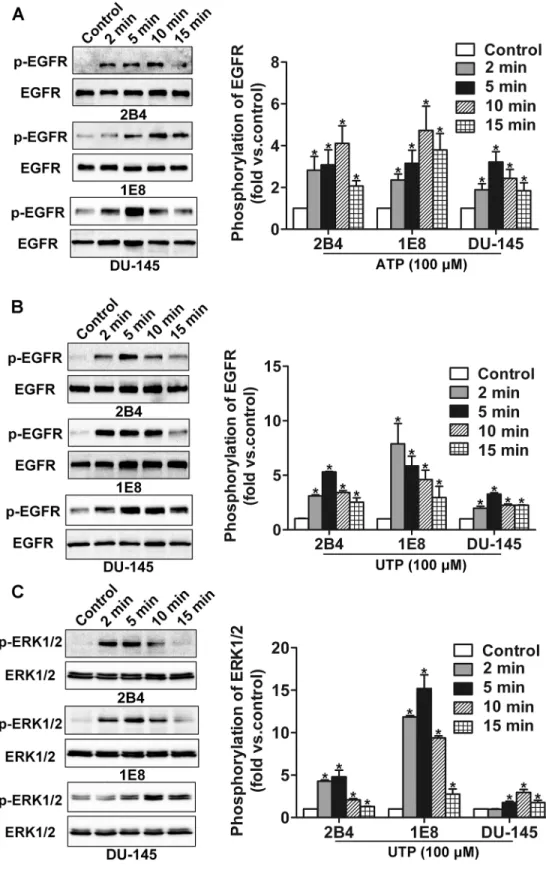
Extracellular ATP or UTP stimulation activates EGFR and ERK1/2
In the present study, after incubation with 100μM ATP, we detected the phosphorylation of
EGFR in 2B4, 1E8 and DU-145 cells by western blotting at 2 min, 5 min, 10 min and 15 min. Our results showed that ATP stimulation resulted in activation of EGFR (Tyr-1068) in prostate cancer cells (Fig 1A). Similar results were observed with UTP treatment (Fig 1B). Our previous study showed that ATP could induce activation of ERK1/2 in prostate cancer 2B4, 1E8 and DU-145 cells [12]. Here, we further found that UTP also stimulated activation of ERK1/2 (Thr202/Tyr204) in 2B4, 1E8 and DU-145 cells (Fig 1C). Among eight P2Y subtype receptors identified in mammalian cells, only P2Y2 receptor can be equally activated by ATP and UTP [14]. Therefore, these results indicate that P2Y2 receptor may be involved in extracellular ATP/UTP-induced activation of EGFR and ERK1/2. The following experiments were only per-formed with ATP treatment.
P2Y2 receptor mediates activation of EGFR and ERK1/2
Fig 1. Phosphorylation of EGFR was detected after ATP or UTP treatment.(A) After treatment with 100μM ATP, phosphorylation of EGFR was detected by western blotting. (B) After treatment with 100μM UTP, phosphorylation of EGFR was detected by western blotting. (C) After treatment with 100μM UTP, phosphorylation of ERK1/2 was detected by western blotting. Results were demonstrated by histograms to quantify the expression levels. Data were presented as means±SD (vertical bars). Three independent
experiments were performed.*p<0.05 vs. control cells.
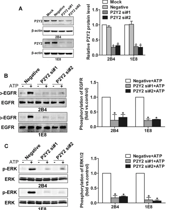
Fig 2. Knockdown of P2Y2 receptor inhibited ATP-induced activation of EGFR and ERK1/2.(A) 2B4 and 1E8 cells were transfected with two different P2Y2 siRNAs (P2Y2 si#1 and P2Y2 si#2) or a control siRNA (Negative), respectively. Knockdown efficiency was determined by western blotting. After transfection with two different P2Y2 siRNAs (P2Y2 si#1 and P2Y2 si#2) or a control siRNA (Negative) respectively, 2B4 and 1E8 cells were incubated with or without 100μM ATP for 5 min. Then the protein was extracted for phosphorylation detection of (B) EGFR and (C) ERK1/2. Results were demonstrated by histograms to quantify the expression levels. Data were presented as means±SD (vertical bars). Three independent experiments were performed.*p<0.05.
blotting. Here, we found that ATP treatment induced activation of EGFR and ERK1/2 in con-trol siRNA cells. After knockdown of P2Y2 receptor, the activation of EGFR and ERK1/2 induced by ATP was significantly suppressed (Fig 2B and 2C). Together, these data suggest that the ATP-induced activation of EGFR and ERK1/2 was predominantly regulated by P2Y2 receptor.
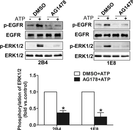
EGFR is involved in ATP-induced activation of ERK1/2
To explore the role of EGFR in ATP-induced activation of ERK1/2, AG1478 (100 nM), a
selec-tive inhibitor of EGFR, was used before ATP treatment.Fig 3demonstrated that ATP induced
activation of EGFR and ERK1/2 in control group (DMSO-treated cells), whereas pretreatment with AG1478 significantly suppressed ATP-induced phosphorylation of EGFR and ERK1/2, suggesting that ATP activates ERK1/2 pathway through EGFR activation.
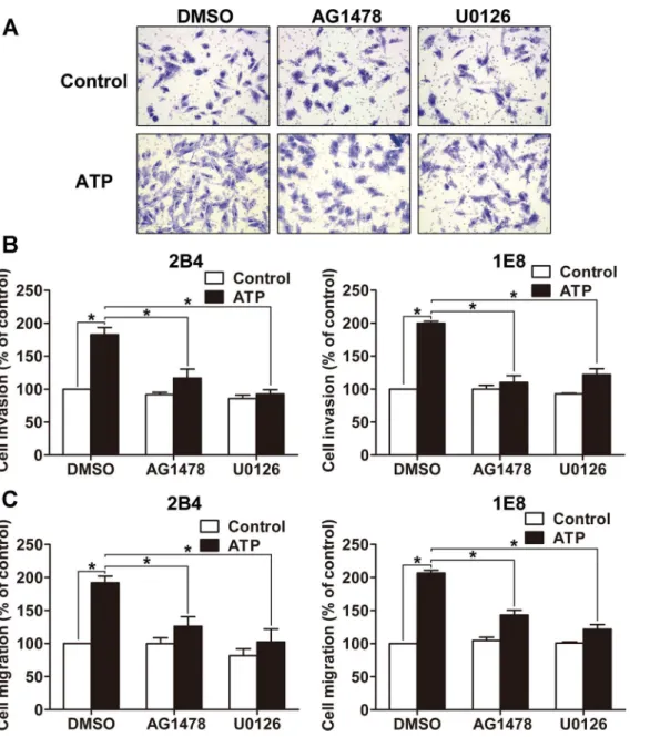
P2Y2-EGFR-ERK1/2 pathway is involved in prostate cancer cell
invasion and migration
Next, to investigate whether EGFR-ERK1/2 pathway mediated ATP-induced invasion and migration, we pretreated 2B4 and 1E8 cells with AG1478 (EGFR inhibitor, 100 nM) and
U0126 (MEK1/2 inhibitor, 10μM) respectively before 100μM ATP stimulation. Invasion
assay and migration assay revealed that ATP treatment enhanced cell invasion and migration in control group (DMSO-treated cells). But in AG1478- or U0126-treated groups, the
ATP-promoted cell invasion and migration was inhibited (Fig 4A–4C). Since P2Y2 receptor
pre-dominantly mediates ATP-induced activation of EGFR and ERK1/2 as described above, and P2Y2 receptor is responsible for ATP-stimulated prostate cancer cell invasion and migration as demonstrated in our previous study [6], the results strongly suggest that P2Y2-EGFR-ERK1/2 pathway is involved in the regulation of prostate cancer cell invasion and migration.
P2Y2-EGFR-ERK1/2 pathway contributes to IL-8 upregulation
Our previous study has shown that ATP and UTP both stimulated the production of IL-8, and knockdown of P2Y2 suppressed the secretion of IL-8 [6]. Here, 2B4 and 1E8 cells were
incu-bated with 100 nM AG1478 (EGFR inhibitor) or 10μM U0126 (MEK1/2 inhibitor)
respec-tively before 100μM ATP stimulation. Using real-time PCR and ELISA assay, we found that
blockade of EGFR activation attenuated ATP-promoted IL-8 expression and secretion, while blockade of ERK1/2 activation also inhibited the expression and secretion of IL-8 (Fig 5A–5D). As ATP induced activation of EGFR and ERK1/2 via P2Y2 receptor, together with the previous results that silencing of P2Y2 with siRNA significantly suppressed ATP-promoted IL-8 pro-duction [6], these data strongly indicate that ATP promotes the expression and secretion of IL-8 via P2Y2-EGFR-ERK1/2 pathway.
Role of IL-8 in ATP-promoted cell invasion and migration
prostate cancer cells was greatly inhibited, suggesting that IL-8 is one of the important players
in the regulation of ATP-promoted cell invasion and migration (Fig 7A and 7B).
Discussion
A number of studies have found that in tumor microenvironment extracellular ATP and its derivatives may act as detrimental signals in tumor progression [15,16]. Our previous study showed that activation of P2Y2 by extracellular ATP promotes cell invasion and metastasis of prostate cancer cellsin vitroandin vivo[6]. Belonging to G protein-coupled receptor (GPCR)
subfamilies, P2Y2 receptors have been reported to couple with multiple intracellular signaling
pathways [17,18]. Most of these pathways are regulated simultaneously by EGFR activation
and involved in tumor invasion and metastasis [19]. However, there is no convincing evidence so far that P2Y2 receptor promotes prostate cancer progression via EGFR. In this study, we revealed that EGFR and ERK1/2 could both be activated by ATP or UTP stimulation. Knock-down of P2Y2 receptor suppressed ATP-induced phosphorylation of EGFR and ERK1/2. In addition, blockade of EGFR suppressed ATP-regulated activation of ERK1/2. Further Fig 3. ATP-induced activation of ERK1/2 was dependent on EGFR activation.After incubation with 100 nM AG1478 (EGFR inhibitor) for 30 min, 2B4 and 1E8 cells were treated with 100μM ATP for 5 min. Phosphorylation of EGFR and ERK1/2 was measured by western blotting. Results were demonstrated by histograms to quantify the expression levels. Data were presented as means±SD (vertical bars). Three independent experiments were performed.*p<0.05.
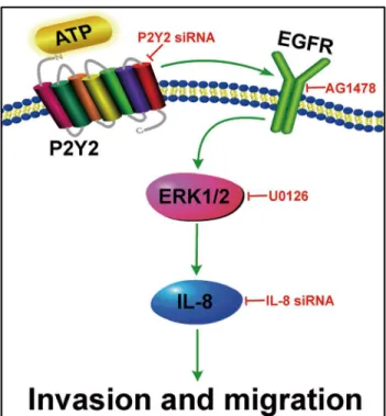
experiments showed that activation of EGFR-ERK1/2 pathway increased ATP-promoted IL-8 production, which then promoted the invasion and migration of prostate cancer cells. Together with our previous study that activation of P2Y2 receptor by ATP promotes cell invasion and metastasis of prostate cancer cells, this study strongly indicates that P2Y2 receptor promotes prostate cancer cell invasion and migration mainly via activation of EGFR-ERK1/2 pathway and upregulation of IL-8 production (Fig 8).
Multiple pathways have been identified downstream of GPCR. There are several possible explanations for the pro-invasive effect after P2Y2 receptor activation. One pathway is that
ATP stimulates Gqα-dependent phospholipase C (PLCb) activity, which generates inositol
Fig 4. Effects of EGFR and ERK1/2 activation on ATP-mediated invasion and migration of prostate cancer cells.(A) Cells were harvested, resuspended and pretreated with AG1478, U0126 or DMSO for 30 min, respectively. Then cells were incubated in Transwell plate with or without 100μM ATP for 12 h, invaded/migrated cells were stained with crystal violet and observed under a microscope at × 200 magnification. (B) Effects of AG1478 and U0126 on the invasion of 2B4 and 1E8 cells. (C) Effects of AG1478 and U0126 on the migration of 2B4 and 1E8 cells. Results were demonstrated by histograms, and data were presented as means±SD (vertical bars). Three independent experiments were performed.*p<0.05.
1,4,5-trisphosphate (IP3) and diacylglycerol (DAG), resulting in an elevation in the intracellu-lar calcium concentration and DAG-dependent activation of protein kinase C (PKC), respec-tively. PLCb2 has been shown to be important for breast cancer cell migration [20]. Changes in intracellular Ca2+concentration alter essential processes in tumor cell migration and invasion [21]. PKC regulates metastasis through phosphorylation of many key proteins in different steps of metastasis. All of these regulatory elements are actively involved in our prostate cancer studies. In our previous studies, we observed remarkable accumulation of IP3 and significant intracellular Ca2+mobilization in prostate cancer cells after ATP treatment [22]. This is obvi-ously due to activation of PLCb by G-protein coupled P2Y2 receptor.
Another pathway is that the P2Y2 receptor can interact withαVβ3/5integrins via an
extra-cellularly oriented RGD domain to regulate the activities of G12-dependent Rho, Go-dependent
Rac, LIM kinase, and cofilin, proteins that regulate actin cytoskeletal rearrangements which are important features of cell migration [23]. We previously demonstrated that ATP induced activation of Rac1 and Cdc42, promoted the formation of lamellipodia and filopodia, and increased the motility of prostate cancer cells [12], indicating a possible role of P2Y2 receptor in prostate cancer cell motility.
Fig 5. IL-8 production was increased after activation of EGFR and ERK1/2 by ATP.2B4 and 1E8 cells were pretreated with AG1478, U0126 or DMSO for 30 min, respectively. Then cells were incubated with or without 100μM ATP for 12 h. (A) and (B) mRNA level of IL-8 was detected by real-time PCR. (C) and (D) Protein level of IL-8 in cell supernatant was examined by ELISA assay. Results were demonstrated by histograms to quantify the expression levels. Data were presented as means±SD (vertical bars). Three independent experiments were performed.*p<0.05.
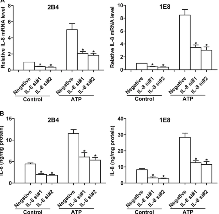
P2Y2 receptor may also interact with EGFR to activate ERK1/2 pathway since there are some evidences that transactivation of P2Y2 receptor and EGFR exists in some cell types, e.g., membrane distribution of the P2Y2 transregulated by EGFR in blood vessel smooth muscle cells [24], promotion of the formation of EGFR/ErbB3 heterodimers by P2Y2 receptor in sali-vary gland cells [25]. Phosphorylation of EFGR by P2Y2 receptor is thought mainly due to Src-dependent recruitment of P2Y2 receptor to a signaling complex containing EGFR in response Fig 6. Effects of IL-8 siRNAs on ATP-mediated IL-8 production.2B4 and 1E8 cells were transfected with two different IL-8 siRNAs (IL-8 si#1 and IL-8 si#2) or a control siRNA (Negative), respectively. Cells were treated with or without 100μM ATP for 12 h. (A) IL-8 mRNA expression was detected by real-time PCR, and (B) IL-8 secretion was measured by ELISA assay. Results were demonstrated by histograms to quantify the expression levels. Data were presented as means±SD (vertical bars). Three independent experiments were performed.*p<0.05 vs. Negative.
to P2Y2 ligands [26]. In the present study, we revealed that EGFR and ERK1/2 could be activated by extracellular ATP or UTP stimulation. Knockdown of P2Y2 receptor suppressed ATP-induced phosphorylation of EGFR and ERK1/2. To our knowledge, our present data represent the first example that EGFR participates in P2Y2 receptor-regulated cell invasion of prostate cancer.
It is well known that EGFR participates in diverse cellular processes such as growth,
differ-entiation, tumorigenesis, invasion and metastasis of cancer [27,28]. Increased EGFR
expres-sion is often detected in prostate cancer, and is associated with poor prognosis [29].
Monoclonal antibody against EGFR has gained an effective improvement in patients with pros-tate cancer [30]. As one subfamily of MAPKs, ERK1/2 is the best characterized cytoplasmic kinase activated by EGFR, and inhibition of ERK1/2 has been utilized as a biomarker for EGFR inhibitor action [31]. Inhibition of ERK1/2 pathway also has been considered as an important treatment strategy for prostate cancer [32,33]. In this study, we demonstrated a significant function of P2Y2-EGFR-ERK1/2 pathway in mediating prostate cancer cell invasion and migration. Together with our previous observation that extracellular ATP is an important Fig 7. Knockdown of IL-8 attenuated ATP-mediated invasion and migration of prostate cancer cells.After knockdown of IL-8 by siNRA, cells were treated with or without 100μM ATP, and then subjected to (A) invasion assay and (B) migration assay. Results were demonstrated by histograms, and data were presented as means±SD (vertical bars). Three independent experiments were performed.*P<0.05.
pro-invasive agent within tumor microenvironment, this may help to elucidate the regulating mechanism(s) underlying EGFR activity in prostate cancer progression.
IL-8 has been found to be highly expressed in androgen-independent metastatic cell lines, such as PC-3 cell line [9]. Clinical studies also reveal that IL-8 production is elevated in tumor tissue and serum of patients with prostate cancer, and there is a direct correlation between high level of IL-8 and tumor progression [34]. Increase of IL-8 production is associated with tumor angiogenesis, proliferation and metastasis of prostate cancerin vitroandin vivo[35].
Here, our study proves that ATP upregulated the expression and secretion of IL-8 via P2Y2-EGFR-ERK1/2 pathway, and IL-8 production contributed to ATP-promoted invasion and migration of prostate cancer cells.
In summary, our results demonstrate that P2Y2 receptor upregulates IL-8 production, thereby promoting the invasion and migration of prostate cancer cells. Cross-talking with EGFR and subsequently activation of ERK1/2 pathway may be one of the important mecha-nisms of P2Y2 receptor function. Therefore, therapies that target P2Y2-EGFR-ERK1/2 path-way may provide effective treatment strategies for prostate cancer.
Acknowledgments
We thank Professor Jian Zhang for providing helpful discussion for the finalization of the manuscript.
Author Contributions
Conceived and designed the experiments: WHL WGF XXT. Performed the experiments: WHL XXT YQ. Analyzed the data: WHL XXT WGF. Contributed reagents/materials/analysis tools: WHL YQ. Wrote the paper: WHL HQZ XXT WGF. Revised the paper: WGF XXT.
Fig 8. Diagram depicting the involvement of P2Y2-EGFR-ERK1/2 pathway and IL-8 upregulation in ATP-promoted invasion and migration of prostate cancer cells.
References
1. Siegel R, Ma J, Zou Z, Jemal A. Cancer statistics, 2014. CA: a cancer journal for clinicians. 2014; 64 (1):9–29. Epub 2014/01/09. doi:10.3322/caac.21208PMID:24399786.
2. Gupta GP, Massague J. Cancer metastasis: building a framework. Cell. 2006; 127(4):679–95. Epub 2006/11/18. doi:10.1016/j.cell.2006.11.001PMID:17110329.
3. Burnstock G, Verkhratsky A. Long-term (trophic) purinergic signalling: purinoceptors control cell prolif-eration, differentiation and death. Cell Death Dis. 2010; 1:e9. Epub 2010/01/01. doi:10.1038/cddis. 2009.11cddis200911[pii]. PMID:21364628; PubMed Central PMCID: PMC3032501.
4. Burnstock G. Therapeutic potential of purinergic signalling for diseases of the urinary tract. BJU interna-tional. 2011; 107(2):192–204. Epub 2011/01/07. doi:10.1111/j.1464-410X.2010.09926.xPMID: 21208364.
5. Chen L, He HY, Li HM, Zheng J, Heng WJ, You JF, et al. ERK1/2 and p38 pathways are required for P2Y receptor-mediated prostate cancer invasion. Cancer letters. 2004; 215(2):239–47. Epub 2004/10/ 19. doi:10.1016/j.canlet.2004.05.023PMID:15488643.
6. Li WH, Qiu Y, Zhang HQ, Liu Y, You JF, Tian XX, et al. P2Y2 receptor promotes cell invasion and metastasis in prostate cancer cells. British journal of cancer. 2013; 109(6):1666–75. Epub 2013/08/24. doi:10.1038/bjc.2013.484PMID:23969730; PubMed Central PMCID: PMC3776994.
7. Matsushima K, Baldwin ET, Mukaida N. Interleukin-8 and MCAF: novel leukocyte recruitment and acti-vating cytokines. Chemical immunology. 1992; 51:236–65. Epub 1992/01/01. PMID:1567543.
8. Green AR, Green VL, White MC, Speirs V. Expression of cytokine messenger RNA in normal and neo-plastic human breast tissue: identification of interleukin-8 as a potential regulatory factor in breast tumours. International journal of cancer Journal international du cancer. 1997; 72(6):937–41. Epub 1997/11/05. PMID:9378554.
9. Araki S, Omori Y, Lyn D, Singh RK, Meinbach DM, Sandman Y, et al. Interleukin-8 is a molecular deter-minant of androgen independence and progression in prostate cancer. Cancer research. 2007; 67 (14):6854–62. doi:10.1158/0008-5472.can-07-1162PMID:WOS:000248319000039.
10. Liu Y, Zheng J, Fang W, You J, Wang J, Cui X, et al. Identification of metastasis associated gene G3BP by differential display in human cancer cell sublines with different metastatic potentials G3BP as highly expressed in non-metastatic. Chinese medical journal. 2001; 114(1):35–8. Epub 2002/01/10. PMID: 11779432.
11. Livak KJ, Schmittgen TD. Analysis of relative gene expression data using real-time quantitative PCR and the 2(T)(-Delta Delta C) method. Methods. 2001; 25(4):402–8. doi:10.1006/meth.2001.1262 PMID:WOS:000173949500003.
12. Zhang Y, Gong LH, Zhang HQ, Du Q, You JF, Tian XX, et al. Extracellular ATP enhances in vitro inva-sion of prostate cancer cells by activating Rho GTPase and upregulating MMPs expresinva-sion. Cancer let-ters. 2010; 293(2):189–97. Epub 2010/03/06. doi:10.1016/j.canlet.2010.01.010PMID:20202742.
13. Albini A, Iwamoto Y, Kleinman HK, Martin GR, Aaronson SA, Kozlowski JM, et al. A rapid in vitro assay for quantitating the invasive potential of tumor cells. Cancer research. 1987; 47(12):3239–45. Epub 1987/06/15. PMID:2438036.
14. Molliver DC, Cook SP, Carlsten JA, Wright DE, McCleskey EW. ATP and UTP excite sensory neurons and induce CREB phosphorylation through the metabotropic receptor, P2Y2. European Journal of Neu-roscience. 2002; 16(10):1850–60. doi:10.1046/j.1460-9568.2002.02253.xPMID:
WOS:000179537800003.
15. Pellegatti P, Raffaghello L, Bianchi G, Piccardi F, Pistoia V, Di Virgilio F. Increased level of extracellular ATP at tumor sites: in vivo imaging with plasma membrane luciferase. PloS one. 2008; 3(7):e2599. Epub 2008/07/10. doi:10.1371/journal.pone.0002599PMID:18612415; PubMed Central PMCID: PMC2440522.
16. Yokdang N, Tellez JD, Tian H, Norvell J, Barsky SH, Valencik M, et al. A role for nucleotides in support of breast cancer angiogenesis: heterologous receptor signalling. British journal of cancer. 2011; 104 (10):1628–40. Epub 2011/04/21. doi:10.1038/bjc.2011.134PMID:21505453; PubMed Central PMCID: PMC3101911.
17. Buzzi N, Bilbao PS, Boland R, de Boland AR. Extracellular ATP activates MAP kinase cascades through a P2Y purinergic receptor in the human intestinal Caco-2 cell line. Biochimica et biophysica acta. 2009; 1790(12):1651–9. Epub 2009/10/20. doi:10.1016/j.bbagen.2009.10.005PMID:19836435.
19. De Luca A, Carotenuto A, Rachiglio A, Gallo M, Maiello MR, Aldinucci D, et al. The role of the EGFR signaling in tumor microenvironment. Journal of cellular physiology. 2008; 214(3):559–67. Epub 2007/ 09/27. doi:10.1002/jcp.21260PMID:17894407.
20. Bertagnolo V, Benedusi M, Brugnoli F, Lanuti P, Marchisio M, Querzoli P, et al. Phospholipase C-beta 2 promotes mitosis and migration of human breast cancer-derived cells. Carcinogenesis. 2007; 28 (8):1638–45. Epub 2007/04/13. doi:10.1093/carcin/bgm078PMID:17429106.
21. Prevarskaya N, Skryma R, Shuba Y. Calcium in tumour metastasis: new roles for known actors. Nature reviews Cancer. 2011; 11(8):609–18. Epub 2011/07/23. doi:10.1038/nrc3105PMID:21779011.
22. Fang WG, Pirnia F, Bang YJ, Myers CE, Trepel JB. P2-purinergic receptor agonists inhibit the growth of androgen-independent prostate carcinoma cells. The Journal of clinical investigation. 1992; 89(1):191– 6. Epub 1992/01/01. doi:10.1172/JCI115562PMID:1309535; PubMed Central PMCID: PMC442836.
23. Bagchi S, Liao Z, Gonzalez FA, Chorna NE, Seye CI, Weisman GA, et al. The P2Y2 nucleotide receptor interacts with alphav integrins to activate Go and induce cell migration. The Journal of biological chem-istry. 2005; 280(47):39050–7. Epub 2005/09/28. doi:10.1074/jbc.M504819200PMID:16186116.
24. Norambuena A, Palma F, Poblete MI, Donoso MV, Pardo E, Gonzalez A, et al. UTP controls cell sur-face distribution and vasomotor activity of the human P2Y2 receptor through an epidermal growth factor receptor-transregulated mechanism. The Journal of biological chemistry. 2010; 285(5):2940–50. Epub 2009/12/10. doi:10.1074/jbc.M109.081166PMID:19996104; PubMed Central PMCID: PMC2823411.
25. Ratchford AM, Baker OJ, Camden JM, Rikka S, Petris MJ, Seye CI, et al. P2Y2 nucleotide receptors mediate metalloprotease-dependent phosphorylation of epidermal growth factor receptor and ErbB3 in human salivary gland cells. The Journal of biological chemistry. 2010; 285(10):7545–55. Epub 2010/ 01/13. doi:10.1074/jbc.M109.078170PMID:20064929; PubMed Central PMCID: PMC2844202.
26. Liu J, Liao Z, Camden J, Griffin KD, Garrad RC, Santiago-Perez LI, et al. Src homology 3 binding sites in the P2Y2 nucleotide receptor interact with Src and regulate activities of Src, proline-rich tyrosine kinase 2, and growth factor receptors. The Journal of biological chemistry. 2004; 279(9):8212–8. Epub 2003/12/13. doi:10.1074/jbc.M312230200PMID:14670955.
27. Velu TJ, Beguinot L, Vass WC, Willingham MC, Merlino GT, Pastan I, et al. Epidermal-growth-factor-dependent transformation by a human EGF receptor proto-oncogene. Science. 1987; 238
(4832):1408–10. Epub 1987/12/04. PMID:3500513.
28. Turner T, Chen P, Goodly LJ, Wells A. EGF receptor signaling enhances in vivo invasiveness of DU-145 human prostate carcinoma cells. Clinical & experimental metastasis. 1996; 14(4):409–18. Epub 1996/09/01. PMID:8878415.
29. Schlomm T, Kirstein P, Iwers L, Daniel B, Steuber T, Walz J, et al. Clinical significance of epidermal growth factor receptor protein overexpression and gene copy number gains in prostate cancer. Clinical cancer research: an official journal of the American Association for Cancer Research. 2007; 13(22 Pt 1):6579–84. Epub 2007/11/17. doi:10.1158/1078-0432.CCR-07-1257PMID:18006757.
30. Cathomas R, Rothermundt C, Klingbiel D, Bubendorf L, Jaggi R, Betticher DC, et al. Efficacy of cetuxi-mab in metastatic castration-resistant prostate cancer might depend on EGFR and PTEN expression: results from a phase II trial (SAKK 08/07). Clinical cancer research: an official journal of the American Association for Cancer Research. 2012; 18(21):6049–57. Epub 2012/09/15. doi:10.1158/1078-0432. CCR-12-2219PMID:22977195.
31. Roberts PJ, Der CJ. Targeting the Raf-MEK-ERK mitogen-activated protein kinase cascade for the treatment of cancer. Oncogene. 2007; 26(22):3291–310. Epub 2007/05/15. doi:10.1038/sj.onc. 1210422PMID:17496923.
32. Johnson GL, Lapadat R. Mitogen-activated protein kinase pathways mediated by ERK, JNK, and p38 protein kinases. Science. 2002; 298(5600):1911–2. Epub 2002/12/10. doi:10.1126/science.1072682 PMID:12471242.
33. Kinkade CW, Castillo-Martin M, Puzio-Kuter A, Yan J, Foster TH, Gao H, et al. Targeting AKT/mTOR and ERK MAPK signaling inhibits hormone-refractory prostate cancer in a preclinical mouse model. The Journal of clinical investigation. 2008; 118(9):3051–64. Epub 2008/08/30. doi:10.1172/JCI34764 PMID:18725989; PubMed Central PMCID: PMC2518074.
34. Lehrer S, Diamond EJ, Mamkine B, Stone NN, Stock RG. Serum interleukin-8 is elevated in men with prostate cancer and bone metastases. Technology in cancer research & treatment. 2004; 3(5):411. Epub 2004/09/30. PMID:15453805.