Osteocyte-Derived Insulin-Like Growth Factor
I Is Not Essential for the Bone Repletion
Response in Mice
Kin-Hing William Lau1,2, David J. Baylink1, Matilda H.-C. Sheng1*
1Division of Regenerative Medicine, Department of Medicine, Loma Linda University School of Medicine, Loma Linda, California, United States of America,2Musculoskeletal Disease Center, Jerry L. Pettis Memorial VA Medical Center, Loma Linda, California, United States of America
Abstract
The present study sought to evaluate the functional role of osteocyte-derived IGF-I in the bone repletion process by determining whether deficient expression ofIgf1in osteocytes would impair the bone repletion response to one week of dietary calcium repletion after two weeks of dietary calcium deprivation. As expected, the two-week dietary calcium depletion led to hypocalcemia, secondary hyperparathyroidism, and increases in bone resorption and bone loss in bothIgf1osteocyte conditional knockout (cKO) mutants and WT control mice. Thus, conditional disruption ofIgf1in osteocytes did not impair the calcium depletion-in-duced bone resorption. After one week of calcium repletion, both cKO mutants and WT lit-termates showed an increase in endosteal bone formation attended by the reduction in osteoclast number, indicating that deficientIgf1expression in osteocytes also did not have deleterious effects on the bone repletion response. The lack of an effect of deficient osteo-cyte-derived IGF-I expression on bone repletion is unexpected since previous studies show that theseIgf1osteocyte cKO mice exhibited impaired developmental growth and displayed complete resistance to bone anabolic effects of loading. These studies suggest that there is a dichotomy between the mechanisms necessary for anabolic responses to mechanical loading and the regulatory hormonal and anabolic skeletal repletion following low dietary calcium challenge. In conclusion, to our knowledge this study has demonstrated for the first time that osteocyte-derived IGF-I, which is essential for anabolic bone response to mechan-ical loading, is not a key regulatory factor for bone repletion after a low calcium challenge.
Introduction
Mammalian bones have two remarkable properties that are essential for the maintenance of mechanical integrity of the skeleton: First, they constantly replace damaged or fatigued bony tissues with new and mechanically sound bone through a cellular process known as the bone remodeling. Bone remodeling is accomplished through collaborative and sequential actions of osteoclasts and osteoblasts to mediate bone resorption and formation, respectively, in a OPEN ACCESS
Citation:Lau K-HW, Baylink DJ, Sheng MH-C (2015) Osteocyte-Derived Insulin-Like Growth Factor I Is Not Essential for the Bone Repletion Response in Mice. PLoS ONE 10(1): e0115897. doi:10.1371/journal. pone.0115897
Academic Editor:Pierre J Marie, Inserm U606 and University Paris Diderot, FRANCE
Received:July 3, 2014
Accepted:November 28, 2014
Published:January 30, 2015
Copyright:This is an open access article, free of all copyright, and may be freely reproduced, distributed, transmitted, modified, built upon, or otherwise used by anyone for any lawful purpose. The work is made available under theCreative Commons CC0public domain dedication.
Data Availability Statement:All relevant data are within the paper.
coupled manner [1]. Accordingly, the increase in bone resorption is followed by the increase in compensatory bone formation of equal magnitude, such that there is no loss in bone mass or mechanical integrity as the result of bone remodeling. Second, mammalian bones can adjust their shape and size to adapt to changes in mechanical stresses by removing bone mass from bone sites of reducing mechanical load and promoting bone formation at sites where
mechanical stress is increased [2]. Contrary to the coupled bone formation in bone
remodeling, the loading-induced bone formation is not spatially or temporally coupled to
resorption [2].
In addition to providing mechanical supports and protection for internal organs,
mammali-an bones also serve as the skeletal reservoir of essential bone minerals [3]. When there is an
ur-gent bodily need for bone minerals (e.g., dietary calcium insufficiency or lactation), bone resorption is increased to release bone minerals to maintain bone mineral homeostasis. When the demand dissipates (e.g., dietary calcium repletion or weanling), the lost bone mass is rapid-ly regenerated through a process known as the bone repletion. The bone repletion is the guard-ian of bone maintenance and is an important bone repair mechanism to restore bone mass in
time when the coupling process of bone formation to resorption is subverted [4,5]. Defective
bone repletion would eventually lead to osteoporosis and an increased fracture risk [6]. Bone
depletion/repletion process is mechanistically similar to bone remodeling [4], with the
excep-tion that the bone repleexcep-tion process is spatially or temporally uncoupled to the bone depleexcep-tion and it occurs only after removal of the mineral stress. The bone repletion, along with the osteo-genic response to loading and the healing of fractures, are the three major physiological bone regenerative processes. Thus, a better understanding of the bone repletion process is essential for our overall understanding, not only of the pathophysiology of osteoporosis, but also of the mechanism(s) regulating bone regeneration.
Recent studies have implicated an important regulatory role for osteocytes in the bone
for-mation response to mechanical loading and in bone remodeling [7,8,9]. The osteocyte has an
extensive network of dendrites extending to and making contact with other osteocytes, perios-teal and endosperios-teal lining cells, bone surface osteoblasts and osteoclasts, and bone marrow cells
through an interconnected, widespread, canaliculi system [10]. This network also allows
solu-ble paracrine signaling molecules migrate freely from the osteocyte to“communicate”with
other bone cells. Thus, osteocytes are ideally positioned in the bone to sense mechanical and
biochemical signals to regulate the loading-induced bone formation and bone remodeling [11].
The mechanism of the osteocyte regulation of bone remodeling likely involves osteocyte-de-rived signaling mediators. Osteocytes have been shown to release paracrine factors, such as
sclerostin (SOST) [12] and receptor activator of nuclear factor-κB ligand (RANKL) [13,14], to
locally regulate bone formation and resorption, respectively. Osteocytes also secrete endocrine effectors, such as fibroblast growth factor-23 (FGF-23), to regulate phosphate and vitamin D
homeostasis in the kidneys [15,16]. We are interested in osteocyte-derived insulin-like growth
factor (IGF)-I as the soluble osteocyte-derived signaling factor for the following reasons: 1)
Bone is a major target organ for IGF-I [17,18], which not only promotes bone formation and
developmental bone growth [19,20] but also enhances bone resorption [21]; 2) IGF-I is a
criti-cal signaling mediator of the bone formation response to PTH [22] and a key mediator of the
PTH/PTHrP receptor signaling in the osteocyte regulation of periosteal bone formation and
intracortical remodeling [23,24]; and 3) osteocytes express substantial amounts of IGF-I [25];
and conditional disruption of theIgf1gene in osteocytes impaired developmental bone growth
that was associated with suppression in bone turnover [26]. Our interest in osteocyte-derived
IGF-I is further enhanced by the finding that osteocyte-derived IGF-I is an essential mediator
of the osteogenic response to loading [27]. We envisioned that osteocyte-derived IGF-I could
also be a key regulatory mediator in bone remodeling, especially in the bone repletion process. Competing Interests:The authors have declared
The present study sought to test the hypothesis that deficient expression ofIgf1in osteocytes would impair the bone repletion process using a mouse dietary calcium depletion/repletion model. The calcium depletion-repletion model was used, because: 1) dietary calcium restriction triggers increases in bone resorption along with suppression of bone formation and
mineraliza-tion [28]; 2) dietary calcium repletion rapidly regenerates the bone mass that was lost during
calcium depletion [29]; and 3) the increased bone formation upon dietary calcium repletion is
one of the most physiologically relevant bone repletion processes.
Methods
Animals
The osteocyteIgf1conditional knockout (cKO) mice were generated, as previously described
[26], by crossbreedingIgf1loxp/loxptransgenic mice with DMP1-Cretransgenic mice, in which
theCretransgene was driven by the 9.6-kb DMP1 promoter. Four-week-old female osteocyte
Igf1cKO mice and WT littermates were used for this work. Calcium deficiency was created
through dietary calcium deprivation by subjecting mice for two weeks to a diet with low (<
0.01%) calcium content (Harlan, Madison, WI). Weanling mice were used, because previous studies have shown that weanling rodents are highly susceptible to the calcium depletion stress with large decreases in bone mass along with corresponding changes in bone resorption and
formation parameters [4,5,28,30,31,32,33,34,35]. The experimental design of this calcium
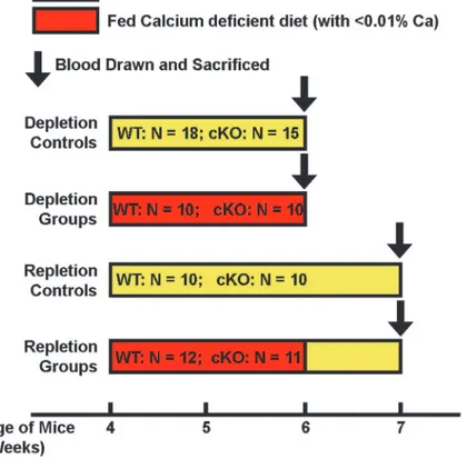
de-pletion/repletion protocol is shown inFig. 1. A group of mice of each genotype were fed a diet
containing 1.2% calcium (Harlan) in parallel as the control group for depletion. To evaluate the effect of dietary calcium repletion, a separate group of cKO mice and corresponding WT littermates were fed the diet containing 1.2% calcium for up to 7 days immediately after the two-week of dietary calcium depletion. Another group of mice of each genotype was fed a diet containing 1.2% calcium throughout the entire three weeks of the experiment as repletion con-trol groups. All animals had free access to water and food, and were housed in groups of 4 mice per cage in a 12-hr light/12-hr dark cycle. All animal use protocols were approved by the Insti-tutional Animal Care and Use Committee (IACUC) of Loma Linda University and also by the Animal Care and Use Review Office (ACURO) of the United States Department of Army.
Plasma biomarkers of bone turnover
Blood was drawn at noon of the day of sacrifice. Briefly, animals were euthanized by inhalation of an overdose of carbon dioxide. Blood was collected through cardiac puncture using a 25-G needle into heparinized tubes. Plasma was prepared by low-speed centrifugation (3,000 × g for
15 min at 4°C), and each plasma sample was stored in aliquots at−80°C until assay. Plasma
lev-els of c-terminal telopeptide of type I collagen (CTx), procollagen type I N-terminal propeptide
(P1NP), FGF-23, and PTH(1–84)were measured with respective Enzyme-Linked
ImmunoSor-bent Assay (ELISA) kit; purchased from either Immunodiagnostics Systems (Fountain Hills, AZ) or Immunoptics (San Clemente, CA). Plasma calcium and phosphorus levels were deter-mined with commercial colorimetric assays obtained from Biovision (Milpitas, CA). All assays were performed according to instruction provided by the respective supplier. The color intensi-ty of the product in each ELISA was analyzed by a Synergy HT Biotek microplate reader with the Gen5 software (BioTek Instruments, Winooski, CT).
Quantitative RT-PCR
After removal of muscles and connective tissue layers, tibias were frozen at−80°C until
liquid nitrogen. Total RNA was isolated from bone powders using the Qiazol Lysis Reagent and purified with RNeasy Mini kit (Qiagen, Valencia, CA). The quality of each RNA sample was assessed with Agilent 2100 Bioalanyzer (Agilent Technologies Inc., Santa Clara, CA) using the RNA 6000 nano-Kit (Agilent Technologies Inc.). Only RNA samples with high quality of RNA were used in subsequent RT-PCR assays. The cDNA was generated from each RNA sam-ple by reverse transcription using the ThermoScript RT kit (Invitrogen, La Jolla). The relative level of each gene-of-interest was determined with the SyBr Green PCR kit (Applied Biosys-tems) in an ABI 7500 Fast cycler (Applied Biosystems, Foster City, CA). The PCR primer set
sequences of each gene-of-interest are shown inTable 1.
pQCT analysis
Femurs after dissection were fixed in 10% formalin overnight and were stored in phosphate buffered saline (PBS) at 4°C until analyses. The length of each femur was measured with a digital caliper. pQCT measurements were performed with the STRATEC XCT 960M (Norland Medical Systems, Ft. Akinson, WI) and analyzed with the version 6.00 software program
provided by the manufacturer as described previously [26]. Briefly, two slices per femur at the
Figure 1. Schematic representation of the experimental design of the dietary calcium depletion and repletion experiment.The indicated numbers of 4-week-old femaleIgf1osteocytic cKO mutants and age-and gender-matched WT littermates were each divided into four groups: 1) depletion control groups, which were fed the calcium sufficient diet (with 1.2% calcium) for 2 weeks; 2) depletion experimental groups, which were fed the calcium deficient diet (with<0.01% calcium) for the same 2 weeks; 3) repletion control groups,
which were fed the calcium sufficient diet for 3 weeks; and 4) repletion experimental groups, which were fed the calcium deficient diet for two weeks followed by the calcium sufficient diet for one week. At the end of each regimen, all mice were sacrificed and blood drawn for measurements of plasma parameters. Femurs were isolated, cleaned, fixed with formalin and subjected pQCT and histomorphometry measurements. Tibias were isolated, and total RNA was isolated for real-time RT-PCR measurements.
position of 20% and 50% of the total femur length from the distal end, which corresponded to metaphysis and mid-point of the femur shaft, respectively, were scanned. The inner and outer
thresholds were set at 230 and 630 mg/cm3, respectively, to distinguish the cortical
compart-ment from the trabecular compartcompart-ment of bones of mice fed a normal calcium-containing diet. To adjust for the reduced bone mineral density (BMD) in bone of mice that fed the
calcium-de-ficient diet, the inner and outer thresholds were re-set at 180 and 400 mg/cm3, respectively.
Bone histomorphometry
Static bone histomorphometry of femurs was determined as previously described [36]. Trabecular
bone parameters were measured at the secondary spongiosa of the femur. To determine endosteal bone formation parameters, mice were injected with 25 mg/kg body weight of demeclocycline and 25 mg/kg body weight of tetracycline at day 6 and day 1 prior to euthanasia (i.e., a 5 day inter-labeling time). Twenty four hours after the second injection, mice were euthanized and femurs were removed and immediately fixed in 10% formalin. Bone was subsequently dehydrated in ethanol, infiltrated, and embedded into methylmethacrylate (MMA). Longitudinal thin sections
(7.5mm in thickness) were obtained with theLeicaMicrotome (Leica Microsystems, Inc., Buffalo
Grove, IL). A set of bone specimens was demineralized for 21 days in 14% EDTA solution, em-bedded into paraffin wax, and sectioned. All histomorphometric measurements was performed with an Olympus fluorescent microscope BX51 (Tokyo, Japan) using the Osteomeasure digita-lized system (OsteoMetrics, Atlanta, GA). Total tetracycline labeled surface (L.Pm) was the sum of double labeled surface length (dL.Pm) and one half of the single labeled surface length (sL.Pm). The mineral apposition rate (MAR) was the distance of the double labels divided by the inter-labeling time (5 days). Bone formation rate (BFR) was calculated by multiplying MAR by L.Pm.
Statistical Analysis
Results were shown as meanstandard error of the mean (SEM). Each mouse strain (WT mice
and cKO mutants) has two experimental groups (the depletion group and the repletion group) and two age- and gender-matched corresponding control groups. Statistical significance of the
differences in each mouse strain was determined with two-tailedStudent’st-test by comparing
the effect on the experimental group with that on corresponding control group. The individual effect of the genotype (WT vs. cKO) or the diet (normal vs. calcium-deficient diet) as well as the interaction between genotype and diet were determined on all four groups together by two-way
analysis of variance (ANOVA). The difference was considered significant, when P<0.05.
Results
Effects of deficient
Igf1
expression in osteocytes on plasma levels of
bone minerals, PTH, and FGF-23 after two-week dietary calcium
depletion
Under basal conditions (fed a diet containing sufficient amounts of calcium), plasma level of
PTH, but not that of calcium or phosphorus, of osteocyteIgf1cKO mutant mice were
Table 1. Sequence of PCR primer sets for the test mouse genes.
Gene Accession Number Forward Primer Reverse Primer
Actin NM_007393 5’caggcattgctgacagga 5’tgctgatccacatctgctgg
Opg MMU94331 5’cacacacactggggactctg 5’cagctgtgaggagaggaagg
Rankl AF019048.1 5’cacagccctctctcttgagc 5’gactgtgacccccttccata
Sost NM_024449 5’tcaggaatgatgccacagag 5’tgtcaggaagcgggtgtagt
significantly higher than those of corresponding WT control mice (Table 2). After two weeks of dietary calcium deprivation, the plasma calcium level of both WT control mice and cKO
mutants was reduced significantly by 8% each (Fig. 2A), and was attended by the
correspond-ing 4-fold increase in plasma PTH level over the basal level (Fig. 2B). These findings confirm
the previous studies that rodents fed a low calcium diet at weanling developed hypocalcemia
and secondary hyperparathyroidism [28,30,34,35] and indicate that osteocyte-derived IGF-I
Table 2. Comparison of effects of the two-week dietary calcium depletion on plasma levels of Ca, P, PTH, and FGF-23 level in 4 weeks-old female osteocyteIgf1conditional knockout (cKO) mice with those in age- and sex-matched WT control mice.
Plasma parameters
Calcium-containing Control Diet
Calcium Deficient Diet Two-way ANOVA
WT control mice cKO mice WT control mice cKO mice Diet Gene Interaction
Ca, mg/dl 12.50.3 (n = 10) 12.70.5 (n = 8) 11.50.3**(n = 12) 11.70.2**(n = 7) P<0.01 P = N.S. P = N.S.
PTH, pg/ml 384 (n = 8) 539a(n = 6) 194
16***(n = 10) 24739***(n = 7) P<0.01 P = N.S. P = N.S.
Phosphorus,μM 7.30.4 (n = 14) 8.30.5 (n = 8) 11.01.0*(n = 8) 8.30.5 (n = 6) P<0.01 P = N.S. P<0.01
FGF-23, ng/ml 38638 (n = 10) 43834a(n = 4) 49
3***(n = 5) 373***(n = 5) P<0.0001 P = N.S. P = N.S.
aP<0.05, when compared to WT control mice on basal calcium-containing diet.
*P<0.05,**P<0.01; and***P<0.001, compared to corresponding controls on calcium-containing diet of each respective mouse strain.
P = N.S. (not significant), when P>0.05.
doi:10.1371/journal.pone.0115897.t002
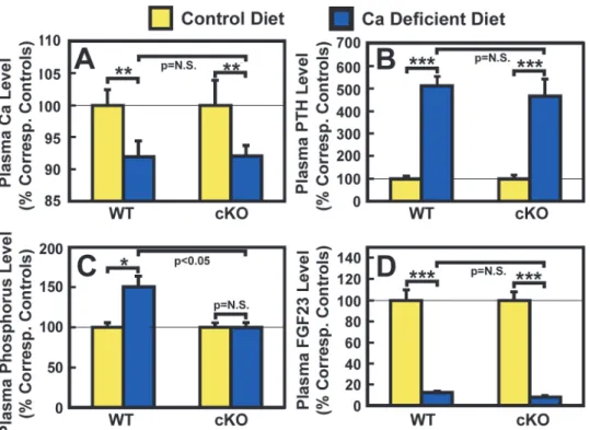
Figure 2. Effects of two-week dietary calcium restriction on plasma calcium (A), PTH (B), phosphorus (C), and FGF23 (D) of osteocyteIgf1cKO mice and those of WT mice.Four-week-old female osteocyte
Igf1cKO mice and age- and gender-matched WT control mice were fed a mouse diet containing either
<0.01% calcium (calcium-deficient diet) or 1.2% calcium (control diet) for two weeks. At the end of the
calcium depletion, blood samples were collected from each mouse for measurements of calcium (Ca), PTH, phosphorus, and FGF23. Results are shown in relative percentage of the respective level of each metabolite in corresponding control mice that were fed the control calcium-containing diet (meanS.E.M). The number of animals in each test group is shown inTable 2.*P<0.05;**P<0.01; and***P<0.001. P = N.S. (not
significant), where P>0.05.
does not play a role in the development of hypocalcemia and secondary hyperparathyroidism in response to the calcium stress. The calcium deficiency increased plasma phosphorus level
slightly but significantly (P<0.05) in WT mice, but not in cKO mutants (Fig. 2C). Consistent
with a previous report that calcium deficiency reduced circulating levels of FGF-23 [37], the
two-week dietary calcium restriction led to highly significant reduction in the plasma level of
FGF-23 in WT control mice and in cKO mutants (Table 2). The reduction in plasma FGF-23
in both mouse strains was similar in the percentage of respective basal level, each by ~60% (Fig. 2D). These findings suggest that osteocyte-derived IGF-I also does not have a regulatory role in the calcium deficiency-mediated changes in circulating levels of phosphorus or FGF-23.
Effects of deficiency in osteocyte-derived IGF-I on the calcium
deficiency-induced resorption
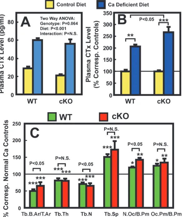
We next evaluated the potential mediating role of osteocyte-derived IGF-I in the calcium defi-ciency-induced bone resorption. We first compared the plasma level of CTx (a bone resorption
biomarker) in osteocyteIgf1cKO mutants with that in age- and gender-matched WT control
mice. Despite a 40% higher basal plasma PTH level in cKO mutants (Table 2), the basal plasma
CTx level in cKO mutants was significantly lower by ~28% (P<0.05) than that in WT control
mice (Fig. 3A), suggesting the higher basal PTH level in cKO mutants did not lead to an
elevat-ed basal bone resorption. However, the two-week calcium depletion significantly increaselevat-ed
plasma CTx level in both cKO mutants and WT control mice (Table 2). When the plasma CTx
level was normalized against respective basal level in each mouse strain (Fig. 3B), the calcium
depletion-induced increase in plasma CTx in cKO mutants was significantly greater than that
in WT mice (16414% increase over basal in cKO mice vs. 1095% increase over basal in WT
mice, P<0.05). The possibility that deficient expression ofIgf1in osteocytes may promote
cal-cium deficiency-induced bone resorption is further supported by the marginally significant
(P = 0.064) genotype effect (Fig. 3A).
We next compared several bone resorption-related static histomorphometric parameters of
trabecular bone between the two mouse strains. As previously reported [26], there were no
sig-nificant differences in trabecular bone area (Tb.B.Ar/T.Ar), number (Tb.N), thickness (Tb. Wi), and separation (Tb.Sp) between WT mice and cKO mutants under basal conditions (Table 3). The number of osteoclasts per bone surface (N.Oc/B.Pm) and the percentage of TRAP positive bone length per bone surface (Oc.Pm/B.Pm)] in cKO mutants under basal con-ditions were slightly (by 2.5% and 14%, respectively) but not significantly lower than those in
WT control mice (Table 3). The dietary calcium depletion reduced Tb.N and Tb.Wi, resulting
in reduction in Tb.B.Ar/T.Ar and increase in Tb.Sp in both mouse strains. The Oc.Pm/B.Pm
was also increased in response to calcium depletion in both mouse strains (Table 3). When the
calcium depletion-induced changes in these parameters were normalized against correspond-ing basal level, the reduction in Tb.N and the increase in N.Oc/B.Pm in cKO mutants were
each significantly greater than those in WT mice (Fig. 3C). The increases in Tb.Sp and in
Oc.Pm/B.Pm in the cKO mutants were also larger, albeit not significant statistically, than those in
WT mice. These findings, along with the larger increase in plasma CTx (Fig. 3B), are consistent
with the interpretation that conditional disruption ofIgf1gene in osteocytes could have led to a
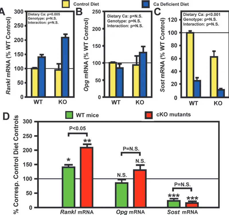
small but significant enhancement in the bone resorption response to calcium deficiency. RANKL and its decoy receptor, osteoprotegerin (OPG), are key regulators of osteoclast dif-ferentiation and activity. To determine the potential mechanism for the small enhanced bone
resorption response in cKO mice, we measured the relative levels ofRanklandOpgmRNA in
the bone (including bone marrow) of cKO mutants and WT control mice after two weeks of
andOpg(Fig. 4B) mRNA levels in bones of osteocyteIgf1cKO mice were not different from those of age- and gender-matched WT control mice. Importantly, the calcium depletion
signif-icantly increasedRanklmRNA, but notOpgmRNA, in bones of both mouse strains (Fig. 4C).
When each mRNA species was normalized against each respective control, the calcium
defi-cient-induced upregulation ofRanklmRNA was significantly larger (P<0.05) in cKO mutants
than in WT controls (Fig. 4D). There was no significant difference in the response of theOpg
Figure 3. Effects of two-week dietary calcium restriction on plasma CTx (A and (B) and on histomorphometric parameters of the femur (C) of osteocyteIgf1cKO mice and those of WT mice.A: The same plasma samples used inFig. 1were assayed for CTx level. The left panel shows the actual CTx levels, and the right panel shows the relative percentage of the corresponding basal CTx levels. The number of animals in each group is between 8 and 10. B: static bone histomorphometric parameters of the femur bone of each mouse. The number of mice per group is shown inTable 3. Results are shown as meanS.E.M. in relative percentage of the respective level of each metabolite in corresponding control mice that were fed the calcium-containing control diet throughout the study.*P<0.05;**P<0.01; and***P<0.001. P = N.S. (not significant), where P>0.05.
mRNA expression to calcium depletion in either mouse strain. Therefore, the effect of calcium
depletion on the ratio ofRanklmRNA/OpgmRNA, which is more important than theRankl
expression level alone in regulating osteoclast differentiation and activity, should also be
simi-lar to that on theRanklmRNA. Accordingly, these findings raise the possibility that the
appar-ent enhanced bone resorption response to calcium depletion in osteocyteIgf1cKO mice may
in part be due to a larger increase in RANKL expression in response to calcium depletion in cKO mutants compared to WT mice.
Previous studies have reported that calcium depletion also led to inhibition of bone
forma-tion in rodents [28,35]. Consistent with these findings, the two weeks dietary calcium depletion
significantly reduced trabecular BFR in WT mice by 45% (1.840.24 vs. 3.900.57 mm2×
103/day, n = 5–6 mice per group, p<0.01). We did not measure the effect of calcium depletion
on trabecular BFR in our osteocyteIgf1cKO mutants, but it is likely that calcium depletion
may also suppress trabecular BFR in these cKO mutants. Because SOST is a major local
para-crine factor produced by osteocytes to regulate bone formation [12], we measured basal bone
levels ofSostmRNA and also assessed the effects of calcium depletion on the bone content of
SostmRNA level in both groups of mice. The osteocyteIgf1cKO mutants had 40% lower basal
SostmRNA levels in the bone than that corresponding WT mice (Fig. 4C), suggesting that
oste-ocyte-derived IGF-I may have a positive regulatory function on the osteocyte expression of
SOST. However, because cKO mutants had higher basal plasma PTH level (Table 2), which is a
negative regulator ofSostexpression in the bone [38], an alternative possibility is that the lower
basalSostmRNA level in cKO mutants was a direct consequence of the elevated basal plasma
PTH level. Importantly, calcium depletion drastically reduced theSostmRNA content by 80–
90% in bones of both mouse strains (Fig. 4C). It is conceivable that the large increase in plasma
PTH in response to calcium depletion (Table 2andFig. 2B) may have a key role in the observed
drastic reduction in the bone content of SOST in both groups of mice (Fig. 4C&D).
Neverthe-less, when theSostmRNA level in the calcium depletion group of each mouse strain was
nor-malized against respective control group, there was no significant difference in the suppressive
effect of calcium depletion onSostmRNA expression in either mouse strain (Fig. 4D). It is
noteworthy that despite the dramatic reduction in SOST with calcium depletion, there was still a highly significant reduction in BFR in both groups of mice.
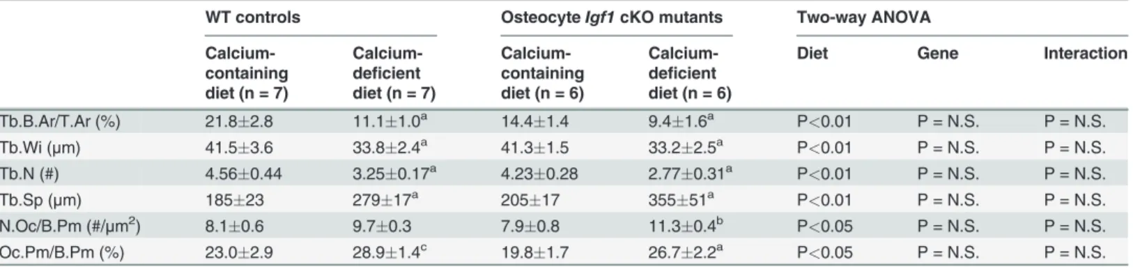
Table 3. Effects of the two-week dietary calcium depletion on static bone histomorphometric parameters of trabecular bone at the secondary spongiosia of the distal femur.
WT controls OsteocyteIgf1cKO mutants Two-way ANOVA
Calcium-containing diet (n = 7)
Calcium-deficient diet (n = 7)
Calcium-containing diet (n = 6)
Calcium-deficient diet (n = 6)
Diet Gene Interaction
Tb.B.Ar/T.Ar (%) 21.82.8 11.11.0a 14.4
1.4 9.41.6a P<0.01 P = N.S. P = N.S.
Tb.Wi (μm) 41.53.6 33.82.4a 41.3
1.5 33.22.5a P<0.01 P = N.S. P = N.S.
Tb.N (#) 4.560.44 3.250.17a 4.23
0.28 2.770.31a P<0.01 P = N.S. P = N.S.
Tb.Sp (μm) 18523 27917a 20517 35551a P<0.01 P = N.S. P = N.S.
N.Oc/B.Pm (#/μm2) 8.10.6 9.70.3 7.90.8 11.30.4b P<0.05 P = N.S. P = N.S. Oc.Pm/B.Pm (%) 23.02.9 28.91.4c 19.8
1.7 26.72.2a P<0.05 P = N.S. P = N.S.
aP<0.001;bP<0.01; andcP<0.05, when compared to corresponding control mice of each respective mouse strain that were fed the basal calcium-containing diet.
P = N.S. (not significant), when P>0.05.
Figure 4. Effects of two-week dietary calcium restriction on theRankland theOpgmRNA levels of osteocyteIgf1cKO mice and those of WT mice.
Total RNA was isolated from bone powders of entire femur of cKO and WT mice after two weeks on calcium-containing or-deficient diet (n = 4 animals per group). mRNA levels ofRanklandOpgby real-time RT-PCR, and are shown as meanSEM. A: theRanklmRNA levels as relative percentage of that in WT control (i.e., on calcium-containing diet); B is theOpgmRNA levels as percentage of that in WT control; and B: comparison of the effect of calcium deficiency onRanklandOpgmRNA in either mouse strain.*P<0.05;**P<0.01; and***P<0.001. P = N.S. (not significant), where P>0.05.
Effects of deficiency of osteocyte-derived IGF-I on calcium
repletion-induced bone formation
Previous studies in growing rodents have indicated that dietary calcium repletion would
rapid-ly stimulate bone formation to replace the bone mass lost during calcium depletion [4,5,31,32].
Thus, we next sought to determine whether deficientIgf1expression in osteocytes would alter
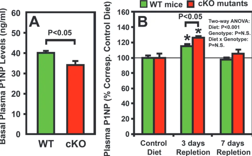
the bone repletion response. We compared plasma level of P1NP (a bone formation
biomark-er) of osteocyteIgf1cKO mice with that of corresponding WT control mice after 3 or 7 days of
calcium repletion. The cKO mutants not only have 28% lower basal plasma CTx level than WT
control mice (Fig. 3A), but also have 15% lower basal plasma P1NP level than corresponding
WT mice (Fig. 5A). These findings are consistent with our previous conclusion that basal bone
remodeling rate in osteocyteIgf1cKO mice is lower than WT mice during developmental
growth [26]. The plasma P1NP level in both cKO mutants and WT control mice was each
sig-nificantly increased at day 3 of dietary calcium repletion (Fig. 5B). In addition, the increase in
plasma P1NP was slightly but significantly (P<0.05) greater in cKO mutants than that in WT
control mice. There was no significant genotype effect (i.e., effects that were due to deficient os-teocyte-derived IGF-I) nor was there an interaction between dietary calcium and osteocyte-Figure 5. Effects of one-week of dietary calcium repletion on plasma P1NP level in osteocyteIgf1cKO mutants and in corresponding WT mice.
Four-week-old osteocyteIgf1cKO mice (n = 8) and age- and gender-matched WT control mice (n = 10) were fed either the deficient or the calcium-containing diet for two weeks followed by 1 week calcium-calcium-containing diet. Plasma samples were obtained at the beginning of the experiment (basal), and after three days and 7 days of calcium repletion. Results are shown as meanSEM. A shows the actual basal level of P1NP; and B shows the effect of the dietary calcium repletion in the two strains, which is reported as relative percentage changes of the basal level of WT control mice fed the calcium-containing diet for east indicated test time period.*P<0.05.
derived IGF-I deficiency on plasma P1NP level (Fig. 5B). The plasma P1NP level of either mouse strain was reverted back toward respective basal level at the end of the 7 days calcium repletion.
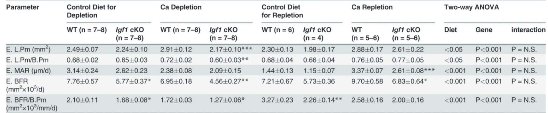
We next determined endocortical bone formation histomorphometric parameters at the
mid-point of femurs of both mouse strains.Table 4shows that basal endosteal BFR of cKO
mutants was significantly lower (by 26%, P<0.05) than that of WT control mice. The lower
basal endosteal BFR in cKO mutants remained significant even after normalization against bone surface length (BFR/B.Pm). The basal tetracycline labeling surface length (L.Pm) and basal mineral apposition rate (MAR) were also lower by 10% and 17%, respectively, but these differences did not reach statistically significant levels. The two-week calcium depletion
signifi-cantly reduced endosteal MAR and BFR in both mouse strains (Table 4). L.Pm was increased
(by 17%, P<005) in WT control mice but not in cKO mutants (Table 4andFig. 6). In addition,
the calcium depletion also reduced periosteal BFR in both WT mice (reduced to 574% of that
of corresponding WT control mice, n = 6 per group, p<0.05) and cKO mutants (reduced to
396% of that of corresponding cKO control mice, n = 5 mice per group, p<0.05), indicating
that dietary calcium deprivation also suppressed bone formation at the periosteal bone surface. The one-week calcium repletion increased MAR by 40% in the WT and 25% in cKO mice. Calcium repletion resulted in 20% increase in L.Pm (P = 0.07) and 28% increase in L.Pm/B.Pm
(P<0.05) in cKO mutants but had no effect on L.Pm in WT mice. When the bone formation
parameters were normalized against each respective basal level (Fig. 6), the calcium
repletion-induced increases in these bone formation parameters, with the exception of MAR, did not dif-fer significantly between the two mouse strains. Thus, deficient IGF-I expression in osteocytes does not have a significant impact on the bone repletion response.
Effects of deficiency in osteocyte-derived IGF-I on changes in bone size
and bone mineral density (BMD) in response to calcium depletion/
repletion
To determine if deficient expression of osteocyte-derived IGF-I would have an effect on bone size and BMD (two key determinants of bone strength) in response to bone depletion/tion, we compared the consequence of the two-week calcium depletion and the one-week reple-tion on the various bone size, bone mineral content (BMC), and BMD parameters in femurs of
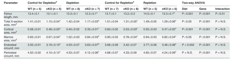
osteocyteIgf1cKO mutants with those of WT control mice. Consistent with our previous
re-port that deficient osteocyteIgf1expression impaired developmental bone growth [26], the
cKO mutants had ~3% shorter femur, 13% decrease in total cross-sectional bone area, 12%
re-duction in cortical area, 13% decrease in marrow area, and 7–8% each decreases in endosteal
and periosteal circumferences compared to WT control mice (Table 5). The two-week calcium
depletion reduced significantly total cross-section area, cortical area, and periosteal circumfer-ence, and increased marrow area and endosteal circumference in both mouse strains. The one-week repletion almost completely reversed the calcium depletion-induced effects on these bone size parameters and returned them to levels approaching those of the corresponding control
mice of each mouse strain (Table 5). To evaluate whether deficientIgf1expression in osteocytes
would affect calcium depletion/repletion-induced effects on bone length and size, we compared the relative % change in each parameter from each respective control basal value between cKO mutants and WT control mice. The effects of two-week calcium depletion on each parameter,
other than periosteal circumference, were significantly (p<0.05) greater in cKO mutants than
in WT mice (Fig. 7). In contrast, the effects of one-week calcium depletion on each test
param-eter were not significantly different between the two mouse strains. Thus, it is likely that the
Table 4. Effects of two weeks of dietary calcium depletion followed by one week of dietary calcium repletion on dynamic bone formation histomorphometric parameters of at the endosteal surface of the secondary spongiosia of the distal femur.
Parameter Control Diet for Depletion
Ca Depletion Control Diet
for Repletion
Ca Repletion Two-way ANOVA
WT (n = 7–8) Igf1cKO
(n = 7–8)
WT (n = 7–8) Igf1cKO
(n = 7–8)
WT (n = 6) Igf1cKO (n = 4)
WT (n = 5–6)
Igf1cKO (n = 5–6)
Diet Gene interaction
E. L.Pm (mm2) 2.49
0.07 2.240.10 2.910.12 2.170.10*** 2.300.13 1.980.17 2.880.17 2.610.22 <0.05 P<0.001 P = N.S. E. L.Pm/B.Pm 0.680.02 0.650.03 0.720.02 0.600.03** 0.680.04 0.660.04 0.760.05 0.770.05 <0.05 P<0.001 P = N.S. E. MAR (μm/d) 3.140.24 2.620.23 2.380.08 2.090.15 1.440.13 1.150.07 3.370.07 2.610.08*** <0.001 P<0.001 P = N.S. E. BFR
(mm2×103/d) 7.760.57 5.770.37* 6.950.18 4.560.27** 7.210.67 5.730.36 9.700.58 6.830.64*
<0.001 P<0.001 P = N.S.
E. BFR/B.Pm
(mm2×103/mm/d) 2.100.11 1.680.08* 1.720.03 1.270.06* 3.270.23 2.260.14** 2.580.16 2.000.16
<0.001 P<0.001 P = N.S.
E = endosteal; L.Pm = tetracycline labeling surface (dL.Pm + ½ sL.Pm); B.Pm = bone surface; MAR = mineral apposition rate; BFR = bone formation rate. P = N.S. (not significant), when P>0.05.
*P<0.05;**P<0.01;***P<0.001, when compared to respective control mice on the control diet.
doi:10.1371/journal.pone.0115897.t004
Osteocyte
-Derived
IGF-I
Is
Not
Essenti
al
for
Bone
Repletion
ONE
|DOI:10.137
1/journal.p
one.0115897
January
30,
two-way ANOVA (Table 5) on each parameter was due primarily to differences seen during depletion but not during repletion.
The effects of calcium depletion/repletion on BMC and BMD are summarized inTable 6.
Consistent with our previous report [26], femurs of cKO mutant mice fed a normal calcium
diet throughout had ~10% reduction each in total and cortical BMC, as well as in total, trabecu-lar, and cortical BMDs, when compared to WT control mice. The two-week calcium depletion reduced, whereas the one-week calcium repletion increased, total and cortical (but not
trabecu-lar) BMC and BMD in both mouse strains (Table 6). When BMC and BMD values are
normal-ized against each corresponding basal value, the depletion-mediated reduction in cortical BMC and BMD (but not depletion-related decreases in total or trabecular BMC and BMD) in cKO
mutants was significantly (P<0.05) bigger than those in WT mice (Fig. 8). However, there was
no significant mouse strain-related difference in the repletion-induced restoration of BMC and
BMD in either cortical or trabecular bones (Fig. 8). The fact that deficient expression ofIgf1in
osteocytes did not have an effect on the repletion-induced restoration of BMC and BMD sug-gests that osteocyte-derived IGF-I is probably not a key regulator of the bone repletion-mediat-ed bone formation.
Discussion
We have recently reported that disruption ofIgf1gene in osteocytes impaired developmental
bone growth that was associated with suppression in bone remodeling [26]. Because the bone
remodeling process is mechanistically similar to the bone depletion/repletion response [4], we
had originally predicted that conditional deletion ofIgf1in osteocytes would also negatively
affect the bone depletion/repletion process. To our surprise, disruption ofIgf1expression in
osteocytes did not have significant negative effects on bone depletion or repletion. This conclu-sion was based on two key findings: First, two weeks of dietary calcium restriction induced hy-pocalcemia and the associated secondary hyperparathyroidism to similar levels in both
osteocyteIgf1cKO mutant mice and age- and gender-matched WT control mice (Table 2and
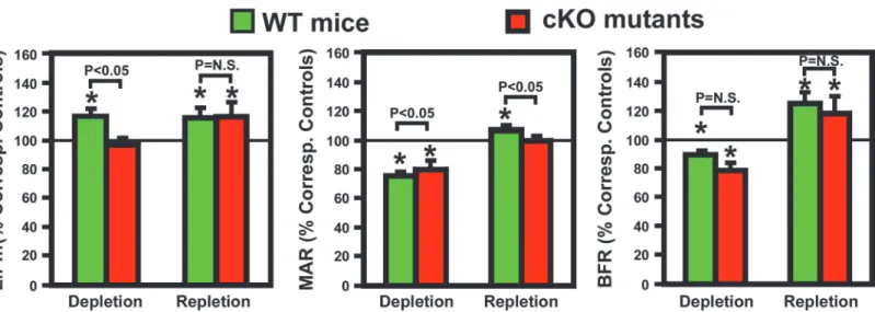
Fig. 2). These effects led to an increase in bone resorption [as reflected by the significant Figure 6. Effects of dietary calcium depletion and subsequent repletion on dynamic histomorphometric parameters of bone formation in femurs of osteocyteIgf1cKO mutants and corresponding WT mice.Left panel: the bone mineralizing surface, shown as relative percentage of TLS of
corresponding controls (mice of corresponding mouse strain fed the calcium-containing diet throughout); middle panel: the bone mineralization apposition rate; and right panel: the calculated bone formation rate per bone surface. Results are shown as meanSEM. The number of mice per group is shown in
Table 4.*P<0.05.
Table 5. Comparison of cortical cross-sectional bone parameters at the mid-shaft of femurs between WT andIgf1cKO mice after two-week dietary calcium depletion and/or after one-week dietary calcium repletion.
Parameter Control for Depletion# Depletion Control for Repletion# Repletion Two-way ANOVA
WT (n = 5) cKO (n = 5) WT (n = 8) cKO (n = 7) WT (n = 7) cKO (n = 8) WT (n = 5) cKO (n = 6) Diet Gene Interaction
Femur length, mm
13.40.1 13.10.1 13.30.1 12.30.1c 13.70.1 13.20.2 14.00.1 13.30.1a P<0.001 P<0.001 P<0.01
Total X-section
area, mm2 1.410.01 1.150.04 c
1.420.04 1.170.03a 1.510.04 1.310.05c 1.490.05 1.290.06c P<0.05 P<0.001 P = N.S.
Cortical
area, mm2 0.580.01 0.480.03 c
0.400.02 0.330.01c 0.600.02 0.530.03c 0.550.03 0.470.02c P<0.001 P<0.001 P = N.S.
Marrow
area, mm2 0.830.01 0.670.04 c
1.020.03 0.840.03a 0.900.03 0.780.04c 0.940.03 0.820.04c P<0.05 P<0.001 P = N.S.
Endosteal circumf, mm
3.550.01 3.190.10c 4.030.07 3.630.07b 3.680.09 3.420.07 3.770.06 3.460.08c P = 0.002 P<0.001 P = N.S.
Periosteal circumf, mm
4.520.02 4.100.12c 4.530.07 4.120.06c 4.680.07 4.330.09 4.650.07 4.240.09c P = N.S. P<0.001 P = N.S.
Results are shown as meanSEM.
#The control group are mice receiving normal calcium-containing diet throughout. aP<0.001;bP<0.01;cP<0.05; when compared to corresponding WT littermates.
P = N.S., not significant, when P>0.05.
doi:10.1371/journal.pone.0115897.t005
Osteocyte
-Derived
IGF-I
Is
Not
Essenti
al
for
Bone
Repletion
ONE
|DOI:10.137
1/journal.p
one.0115897
January
30,
increases in plasma CTx and bone resorption histomorphometry parameters (Fig. 3)], which
in turn resulted in significant loss of BMC (Table 4) and BMD (Table 4andFig. 7) in both
mouse strains. In addition, the calcium depletion suppressed BFR in both cKO mutants and
WT mice to the similar extent (Fig. 6). Second, dietary calcium repletion acutely increased
bone formation [as indicated by an elevated level of plasma P1NP after 3 days of dietary
calci-um repletion (Fig. 5B) and increases in MAR and BFR (Fig. 6)] in both cKO mutants and WT
control mice. This repletion-induced increase in bone formation returned the BMC and BMD
level of each mouse strain almost completely back to their respective basal levels (Fig. 7). Thus,
inasmuch as osteocyte-derived IGF-I is a key positive regulator of bone remodeling, it does not appear that osteocyte-derived IGF-I is an important regulator of the bone response to dietary calcium depletion and repletion.
Interestingly, conditional disruption ofIgf1in osteocytes not only did not impair, but in fact
slightly and significantly enhanced the calcium depletion-induced bone resorption response.
Accordingly, the osteocyteIgf1cKO mutant mice exhibited significantly greater (by 150–
200%) increases in plasma CTx and in N.Oc/B.Pm, along with significant reductions in Tb.N (Fig. 3) and cortical BMD (Fig. 7), in response to the two weeks of calcium depletion than cor-responding WT control mice. The larger bone resorption response to calcium depletion in cKO mutants is consistent with the previous report that calcium depletion exerted greater
in-creases in bone resorption and bone loss inIgf1global KO mice [30]. The molecular
Figure 7. Effects of calcium depletion and repletion on bone size parameters in femurs of osteocyteIgf1cKO mutants and corresponding WT mice.With the exception of the femur length, all bone size parameters were measured at the mid-diaphysis of femur by pQCT. Results are shown as relative percentage of corresponding control mice (meanS.E.M.). The number of mice per group is shown inTable 6.*P<0.05; and***P<0.001.
Table 6. Comparison of bone mineral content (BMC) and density (BMD) of the mid-point femurs between WT andigf1KO mice during calcium depletion or repletion.
Parameters Control for Depletion Depletion Control for Repletion Repletion Two-way ANOVA
WT (n = 18) cKO (n = 15) WT (n = 10) cKO (n = 10) WT (n = 5) cKO (n = 6) WT (n = 12) cKO (n = 11) Diet Gene Interaction
total BMC (mg) 0.780.02 0.700.03c 0.560.02 0.460.03b 0.810.04 0.660.06 0.760.02 0.620.02a P<0.001 P<0.001 P = N.S. cort BMC (mg) 0.570.02 0.500.02c 0.28
0.03 0.210.02 0.600.04 0.490.05 0.530.02 0.410.02a P<0.001 P<0.001 P = N.S. trab BMC (mg)* 0.170.01 0.160.01 0.220.01 0.170.01b 0.16
0.01 0.110.02c 0.18
0.01 0.160.01c P = 0.001 P = 0.001 P = 0.06 total BMD (mg/mm3) 486
6 4896 34511 3418 51011 5377 4426 4428 P<0.001 P = N.S. P = N.S. cort BMD (mg/mm3) 9496 9419 79411 7658c 96913 97616 9078 87711c P<0.001 P = 0.018 P = N.S. Trab BMD (mg/mm3)
* 1404 1454 1503 1404a 145
5 1506 1404 1493 P = N.S. P = N.S. P = 0.059
Data are shown as MeanSEM.
#The control group are mice receiving normal calcium-containing diet throughout. aP<0.001;bP<0.01;cP<0.05; when compared to corresponding WT littermates.
P = N.S., not significant, when P>0.05.
*Measured at metaphysis area immediately from secondary spongiosia.
doi:10.1371/journal.pone.0115897.t006
Osteocyte
-Derived
IGF-I
Is
Not
Essenti
al
for
Bone
Repletion
ONE
|DOI:10.137
1/journal.p
one.0115897
January
30,
mechanism responsible for the larger bone resorption response to calcium depletion in
osteo-cyteIgf1cKO mice is unclear. However, the dietary calcium restriction increasedRanklmRNA
levels in the bone in both mouse strains, but the calcium deficiency-induced increase inRankl
mRNA (but notOpgmRNA) in bones of cKO mutants was almost 2-fold of that in bones of
WT control mice (Fig. 4). Because the effect of the same dietary calcium deprivation on the
bone levels ofOpgmRNA, the soluble decoy receptor of RANKL, in cKO mutants was not
sig-nificantly different from that in WT mice, it is likely that the larger bone resorption response to
calcium depletion in osteocyteIgf1cKO mice is in part due to bigger calcium
depletion-in-duced local RANKL production in bone. These results would suggest that osteocyte-derived IGF-I is a negative regulator of the calcium-induced bone resorption and that its inhibitory ac-tion is mediated in part through suppression of RANKL (without affecting OPG) in the bone.
These findings are unexpected, since previous studies reported that deficiency in IGF-I inIgf1
global KO mice reduced osteoclastogenesis through suppression ofRanklexpression in the
bone [21]. In addition, the finding that our osteocyteIgf1cKO mice exhibited a larger response
in osteoclastogenesis and bone resorption to calcium depletion contradicts the previous
find-ings that the IGF-I derived from osteoblasts [39] or other cell types [21] stimulates
osteoclasto-genesis (and thereby bone resorption) through upregulation of theRanklexpression. It is
possible that IGF-I under different context (e.g., derived from osteoblasts vs. from osteocytes) may have different actions on osteoclastogenesis and bone resorption. Thus, understanding of the exact molecular mechanism that directs the negative regulatory action of osteocyte-derived
IGF-I on the calcium depletion-inducedRanklexpression and osteoclastogenesis is important,
but it would require much additional studies.
Figure 8. Effects of calcium depletion and repletion on BMD parameters in femurs of osteocyteIgf1cKO mutants and corresponding WT mice.
BMD parameters were determined by pQCT. Total BMD was performed on the entire bone; cortical BMD was measured at the mid-diaphysis; and Trabecular BMD was determined at the site corresponding to secondary spongiosa. Results are shown as relative percentage of corresponding control mice (meanS. E.M.). The number of mice per group is shown inTable 6.*P<0.05; and***P<0.001.
The more intriguing finding of this study perhaps is the lack of an adverse effect of deficient expression of osteocyte-derived IGF-I on the bone repletion process. Accordingly, the one-week dietary calcium repletion in cKO mutants appeared to be equally effective in increasing
the plasma (Fig. 5) and dynamic bone histomorphometric (Fig. 6) bone formation parameters
as WT littermates. The relative extent of the return of bone mass (Fig. 7) and BMD (Fig. 8)
after the one-week calcium repletion was also similar in both mouse strains. The lack of a nega-tive effect of deficient expression of osteocyte-derived IGF-I on the bone repletion process is surprising, especially in light of our recent discovery that the two-week regimen of four-point bending mechanical loading exercise failed to elicit the bone formation response in these same
osteocyteIgf1cKO mutants [27], which implicates an essential role of osteocyte-derived IGF-I
in loading-mediated bone formation. The bone repletion response and the anabolic response to mechanical loading are both important physiological guardians of maintenance of bone
mass and strength. Therefore, our findings that target disruption ofIgf1gene in osteocytes had
no discernable negative effects on the bone depletion process even though this osteocytic growth factor is an essential mediator of mechanosensitivity in the bone suggest a dichotomy between the mechanisms necessary for the osteogenic response to loading and the regulatory hormonal and anabolic skeletal repletion following low dietary calcium challenge.
The mechanistic reason(s) contributing to the lack of a significant regulatory role of osteo-cyte-derived IGF-I in bone repletion after a severe calcium stress is unclear. In this regard, we
have proposed a model (Fig. 9A) that may provide mechanistic insights into why deficient
oste-ocyte-derived IGF-I did not have significant effects on the bone repletion process. In this model, it is well established that the calcium stress caused secondary hyperparathyroidism and
elevated circulating levels of 1,25(OH)2D3[3,28,31,33,34]. Accordingly, we have demonstrated
that calcium depletion markedly increased plasma PTH levels in both cKO mutants and WT
mice. Although we did not measure plasma levels of 1,25(OH)2D3in this study, it is reasonable
to suspect that the elevated plasma PTH, which is a potent activator of renal production of 1,25
(OH)2D3, as well as the large reduction in plasma FGF23, which is a potent suppressor of renal
production of 1,25(OH)2D3[15,16], occurred during calcium depletion would result in a large
increase in the circulating level of 1,25(OH)2D3in both cKO mutants and WT mice. Both of
these systemic hormones are generally accepted to be the primary driving force for the bone
re-sorption response during the calcium stress [4,5,28,31], since these systemic hormones,
Figure 9. Proposed cellular mechanisms contribute to bone repletion-induced bone formation (A) or to loading-induced bone formation (B). Please refer to text for details.
especially PTH, are potent stimulators of bone turnover. Our previous studies [26] have impli-cated that osteocyte-derived IGF-I plays a regulatory role in the local bone remodeling process, as deficient osteocyte-derived IGF-I significantly suppressed bone turnover in growing mice. However, we postulate that the local effect of deficient osteocyte-derived IGF-I on bone remod-eling might have been masked by the huge systematic effects of hyperparathyroidism on bone turnover and bone loss. Accordingly, it is conceivable that osteocyte-derived IGF-I is a local regulator of bone remodeling and does not participate in systemic regulation of bone turnover, which is mediated largely through systemic effectors, such as PTH and 1,25(OH)2D3. In any event, all three major cell types (osteocytes, osteoblasts, and osteoclasts) in the bone express
re-ceptors for PTH and for 1,25(OH)2D3(VDR) [40,41,42,43], and both PTH and 1,25D are not
only activators of osteoblasts, but can also activate osteoclast differentiation. We postulate that under the condition of calcium deficiency, the two systemic hormones favor bone resorption
over bone formation through a poorly understood mechanism [5,28]. We also speculate that
the reduced sensitivity of osteoblasts to PTH and 1,25(OH)2D3under conditions of insufficient
bone minerals is an important physiological protective mechanism, in that bone minerals are used for the maintenance of bone mineral homeostasis rather than for mineralization of newly formed bone matrix. However, when the calcium deficiency is corrected by repletion, the rapid
increase in the circulating calcium would rapidly induce osteoclast apoptosis [4,32] to inhibit
further bone resorption, but the circulating levels of PTH and 1,25(OH)2D3during the early
phase of calcium repletion remain elevated [3,4,31]. Accordingly, these two systemic
hor-mones, in the presence of sufficient calcium, would favor bone formation over resorption,
lead-ing to rapid increases in bone formation durlead-ing repletion (Fig. 9A).
This model may also account for the apparent dichotomy between the mechanisms neces-sary for anabolic responses to mechanical loading and the regulatory hormonal and anabolic
skeletal repletion following the low dietary calcium challenge (Fig. 9B). Accordingly, we
postu-late that, upon mechanical loading, osteocytes (the primary mechanosensing cell type in the
bone [8]) reduce production and release of SOST (an inhibitor of the Wnt signaling). This
would lead to activation of canonical Wnt signaling in nearby osteoblasts, resulting in
subse-quent increases in local bone formation [8,12]. The loading strains also enhance local
produc-tion and release of IGF-I from osteoblasts and also upregulate IGF receptors in osteoblasts
[44]. These actions then further enhance the osteogenic response to loading. The mechanical
loading also reduces osteocytic RANKL production, which leads to reduction in osteoclast
dif-ferentiation and resorption activity [45]. We postulate that the loading-mediated activation of
osteoblasts also reduce osteoblastic production of RANKL, leading to further suppression in osteoclast differentiation, activity, and thereby bone resorption. Consequently, the overall effect of mechanical loading is activation of osteoblasts and bone formation and suppression of oste-oclastic resorption. Because osteocyte-derived IGF-I is essential and also required for these
loading-induced anabolic effects, disruption ofIgf1in osteocytes would abolish the
loading-in-duced anabolic effects in the bone [27]. We believe that the local loading-induced bone
forma-tion process shown inFig. 9Bremains operational during bone depletion/repletion, but the
magnitude of these local bone effects is much lower than that of the systemic-induced bone ef-fects. However, we should note that in the case of calcium repletion, the SOST levels in the
bone following calcium depletion are likely to have already been very low (Fig. 4 C&D). The
should emphasize that evidence supporting this model is largely circumstantial, but neverthe-less, it is an interesting model and is worthy of further investigation.
In summary, this study has demonstrated for the first time that although osteocyte-derived
IGF-I is essential in determining the bone mechanosensitivity [27], this osteocyte-derived bone
growth factor does not play a major role in the regulation of bone repletion response. Conse-quently, there is a dichotomy between the mechanisms necessary for anabolic responses to me-chanical loading and the regulatory hormonal and anabolic skeletal repletion following low dietary calcium challenge. It would be important to understand not only the molecular mecha-nism by which it determines the osteogenic response to loading but also the mechamecha-nism that regulates bone repletion in order to have a more complete overall understanding of how bone regeneration is regulated.
Acknowledgments
The authors thank Professors Derek LeRoith and Lynda Bonewald for providing us with
breed-ing pairs of theIgf1flox/floxmice and DMP1-Cretransgenic mice, respectively. The authors
ac-knowledge the excellent technical assistance of Dr. Xiao-Dong Zhou, Mr. Adam Finck, and Ms. Denise Rodriguez. The views, opinions and/or findings contained in this report are those of the authors and should not be construed as an official Department of the United States Army position, policy or decision unless so designated by other documentation. In conducting research using animals, the investigators adhered to the Animal Welfare Act Regulations and other Federal statutes relating to animals and experiments involving animals and the principles set forth in the current version of the Guide for Care and Use of Laboratory Animals, National Research Council. In addition, in conducting work involving the use of recombinant DNA the investigators adhered to the current version of the National Institutes of Health (NIH) Guide-lines for Research Involving Recombinant DNA Molecules.
Author Contributions
Conceived and designed the experiments: KHWL DJB MHCS. Performed the experiments: KHWL MHCS. Analyzed the data: KHWL DJB MHCS. Wrote the paper: KHWL DJB MHCS.
References
1. Manolagas SC (2000) Birth and death of bone cells: basic regulatory mechanisms and implications for the pathogenesis and treatment of osteoporosis. Endocr Rev 21: 115–137. doi:10.1210/er.21.2.115
PMID:10782361
2. Robling AG, Castillo AB, Turner CH (2006) Biomechanical and molecular regulation of bone remodel-ing. Annu Rev Biomed Eng 8: 455–498. doi:10.1146/annurev.bioeng.8.061505.095721PMID:
16834564
3. Akesson K, Lau KH, Johnston P, Imperio E, Baylink DJ (1998) Effects of short-term calcium depletion and repletion on biochemical markers of bone turnover in young adult women. J Clin Endocrinol Metab 83: 1921–1927. doi:10.1210/jc.83.6.1921PMID:9626120
4. Stauffer M, Baylink D, Wergedal J, Rich C (1972) Bone repletion in calcium deficient rats fed a high cal-cium diet. Calcif Tissue Res 9: 163–172. doi:10.1007/BF02061954PMID:5045998
5. Drivdahl RH, Liu CC, Baylink DJ (1984) Regulation of bone repletion in rats subjected to varying low-calcium stress. Am J Physiol 246: R190–196. PMID:6696144
6. Ravn P, Rix M, Andreassen H, Clemmesen B, Bidstrup M, et al. (1997) High bone turnover is associat-ed with low bone mass and spinal fracture in postmenopausal women. Calcif Tissue Int 60: 255–260. doi:10.1007/s002239900225PMID:9069162
7. Atkins GJ, Findlay DM (2012) Osteocyte regulation of bone mineral: a little give and take. Osteoporos Int 23: 2067–2079. doi:10.1007/s00198-012-1915-zPMID:22302104
9. You L, Temiyasathit S, Lee P, Kim CH, Tummala P, et al. (2008) Osteocytes as mechanosensors in the inhibition of bone resorption due to mechanical loading. Bone 42: 172–179. doi:10.1016/j.bone.2007. 09.047PMID:17997378
10. Bonewald LF (2011) The amazing osteocyte. J Bone Miner Res 26: 229–238. doi:10.1002/jbmr.320
PMID:21254230
11. Polig E, Jee WS (1990) A model of osteon closure in cortical bone. Calcif Tissue Int 47: 261–269. doi:
10.1007/BF02555907PMID:2257518
12. Winkler DG, Sutherland MK, Geoghegan JC, Yu C, Hayes T, et al. (2003) Osteocyte control of bone for-mation via sclerostin, a novel BMP antagonist. EMBO J 22: 6267–6276. doi:10.1093/emboj/cdg599
PMID:14633986
13. Nakashima T, Hayashi M, Fukunaga T, Kurata K, Oh-Hora M, et al. (2011) Evidence for osteocyte regu-lation of bone homeostasis through RANKL expression. Nat Med 17: 1231–1234. doi:10.1038/nm. 2452PMID:21909105
14. Xiong J, Onal M, Jilka RL, Weinstein RS, Manolagas SC, et al. (2011) Matrix-embedded cells control osteoclast formation. Nat Med 17: 1235–1241. doi:10.1038/nm.2448PMID:21909103
15. Quarles LD (2012) Skeletal secretion of FGF-23 regulates phosphate and vitamin D metabolism. Nat Rev Endocrinol 8: 276–286. doi:10.1038/nrendo.2011.218PMID:22249518
16. Feng JQ, Clinkenbeard EL, Yuan B, White KE, Drezner MK (2013) Osteocyte regulation of phosphate homeostasis and bone mineralization underlies the pathophysiology of the heritable disorders of rickets and osteomalacia. Bone 54: 213–221. doi:10.1016/j.bone.2013.01.046PMID:23403405
17. Mohan S, Baylink DJ (1991) Bone growth factors. Clin Orthop Relat Res: 30–48. PMID:1993386 18. Grinspoon SK, Baum HB, Peterson S, Klibanski A (1995) Effects of rhIGF-I administration on bone
turn-over during short-term fasting. J Clin Invest 96: 900–906. doi:10.1172/JCI118137PMID:7543494 19. Behringer RR, Lewin TM, Quaife CJ, Palmiter RD, Brinster RL, et al. (1990) Expression of insulin-like
growth factor I stimulates normal somatic growth in growth hormone-deficient transgenic mice. Endocri-nology 127: 1033–1040. doi:10.1210/endo-127-3-1033PMID:2387246
20. Mohan S, Richman C, Guo R, Amaar Y, Donahue LR, et al. (2003) Insulin-like growth factor regulates peak bone mineral density in mice by both growth hormone-dependent and -independent mechanisms. Endocrinology 144: 929–936. doi:10.1210/en.2002-220948PMID:12586770
21. Wang Y, Nishida S, Elalieh HZ, Long RK, Halloran BP, et al. (2006) Role of IGF-I signaling in regulating osteoclastogenesis. J Bone Miner Res 21: 1350–1358. doi:10.1359/jbmr.060610PMID:16939393 22. Bikle DD, Wang Y (2012) Insulin like growth factor-I: a critical mediator of the skeletal response to
para-thyroid hormone. Curr Mol Pharmacol 5: 135–142. PMID:21787292
23. Rhee Y, Allen MR, Condon K, Lezcano V, Ronda AC, et al. (2011) PTH receptor signaling in osteocytes governs periosteal bone formation and intracortical remodeling. J Bone Miner Res 26: 1035–1046. doi:
10.1002/jbmr.304PMID:21140374
24. Saini V, Marengi DA, Barry KJ, Fulzele KS, Heiden E, et al. (2013) Parathyroid hormone (PTH)/PTH-lated peptide type 1 receptor (PPR) signaling in osteocytes regulates anabolic and catabolic skeletal re-sponses to PTH. J Biol Chem 288: 20122–20134. doi:10.1074/jbc.M112.441360PMID:23729679 25. Lean JM, Mackay AG, Chow JW, Chambers TJ (1996) Osteocytic expression of mRNA for c-fos and
IGF-I: an immediate early gene response to an osteogenic stimulus. Am J Physiol 270: E937–945. PMID:8764176
26. Sheng MH, Zhou XD, Bonewald LF, Baylink DJ, Lau KH (2013) Disruption of the insulin-like growth fac-tor-1 gene in osteocytes impairs developmental bone growth in mice. Bone 52: 133–144. doi:10.1016/ j.bone.2012.09.027PMID:23032105
27. Lau KH, Baylink DJ, Zhou XD, Rodriguez D, Bonewald LF, et al. (2013) Osteocyte-derived insulin-like growth factor I is essential for determining bone mechanosensitivity. Am J Physiol Endocrinol Metab 305: E271–281. doi:10.1152/ajpendo.00092.2013PMID:23715728
28. Stauffer M, Baylink D, Wergedal J, Rich C (1973) Decreased bone formation, mineralization, and en-hanced resorption in calcium-deficient rats. Am J Physiol 225: 269–276. PMID:4722387
29. Hamalainen MM (1994) Bone repair in calcium-deficient rats: comparison of xylitol+calcium carbonate with calcium carbonate, calcium lactate and calcium citrate on the repletion of calcium. J Nutr 124: 874–881. PMID:8207545
30. Kasukawa Y, Baylink DJ, Wergedal JE, Amaar Y, Srivastava AK, et al. (2003) Lack of insulin-like growth factor I exaggerates the effect of calcium deficiency on bone accretion in mice. Endocrinology 144: 4682–4689. doi:10.1210/en.2003-0745PMID:12960002
32. Liu CC, Rader JI, Gruber H, Baylink DJ (1982) Acute reduction in osteoclast number during bone reple-tion. Metab Bone Dis Relat Res 4: 201–209. doi:10.1016/0221-8747(82)90019-4PMID:6897558 33. Tanimoto H, Lau KH, Nishimoto SK, Wergedal JE, Baylink DJ (1991) Evaluation of the usefulness of
serum phosphatases and osteocalcin as serum markers in a calcium depletion-repletion rat model. Cal-cif Tissue Int 48: 101–110. doi:10.1007/BF02555874PMID:1849444
34. Thompson ER, Baylink DJ, Wergedal JE (1975) Increases in number and size of osteoclasts in re-sponse to calcium or phosphorus deficiency in the rat. Endocrinology 97: 283–289. doi: 10.1210/endo-97-2-283PMID:1157752
35. Yoo A, Tanimoto H, Akesson K, Baylink DJ, Lau KH (1998) Effects of calcium depletion and repletion on serum insulin-like growth factor I and binding protein levels in weanling rats. Bone 22: 225–232. doi:
10.1016/S8756-3282(97)00277-9PMID:9514215
36. Sheng MH, Baylink DJ, Beamer WG, Donahue LR, Lau KH, et al. (2002) Regulation of bone volume is different in the metaphyses of the femur and vertebra of C3H/HeJ and C57BL/6J mice. Bone 30: 486– 491. doi:10.1016/S8756-3282(01)00693-7PMID:11882462
37. Rodriguez-Ortiz ME, Lopez I, Munoz-Castaneda JR, Martinez-Moreno JM, Ramirez AP, et al. (2012) Calcium deficiency reduces circulating levels of FGF23. J Am Soc Nephrol 23: 1190–1197. doi:10. 1681/ASN.2011101006PMID:22581996
38. Bellido T (2006) Downregulation of SOST/sclerostin by PTH: a novel mechanism of hormonal control of bone formation mediated by osteocytes. J Musculoskelet Neuronal Interact. 2006/12/23 ed. pp. 358– 359. PMID:17185824
39. Zhang M, Xuan S, Bouxsein ML, von Stechow D, Akeno N, et al. (2002) Osteoblast-specific knockout of the insulin-like growth factor (IGF) receptor gene reveals an essential role of IGF signaling in bone matrix mineralization. J Biol Chem 277: 44005–44012. doi:10.1074/jbc.M208265200PMID:12215457 40. Jongen JW, Willemstein-van Hove EC, van der Meer JM, Bos MP, Juppner H, et al. (1996)
Down-regu-lation of the receptor for parathyroid hormone (PTH) and PTH-related peptide by PTH in primary fetal rat osteoblasts. J Bone Miner Res 11: 1218–1225. doi:10.1002/jbmr.5650110905PMID:8864895 41. Ryan JW, Reinke D, Kogawa M, Turner AG, Atkins GJ, et al. (2013) Novel targets of vitamin D activity
in bone: action of the vitamin D receptor in osteoblasts, osteocytes and osteoclasts. Curr Drug Targets 14: 1683–1688. PMID:24010964
42. Rhee Y, Lee EY, Lezcano V, Ronda AC, Condon KW, et al. (2013) Resorption controls bone anabolism driven by parathyroid hormone (PTH) receptor signaling in osteocytes. J Biol Chem 288: 29809– 29820. doi:10.1074/jbc.M113.485938PMID:23963454
43. Ben-Awadh AN, Delgado-Calle J, Tu X, Kuhlenschmidt K, Allen MR, et al. (2014) Parathyroid hormone receptor signaling induces bone resorption in the adult skeleton by directly regulating the RANKL gene in osteocytes. Endocrinology: en20141046. doi:10.1210/en.2014-1046PMID:24877630
44. Lau KH, Kapur S, Kesavan C, Baylink DJ (2006) Up-regulation of the Wnt, estrogen receptor, insulin-like growth factor-I, and bone morphogenetic protein pathways in C57BL/6J osteoblasts as opposed to C3H/HeJ osteoblasts in part contributes to the differential anabolic response to fluid shear. J Biol Chem 281: 9576–9588. doi:10.1074/jbc.M509205200PMID:16461770