i
Computational algorithms for image analysis:
Applications on human vocal tract
and silhouette
Dissertation submitted in fulfillment of the requirements for the degree of Doctor in Informatics Engineering by the
Faculdade de Engenharia da Universidade do Porto
Maria João Medeiros de Vasconcelos BSc in Applied Mathematics for Technology by Faculdade de Ciências da Universidade do Porto (2002)
MSc in Applied Statistics and Modelling by Faculdade de Engenharia da Universidade do Porto (2006)
Supervisor
João Manuel Ribeiro da Silva Tavares
Associate Professor of the Department of Mechanical Engineering Faculdade de Engenharia da Universidade do Porto
Co-supervisor
Miguel Fernando Paiva Velhote Correia
Auxiliary Professor of the Department of Electrical and Computer Engineering Faculdade de Engenharia da Universidade do Porto
ii
The present Thesis belongs to the field of Computer Vision, more specifically segmentation and analysis of objects represented in images. While Computer Vision seeks to make useful decisions about real objects and scenes based on images through the construction of artificial systems, object segmentation and analysis aims to construct capable models to efficiently characterize objects and perform segmentation in new images.
This Thesis aims to present computational algorithms for object segmentation and analysis in images suitable for application on objects such as the human vocal tract and silhouette.
The main objective for studying the vocal tract in images is to better understand the vocal tract morphology and the involved movements during speech production of the European Portuguese language. Consequently, methodologies based on statistical deformable models, namely active shape models and active appearance models, were developed to represent the vocal structures from a global perspective in magnetic resonance images.
The suggested models made it possible to obtain a realistic simulation of the vocal tract during speech production as well as efficiently perform segmentation of vocal tract in new images. Furthermore, the use of such image analysis techniques can allow for obtaining quantitative measures with higher precision and are particularly advantageous when speech therapists and imaging specialists need to analyze a large volume of data.
Regarding the human silhouette analysis, four background subtraction models were studied to segment moving silhouettes in image sequences , with different levels of complexity. In addition, and following the same methodology used for modeling the vocal tract, an active silhouette model was also developed, using information about the contour of the silhouette together with anatomical stick points and combining the shape model with its gray level profiles with the purpose of segmenting the modeled silhouettes in new images.
The results obtained from the application of the background subtraction models in four different datasets suggested that the best model depends on the complexity of the images. Moreover, the good results obtained through the use of the active silhouette model built to perform human shape segmentation in new images strongly suggests that this type of deformable model can be successfully used in this task. The main contribution accomplished regarding the modeling of human silhouettes in images is that it allows for building an active shape model that gathers the necessary information independent of the walking direction of the subject.
In conclusion, the identification and analysis of human structures are complex tasks, since their shapes are not constant and vary through time; however, techniques of Computer Vision and objects modeling can assist in their achievement as is demonstrated throughout this Thesis. To conclude, the application of the developed models in images allows realistic simulations of the human vocal tract and silhouette, making possible their competent segmentation and characterization.
Keywords: Image Processing and Analysis, Human Vocal Tract, Human Silhouette, Object Segmentation, Object Modeling, Statistical Modeling.
iv
Sumário
A presente Tese pertence ao domínio da Visão por Computador, mais especificamente à segmentação e análise de objetos representados em imagens. Enquanto a Visão Computacional procura efetuar decisões sobre objetos reais e cenas baseado em imagens através da construção de sistemas artificiais, a segmentação e análise de imagem procura construir modelos capazes de caracterizar eficientemente objetos e efetuar segmentação em novas imagens.
Esta Tese apresenta algoritmos computacionais para segmentação e análise de imagens para aplicação em estruturas como o tracto vocal e a silhueta humana.
O objetivo do estudo do tracto vocal em imagens advém da necessidade de melhor compreender a morfologia do tracto vocal assim como os movimentos envolvidos particularmente na articulação da fala no Português Europeu. Consequentemente, foram desenvolvidas metodologias para representação global das estruturas vocais através de imagens obtidas por ressonância magnética, baseadas em modelos estatísticos deformáveis, nomeadamente modelos de forma e aparência ativa.
Os modelos desenvolvidos permitiram simular de forma realista o tracto vocal durante a articulação da fala assim como efetuar a sua segmentação em novas imagens. Para além disso, a utilização de tais técnicas de análise de imagem permitiram a obtenção de medidas quantitativas de maior exatidão e são particularmente vantajosas quando terapeutas da fala e imagiologistas necessitam de analisar grandes volumes de dados.
Em relação à análise da silhueta humana, foram estudados quatro métodos de subtração de fundos para segmentação de objetos em movimento em sequências de imagens com diferentes níveis de complexidade. Foram ainda desenvolvidos modelos de silhueta ativos, que utilizam a informação do contorno da silhueta conjuntamente com pontos anatómicos, com o objetivo de segmentar as silhuetas modeladas em novas imagens, através da combinação do modelo da forma com os seus perfis de cinzento.
Os resultados obtidos pela aplicação dos métodos de sub tração de fundos em quatro bases de imagens distintas sugerem que o modelo ideal depende fortemente da complexidade da imagem em causa. Os bons resultados obtidos pela aplicação dos modelos de silhueta ativos para segmentação de silhuetas humanas em novas imagens demostram que este tipo de modelos deformáveis pode ser utilizado nesta tarefa. O principal resultado obtido em relação à modelação da silhueta humana através de imagens concerne ao facto do modelo sugerido permitir construir um modelo da forma ativo que reúne a informação da silhueta independentemente da direção do movimento do sujeito.
Em conclusão, a análise automática do tracto vocal e da silhueta humana em imagens são tarefas complexas, pois estas estruturas apresentam formas complexas bem como variáveis; no entanto, a Visão por Computador e a modelação de objetos podem ser utilizadas de forma a auxiliar tais tarefas, como se demonstra ao longo desta Tese. Assim, os modelos desenvolvidos permitem simular a forma do tracto vocal e a silhueta humana assim como efetuar com sucesso a segmentação e caracterização de tais estruturas em novas imagens.
Palavras-Chave: Processamento e Análise de Imagem, Tracto Vocal Humano, Silhueta Humana, Segmentação de Objetos, Modelação de Objetos, Modelação Estatística.
vi
Acknowledgment
I would like to express my gratitude to my supervisor, Prof. João Manuel R. S. Tavares, for his continuous support throughout the project and encouragement for scientific production. Prof. Tavares incentive made me choose for the research career.
I would also like to thank to my co-supervisor, Prof. Miguel F. P. V. Correia for his availability whenever I needed.
To Sandra Ventura, colleague in this journey, by the collaboration in the work related with vocal tract modeling and opportunity to develop such models.
To my colleagues and friends from L304 especially Carla and Andreia for their presence and support during these years.
To my friends Isabel, Liliana, Camila, Rita, Octávio and Cláudia for the encouragement.
To my parents, my brother, to Luís and my family for the patience and for believing in me. To my grandmother that never understood why I didn’t finish my studies like everybody else, this is for you.
This work was supported by the PhD grant SFRH/28817/2006 from Fundação para a Ciência e Tecnologia (FCT), in Portugal.
vii
Contents
1 Introduction...1
1.1. Objectives ...4
1.2. Organization of the Thesis ...5
1.3. Contributions ...6 1.4. List of Publications...7 1.4.1. Book Chapters ...7 1.4.2. Journal Articles...8 1.4.3. Conference Papers ...9 1.4.4. Conference Abstracts...10
1.5. Organization of Scientific Events ...11
2 Image Analysis of the Human Vocal Tract and Silhouette ...12
2.1. Vocal Tract ...13
2.1.1. Anatomy ...15
2.1.2. Imaging Techniques ...16
2.1.3. Vocal Tract Models ...18
2.1.4. Studied Languages...21 2.1.5. Applications...23 2.2. Human Motion ...24 2.2.1. Surveys ...24 2.2.2. Motion Detection ...27 Temporal Information ...27 Spatial Information ...28 Spatio-Temporal Information...29 2.2.3. Motion Tracking ...29
Model-based Tracking...30 Active-Contour-based Tracking ...33 Feature-based Tracking ...35 2.2.4. Motion Understanding ...37 2.2.5. Motion Datasets ...40 2.2.6. Challenges ...43
3 Vocal Tract Active Models: Application to the European Portuguese Language ...44
3.1. European Portuguese Language ...45
3.2. Point Distribution Model...48
3.2.1. Active Shape Model ...49
3.2.2. Active Appearance Model ...50
3.3. Image Datasets ...51 3.3.1. 1.5T Dataset...51 3.3.2. 3.0T Sounds Dataset ...52 3.3.3. 3.0T Sequences Dataset ...53 3.4. Models ...54 3.4.1. Implementation ...54
3.4.2. Tongue Shape Model...55
3.4.3. Vocal Tract Model and Sounds Reconstruction ...57
3.4.4. Vocal Tract Active Models on 1.5T MR Images ...62
3.4.5. Vocal Tract Active Models on 3.0T MR Images ...71
3.4.6. Application Example ...82
3.5. Discussion and Conclusion ...83
4 Silhouette Models ...86
4.1.1. NADA...87
4.1.2. CASIA-A ...88
4.1.3. CAVIAR ...88
4.1.4. CASIA-B ...89
4.2. Background Subtraction Models ...90
4.2.1. Simple Difference Model ...90
4.2.2. Running Average Model ...90
4.2.3. Mixture of Gaussians Model ...91
4.2.4. Foreground Object Detection Model ...91
4.2.5. Human Silhouette Extraction ...92
4.3. Active Silhouette Model...93
4.4. Segmentation Quality Assessment ...97
4.4.1. F-measure ...98
4.4.2. Euclidean Distance ...98
4.4.3. Hausdorff Distance ...99
4.5. Implementations ...99
4.6. Segmentation Results ...100
4.6.1. Background Subtraction Models ...100
4.6.2. Active Silhouette Model ...114
4.7. Discussion and Conclusion ...119
5 Conclusion and Future Work ...122
5.1 Application in Studying the Human Vocal Tract ...122
5.2 Application on Human Silhouette...124
5.3 Future Work ...126
x
List of Figures
Figure 2.1 – Example of an MR sagittal slice demonstrating the vocal tract organs (from [Ventura, Freitas, et al. 2011]). ...15
Figure 2.2 – Human motion analysis framework. ...24
Figure 3.1 – Examples of images from the 1.5T (left) and 3.0T (right) datasets. ...54
Figure 3.2 – Landmark points considered to build the tongue shape model. ...55
Figure 3.3 – Effects of varying each of the first four modes of variation of the tongue model (mean ± 2 standard deviation). ...56
Figure 3.4 – a) Training image, b) landmark points selected, c) image labeled with the overlapped landmark points selected...57
Figure 3.5 – Effects produced by the variation of each of the first four modes of variation of the vocal tract model built (mean ± 2 standard deviation). ...59
Figure 3.6 – Reconstruction of the EP speech sounds [s], [z], [u] and [i]: a) original shape, b) reconstructed shape and c) both shapes overlapped. ...60
Figure 3.7 – Effects of varying each of the first six modes of variation of the model built for the vocal tract’s shape (mean ± 2 standard deviation). ...64
Figure 3.8 – Testing image with the initial position of the mean shape of the model built overlapped and after 4, 9 and 14 iterations of the segmentation process by an active shape model. ...65
Figure 3.9 – Testing images with the initial position of the mean shape model built overlapped (left) and the final results of the segmentation process by an active shape model (right). ...66
Figure 3.10 – First three modes of texture variation of the active appearance model built for the vocal tract’s shape (mean ± 2 standard deviation). ...68
Figure 3.11 – First three modes of appearance variation of the active appearance model built for the vocal tract’s shape (mean ± 2 standard deviation). ...68
Figure 3.12 – Results after the 1st, 7th, 12th and 20th iterations of the segmentation process using one active appearance model built for the vocal tract. ...69
Figure 3.13 – Testing images with the initial position of the mean shape model built overlapped (left) and the final results of the segmentation process obtained by an active appearance model (right). ...70
Figure 3.14 – a) Training image, b) landmark points selected, c) image labeled with the overlapped landmark points selected...71
Figure 3.15 – Effect on the vocal tract by varying (±2standard deviation) each of the first six modes of variation of the model built. ...74
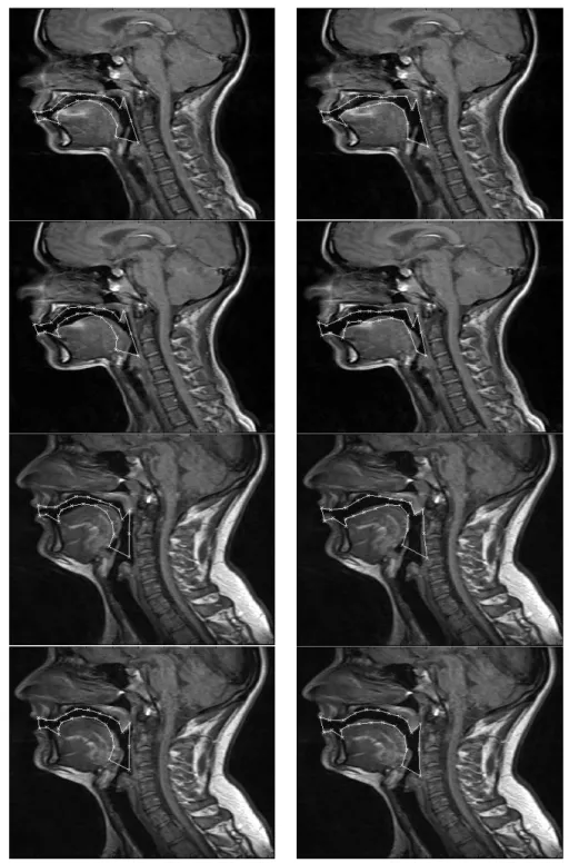
Figure 3.16 – Test image of female (top row) and male (bottom row) subjects overlapped with the mean shape model built and after some iterations of the segmentation process of the active shape model built. ...74
Figure 3.17 – Four test images overlapped with the mean shape model built (left) and after the conclusion of the segmentation process by the active shape model built (right). ...75
Figure 3.18 – Influence of the first 3 modes of texture variation of the active appearance model built (mean ± 2 standard deviation). ...77
Figure 3.19 – Influence of the first 3 modes of appearance variation of the active appearance model built (mean ± 2 standard deviation). ...78
Figure 3.20 – Segmentation process of two test images by the active appearance model built for the vocal tract. ...79
Figure 3.21 – Four test images overlapped with the mean shape model built (left), final results of the segmentation process by the active appearance model built (middle) and correspondent original images (right). ...80
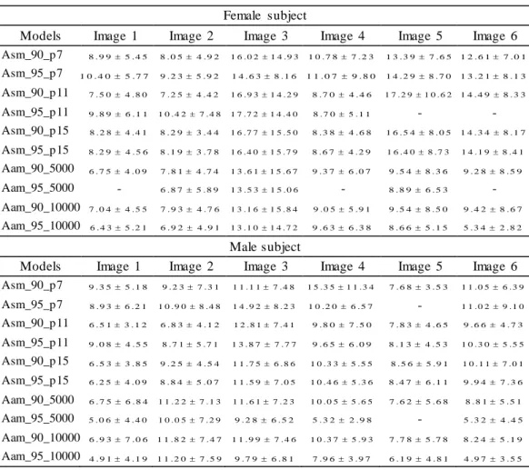
Figure 3.22 – Mean errors (in pixels) and standard deviations of the segmentations obtained by the deformable models built for the vocal tract of the female subject. ...81
Figure 3.23 – Mean errors (in pixels) and standard deviations of the segmentations obtained by the deformable models built for the vocal tract of the male subject...81
Figure 3.24 – a) Landmark points positions, b) landmark points selected, c) image labeled with the overlapped landmark points selected. ...82
Figure 4.1 – Examples of images extracted from the NADA image sequence. ...87
Figure 4.2 – Examples of images extracted from the CASIA-A image sequence. ...88
Figure 4.3 – Examples of images extracted from the CAVIAR image sequence. ...88
Figure 4.4 – Examples of images extracted from the CASIA-B image sequence, with different subjects and images taken from different views. ...89
Figure 4.5 – A silhouette example a); silhouette contour extracted from a), b); and 100 contour points extracted from the silho uette a). ...93
Figure 4.6 – Example of landmark points considered in the four directions (0º, 36º, 54º and 90º) to build the model. ...95
Figure 4.7 – First four modes of variation of the PDM built (mean shape ± 1 std)...97
Figure 4.8 – Three images from the NADA image sequence, the respective silhouette ground truth and segmentation results using the different background subtraction models. ...101
Figure 4.9 – Mean F- measures obtained using the different studied segmentation models for each test image of the NADA ima ge sequence. ...102
Figure 4.10 – Mean Hausdorff distances obtained using the different studied segmentation models for each test image of the NADA image sequence. ...102
Figure 4.11 – Mean Euclidean distances obtained using the different studied segmentation models for each test image of the NADA image sequence. ...102
Figure 4.12 – Three images of the CASIA-A image sequence, the respective silhouette ground truth and segmentation results using the different background subtraction models. ...105
Figure 4.13 – Mean F- measures obtained using the different studied segmentation models for each test image of the CASIA-A image sequence. ....106
Figure 4.14 – Mean Hausdorff distances obtained using the different studied segmentation models for each test image of the CASIA-A image sequence. ....106
Figure 4.15 – Mean Euclidean distances obtained using the different studied segmentation models for each test image of the CASIA-A image sequence. ....106
Figure 4.16 – 16th, 17th and 18th test images of the CASIA-A image sequence and the respective segmentation results using the running average model...107
Figure 4.17 – Three images from the CAVIAR image sequence, the respective silhouette ground truth and segmentation results using the different background subtraction models. ...109
Figure 4.18 – Mean F- measure obtained using the different studied segmentation models for each test image of the CAVIAR image sequence. ...110
Figure 4.19 – Mean Hausdorff distances obtained using the mixture of Gaussian models for each test image of the CAVIAR image sequence. ...110
Figure 4.20 – Mean Euclidean distances obtained using the mixture of Gaussian models for each test image of the CAVIAR image sequence. ...110
Figure 4.21 – Three images from the CASIA-B image sequences from one direction, 0º, the respective silhouette gro und truth and segmentation results using the background subtraction models. ...112
Figure 4.22 – Three images from the CASIA-B image sequences from different directions (36º, 54º and 90º), the respective silhouette ground truth and segmentation results using the background subtraction models. ...113
Figure 4.23 – Example of the iteration process using an active shape model in a new image in the first row (different image sizes correspond to different resolutions), and, in the second row, the initial and final (i.e. the segmentation result) positions of the model...115
Figure 4.24 – Examples of segmentation results obtained in images for the 4 directions studied. ...115
Figure 4.25 – Mean error distribution according the landmark point set (red lines are the median values and red + the outliers). ...116
Figure 4.26 – Mean error distributions according to the direction of the subjects and considering all the 113 landmark points (red lines are the median values and red + the outliers). ...117
Figure 4.27 – Mean error distributions according to the direction of the subjects and considering only the landmark points from the contour (red lines are the median values and red + the outliers). ...118
Figure 4.28 – Mean error distributions according to the direction of the subjects and considering the anatomical landmark points (red lines are the median values and red + the outliers). ...118
xv
List of Tables
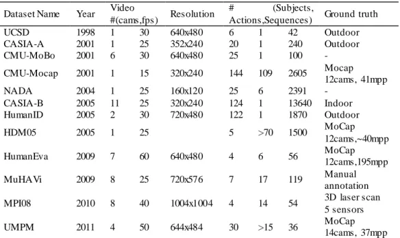
Table 2.1 – Summary of most relevant datasets for video-based human analysis...41
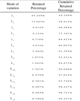
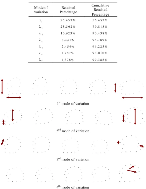
Table 3.1 – First seven modes of variation of the model obtained and their retained percentages. ...56
Table 3.2 – First 16 modes of variation of the model b uilt and their retained percentages...58
Table 3.3 – Errors obtained of the reconstructed shapes. ...60
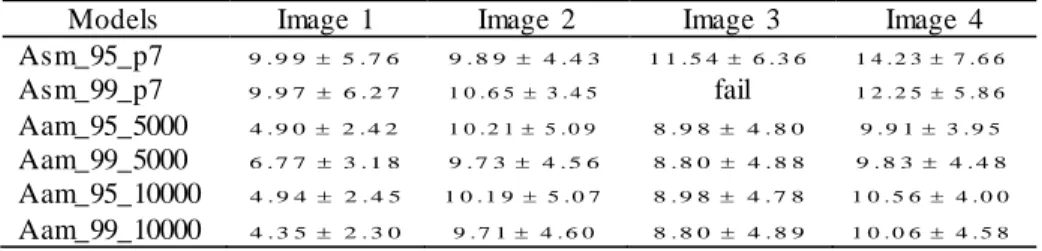
Table 3.4 – First 15 modes of variation of the model built for the vocal tract’s shape and their retained percentages...63 Table 3.5 – Mean and standard deviation (mean ± std) errors of the segmentations obtained from the testing images by the statistical models built. .65
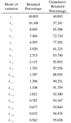
Table 3.6 – Retained percentages along the initial 17 modes of variation of the model built for the vocal tract. ...73
Table 3.7 – Mean and standard deviation (mean ± std) errors of the shapes segmented by the deformable models built. ...76
Table 4.1 – Summarized table of the data used to build and test the ASM.93
Table 4.2 – Retained and cumulative percentage of the modes of variation of the silhouette model. ...96
Table 4.3 – Mean and standard deviation (mean ± std) errors of the segmentations obtained using the NADA image sequence for different segmentation models...103
Table 4.4 – Mean and standard deviation (mean ± std) errors of the segmentations obtained using the CASIA-A image sequence for different segmentation models...107
Table 4.5 – Mean and standard deviation (mean ± std) errors of the segmentations obtained using the CAVIAR image sequence for different segmentation models...111
Table 4.6 – Mean and standard deviations (mean ± std) of the F-measures (%) obtained using the different segmentation models for different directions studied. ...114
Table 4.7 – Mean and standard deviation (mean ± std) errors of the mean Euclidean distributions according the direction of the subjects and the considered points. ...119
xvii
Acronyms
1.5T – 1.5 Tesla
3.0T – 3.0 Tesla
AAM – Active Appearance Model
ACL – Anterior Cruciate Ligament
ASM – Active Shape Model
CASIA – Institute of Automation Chinese Academy of Sciences
CMU – Carnegie Mellon University
CT – Computed Tomography
EMA – Electromagnetic Articulography
EP – European Portuguese
FIPM – Football Interaction and Process Model
FN – False Negatives
FP – False Positives
GMM – Gaussian Mixture Model
HBA – Human Behavior Analysis
HBU – Human Behavior Understanding
HOG – Histogram of Orientated Gradients
ICP – Iterative Closest Point
IPA – International Phonetic Alphabet
KTH – Kungliga Tekniska Hogskolan
MICA – Modified Independent Component Analysis
MoCap – Motion Capture
MPI08 – Indoor Motion Capture Dataset
MRI – Magnetic Resonance Imaging
MuHAVi – Multicamera Human Action Video Dataset
OCR – Optical Character Recognition
P – Precision
PCA – Principal Component Analysis
PDM – Point Distribution Model
R – Recall
SIFT – Scale Invariant Feature Transformation
TN – True Negatives
TP – True Positives
1
1
Introduction
The domain of Computer Vision seeks to make useful decisions about real objects and scenes based on images. It is a multidisciplinary domain of science and technology that depends on the information taken from images for designing artificial systems that aim to simulate human vision [Ballard et al. 1982].
The evolution of this domain is strongly influenced by the need for identifying, tracking and analyzing objects in an image or a sequence of images. In order to do this, it is necessary to perform tasks such as object modeling, segmentation, tracking and analysis [Szeliski 2010]. Segmentation and analysis of objects represented in images are two of the more studied and developed tasks in computer vision, wherein various methodologies have been used to build models capable of efficiently characterizing objects.
In this Thesis, particular attention is given to the use of deformable models in image analysis, which include segmentation techniques such as template matching, active contours, deformable templates, statistical methods, level set methods and physical methods [Zhang 2001; Tavares et al. 2009].
Template matching consists of comparing the template images with the new image and searching for similarities between the two images [Schalkoff 1989; Carvalho et al. 2005]. For example, in [Carvalho et al. 2005] a template of a human eye is used to segment the eye into new images through image correlation.
The use of deformable models in image analysis and interpretation was first introduced by [Kass et al. 1988], in which snakes are presented. A snake is an active contour that adjusts to a given object through a combination of internal and external forces, where the internal forces translate the flexibility and stretch, and the point at which the external forces pull the contour towards relevant areas of the image. The adjustments of the active contour are stopped when a minimal energy state is reached, typically when it finds the object border.
Other types of deformable models are deformable templates, which use templates described by parametric functions [Carvalho et al. 2007]. The geometrical templates are defined by parameters which describe the expected geometrical shape of the object and interact dynamically with the image during the segmentation process, similarly as with snakes. For instance, in [Yuille et al. 1992], the authors build a model to detect eyes in images, where the eye is represented by a circle describing the iris, two parabolic curves describing eyelids and also the intensity of these regions. The combination of all these characteristics typically translates into a model with high number of parameters, making the construction of deformable templates comp lex whenever a new object type needs to be modeled [Tien et al. 2000].
Statistical models are also included in the category of deformable models. An example of such modeling technique is given in [Carvalho et al. 2007] to identify skin areas in an image. For this, sample images of skin are used to build a statistical model for posterior skin segmentation that indicates the probability of the pixels of the new image to be associated with human skin. Another example of statistical modeling are the Point Distribution Models (PDMs) that were initially proposed by [Cootes, Taylor, et al. 1992] to model objects based on its statistical analysis. These models are obtained through the analysis of the statistics of the coordinates of the landmarks that represent the deformable object under study: after aligning the object shapes, a principal component analysis is made and the mean shape of the object and the main modes of its variation are obtained.
Active Shape Models [Cootes and Taylor 1992] and Active Appearance Models [Cootes et al. 1998] use PDMs to segment and recognize the modeled objects in new images. Both models use a combination of the statistical shape model with the gray levels of the object’s landmarks.
The idea of considering physical constraints in object modeling has been suggested and used by several authors. In [Pentland et al. 1991], the authors present physical-based solution for modeling objects. The approach is based on the finite element method and parametric solid modeling using implicit functions. Also in [Gonçalves et al. 2009] a physical approach based on the finite element method is used to segment an object and simulate its deformation. For this, an initial contour is manually defined that automatically evolves until it converges to the border of the desired object.
Another possibility to perform image segmentation of objects is to use level set methods, introduced by [Osher et al. 1988]. The main idea behind these methods is to represent the moving contour using a signed function whose zero corresponds to the actual contour. Then, by tracking the zero level set of the function adopted in the modeling it is possible to derive a similar flow for the implicit surface. A survey of algorithms that combine statistical techniques with level set methods can be found in [Cremers et al. 2007]. For example, in [Ma et al. 2013] a level set based algorithm is proposed to reconstruct the 3D s hape of the bladder using cross-sectional boundaries in magnetic resonance images.
The analysis of objects in images has been encouraged by the improvement of human/machine interaction in several applications, covering fields from industrial inspection, optical character recognition (OCR), medical imaging, surveillance or fingerprint recognition and biometrics. In industrial inspection it is used mostly for quality control purposes or defect recognition [Agin 1980; Klein et al. 1994; Campos et al. 2010]. Regarding OCR, examples of applications include reading handwritten postal codes on letters and automatic number plate recognition [Matan et al. 1992; Fahmy 1994; Volna et al. 2013]. Applications in medical imaging include performing image registration [Ayache 1998; Damas et al. 2011]. Computer Vision can also aid in the designing of surveillance systems for detecting and monitoring intruders or analyzing highway traffic [Sage et al. 1999; Norouznezhad et al. 2008]. Fingerprint recognition and biometrics has been used extensively for automatic access authentication and forensic applications [Junta Doi et al. 2004; Garibotto 2009; Nadipally et al. 2013].
1.1. Objectives
The subject of object analysis in Computer Vision has been developing in recent decades; especially in the domains of the analysis of objects in medical images and the human body, two of its most active fields.
The identification and analysis of human structures are complex tasks, since their shapes are not constant and vary through time; however, techniques of Computer Vision and objects modeling can assist in their achievement as o ne aim to demonstrate throughout this Thesis.
This Thesis is dedicated to developing computational algorithms for object segmentation and analysis in images. The human vocal tract and silhouettes were the objects selected to be analyzed in this Thesis. Hence, the objectives defined in this project included:
Review the existing algorithms for image analysis used to characterize and segment the human vocal tract and the human silhouette;
Analyze the need to develop methods for application in objects such as the vocal tract and human silhouette and define suitable clues that can be used to enhance the segmentation;
Develop new computational algorithms for characterizing such objects in images, particularly highlighting techniques based on the modeling of the geometrical shape of the object as well as its behavior;
Test the developed algorithms for segmenting the objects in new images and analyze the segmentation results, both qualitatively and quantitatively; compare the algorithms with existing ones and find the positive aspects and drawbacks of these algorithms.
1.2. Organization of the Thesis
After this introduction, in Chapter 2 the state-of-the-art of computational algorithms for image analysis used for studying the human vocal tract and human motion analysis are reviewed. A brief description and explanation are provided for the vocal tract anatomy and the most common imaging techniques used to acquire images of the complete vocal tract. The most promising methods used to represent its shape, including a summary of speech production studies in various languages available at the moment are also provided as well as the importance of vocal tract modeling. Regarding application on human motion analysis, the most important related research is presented, together with a description of the methodologies used for human detection, tracking and understanding; existing applications and existing datasets are also referred to and challenges are pointed out.
Chapter 3 presents the developed methodology to segment the shape of the vocal tract in new images for speech production assessment. A description is provided for the sounds of European Portuguese language and Point Distribution Models, Active Shape Models and Active Appearance Models. The image datasets and the Magnetic Resonance Imaging protocols used to build the models are also described. The segmentation results of the various active shape models developed for the study of the shape and appearance of the vocal tract shape are also presented and discussed, as well as an example of their application to actual studies.
The methodologies developed to segment silhouettes from images sequences are reported in Chapter 4. Four different background subtraction models are addressed in this chapter and an active silhouette model built is presented. Different image sequences were used for testing the developed methods and quantitative results are presented and discussed.
Finally, in Chapter 5 the main conclusions are drawn and suggestions for future research are given.
1.3. Contributions
Throughout this project, computational algorithms for image analysis were developed for application on human vocal tract and silhouettes. During this period, one book chapter was published and another was accepted for publication; six papers derived from the Thesis have been published in peer-reviewed journals; additionally, four papers and eight abstracts have been included in conference proceedings. In addition one symposium was organized during this Thesis.
The main contributions of this Thesis can be summarized as the following:
A comprehensive review of the current computational algorithms for image analysis that have been used for the study of the human vocal tract during speech production and for the study of human motion;
The development of two methodologies based on deformable models, namely active shape models and active appearance models, that allow for characterizing the shape of the vocal tract for speech production assessment of European Portuguese language in magnetic resonance images;
The application of the developed methods for the modeling of the vocal tract, a study of the best parameters to use in each model depending on the quality of the images as well as the qualitative and quantitative evaluation of the segmentation results by using the models referred to;
A comparison between the performances of active shape models and active appearance models and discussion on the advantages and disadvantages of each of these models;
The study of models using images with quality 1.5T and 3T (super ior) and posterior evaluation and comparison of the segmentation results;
Presentation of a realistic use case of application of the previous methodology that helps imaging experts and speech therapists by effectively reducing the amount of time spent on manually segmenting the vocal tract in new images;
The study of four methodologies based on background subtraction models to perform segmentation of the human silhouette in new images;
The development of a methodology based on active shape models for characterizing the silhouette of a human subject from an image sequence, which can be used later to perform their segmentation in new images; in addition to information on the contour of the silhouette, the developed method also integrates information on specific anatomical points such as the position of the head, shoulders, elbows, right and left hip positions, knees and feet;
An application of the previously mentioned methods for the modeling of the human silhouette in four different datasets, a study of the best parameters to use in each model depending on the quality of the images as well as the qualitative and quantitative evaluation of the segmentation results;
A comparison between the performances of the models and discussion on the advantages and disadvantages of using each model built.
1.4. List of Publications
In the scope of this Thesis, the following publications were produced:
1.4.1. Book Chapters
M.J.M. Vasconcelos, J.M.R.S. Tavares. Human Motion Segmentation using Active Shape Models. Accepted in Computational and
Experimental Biomedical Sciences: Methods & Applications, Lecture
Notes in Computational Vision and Biomechanics, Springer, October 2013.
S.M. Rua Ventura, M.J.M. Vasconcelos, D.R.S. Freitas, I.M.A.P. Ramos, J.M.R.S. Tavares. Speaker-specific articulatory assessment and measurements during Portuguese speech production based on Magnetic
Resonance Images. In Language Acquisition, ISBN: 978-1-61209-569-1, Nova Science Publishers, Inc., pp. 117-138, May 2012.
1.4.2. Journal Articles
M.J.M. Vasconcelos, S.M. Rua Ventura, D.R.S. Freitas, J.M.R.S. Tavares. Inter-speaker speech variability assessment using statistical deformable models from 3.0 Tesla magnetic resonance images.
Proceedings of the Institution of Mechanical Engineers, Part H: Journal of Engineering in Medicine, ISSN: 0954-4119 (print) - 2041-3033
(online), Professional Engineering Publishing, DOI: 10.1177/0954411911431664, Volume 226, Issue 3, pp. 185-196, March 2012.
M.J.M. Vasconcelos, S.M. Rua Ventura, D.R.S. Freitas, J.M.R.S. Tavares. Towards the Automatic Study of the Vocal Tract from Magnetic Resonance Images. Journal of Voice, ISSN: 0892-1997, Elsevier, DOI: 10.1016/j.jvoice.2010.05.002, Vol. 25, No. 6, pp. 732-742, November 2011.
M.J.M. Vasconcelos, S.M. Rua Ventura, D.R.S. Freitas, J.M.R.S. Tavares. Using Statistical Deformable Models to Reconstruct Vocal Tract Shape from Magnetic Resonance Images. Proceedings of the
Institution of Mechanical Engineers, Part H: Journal of Engineering in Medicine, ISSN: 0954-4119 (print) - 2041-3033 (online), Professional
Engineering Publishing, DOI: 10.1243/09544119JEIM767, Volume 224, Number 10 / 2010, pp. 1153-1163, 2010.
J.M.R.S. Tavares, F.J.S. Carvalho, F.P.M. Oliveira, I.M.S. Reis, MJ.M. Vasconcelos, P.C.T. Gonçalves, R.R. Pinho, Z. Ma. Computer Analysis of Objects’ Movement in Image Sequences: Methods and Applications.
International Journal for Computational Vision and Biomechanics,
ISSN: 0973-6778, Serials Publications, Vol. 2, No. 2, pp. 209-220, July-December 2009.
M.J.M. Vasconcelos, J.M.R.S. Tavares. Segmentation Methods for Human Motion Analysis from Image Sequences. ICCES, ISSN: 1933-2815, Tech Science Press, DOI: 10.3970/icces.2009.010.003, Vol. 10, No. 1, pp. 3-4, 2009.
M.J.M. Vasconcelos, J.M.R.S. Tavares. Methods to Automatically Build Point Distribution Models for Objects like Hand Palms and Faces Represented in Images. Computer Modeling in Engineering & Sciences, DOI: 10.3970/cmes.2008.036.213, Tech Science Press, ISSN: 1526-1492 (print) - 1526-1506 (online), vol. 36, no. 3, pp. 213-241, 2008.
1.4.3. Conference Papers
S. R. Ventura, M. J. M. Vasconcelos, D. R. Freitas, I. M. Ramos, J.M.R.S. Tavares. Speech Articulation Assessment Using Dynamic Magnetic Resonance Imaging Techniques. In VipIMAGE 2011 - III ECCOMAS
Thematic Conference on Computational Vision and Medical Image Processing, Real Marina Hotel & Spa, Olhão, Algarve, Portugal, 12-14
October 2011, ISBN: 978-0-415-68395-1, e-ISBN: 978-0-203-85830-1, Taylor and Francis, pp. 225-231.
M.J.M. Vasconcelos, S.R. Ventura, J.M.R.S. Tavares, D. R. Freitas. Analysis of Tongue Shape and Motion in Speech Production using Statistical Modeling. In SEECCM 2009 - 2nd South-East European
Conference on Computational Mechanics, ISBN: 978-960-254-683-3, pp.
96-103., 22-24 June 2009, Island of Rhodes, Greece.
M.J.M. Vasconcelos, J.M.R.S. Tavares. Métodos de Segmentação de Imagem para Análise da Marcha. In 3º Congresso Nacional de
Biomecânica, ISBN: 978-989-96100-0-2, pp. 563-564, Instituto
Politécnico de Bragança, Bragança, Portugal, 11-12 Fevereiro 2009.
M.J.M. Vasconcelos, J.M.R.S. Tavares. Human Motion Analysis: Methodologies and Applications. In CMBBE 2008 - 8th International
Symposium on Computer Methods in Biomechanics and Biomedical Engineering, 6 pag., Porto, Portugal, 27th February-1st March 2008.
1.4.4. Conference Abstracts
M.J.M. Vasconcelos, J.M.R.S. Tavares. Human Motion Segmentation using Active Shape Models. In ICCEBS2013 - International Conference
on Computational and Experimental Biomedical Sciences, 1 pag., Hotel
Marina Atlântico, Ponta Delgada, S Miguel Island, Azores, October 20-22, 2013.
M.J.M. Vasconcelos, J.M.R.S. Tavares. Segmentation Methods for Human Motion Analysis from Image Sequences. In colloquium 511 -
Biomechanics of Human Motion, New Frontiers of Multibody, Techniques for Clinical Applications, pp.19, University of the Azores, Ponta Delgada,
Azores, Portugal, March 9-12, 2011.
M.J.M. Vasconcelos, S.M. Ventura, D.R.S. Freitas, J.M.R.S. Tavares. Modelling and Segmentation of the Vocal Track during Speech Production by using Deformable Models in Magnetic Resonance Images. In 6th World Congress on Biomechanics, pp. 538, Singapore Suntec Convention Centre, 1-6 August 2010.
M.J.M. Vasconcelos, S.M. Rua Ventura, D.R.S. Freitas, J.M.R.S. Tavares. Segmentation of the Vocal Tract in Magnetic Resonance Images using Deformable Models. In ICCES'10 - International Conference on Computational & Experimental Engineering and Sciences, 28 March - 1 April 2010, Las Vegas, USA.
J.M.R.S. Tavares, M.J.M. Vasconcelos, R.R. Pinho. Motion Tracking in Images based on Stochastic Filters and Optimization. In CMBBE2010 -
9th International Symposium on Computer Methods in Biomechanics and Biomedical Engineering, ISBN: 978-0-9562121-3-9, Arup, 1 pag., Westin
Hotel, Valencia, Spain, 24-27 February, 2010.
M.J.M. Vasconcelos, J.M.R.S. Tavares. Segmentation Methods for Human Motion Analysis from Image Sequences. In ICCES'09 -
International Conference on Computational & Experimental Engineering and Sciences, ISBN-10: 0-9717880-9-X, ISBN-13: 978-0-9717880-9-1,
M.J.M. Vasconcelos, J.M.R.S. Tavares. Methodologies for Human Detection in Image Sequences. In 3DMA-'08 - 10th Meeting of the
technical group on '3D Analysis of Human Movement' of the International Society of Biomechanics, 2 pag., Santpoort-Amsterdam, the Netherlands,
October 28th - 31st, 2008.
M.J.M. Vasconcelos, J.M.R.S. Tavares Image Segmentation for Human Motion Analysis: Methods and Applications. In 8th. World Congress on
Computational Mechanics (WCCM8) / 5th. European Congress on Computational Methods in Applied Sciences and Engineering (ECCOMAS 2008), ISSN: 978-84-96736-55-9, 2 pag., Venice, Italy, June
30 - July 5, 2008.
1.5. Organization of Scientific Events
During this PhD project, the following symposium was organized:
J.M.R.S. Tavares. Y. Zhang, M.J.M Vasconcelos. Image Processing and Analysis. Symposium within the International Conference on
Computational Experimental Engineering & Sciences (ICCEES) 2009,
12
2
Image Analysis of the Human
Vocal Tract and Silhouette
The task of finding and identifying objects in an image is trivial for humans, despite the fact that the object image can vary depending on its viewpoint or size. Even if the object is occluded or the image quality is low, humans can still easily recognize it. It is a natural task that we are prepared for from the moment we are born.
Computer vision studies how to reconstruct, interpret and understand a 3D scene from its 2D images in terms of the properties of the objects present in the scene [Schalkoff 1989]. Therefore, the ultimate goal of computer vision is to model and replicate human vision using computational algorithms at different levels. For this, it is necessary to combine the knowledge of distinct fields such as computer sciences, electrical engineering, mathematics and biology in order to understand and simulate the human vision system [Szeliski 2010].
In addition, the ability to extract points from an image that can characterize an object in an image or image sequences is of extreme importance for the computer vision field. These characteristics may involve many tasks in image analysis such as object detection, shape recognition, image registration and object understanding. Motivated by its wide range of applications, object analysis has been evolving considerably over recent decades, with various examples of
applications found in medical imaging, human gait analysis or surveillance systems [Umbaugh 2010].
Considering this background, this Thesis focuses on the development of computer algorithms for image analysis. Particular attention is given to two applications: the human vocal tract and human motion. Therefore, this chapter is dedicated to reviewing the state-of-the-art of computational algorithms for image analysis that have been used for both applications.
Regarding the subject of the vocal tract, presented throughout the first section of this chapter, the structure is as follows. The vocal tract anatomy will be reviewed, followed by a review of the imaging techniques used to obtain the picture of the complete vocal tract. Then, models that have been used to represent the shape of the vocal tract are described, followed by a summary of speech production studies that have been developed in the various languages. The section ends with some examples of the importance of vocal tract modeling.
The research available on the subject of human motion analysis will be described in the second section. The most current research is presented, followed by the methodologies used to study motion detection. Next, the techniques developed for human motion tracking are described. Current understanding of motion, along with multiple applications in human analysis is addressed next and the existing datasets are referred to. Finally, the challenges that human motion studies still have to overcome are pointed out.
2.1. Vocal Tract
Verbal communication is the most common, familiar and frequently used form of human interaction, which results from the organized and synchronized work of a set of anatomic organs. The articulation is a result of the activity of a set of organs: the vocal tract that modifies the ir position and shape during air expulsion (expiration), producing different sounds and consequently, distinct acoustic representations.
Since the beginning of studies of this nature, the process of speech production has attracted human interest aiming at reaching a deeper understanding and modeling of all the mechanisms involved by taking both morphological and speech acoustic aspects into consideration. The main anatomic aspects and the physiology of the vocal tract are common to all individuals. However, the mechanism engaged in human speech production is complex and unique due to the variety of anatomical structures that compose the vocal tract, implying that any computational modeling developed needs to be flexible so as to permit accurate individual characterizations [Stone 1991; Benesty et al. 2008].
Two different approaches have been used to determine the shape of the vocal tract: direct methods based on geometrical measurements of the vocal tract; and indirect methods based on acoustic inversio n [Ball et al. 2008]. Among the direct measurement methods, several imaging methods have been used to obtain a complete picture of the vocal tract, like X-ray Radiography, Computed Tomography or Magnetic Resonance Imaging [Thimm et al. 1999; Ventura et al. 2009; Bakhshaee et al. 2013]. Indirect methods, in contrast, determine the vocal tract shape through acoustic data, either from a speech signal or from the acoustic response of the vocal tract. As image analysis is the basis of this Thesis, this dissertation will mainly focus on works based on direct approaches.
By assembling the former facts, it is straightforward that the study of the speech production is a multidisciplinary subject. To name a few, it involves subjects like: medicine, with the anato mic and functional study of the vocal tract organs; engineering, particularly informatics with the construction of the vocal tract models for speech and acoustics analysis; medical imaging, with the improvement and application of computational image techniques that can be used in the study of the vocal tract during speech production; phonetics, in the study of the production and perception of speech and sounds; and speech therapy, with the assessment of anatomic and physiological aspects related to communication disorders, language and speech [Beautemps et al. 1995; Baer et al. 1991; Apostol et al. 1999].
2.1.1. Anatomy
Various organs play important roles in the production of numerous speech sounds, functioning in an organized, i.e. articulated, manner in order to change the shape and length of a set of air cavities - the vocal tract. Most of these organs, named articulators, are soft-tissues that execute active movements during speech production, such as the lips, tongue and velum [Benesty et al. 2008; Ventura 2012].
Sagittal data is particularly useful in the study of the entire vocal tract anatomy [Ventura et al. 2010], demonstrating the main aspects of the shape and positions of some articulators, Figure 2.1. The tongue is a large muscular organ covered by mucous membrane, located on the floor of the mouth, which is attached by muscles to the hyoid bone, mandible, styloid processes, and pharynx. Besides its key role in mastication and swallowing, the tongue has an important function in speech production because it is the articulator with the most mobility and flexibility. Its main mass is composed of a set of muscles, which permits the elongation and constriction of the entire tongue or of specific parts allowing the articulation of sounds. The tongue’s structure presents a tip, which usually rests against the incisors, and margin, body, dorsum, inferior surface and root.
Figure 2.1 – Example of an MR sagittal slice demonstrating the vocal tract organs (from [Ventura, Freitas, et al. 2011]).
2.1.2. Imaging Techniques
Imaging techniques are methods that allow for obtaining an image of the interior of the vocal tract, and greater understanding of the positions and movements of the vocal tract organs [Stone 1991]. Two types of imaging techniques exist: structural, where the image of the structures is obtained; and tracking, where tracks are attached to important points of the vocal tract.
Examples of structural imaging techniques are X-ray Radiography, Computed Tomography (CT) and Magnetic Resonance Imaging (MRI), whereas tracking techniques include X-ray Microbeam and Electromagnetic Articulography (EMA) [Ball et al. 2008].
Radiography is a classical reference technique to study speech production. Some years after the discovery of X-rays in 1895, radiographic images of the upper vocal tract were taken by phoneticians [Harshman et al. 1977]. Since then, many techniques have been developed, from still pictures to video.
The advantages of using such an imaging technique include the possibility of obtaining the full sagittal view of vocal tract articulators during running speech with optimal temporal resolution, about fifty images per second. However, due to ethical concerns of the potential side effects of radiation exposure inflicted on the examiners, X-ray imaging technology is now rarely practiced [Xue et al. 2006]. Another limitation of this technique is related to the fact that volumetric information cannot be obtained, since X-ray can only take plain images of the speech mechanisms [Xue et al. 2006].
Nonetheless, studies have been developed using x-ray imaging, but they rely on existing images and databases previously acquired. The ATR X-ray film database is the largest X-ray database available for speech research [Munhall et al. 1995], with 25 different films of 55 minutes in total, it offers nearly 100,000 images. Other cineradiographic databases exist, such as the French database [Arnal et al. 2000], developed by the Strasbourg Institute of Phonetics and the Grenoble Institute of Speech Communication. Research by [Thimm et al. 1999;
Höwing et al. 1999; Fontecave et al. 2005; Fontecave Jallon et al. 2009] are examples of studies that combine x-ray images to comprehend speech production phenomena.
Another imaging technique used to acquire shape information about the vocal tract is Computed Tomography [Perrier et al. 1992; Inohara et al. 2010; Bakhshaee et al. 2013]. This imaging technique allows making the distinction between bones, soft tissues and air, but does not allow for discriminating different soft tissues. Moreover, the scanning speed of image acquisition is much higher than MRI. On the other hand, it has the serious disadvantage of requiring significant ionizing radiation doses, and for this reason, few studies adopt this imaging technique.
From the imaging modalities that have been used to study the vocal tract’s shape and articulators, MRI has been the most commonly accepted. Its key advantages include the quality and resolution of soft-tissues and the use of non-ionizing radiation [Avila-Garcia et al. 2004; Engwall 2003]. In addition, it allows for morphologic measurements in static as well as dynamic studies [Avila-Garcia et al. 2004; Engwall 2003]. Its main drawback is that a supine position is generally required that might interfere with normal position of the vocal tract during speech [Engwall 2000a].
Due to the lengthy data acquisition time of the early MR imaging systems, the first studies were restricted to vowels and some consonants [Baer et al. 1991; Narayanan et al. 1995]. The first comprehensive body of dimensional data on the vocal tract employing MR technology was presented by [Baer et al. 1987; Baer et al. 1991]. Another study [Crary et al. 1996] describes a dynamic MRI technique that offers several promising features for studying the configuration of the vocal tract.
With cutting-edge MR improvements, a proper 3D description on the vocal tract geometry of a speaker can be reached, both in terms of good image contrast and temporal resolution. Also, with the emerging development of rapid imaging techniques, such as synchronized sampling methods [Bresch et al. 2006] or tagged
cine-MR [Parthasarathy et al. 2007; Stone et al. 2001], the acquisition of image data regarding articulatory movements became possible. Nowadays, the acquisition of three-dimensional MR image sequences has been steady [Masaki et al. 2008] and, consequently, there are enormous expectations for attaining of image data on speech production in a more efficient and repeatable manner.
An X-ray microbeam is a tracking technique that uses very small doses of X-rays to record the movement of pellets attached to the tongue. It is a technique with little risk to the subject and it has also been used to investigate both normal and disordered speech. However, the images obtained by it show only a projection by volume, turning contour extraction very difficult. This technique is now rarely used, and like radiography, recent studies are based on previously acquired databases [Fujimura et al. 1973; Dang et al. 2002].
The other tracking technique, EMA, also provides information on speech kinematics [Perkell et al. 1992; Fitzpatrick et al. 2002; J. Kim et al. 2014]. EMA has the advantage of providing the same information as the X-ray microbeam, with higher temporal resolution but without using radiation. Another advantage is that it allows for a cleaner speech audio-recording environment. The main disadvantage, as in X-ray microbeam, is that EMA requires placing small connector coils in and on the speaker’s mouth, with the main problem being that relatively few subjects can tolerate this due to the sensitivity of the soft palate.
2.1.3. Vocal Tract Models
Information about the vocal tract shape and dimensions are essential to a full understanding of the articulatory and acoustical processes involved in speech production. The search for realistic and precise models to represent the vocal tract is long and several methods have been studied as reported below.
In [Harshman et al. 1977], the authors present a model to describe the tongue shapes of English vowels for five speakers. The model is based on a full x-ray measurement procedure that is reduced to a few underlying components by
means of the statistical techniques of factor and principal-component analysis. A year later, [Shirai et al. 1978] used statistical analysis of real data to describe the position of the articulatory organs. In [Maeda 1988], a factor analysis of the lateral shapes of the vocal tract is described.
The procedure to obtain the vocal tract shape [Story et al. 1996] from MR image sets included the segmentation of the airspace from the surrounding tissue, shape-based interpolation to generate the reconstruction of the airspace and analysis of the cross-sectional area. In [Kagawa et al. 1997], the authors model the vocal tract wall by an assemblage of spline functions, which is deformable around the points of interest. Also, in [Stone et al. 1997] a principal component analysis was used to examine sagittal tongue contours for five English vowels constructed from ultrasound images. In [Thimm et al. 1999] the segmentation of the vocal tract is done in an iterative manner. First, the teeth were tracked using two specialized histogram normalization techniques combined with a pattern- matching algorithm that also gives the position of the palate. Then, the normalization of the position of the vocal tract is used to track the throat and the lips. Finally, background subtraction is used to enhance the contrast of the tongue and configure its deformation. The referred segmentation procedure was optimized for X-ray images showing the side view of the vocal tract.
In 2000, a 3D tongue model was developed by [Engwall 2000b] within the
Kungliga Tekniska Hogskolan (KTH) 3D vocal tract project using manually
extracted tongue contours from MR images of a reference subject producing 43 sustained Swedish articulations. The extraction of the articulatory model’s parameters was done by decomposing the geometrical points of the tongue in linear components, through a Linear Component Analysis, where the factors to be extracted were imposed on the model using MR images articulatory measures. Two years later, in [Badin et al. 2002], a database of 3D geometrical description of tongue, lips and face was established for a speaker sustaining a set of French allophones. For this, data from MRI, along with a video with and without a jaw splint were used. An important finding of this research was that, most 3D geometry of tongue, lips and face could be predicted from their midsagittal contours, at least for speech assessment purposes. Indeed, the knowledge acquired from midsagittal data and from traditio nal 2D models is far from obsolete. A
complement of the previous models was presented in [Serrurier et al. 2005] based on the same French subject, achieving a final articulatory model of the shape of the complete vocal and nasal tracts. For this research the 3D surface that defines each organ of the vocal tract was extracted from MRI and CT images, where the 2D contours were manually extracted from the corresponding images and later expanded to 3D. Then, principal component analysis was applied to the set of organ surfaces to uncover the two main uncorrelated articulatory degrees of freedom for the velum’s movement.
A region growing technique was also explored to model the vocal tract shape [Behrends et al. 2003]. Here the authors first matched teeth phantoms to the MRI dataset to perform the segmentation and reduce human time expense. The segmentation method places a seed inside the vocal tract that expands until it reaches its walls. The expansion is based on gray- level comparison between the mean gray-level value of the segmented region and the neighborhood pixels of its contour until a defined difference value is achieved. The vocal tract midline is also computed by using a modified 1D-Kohonen algorithm to calculate the characteristic area functions. Later in [Carbone et al. 2008], the same technique was used to segment 100 2D vocal tract contours over a European Portuguese Database achieving Pratt Indices from 84% to 100%. A recent study [Silva et al. 2013] also uses region growing to segment the vocal tract, this time on real-time MR image sequences.
In [Mollaei et al. 2008], the authors use radiography images to obtain the vocal tract shape and calculate the median line through center of gravity of the contours of the vocal tract. To obtain the vocal tract shape model, the authors followed the approach of [Maeda 1988; Beautemps et al. 1995] and presented a model based on simple polynomials.
A semi automatic technique for facilitating the extraction of vocal tract contours is described in [Fontecave Jallon et al. 2009]. The method combined the manually acquired geometrical data for a small number of key images and used a similarity measure based on the low- frequency Discrete Cosine Transform components of the images to automatically index the other images. Finally, the acquired contours are combined to reconstruct the movements of the entire vocal tract. In [Bresch et al. 2008] an unsupervised regional segmentation technique was
adopted to track the contours of about a dozen tract variables, as the lip and velum aperture or the tongue tip constriction.
More recently, CT images were used to create models of the vocal tract in [Bakhshaee et al. 2013]. The intensity contrast between air and tissue is high in CT images, so the vocal tract boundaries were clearly identified. A semi automatic segmentation procedure was used based on a region-based sectioning method: the threshold values were determined after an indication of the user of the region of interest.
Statistical models were also used to represent the vocal tract shape. In [Avila-Garcia et al. 2004] active shape models and Hough transform were employed to extract the shape of dynamic MR images. In contrast, shape deformation techniques to define and extract the vocal tract in static MR images are presented in [Vasconcelos et al. 2010; Vasconcelos et al. 2011; Vasconcelos et al. 2012]. Active Shape Models (ASM) [Cootes et al. 1995] and Active Appearance Models (AAM) [Cootes et al. 1998] are used to define the vocal tract shape model. Details on these models are given in the next chapter of this Thesis. Last year, in [Raeesy et al. 2013] a method of automatic landmark tagging by recursive boundary subdivision was applied to obtain the corresponding sets of landmarks on the vocal tract contours. Here, an active-orientated shape model technique was adopted to recognize and delineate the shape of the vocal tract in standardized MR images. To avoid the task of manually positioning the landmarks, a recursive boundary subdivision approach [Rueda et al. 2011] was used.
2.1.4. Studied Languages
The study of the vocal tract has been used for speech assessment in many different languages, namely English [Masaki et al. 1996], Swedish [Engwall et al. 2000], French [Soquet et al. 1996], Japanese [Takemoto et al. 2004], German, European Brazilian [Gregio 2006; Pontes et al. 2009] and European Portuguese (EP) [Martins et al. 2008; Ventura et al. 2009].
For the English, [Harshman et al. 1977] describes the tongue shape of ten English vowels for five subjects. An inventory of speaker-specific, three dimensional, vocal tract air space shapes was done by [Story et al. 1996], corresponding to a particular set of vowels and consonants, namely 12 vowels, 3 nasal and 3 plosives.
Regarding the Swedish language, in [Engwall et al. 2000], a tongue model was developed for MR images of a reference subject producing 43 artificially sustained Swedish articulations.
In [Badin et al. 2002], the geometry of vocal organs is measured on one subject uttering a corpus of sustained articulations in French. Later, a more complete study used a corpus of 46 French phonemes [Serrurier et al. 2005]. Also focusing on French, in [Clément et al. 2007], MR images of the vocal tract were obtained from one subject during sustained production of three French point vowels with short scanning duration. The manually traced boundaries of the vocal tract served to obtain estimates of the area functions, which were later used as input for a speech simulation system.
Also, in [Takemoto et al. 2004], five sets of volume data were acquired to extract the vocal tract shape during sustained production of the Japanese vowels from one subject. Two MRI corpora of one male subject were acquired for German [Birkholz et al. 2006]: the first from sustained phonemes of 18 sagittal slices; and the second from dynamic sequences of three utterances.
The first study concerning European Portuguese (EP) language dates back to 1997 [Teixeira et al. 1997], in which a software tool is presented to study Portuguese vowels. Since then, other studies from the same group have been presented [Teixeira et al. 2001; Teixeira et al. 2005; Martins et al. 2008]. Meanwhile, and since research on EP remained scarce, other studies have been presented [Ventura et al. 2008; Ventura et al. 2009; Vasconcelos et al. 2010; Ventura, Freitas, et al. 2011; Vasconcelos et al. 2012; Ventura 2012].

![Figure 3.6 – Reconstruction of the EP speech sounds [s], [z], [u] and [i]: a) original shape, b) reconstructed shape and c) both shapes overlapped](https://thumb-eu.123doks.com/thumbv2/123dok_br/15236739.1022625/78.893.174.705.136.773/figure-reconstruction-speech-sounds-original-reconstructed-shapes-overlapped.webp)