FACULDADE DE CIÊNCIAS UNIVERSIDADE DE LISBOA DEPARTAMENTO DE BIOLOGIA VEGETAL
TOWARDS CARDIAC CELL THERAPY
Pedro Daniel dos Santos Simões
Tese de Doutoramento em Biologia
na especialidade de Biologia Molecular
FACULDADE DE CIÊNCIAS UNIVERSIDADE DE LISBOA DEPARTAMENTO DE BIOLOGIA VEGETAL
TOWARDS CARDIAC CELL THERAPY
Pedro Daniel dos Santos Simões
Tese de Doutoramento em Biologia
na especialidade de Biologia Molecular
Orientada pela
Professora Doutora Teresa Ramos
e
Professora Doutora Graça Fialho
2007
Primary culture of UCWJ cells
To my son Miguel.
To all who have dedicated
their lives to Science …
Palavras-chave: Miogénese, apoptose, cardiomiocito, cadeia pesada beta da miosina, progenitores cardíacos
O presente trabalho pretende contribuir para o desenvolvimento de um modelo humano de cardiomiogénese, in vitro, o qual, constitui um ponto-chave na implementação clínica e rotineira dos métodos de terapia celular.
Nesta dissertação, a primeira parte do trabalho experimental relata a construção de uma molécula capaz de conferir resistência à geneticina em células humanas que apresentem expressão da cadeia pesada β da miosina (βMyhc). A selecção de cardiomiocitos de entre células estaminais indiferenciadas, utilizando uma estratégia semelhante de biologia molecular, já foi descrita no ratinho. No entanto, tanto quanto sabemos, este tipo de estudos nunca foi aplicado em células humanas.
A segunda parte do trabalho experimental é baseada em estudos de diferenciação com linhas humanas de carcinoma embrionário, com o objectivo de obter claros fenótipos cardíacos, como aqueles já observados no ratinho num modelo similar de carcinoma embrionário.
Este tipo de células é conhecido como constituindo bons modelos celulares onde se podem estudar os mecanismos de diferenciação durante a embriogénese. Neste contexto, foram utilizadas e estudadas a nível morfológico e ultra-estrutural, as linhas celulares humanas de carcinoma embrionário NTERA2cl.D1 (NT2/D1) e PA1. Particularmente, a linha celular NT2/D1, é um conhecido sistema de diferenciação ectodérmica, in vitro. Utilizando o Ácido Retinóico e a Proteína Morfogenética do Osso ou a Hexametilenobisacetamida, pode obter-se padrões de diferenciação dorsal (essencialmente neurónios) e padrões de diferenciação ventral, respectivamente. De qualquer modo, muito pouco se sabe acerca destas células humanas poderem também originar derivados da mesoderme como as suas homólogas do ratinho. Assim, neste trabalho, durante o crescimento das células NT2/D1 num meio indutor da angiomiogénese, foram analisadas as características morfológicas e a activação transcricional de genes relevantes na diferenciação cardíaca e endotelial. Os nossos resultados mostram que apesar da tendência natural das células NT2/D1 diferenciarem em linhagens da neuroectoderme, elas também podem activar genes chave de linhagens mesodérmicas.
Key Words: Cardiomyocyte, beta myosin heavy chain, cardiac progenitors, pluripotent embryonal carcinomas, angiomyogenesis
The present work aims to contribute to the understanding of a human model of cardiac myogenesis, in vitro, as, according to our judgment, this is the vital point in order that future human cardiac cell therapies become frequently used as a clinical practice.
In this dissertation, the first part of the experimental data reports the construction of a molecule capable of confering resistance to geneticin in a human cell line expressing the human beta myosin heavy chain (βMyhc) gene. Selection of cardiomyocytes among undifferentiated stem cells, using a molecular biology approach, has been reported in mice. However, to our knowledge, this strategy has never been applied to human cells.
The second part of the experimental work was based on in vitro differentiation studies with pluripotent human embryonal carcinoma (EC) cell lines, with the main goal of obtaining a cardiac phenotype, as previously observed in a mouse pluripotent EC model. Also, it has been reported that these undifferentiated cells are good potential models to study, in vitro, the mechanisms that control differentiation during embryogenesis. In this context, the NTERA2cl.D1 (NT2/D1) and the PA1 human embryonal carcinoma cell lines were used and studied at an ultrastructural and morphological level. Particularly, the NT2/D1 cell line is a well known system of ectodermal differentiation. Retinoic acid (RA) induces a dorsal pattern of differentiation (essentially neurons) and bone morphogenetic protein (BMP) or hexamethylenebisacetamide (HMBA) induces a more ventral (epidermal) pattern of differentiation. However, little is known whether these human cells can give rise to mesoderm derivatives as their counterpart in the mouse. In this work, the morphological characteristics and transcriptional activation of genes pertinent in cardiac muscle and endothelium differentiation were analyzed, during the growth of NT2/D1 cells in an inductive angiomyogenic medium. Our results show that, despite the NT2/D1 cells natural tendency to neuroectodermal lineages, they can also activate genes of mesodermal lineages.
Index
Page
Resumo v
Abstract vii
Index ix
List of abbreviations xiii
Glossary and notes xix
General glossary xix
Embryological glossary xxii
Embryological staging references xxvi
Timelines of heart development xxvii
Gene nomenclature xxxiii
Preface xxxv
Introduction 1
General insight 3
Cardiac embryology 4
Formation of the endocardial tubes 6
Formation of the heart tube 8
Cardiac looping 11
Epicardium and coronary vasculogenesis 13
Sinus venosus 14
Cardiac septation and valve formation 14
The cardiac pacemaker and conduction system 18
The electrocardiogram 20
The connexins 20
Molecular families in cardiogenesis 21
The NK2 homeobox family 21
The GATA zinc-finger transcription factor family 22
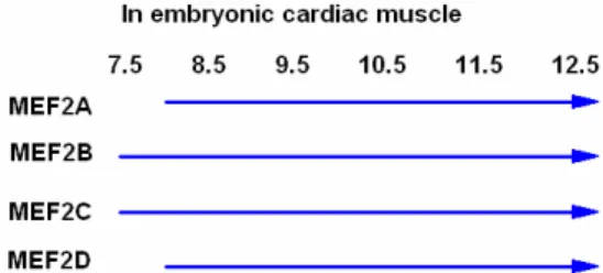
The MEF2 transcription factor family 23
The TGF beta superfamily 24
The Nodal co-receptor Cripto 26
The WNT family 28
The FGF family 31
Axes and gene patternings 34
Cranio-caudal patterning 36
Dorso-ventral patterning 40
Left-right patterning 42
The cardiomyocyte 48
Cardiomyogenic cultures 52
Cell sources for cardiac cell therapy 58
Embryonic stem cells 58
Resident cardiac stem/progenitor cells 60
Muscle cell populations 60
Clinical cardiac regeneration 61
References 66
Aims and technical goals 99
Materials and Methods 101
Nucleic acid extraction 103
DNA extraction from human tissues and cells in culture 103 RNA extraction from human tissues and cells in culture 103 Plasmid DNA extraction with the lysozyme/ boiling miniprep protocol 104
DNA extraction from agarose gels 105
Molecular Biology 105
Polimerase Chain Reaction 105
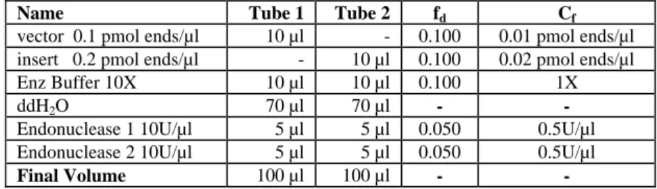
Digestion with restriction endonucleases 107
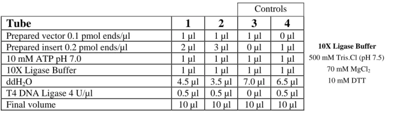
Ligation 108
The β-Myosin heavy chain gene promoter 108
The Neomycin resistance gene 110
The Hygromycin resistance gene 110
The pSKII Multiple Cloning Site primers 111
Cloning 111
Reverse transcription and polymerase chain reaction analysis 113
Expression studies in human rhabdomyosarcoma cells 116
Primary Cultures 118
Embryonal carcinoma and rhabdomyosarcoma cell cultures 118
Preliminary embryonal carcinoma differentiation studies 118 The NT2/D1 cell cultures in inductive angiomyogenic medium 120
Histochemistry and Immunofluorescence 120
Hematoxilin-eosin staining 120 Phalloidin staining 120 Immunofluorescence 121 Ultrastructural analysis 121 Sequencing 121 References 123 Results 125
1. Construction/validation of the pβMyhc-NeoR
-HygR molecule 128
The pβMyhc SalI/BamHI, the NeoR
BamHI/XbaI and the
HygR XbaI/NotI fragments 130
Construction steps 131
The pβMyhc-NeoR
-HygR insert details in the plasmid pSKII 133
βMyhc (or Myh7) expression in RH30 cells 139
Expression studies with the construct pβMyhc-NeoR
-HygR 146
References 151
2. Human cell culture studies 155
2.1 Primary cultures 155
From human myocardium 155
References 167
2.2 Human embryonal carcinomas 169
Preliminary cultures of PA1 and NT2/D1in inductive medium 170 Cultures of NT2/D1 in inductive medium with fibronectin,
5-Azacytidine and BMP2 180
Cultures of NT2/D1 in inductive angiomyogenic medium with
BMP2 190 References 199 Discussion 201 References 215 Conclusions 223 Future Studies 227 Acknowledgments 231
List of abbreviations
ActRII - Activin type II serine/threonine kinase receptor
ActRI - Activin type I serine/threonine kinase receptor or Alk2 ActRIB - Activin type IB serine/threonine kinase receptor or Alk4 Alk2 - Activin receptor-like kinase 2
Alk4 - Activin receptor-like kinase 4
AKT - The same as Protein Kinase B (PKB) AMHC - Atrial myosin heavy chain protein ANF - Atrial natriuretic factor
AP - Anteroposterior
AP1 - Activating protein 1
APH3` - Aminoglycoside 3' phosphotransferase APH(4)Ia - Aminoglycoside 4 phosphotransferase Ia
ASC - Adult stem cell
ATF - Activating transcription factor
AV - Animal-vegetal
AVC - Atrioventricular canal
AVE - Anterior visceral endoderm
5-Aza - 5-Azacytidine
BDNF - Brain-derived neurotrophic factor bFGF/FGF2 - Basic fibroblast growth factor
bHLH - Basic helix-loop-helix (transcription factor)
BM - Bone marrow
BMC - Bone marrow cell
2βME - 2-beta mercaptoethanol BMP - Bone morphogenetic protein BMSC - Bone marrow stem cell BNF - Brain natriuretic factor
BSA - Bovine serum albumin
βTGF - Beta transforming growth factor CaMKII - Calmodulin kinase II
Car - Caronte gene
CAT - Chloramphenicol acetyl transferase cDNA - Complementary deoxyribonucleic acid
Cer - Cerberus gene
CM - Cardiomyocyte
CMV - Cytomegalovirus
COUP-TF - Chicken Ovalbumin Upstream Promoter Transcription Factor CPC - Circulating progenitor cells
CPCS - Cardiac pacemaker and conduction system CREB - Cyclic AMP-responsive element-binding protein CSX - Cardiac-specific homeobox protein
cTnC - Cardiac troponin C gene
Cx - Connexin gene
DBD - DNA-binding domain
DiI - Lipophilic fluorescent dye:
1,1-dioctadecyl-3,3,3’3’- tetramethylindocarbocyanine perchlorate
Dkk - Dickkopf gene
Dll 1 - Delta-like 1 gene DMSO - Dimethyl sulfoxide
DNA - Deoxyribonucleic acid
dpc - Days post coitum
DSL - Delta/Serrate/Lag2 DV - Dorsoventral E - Embryonic day EB - Embryoid body EC - Embryonal carcinoma ECG - Electrocardiogram
ECGS - Endothelial cell growth supplement ECM - Extracellular matrix
EG - Embryonic germ (cells)
EGF - Epidermal growth factor
EGF-CFC - Epidermal growth factor - Cripto/FRL1/Cryptic
EPC - Endothelial progenitor cell
ES - Embryonic stem
ESC - Embryonic stem cell
ET1 - Endothelin 1 protein
EtBr - Ethidium bromide
Ets - E26 transformation specific
FAST - Forkhead activin signal transducer protein FBS - Foetal bovine serum
FGF - Fibroblast growth factor
FGFR - Fibroblast growth factor receptor FITC - Fluorescein isothiocyanate
FRL1 - Fibroblast growth factor receptor ligand 1
Fz - Frizzled gene
GATA - WGATAA DNA binding transcription factors GDF - Growth differentiation factor
GDF1 - Growth differentiation factor 1
HAND - Heart, autonomic nervous system and neural crest derivatives protein hASC - Human adult stem cell
hEC - Human embryonal carcinoma
hEG - Human embryonic germ (cell) hEPC - Human endothelial progenitor cell hESC - Human embryonic stem cell
Hh - Hedgehog gene
hHSC - Human hematopoietic stem cell HFN3β - Hepatocyte nuclear factor-3β HGF - Hepatocyte growth factor hMSC - Human mesenchymal stem cell HSPG - Heparin sulphate proteoglycan HSC - Hematopoietic stem cell HSV - Herpes simplex virus
HUVEC - Human umbilical vein endothelial cell HygR - Hygromycin resistance gene
IGF2 - Insulin-like growth factor 2
ITS - Insulin/transferrin/selenium
IRX - Iroquois homeobox protein IVS - Interventricular septum JNK - c-Jun N-terminal kinase
kb - kilobase = 1000 DNA or RNA base pairs
LBD - Ligand-binding domain
LEF - Lymphoid enhancer factor LIF - Leukemia inhibitory factor LPM - Lateral plate mesoderm
LR - Left-right
LRP - Lipoprotein related protein
MADS - MCM1/Agamous/Deficiens/Serum response factor MAPK - Mitogen-activated protein kinase
MCK - Muscle creatine kinase
mEC - Mouse embryonal carcinoma
MEF - Myocyte-enhancer factor mEG - Mouse embryonic germ (cell) mESC - Mouse embryonic stem cell
MF20 - Antibody against myosin heavy chain MHC - Major histocompatibility complex MIS - “Müllerian inhibiting substance” protein MLC2A - Atrial myosin light chain - 2 protein MLC2V - Ventricular myosin light chain - 2 protein mRNA - Messenger ribonucleic acid
MSC - Mesenchymal stem cell
MYH7 - Beta myosin heavy chain; cardiac ventricular myosin; slow fibbers myosin protein
MyHC - Myosin heavy chain; cardiac ventricular myosin; slow fibbers myosin protein
NEAA - Non essential aminoacids NeoR - Neomycin resistance gene
NFAT - Nuclear factor of activated T cells Oep - Zebrafish one-eyed pinhead gene
pBK - pBluescript plasmid from Stratagene
pβMyhc - Beta myosin heavy chain promoter (human) PCR - Polimerase chain reaction
PCP - Polar cell polarity
PI3K - Phosphoinositide 3-kinase
PKA - cAMP-dependent protein kinase or Protein kinase A PKC - Calmodulin-dependent protein kinase or Protein kinase C PLCγ - Phospholipase C γ isophorm
pSKII(+) - pBluescript II SacI KpnI (+) plasmid from Stratagene
RA - Retinoic acid
Raldh2 - Retinaldehyde dehydrogenase type 2
RAR - all-trans retinoic acid receptor
RARE - RA-response element
RAS - A low-molecular weight G-protein
RBP-J - Immunoglobulin J kappa segment recombination signal sequence binding protein
RNA - Ribonucleic acid
RTK - Receptor tyrosine kinase
RT-PCR - Reverse transcriptase – Polimerase chain reaction
RXR - 9-cis retinoic acid receptor
SAP - SAF-A/B/Acinus/PIAS, DNA binding domain SERCA - Sarcoendoplasmic reticulum calcium ATPase SAGE - Serial analysis of gene expression
sFRP - secreted Fz-related protein SHH - Sonic hedgehog protein
SMAD - Sma/mothers against decapentaplegic protein Smyhc3 - Slow myosin heavy chain 3 gene
SRF - Serum response factor
SURE - Stop unwanted recombination events T - T box transcription factor Brachyuri TBX - T-box transcription factor
TCF - T cell factor
TDGF1 - Teratocarcinoma-derived growth factor 1
TR - Thyroid hormone receptor
TRITC - Tetramethylrhodamine isothiocyanate
UC - Umbilical cord
UCB - Umbilical cord blood
UCWJ - Umbilical cord Whorten`s jelly
UTR - Untranslated region
VDR - Vitamin D receptor
VDRE - Vitamin D response element VEGF - Vascular endothelial growth factor VMHC1 - Ventricle myosinheavy chain 1 protein WNT - Wingless/Int protein
Glossary and notes
General glossary
alleles Different variants of a gene found in the normal
population; as individuals carry two copies of each gene, one on each pair of chromosomes, they may have identical (homozygous) or different (heterozygous) alleles.
amniota Vertebrates which have extraembryonic membranes
(amnion, chorion, yolk sac or placenta): reptiles, birds and mammals.
anamniota Vertebrates which do not have extraembryonic
membranes (amnion, chorion, yolk sac or placenta): fishes and amphibian.
antisense RNA Used in a technique for inactivating a gene using an RNA sequence that complements and hybridizes with the normal RNA made by the gene of interest.
complementary DNA DNA synthesized by copying mRNA, used for gene cloning and production of large quantities of protein. DiGeorge’s syndrome A developmental defect caused by malfunction of the
cardiac neural crest, resulting in abnormalities in the thymus, parathyroids, cardiac outflow tract, aortic arches and face.
dominant negative protein Encodes a protein whose structure has been modified, usually by removing some part of it, so that it inhibits the wildtype protein in a dominant fashion. Used experimentally to eliminate the action of proteins such as secreted molecules or receptors.
downregulation Reduction, not cessation, in the effect being studied, normally, gene expression.
downstream 1. Location of a motif or domain in a gene nearer the 3’ end of the sequence than a particular site. Gene sequences are read from the terminal phosphate (-PO42-), linked to the 5’ carbon, also called
the 5’ end, to the terminal hydroxil (-OH) linked to the 3’ carbon, or the 3’ end;
2. One may say that the gene “a” that is regulated by the product of the gene “b” it is downstream of that gene;
3. Later reactions in a biochemical cascade.
ectopic expression Gene expression in cells where that gene is not normally expressed.
expression Production of mRNA by an activated gene.
growth factor Polypeptide involved in intercellular signalling,
regulating the proliferation and maintenance of cells. in situ hybridization Autoradiographic or histochemical method for
detecting mRNA in tissue sections.
lacZ reporter Bacterial lacZ gene encoding β-galactosidase, which is detected by a blue histochemical reaction product. monoclonal antibody Antibody selected to recognize a specific sequence
(epitope) on the target protein.
oligonucleotide Synthetic DNA or RNA sequence.
polymerase chain reaction Multifold amplification of selected DNA using small part of sequence as primer.
phalloidin A toxin from the mushroom Amanita phalloids that
binds to polymeric and oligomeric actin with high affinity.
phenotype Physical manifestations of a wildtype, mutant or
deleted gene.
promoter Transcription-controlling sequence in a gene.
reporter gene A sequence that encodes a product that can be easily identified and quantified. Fusion of the reporter gene to a particular gene promoter can be used to detect and measure gene expression.
repressor protein Product of a regulatory gene that prevents expression of a target gene(s), usually by binding to the promoter sequence of that gene or by interaction with activators or transcription factors.
rescue Re-establishing a function by adding an exogenous
gene or molecule that is either the same or similar to a missing gene or molecule.
reverse transcriptase reaction Synthesis of cDNA by reverse transcription from mRNA.
signal molecule Molecule carrying information between cells. Usually a growth factor that activates an intracellular signalling pathway, also known as a ligand.
signalling pathway Intracellular cascade of biochemical reactions that stimulates an appropriate cellular response as gene transcription; usually activated by a signal molecule binding to its receptor at the cell surface.
transcript mRNA produced by transcription of a particular gene.
transcription factor Protein that regulates gene transcription often by binding to a specific DNA sequence. Normally classified by the type of binding domain they contain: homeobox, T box, helix-loop-helix, zinc finger, etc.
upregulation Opposite of downregulation.
upstream Opposite direction to downstream.
wildtype Individual with no known mutation in the gene under
Embryological glossary
animal pole Portion of the early embryo marked by the second oocyte polar body, where the main embryonic structures will develop.
animal–vegetal axis Line connecting the animal and vegetal poles of the zygote. Whether this corresponds to any particular axis in the adult is still a matter of debate.
anterior visceral endoderm In mouse: visceral endodermal tissue lining the outer surface of the epiblast at the egg-cylinder stage and is displaced anteriorly at the early primitive-streak stage; probably involved in inducing anterior structures but itself forms extraembryonic structures;
In other mammals and birds, termed the primitive endoderm and the hypoblast, respectively.
blastoderm The two layers, epiblast and hypoblast at the animal pole of the blastula.
blastula First stage of embryogenesis when the egg divides several times to form a hollow ball of cells and a two-layered embryo.
cardioblasts Cells committed to become cardiomyocytes.
cell fate The full spectrum of tissues that embryonic cells may
differentiate into, under normal, unperturbed conditions; Restriction of a multipotential embryonic cell to producing cells of a single tissue type.
conceptus The entire product of conception, including the embryo and extraembryonic membranes.
determinants mRNAs or protein molecules that initiate gene
transcription in the early embryo; also known as maternal determinants and dorsal or ventral determinants.
ectoderm The outermost germ layer; gives rise to epidermis and
neural tube, neural crest, and eye and ear placodes.
endoderm The innermost germ layer; contributes to gut, liver,
respiratory system, urogenital system and endocrine glands.
epiblast During the blastula stage, the blastoderm layer that corresponds spatially to the ectoderm in the gastrulating embryo and gives rise to different germ layers during gastrulation.
extraembryonic mesoderm Portions of the mesoderm that do not contribute to embryonic structures; in amniotes gives rise to vasculature and supporting membranes of yolk sac or placenta.
fate mapping Identification of the precursor cells in the early embryo that will give rise to particular tissues or organs.
floor plate A wedge-shaped group of cells in the ventral neural tube that secretes molecules responsible for the DV organization of particular structures as the spinal cord. gastrulation Highly coordinated process involving stereotypic cell
movements; results in the establishment of the germ layers; embryo at this stage is termed a gastrula.
germ layers Three layers of the embryo, ectoderm, endoderm and
mesoderm established during gastrulation that give rise to the tissues and organ systems of the late embryo.
Hensen's node In amniotes: thickening at anterior tip of the primitive streak; organizer inducing axial structures; in the mouse embryo, often referred to as “the node”; homologous to Spemann’s organizer in Amphibia; named after Viktor Hensen, who first described it in 1876.
hypoblast In chick and rabbit: inner layer of blastoderm; in
zebrafish, the equivalent is the yolk syncytial layer, and in mouse, the visceral endoderm; gives rise to extraembryonic structures.
induction Influence of one group of embryonic cells on the fate of neighbouring cells, usually through the production of diffusible signalling molecules or through direct cell–cell contact.
lateral-plate mesoderm Sheet of mesoderm further away from the axis, lateral to the paraxial and intermediate mesoderm; contributes to body tissues and extraembryonic structures.
mesendoderm Embryonic tissue containing a combination of mesoderm and endoderm cells or of cells that contribute with descendants to both germ layers. Also termed endomesoderm.
mesoderm Middle germ layer, gives rise to skeletal muscles, notochord, skeleton, connective tissue, heart and contributes to the gut and urogenital systems formation.
notochord Early mesodermal structure with important role in
patterning early embryo; precursor of part of the vertebral column.
neural tube Precursor of the central nervous system. It is formed by the rolling up of the dorsal neuroectoderm, during a stage termed neurulation.
neurula Third stage of embryogenesis when the embryo elongates
and the neural tube forms.
neural plate Specialized area of ectoderm that gives rise to the nervous system and neural crest.
notochord A rod of cells forming in the midline dorsal mesoderm. Contributes to the induction of the nervous system; persists in some lower chordates but in all vertebrates is replaced by the vertebral column.
oocyte Unfertilized female gamete (haploid).
organizer A group of cells in the embryo that secretes signal
molecules affecting the differentiation of other tissues. Identified by its ability to induce ectopic structures when grafted into a host embryo; see Spemann’s organizer, Hensen’s node.
paraxial mesoderm Mesoderm on either side of neural tube. Forms presomitic mesoderm, which contributes to the head, and the somites.
precardiogenic mesoderm Also known as cardiogenic plate or cardiac crescent. primitive streak A groove at the posterior end of the embryo that marks
the beginning of gastrulation and the formation of the AP axis. At the posterior end, epiblast cells undergo epithelial to mesenchymal transition to give rise to the mesoderm and definitive endoderm.
situs solitus Normal pattern of LR asymmetry often referred to as “situs”. When completed, and total or partial reversal of the internal organs occurs, it is called “situs inversus totalis” or “heterotaxia”, respectively.
somites Blocks of paraxial mesoderm that form sequentially from anterior to posterior direction after gastrulation. They are precursors of the axial skeleton, all skeletal muscles and the dermis.
trophoblast A multipotent germ layer that can differentiate into
extraembryonic tissues.
vegetal pole Opposite side of early embryo to the animal pole, in some species containing yolky material; gives rise mainly to extraembryonic structures.
Embryological staging references
The following references define the stages of embryological development of the organisms mentioned in this dissertation.
Drosophila melanogaster (fruit fly):
Taxonomy: Kingdom: Animalia; Phylum: Arthropoda; Class: Insecta; Order: Diptera; Family: Drosophilidae
Campos-Ortega, JA and Hartenstein, V:
The Embryonic Development of Drosophila melanogaster.
Springer-Verlag, 1985, Berlin, Heidelberg, New York.
Danio rerio (zebrafish):
Taxonomy: Kingdom: Animalia; Phylum: Chordata; Subphylum: Vertebrata Class: Actinopterigi; Order: Cypriniformes; Family: Cyprinidae
Kimmel, CB; Ballard, WW; Kimmel, SR; Ullmann, B and Schilling, TF:
Stages of embryonic development of the zebrafish.
Dev Dyn, 1995, 203, 253-310. Driever, W:
Introduction to the zebrafish. In: Cell Lineage and Fate Determination.
Ed. S. A. Moody, San Diego: Academic Press, 1999, 371-382.
Xenopus laevis (frog):
Taxonomy: Kingdom: Animalia; Phylum: Chordata; Subphylum: Vertebrata Class: Amphibia; Order: Anura; Family: Pipidae Nieuwkoop, PD and Faber, J:
Normal Table of Xenopus laevis.
North-Holland Publications, 1967, Amsterdam.
Gallus gallus (chick):
Taxonomy: Kingdom: Animalia; Phylum: Chordata; Subphylum: Vertebrata Class: Aves; Order: Galliformes; Family: Phasianidae H. Eyal-Giladi, H and S. Kochav, S:
From cleavage to primitive streak formation: a complementary normal table and a new look at the first stages of the development of the chick. I. General morphology.
Dev Biol, 1976, 49, 321-337. Hamburger, V and Hamilton, HL:
A series of normal stages in the development of the chick embryo.
J Morph, 1951, 88, 49-67.
Mus musculus (mouse):
Taxonomy: Kingdom: Animalia; Phylum: Chordata; Subphylum: Vertebrata Class: Mammalia; Order: Rodentia; Family: Muridae Downs, KM and Davies, T:
Staging of gastrulating mouse embryos by morphological landmarks in the dissecting microscope.
Development, 1993, 118, 1255-1266.
Homo sapiens (human):
Taxonomy: Kingdom: Animalia; Phylum: Chordata; Subphylum: Vertebrata Class: Mammalia; Order: Primates; Family: Hominidae O'Rahilly, R:
Early human development and the chief source of information on staged human embryos.
Timelines of heart development
The following diagrams define the main timeline of cardiac development of the organisms mentioned in this dissertation.
Fruit fly
Gestational age (h) 0 4 8 12 24 20 16 GastrulationMesoderm cells spread dorsally along inside of ectoderm Mesoderm splits into outer and inner layers; cardiac cells are the most dorsal mesoderm cells
Heart cells migrate dorsally after germ band shortens Cells arrive at dorsal midline and fuse to form heart tube Cardial cell differentiation begins
Hatching
Zebrafish
Gestational age (h) 0 8 16 24 48 32 40 Gastrulation begins Gastrulation completeCardiac progenitors migrate toward the embryonic axis Cardiac cells form two tubular primordia in LPM Heart cells move close together and fusion begins Beating begins in individual cells
Definitive heart tube fully formed Rightward looping begins
Looping is completed; all four chambers are visible in the linear heart
Hatching
Adapted from “Genetic control of heart development” Ed. J. S. Altman. HFSP, page 14, Strasbourg, 1997. Differentiation of heart anlage commences
Hatching
Heart is S shaped; atrium, ventricle and sinus venosus visible Looping begins
Myocardial wall complete Endocardial tube formation begins
Fusion of bilateral groups of cardiac cells into a single anlage begins
Gastrulation begins
Cardiac progenitors have migrated to the outer edge of the neural plate
Cardiac progenitors are in the deep layer of the mesoderm next to blastopore lip
Gestational age (h) 0 8 16 24 32 40 48
Frog
Adapted from “Genetic control of heart development” Ed. J. S. Altman. HFSP, page 13, Strasbourg, 1997. Hamb urger a nd Hamilt on S tage s
Heart cells move close together and fusion starts anteriorly in the conotruncal region
Hatching at 20-21 days
Looping complete Rightward looping begins
Fusion complete; definitive heart tube fully formed Beating begins
Cardiac progenitors migrate towards ventral midline to form cardiac crescent
Gastrulation begins
Cardiac progenitors enter mesoderm and spread anterolaterally Primitive streak forms
Cardiac progenitors are in the mid-primitive streak Gestational age (h) 0 12 24 36 48 60 3 14 15 13 12 11 10 9 8 7 6 5 4
Chick
Adapted from “Genetic control of heart development” Ed. J. S. Altman. HFSP, page 12, Strasbourg, 1997.
Mouse
Birth at 19.5 dpc
Fetal circulatory system is complete and functioning Interventricular septum, AV cushions and septa forms Atrial septum and AV canal form
Interventricular sulcus forms
Sinus venosus, atrium and ventricular loop are visible; circulation begins
Heart begins contracting; rightward looping commences Heart cells move close together and fusion starts anteriorly in the conotruncal region
During gastrulation cardiac progenitors migrate anteriorly and ventrally to form cardiac crescent
Primitive streak forms
Before gastrulation cardiac progenitors are in a relatively posterior, distal portion of the epiblast
Gestational age (dpc) 0 4 8 12 10
Compiled from:
1. Netter, FH: The Heart - The Ciba Collection of Medical Illustrations vol 5, 5Ed.
The Case-Hoyt Corp, New York, 1981, 114-126;
2. Sadler, TW: Langman´s Medical Embryology, 8Ed.
Lippincott Williams and Wilkins, USA, 2000, 214-15;
3. Christa Wellman and John McNulty: Development of the human heart, 1996,
in http://www.meddean.luc.edu/lumen/MedED/GrossAnatomy/thorax0/heartdev/main_fra.html;
4. Mark Hill: UNSW Embryology: Carnegie Stages, 2007,
in http://embryology.med.unsw.edu.au/wwwhuman/Stages/Stages.htm;
5. Hunter A, Kaufman MH, McKay A, Baldock R, Simmen MW, Bard JB: An ontology of human developmental anatomy. J Anat, 2003, 203(4): 347-55.
Septated fetal heart; fetal circulatory system is complete and functioning
Truncal swellings appear
Superior and inferior endocardial cushions fuse The interventricular canal is completely obliterated
The outflow tracts (aorta and pulmonary trunk) are completely separated
Fusion complete; a single heart tube is formed Endocardial tubes are formed
C a rn e g ie S tag es Birth at ~266 dpc
The bulboventricular loop is formed; the endocardial cushions and the atrial and interventricular septum start appearing Interventricular sulcus forms
Sinus venosus, atrium and ventricular loop are visible; circulation begins
Heart begins contracting; rightward looping commences Endocardial tubes move close together and fusion starts anteriorly in the conotruncal region
During gastrulation cardiac progenitors migrate anteriorly and ventrally to form cardiac crescent
Primitive streak forms
Before gastrulation cardiac progenitors are in a relatively posterior, distal portion of the epiblast
29-34 29 34-35 30 26-29 28 23-26 27 20-23 26 17-20 25 13-17 24 10-13 23 7-10 22 4-7 21 1-4 20 Pair of somites Age (dpc)
Human
Gestational age (dpc) 0 10 20 30 40 50 60 14 5 6 23 22 21 20 19 18 17 16 15 13 12 10 11 9 8 7Gene nomenclature
The following nomenclature is used for genes described in the text.
Gene names are shown with a first uppercase letter followed by lowercase letters throughout the text; whereas, the “PROTEIN” coded by the gene uses the same name/abbreviation but only in uppercase letters. Ex: Fgf8 gene and FGF8 protein.
Preface
In 2001 an interesting paper was published in Nature magazine with the title “Bone marrow cells regenerate infarcted myocardium”. The news of that research caused me such a curiosity that I started to study the state of the art of that area of knowledge.
Among many interesting things, it could be stressed the “New dogma” of heart remodelling, the importance of Insulin growth factors (IGFs), and the recent findings in cardiac progenitors research.
My PhD project had the title: “Mechanisms of transmural scar formation in the surgical treatment of atrial fibrillation with radiofrequency catheter ablation and microwave. Role of heat induced apoptosis.” Although the apoptosis idea is now a well established fact in heart pathology, it did not seem to raise much interest among the portuguese cardiac surgeons, who were more interested in the characterization of physical variables, like the diffusible temperature gradient of the heat induced by radiofrequency, than by genetic ones.
Accordingly, and in a natural way, my work was slowly becoming more engaged with the general field of cardiac progenitors’ research, than with the surgical technique-related apoptotic research. In view of the above, the title “Towards cardiac cell therapy” has been proposed.
Curiously, the two apparently distant research fields were interconnected with a common need, the development of a human cardiomyocyte cell line from undifferentiated adult pluripotent cells. This would allow both the analysis and experiences, at the cellular level, of the conditions and mechanisms underlying cardiomyocyte apoptosis induced by heat and the understanding and the characterization of cell surface antigens involved on cardiac differentiation, in vitro.
Developing a cell line from an undifferentiated progenitor cell, can be accomplished in different ways, depending on the information available regarding the developmental steps (growth factors and surface antigens) that lead to the differentiation only into a particular cell type. Because such information has just started to become available, and because differentiating embryonic stem cells never give rise to pure cardiomyocyte cultures, the solution is to transform undifferentiated cells with a bioengineered construct containing one
of the gene promoters known to be specifically activated in the cardiac cell lineage, as the promoter of the human cardiac myosin heavy chain gene linked to an antibiotic resistance gene. This strategy has already been done in mice since 1996 and allowed the selection, in an antibiotic conditioned culture, of mouse cardiac lineage-committed cells, among other type of cells, with a purity of more than 99 %. Accordingly, a similar human construct was created and used in an undifferentiated human myoblastic cell line.
Of course that one of the logical potentialities of such a study would be to extend this cellular approach to clinics, in areas of pathologic heart conditioning, in order to regenerate affected myocardium resorting to a cell-based therapy.
In view of the difficulties raised by the usage of human embryonic stem cells for cardiac differentiation studies, pluripotent human embryonal carcinoma cell lines PA1 and NTERA.clD1 were used.
Ultrastructural and microscopic analyses were performed in defined time points along PA1 and NTERA.clD1 differentiation in pertinent inductive media.
We also analyzed the histochemistry, the cytoskeletal actin organization and the gene expression of genes relevant to cardiac muscle and endothelium differentiation, during the growth of NT2/D1 cells in a reported inductive angiomyogenic medium.
Unfortunately, cardiac differentiation from this cell line was never accomplished, and for that reason, the experiments based on the assumption that the created construct may transform and select human pluripotent stem cells that are cardiac committed, are still on hold.
Introduction
General insight
The human cardiac commitment, differentiation and morphogenesis are not completely understood.
In this scenario, the ambitious and already underway “clinical cardiac cell therapy” becomes a procedure without a solid scientific fundament.
A major difficulty in understanding cardiogenesis is related to the use of different vertebrate models to give a descriptive approach of the phenomenon, which contributes, in recent papers, to a confusing variety of terms, including chick, mouse and zebra fish models that do not overlap completely [1]. Following this line of thought, reliable studies based on human embryonic material are limited to few famous collections such as the Carnegie Collection and local collections from some medical universities.
Nevertheless, in the past decade research has progressed from the physiological and functional to the molecular approach, leeding to a deeper understanding of the cardiac functions at genetic and proteomic levels in animal models [2].
In the next pages, the basic aspects of heart development will be shortly described and results of molecular data incorporated whenever relevant.
In order to achieve a more complete integration of the text with the experimental work, a significant effort was made in enlightening the main molecular aspects of embryonic stem cells commitment into cardiac cell lineages. This, we believe, will help to integrate the current state of the art and the development of the goals presented in this thesis. This will also introduce the complex world of cardiac differentiation, a knowledge that is an essential tool when dealing with challenges such as “human cardiac cellular therapy”.
Curiously, the number of possible molecular signalling mechanisms (types or classes) responsible for the formation of an adult heart, are the same, as those responsible for the formation of the whole adult organism, from just one single cell. Furthermore, they are relatively few, when compared to the complex network of embryological processes and to the high number of possible signalling events. In addition, we strongly believe that the sharp molecular cell signaling regulation during development is where researchers should look for, if they want to understand the commitment or the differentiation mechanism of a particular cell lineage, as for instance, the understanding of the appearance of the cardiac cell lineage.
Molecular developmental mechanisms may roughly be classified in four distinct classes: i) cell/cell and cell/ECM related pathways, including the integrins, cadherins, DSL proteins, collagens, fibronectins, laminins, fibrins, elastins and other molecular families; ii) secreted protein factors initiating pathways including Bone morphogenetic proteins (BMPs), Fibroblast growth factors (FGFs), Vascular endothelial growth factors (VEGFs), Wingless/Int (WNT) proteins, Hedgehog (Hh) proteins, Epidermal growth factors (EGFs), beta Transforming growth factors (βTGFs) and others; iii) soluble molecules initiating steroid or steroid-like pathways with steroid hormones and Retinoic acid (RA) as the most important members and iv) ion signalling pathways with those Ca2+-mediated as the most important ones.
All of these signalling possibilities have, associated downstream of the respective pathway, specific transcription factor activities, including: i) basic DNA domain, ii) zinc-coordinating DNA binding domain, iii) helix-turn-helix and iv) β-scaffold with minor groove contact transcription factor activities. They will activate or suppress the transcription of specific target genes, which are, ultimately responsible for cell cycle regulation and cell behaviour itself, resulting in proliferative or quiescent cells and migratory or apoptotic cells, respectively.
Since it would not be reasonable to describe all the constitutive members of these four-group’s pathways in the present dissertation, only those known to be pertinent to cardiogenesis will be mentioned or illustrated in subsequent sections.
Cardiac embryology
In vertebrates, cardiomyogenesis is a complex and highly regulated process, being each event spatially and temporally ordered. The embryonic stem cells involved in migration and differentiation events are of different types and of several origins and perform precise interactions with each other. It is easy to understand that any irregularity during this development phase will result in congenital heart defects that will be diagnosed during pregnancy, at birth or later in life, according to their respective severity. Importantly, malformations of the heart and great vessels account for the most frequent congenital defects in humans, with an incidence of approximately 1 % [3].
The heart, the first definitive organ to be formed in the embryo and the respective cardiovascular system, starts appearing during gastrulation, in the middle of the third week, when simple diffusion is no longer sufficient to fulfil nutritional requirements.
The actual process of gastrulation might be relatively unimportant considering the cellular commitment to a cardiac fate, owing to the fact that early mesodermal cells can adopt several different phenotypes and contribute to different tissues when grafted into new environments. However, the patterning processes and dynamic cell movements of gastrulation allocate heart mesodermal precursors to specific cell niches in a progressive and spatially complex way, creating the site specific milieu for cardiac cell differentiation and morphogenesis. Accordingly, it is our belief, that at this particular moment, the creation of the “appropriate environment” is more important than the “cardiac cell commitment” itself. In other words the “soil” is more important than the “seed”.
Before describing the development of the cardiovascular system, it is useful to mention the appearance of the intraembryonic coelom. It is formed by the confluence of small and initially isolated spaces that appear in the lateral plate mesoderm, resulting in a single horseshoe-shaped cavity, just cranially to the prechordal plate. This event separates the mesoderm into two layers, the parietal or somatopleuric mesoderm in contact with the ectoderm, and the visceral or splanchnopleuric mesoderm in contact with the endoderm.
Cardiac progenitors cells lie on both sides of the midline in the epiblast, immediately lateral to the primitive streak [4, 5]. Then, they migrate throughout the primitive streak, clustering themselves bilaterally in the splanchnopleuric layers of the lateral plate mesoderm [6], in a region known as cardiogenic plates, precardiac mesoderm or primary heart fields.
Recently, it was shown that this group of mesodermal cells express a basic helix-loop-helix transcription factor, the Mesp gene, considered to be the earliest sign of cardiovascular development known to date [7, 8]. This region is located cranial and lateral to the neural plate and will later express other cardiac-related genes like Nkx2.5 (Csx1 in humans) [9, 10], Bmp2 [10, 11], Gata4 [12, 13] and Hand [14]. It is suggested that the helix-turn-helix transcription factor Ets, a RTK activity and a FGF pathway might be the keys for the Mesp1 dependent regulation of Gata4, Nkx2.5 and Hand genes, but that has not yet been proven in mammals [15] (See also Molecular families in cardiogenesis - The FGF family).
Although Nkx2.5 is one of the first genes to be detected in the precardiac mesoderm, it is not expressed in the caudal regions of the heart forming zone. By contrast, Bmp2, Gata4 and Hand are detected in the entire primary heart field. Nevertheless, all of them are not cardiac restricted since they are also expressed in regions outside the heart forming zone.
Despite the fact that these genes are many times referred as being cardiac-specific, the specific and restricted early cardiac progenitor marker is still waiting to be discovered.
A recent study in the chick, using two different techniques, fluorescent carbocyanin labelling and gene expression staining, showed that there is no accurate correspondence between the two labellings in the primary heart fields area, highlighting that the final delineation of the exact precardiac regions is not yet completed [10].
Importantly, the first populations of cells that constitute the primary heart fields will not contribute to all the cells of the adult heart or, in other words, it will be the recruitment of other cardiac progenitors, later in development, that will become part of the final outflow and inflow tract-derived structures.
Formation of the endocardial tubes
At the cardiogenic plate in xenopus and chick, BMP2 [16-18], repressors of Wnt signaling [19, 20] and possibly, later FGF signals [21], coming from the endoderm induce cardiac progenitors to form cardiac myoblasts, a complex network that may occur also in mice [22].
The endoderm that is in direct contact with cardiac mesoderm has been considered by some authors as the heart-inducing tissue, because members of the BMP family are expressed in endoderm, as well as in adjacent ectoderm and extraembryonic tissues [23-25]. Accordingly, many studies in different organisms have shown a key role for the BMP signalling, in specifying and maintaining the myocardial lineage [26-29], as well as the BMP-receptor regulated transcription factors of the Smad family which directly activate genes that encode cardiac transcription factors [30-33] (See also Molecular families in cardiogenesis - The TGFβ superfamily).
Thinking of BMP2 as a possible candidate to be a cardiac inducer molecule in vitro is a particular important point. Indeed, this possibility was the basis for one of the main technical goals of the present work. It will be discussed at lenght in the Discussion section.
During gastrulation, hemangioblasts also appear, in the precardiac splanchnopleuric mesoderm region, ventral to the cranial, horseshoe-shaped portion of the intraembryonic coelom, proliferating and clustering in hemangiogenic islands, known as angiocysts. From this cranial part of the coelom will develop the pericardial cavity. Along in time, those cell clusters will fuse and will form the left and right endocardial tubes, each an endothelial lined tube surrounded by cardiomyoblasts.
Progenitor cells that will be committed to the endocardial and myocardial cell lineages, migrate from within the precardiac mesoderm to become localized just above the endoderm
cell layer and in the developing myocardium, respectively. Curiously, in the chick, these migrating cells may express both myocardial and endocardial markers [34]. These results indicate that cells of the cardiac crescent might be the common precursors for myocardial, endocardial and possibly pericardial cells of the heart. In fact, a study in mice has revealed that all three of the above cell types have activated, in the cardiac crescent, the cardiac transcription factor Nkx2.5 [35]. However, the expression of Nkx2.5 may be more a common characteristic of different populations of cells that coexist in a common region than a marker of a common precursor, which is the same as saying that myocardial and endocardial precursors that reside within the heart-forming region express Nkx2.5. In fact, the endocardial cell layer is constituted by a mosaic of cells with and without Nkx2.5 expression [35]. A possible explanation for this is that endocardial precursors that migrate to the cardiac crescent from Nkx2.5 negative regions will not express Nkx2.5.
Recent experiments in chick, with retrovirus-mediated lineage tagging, have suggested that if there is a common precursor of both myocardial and endocardial cells, then it might be commited to a endocardial and myocardial fate at or before initial primitive streak formation [36]. Conversely, data supporting the existence of a “later” common progenitor comes from studieswith the cardiac mesoderm cell line QCE6 [37]. This cell linewas derived from precardiac mesoderm of the Japanese quail andexhibits a phenotype consistent with its being a cardiac stemcell. If treated with a combination of RA, bFGF, TGFß2, and TGFß3, the QCE6 cells willdifferentiate into two distinct phenotypes, a myocardial and an endocardial endothelial within the same culture.
Consistent with these results, two potential models were formulated [36]. One assuming that mesodermal cells reaching at the heart field are equipotent but their fate into either myocyte or endocardial lineage is possibly defined by local, inductive signals from underlying endoderm. Another model presumes that the heart field consists of two subpopulations already restricted to myocardial or endocardial lineage before migration to the heart field. If these two lineages were already separated when their progenitors migrate to the heart field, the role of endoderm-derived factors would be to continue their terminal differentiation, rather than to act as inductive signals.
This still open question regarding the existence of a common endocardial/myocardial progenitor cell had a strong influence on the design of the differentiation studies presented in this thesis. Accordingly, a pluripotent human embryonal carcinoma cell line was used with a previously described angiomyogenic medium [38] in order to obtain cardiac muscle and/or endothelial cell phenotypes.
In the late presomite embryo, other hemangiogenic islands have also developed in the intraembryonic and extraembryonic mesoderm outside the precardiac region. Soon, they will initiate the vasculogenesis of the left and right dorsal aortas that later will connect the cranial pole of the endocardial tubes via the aortic arches, forming the arterial pole of the heart.
Extraembryonically, vasculogenesis has started in the yolk sac mesoderm, forming, sequentially in time, a plexus of vessels in its wall, the vitteline veins, and later the umbilical veins. Together they will connect the most caudal ends of the endocardial heart tubes, establishing the venous pole of the heart. So, each endocardial tube is continuous cranially with a dorsal aorta, the outflow tract, and caudally with a vitteloumbilical vein, the inflow tract (Fig. 1).
Formation of the heart tube
The cranial and lateral folding of the embryo forces the endocardial tubes into the thoracic cavity. As a result of this change, these tubes come to lie closer and parallel to each other, entering more and more into the pericardial cavity.
By this time, CARP, the first cardiac-specific transcriptional regulator to be known, located downstream of the cardiac homeobox gene Nkx 2.5, starts to be expressed. The Carp gene encodes a nuclear coregulator that contains four ankyrin repeats within its carboxyl terminal end and regulates, negatively, the expression of cardiac genes, including Mlc2v and Anf [39]. The subsequent uniform expression of Carp in the heart is due to the presence of distinct cis 5’ flanking sequences of the Carp gene, which are capable of confering specificity regarding the atrial, left ventricular, right ventricular, and conotruncal segments of the heart. Carp may be transcriptional regulated in a cooperative way by Nkx2.5 and GATA4. In fact, the proximal upstream regulatory region of the gene contains an essential GATA4 binding site, which suggests that Nkx2.5 controls CARP expression, at least in part, through GATA4 [40].
At this stage, cardiac progenitors are migrating ventrally, on a graded distribution of fibronectin along the craniocaudal axis, deposited in the endocardial-endodermal interface [41].
The plexiform region of the bilateral cardiac primordia, still separating the right and left endocardial tubes from each other, gradually disappears, resulting in fusion in a craniocaudal direction. At this stage, the single straight heart tube comes to lie, completely, within the pericardial cavity, attached dorsally by a single fold of tissue, the dorsal mesoderm. During this process, correct differentiation of embryonic endoderm is crucial for proper migration
and morphogenesis. Affecting the endoderm-acting genes, as for instance, Gata4, partially disrupt this process, leading to a condition known as cardia bifida [42, 43].
Meanwhile, the mesodermal tissue surrounding the heart tube shows a particular organization in three different layers (Fig. 4). The inner layer, immediately around endothelium, is initially a thick acellular matrix that because of its histological staining (stains lightly) has been called cardiac jelly. The next layer is densely nucleated at first, only a few cells thick. The third and outer layer consists of cuboid epithelial cells which also line the remainder of the pericardial cavity. The second and third layers together are generally referred to as the myocardium, separated from the endocardial tube by the cardiac jelly[44]. The jelly accumulates in the AV junction and in the OFT and progressively disappears from the ventricular and atrial chambers [45].
The human embryo has now 7 somites, is about 2.2 mm long and is approximately 23 days old. It is at about this time, or somewhat earlier that the heart begins to beat.
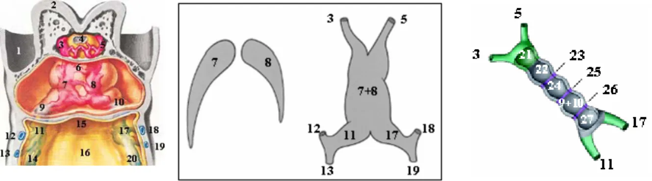
Figure 1 - At left, ventral dissection at the cardiogenic region of a human embryo with approximately 22 days of development (left). Adapted from Netter, FH: The Heart - The Ciba Collection of Medical Illustrations vol 5, 5Ed. The Case-Hoyt Corp, New York, page 116, 1981. Schematic representation of the process of heart tube formation originated by the fusion of the endocardial tubes (middle). Adapted from Gittenberger-de Groot, AC et
al, Pediatric Research, 57(2), page 170, 2005. Schematic representation of segments and rings present in the
embryonic linear heart tube (right). Adapted from Moorman, AF and Christoffels, VM, Physiological Reviews, 83, page 1225, 2003.
Legend: 1-Amnion; 2-Forebrain; 3-Right aortic arch; 4-Buccopharyngeal membrane; 5-Left aortic arch; 6-Communication between right and left endocardial tubes; 7-Right endocardial tube; 8-Left endocardial tube; 9-Primitive right atrium; 10-9-Primitive left atrium; 11-Right sinus horns; 12 and 13-Right vitelline veins; 14-Right umbilical veins 15-Foregut; 18-Yolk sac; 17-Leftt sinus horns; 18 and 19-Left vitelline veins; 20-Left umbilical veins; 21-Aortic sac; 22-Primitive right ventricle; 23-Primary ring; 24-Primitive left ventricle; 25-Atrioventricular ring; 26-Sinoatrial ring; 27-Sinus venosus.
The newly formed heart tube expresses genes that already show a cranial (ventricular) and caudal (atrial) specification, but only during the next stage of cardiac looping, phenotypic differences in myocardium may be observed [1].
It can be divided, from an cranial to caudal direction, into several regions, the left and right dorsal aortas, the aortic sac, the bulbus cordis (truncus arteriosus, conus cordis, the ventriculoarterial ring and the primitive right ventricle), the interventricular primary ring, the primitive left ventricle, the atrioventricular ring, the primitive atria, the sinoatrial ring and the left and right sinus venosus (Fig. 1, right).
The steps that occur after the initial segmentation of the embryonic heart represented in Fig. 1 (right) are interpreted according to two different models, namely the “segmental model” and the “balooning model”. The former defends the concept that the adult atria and ventricles derives, rigorously, from expansion and differentiation of the respective heart tube segment, each of them functioning as somites, morphogenetic units separated by boundary interfaces. The latter, considers that the heart’ s definitive chambers arise from local expansion and differentiation of certain heart tube regions, rather than from an exact tight segmental development [46]. In my opinion, the differences between these two models are more a matter of interpretation of the “segmental model” in a non-classic way. Thus, if each segment of the linear heart tube is not considered to have restricted frontiers and that a single segment may become part of more than one cardiac cavity as well as that the development of a certain chamber may occur by dorsal or ventral expansion of that segment , “the ballooning model”, becomes a flexible interpretation of the “segmental model”. Both models are consistent with studies using whole embryo techniques, which indicate that all of the components and myocardium of the developing heart appear from primary cardiogenic fields [47]. However, later experiments have suggested that the atrioventricular canal, atria and conotruncus are added secondarily to the straight heart tube during looping, concomitantly with addition of new myocardium [48, 49]. The atria are added progressively from the caudal primary heart fields bilaterally, while the myocardium of the conotruncus is elongated from a “secondary heart field” or “anterior heart field” [50-53], located beneath the floor of the foregut, in the prepharyngeal mesoderm.
Cardiac looping
Because the two ends of the heart tube are “fixed”, it is forced to bend in order to adapt itself to the available pericardial space. Meanwhile, perforations appear in the dorsal mesoderm, leading to its disappearance as the openings increase in size, allowing the heart tube to lie free within the pericardial cavity. The cranial portion of the tube bends ventrally and to the right, and the atrial portion shifts dorsally and to the left, regulated by genes that are essential for left-right programming [54]. This is named cardiac looping and is complete by the day 28 (Fig. 2 and 3).
Figure 2 - Schematic representation of the cardiac looping event. Adapted from Sadler, TW: Langman´s Medical Embryology, 8Ed. Lippincott Williams and Wilkins, USA, pages 214-15, 2000.
While the cardiac loop is forming, changes in the heart tube are concerned mainly with the development and expansion of the future heart chambers (Fig. 3). The atrial component, initially a paired structure outside the pericardial cavity, forms a common atrium and is incorporated into the pericardial cavity. The atrioventricular junction remains narrow and forms the atrioventricular canal, which connects the common atrium and the early embryonic left ventricle. The bulbus cordis is narrow except for its proximal third that will form the trabeculated part of the right ventricle. The conus cordis will form the outflow tracts of both ventricles. The distal part of the bulbus, the truncus arteriosus, will form the roots and the proximal portion of the aorta and pulmonary artery while the bulboventricular sulcus remains narrow and is called the primary interventricular foramen. At the venous end of the heart, the expanding common atrium forces the convergence of the paired sinus venosus. At the end of loop formation, the heart tube begins to form the primitive trabeculae in two sharply defined areas along the ventral border of the heart tube in the primitive right and left ventricles, which will invade the cardiac jelly and later the myocardium (Fig. 3 and 4).
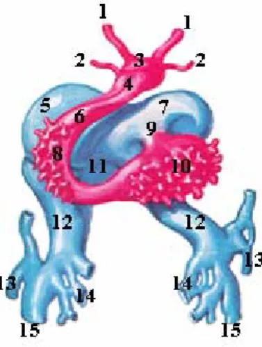
Figure 3 - Schematic drawing of the endocardial tube with myocardium removed. Adapted from Netter, FH: The Heart - The Ciba Collection of Medical Illustrations vol 5, 5Ed. The Case-Hoyt
Corp, New York, page 118, 1981;
Legend: 1-Aortic arches I; 2-Aortic arches II; 3- Aortic sac; 4-Truncus arteriosus; 5-Right atrium; 6-Conus cordis; 7-Left atrium; 8-primitive right ventricle; 9-Atrioventricular canal; 10-Primitive left ventricle; 11-Sinus venosus; 12-Right and left sinus horns; 13-Right and left posterior cardinal veins; 14-Right and left vitelline veins; 15-Right and left umbilical veins.
Figure 4 - Sagittal dissection of a human embryo with approximately 25 days of development. Adapted from Netter, FH: The Heart - The Ciba Collection of Medical Illustrations vol 5, 5Ed. The Case-Hoyt Corp, New York, page 118, 1981;
Legend: 1-Forebrain; 2-Buccopharyngeal membrane; 3-Aortic arch I; 4-Aortic arch II; 5-Aortic sac;
6-Truncus arteriosus; 7-Conus cordis; 8-Primitive right ventricle;
9-Primitive left atrium; 10-Primitive left ventricle; 11-Pericardial cavity; 12-Myocardium; 13-Cardiac jelly; 14-Proepicardial tissue; 15-Sinus venosus; 16-Septum transversum; 17-Hepatic diverticulum; 18-Left anterior cardinal vein; 19-Left common cardinal vein; 20-Left vitelline vein;
21-Left umbilical vein; 22-Left posterior cardinal vein; 23-Yolk sac;
24-Dorsal aorta; 25-Hindgut;
26-Right umbilical vein; 27-Allantois;
28-Umbilical arteries; 29-Left umbilical vein; 30-Cloacal membrane.
The distal portion of the bulbus cordis, initially on the right side of the pericardial cavity, is now located in a more central position, as a result of two dilatations of the atrium, bulging on each side of the bulbus (Fig. 3). At this time, although the heart still consists basically of a single tube, its external appearance already suggests its future four-chambered condition, and now it completely occupies the pericardial cavity.
The human embryo is now about 3.2 mm long, with 20 somites and approximately 25 days old.
Epicardium and coronary vasculogenesis
Around the 24th day of development one can distinguish three cell types in the heart, cardiomyocytes, mesenchymal cells and endocardial cells. Approximately, by this time, a new layer of epithelial cells starts to populate the myocardial surface of the heart which soon will be called the epicardium. This new layer of cells derives from specific proepicardial villi in the proepicardial serosa, as well as from the deeper portions of the adjacent pericardial tissue. These proliferative cells (as shown by simple bromodeoxyuridine experiments) are localized just behind the limit between the liver and the sinus venosus, in a region called “Proepicardial tissue” [55, 56] (Fig. 4). This phase begins when the cardiac tube is already formed and looped. The epicardium-forming cells give rise to the cellular elements of the subepicardial and intermyocardial connective tissues, of the coronary vasculature and blood. These cellular elements include fibroblasts, mesenchymal cells, smooth muscle cells, pericytes, angioblasts, endocardial cells and even a small number of cardiomyocytes [45, 55, 56].
Proepicardial-derived angioblasts differentiate into endothelial cells and assemble into a primitive capillary network that is the basis of the coronary vasculogenesis, which is not yet connected to the systemic circulation. This primitive network subsequently expands in an epi-to-endocardial direction and towards the base of the heart from pre-existing capillaries, the coronary angiogenesis, probably connecting to the developing aorta. Only after perfusion, this plexus become remodelled into larger vessels and respective branches which now become muscularized with proepicardial-derived smooth muscle cells, starting to assume the specific properties of coronary arteries or veins [45].
It is also hypothesied that multipotent proepicardial and/or neural crest-derived cells have an essential role in the induction and differentiation of cardiomyogenic progenitors into cells of the cardiac conduction system [57]. The mechanism by which the proepicardial cells starts invading the myocardium is via the release of groups of cells from the proepicardial
protrusions into the pericardial cavity and subsequent attachment to the myocardial surface [56]. Aditionally, it is important to refer the distal portion of the outflow tract, the truncus arteriosus, a region in which the epicardium does not derive from the proepicardial serosa. Instead, the epicardium is probably derived from the pericardial mesothelium at the junction between the outflow tract and the dorsal wall of the pericardial cavity [45].
Sinus Venosus
Approximatelly around the 25th day of development, the sinus venosus receives three pairs of veins, namelly the vitelline, the umbilical and the common cardinal veins. The first enters the floor of the sinus, medially, the second enters the sinus horns coming from below, and the third also enters the horns but coming from above. At first, communication between the sinus and the atrium is wide (Fig. 3). Later, however, the entrance of the sinus (the sinoatrial opening) shifts to the right, due to the shunting of blood to the right, until the sinus venosus communicates with only the right atrium. The development of anastomotic channels between the right and left systemic veins and the preferential blood flow to the right side, makes the right sinus horn and its proximal cardinal and vitelline veins gain in importance, whereas their left counterparts become greatly reduced in size. At the same time, the right sinus horn attains a more vertical position and becomes incorporated into the right atrium to form the smooth-walled part of it [58]. With obliteration of the left umbilical and vitelline veins and later the left common cardinal vein, all that remains of the left sinus horn is the oblique vein of the left atrium and the coronary sinus. The sinoatrial opening is now flanked in each side by the right and left venous valves. Superiorly, the valves join each other to form a single fold, the septum spurium. As the right sinus horn incorporates into the wall of the atrium, the left valve and septum spurium fuse with the developing atrial septum. The superior portion of the right venous valve disappears entirely and the inferior portion develops into the valve of the inferior vena cava and the valve of the coronary sinus [58, 59].
Cardiac septation and valve formation
During the next period (Fig. 5), that takes approximately 10 days, between the 27th and 37th day of development, no major changes in the external appearance of the heart occurs, however, its relative position keeps changing because of the changing curvature of the embryo and the growth of neighbouring organs.