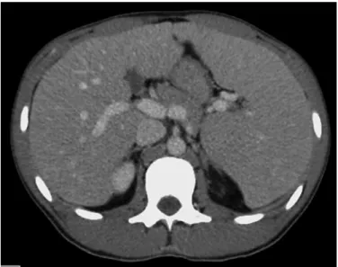
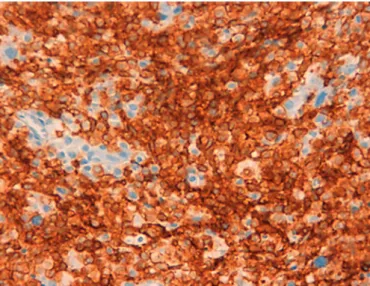
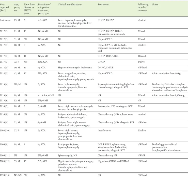
Hepatosplenic T-Cell Lymphoma: A Rare Complication of Monotherapy with Thiopurines in Crohn’s Disease
Texto
Imagem
Documentos relacionados
But composition, that is to say, what a particular body contains, and the internal structure of a molecule or a material, can change depending on the solvent and the whole
We report a case of a 26-year-old male patient with a rapid and fatal reactivation of a known chronic case of Chagas’ disease, manifested by meningoencephalitis, which lead to
Como referido no ponto anterior, é necessário previamente a verificação da RM. De modo a proceder à dispensa propriamente dita é importante perceber se se trata de medicação
benjamina seria a próxima escolha para isolamento de substâncias ativas, pois: apresentou considerável atividade biológica CIM de 312,50 µg/mL, sendo esta a fração mais ativa
Conclusion: The electrophysiological measures obtained from Frequency-following Response using the speech stimulus / da/ in normal-hearing adults without hearing complaints
Thus, the objective of this study was to analyze the measures of the fundamental frequency (f 0 ), the measure of the contact quotient (CQ), the electroglotographic jitter and
RESUMO - O presente estudo teve como objetivo analisar os dados sobre suicídio no município de Mineiros – GO, popularmente conhecido como de alto índice para o suicídio,
