Ana Margarida Carvalho Guerra
CARBAPENEMASE-PRODUCING
ENTEROBACTERIACEAE: FROM COLONIZATION TO
INFECTION
Dissertação do 2º Ciclo de Estudos Conducente ao Grau de Mestre em Análises Clínicas
Trabalho realizado sob a orientação da Professora Doutora Luísa Peixe (Faculdade de Farmácia, Universidade do Porto) e coorientação da Doutora Ângela Novais (Faculdade de Farmácia, Universidade do Porto).
É AUTORIZADA A REPRODUÇÃO INTEGRAL DESTA DISSERTAÇÃO APENAS PARA EFEITOS DE INVESTIGAÇÃO, MEDIANTE DECLARAÇÃO ESCRITA DO INTERESSADO, QUE A TAL SE COMPROMETE.
Ao meu avô, Por nos ter ensinado a lutar.
“Remember to look up at the stars and not down at your feet Try to make sense of what you see and Wonder about what makes the universe exist Be curious”
AGRADECIMENTOS (ACKNOWLEDGMENTS)
A escrita desta dissertação revelou-se um verdadeiro desafio, tendo sido o contributo de diversas pessoas essencial para a sua realização. Aqui expresso os meus sinceros agradecimentos:
À Professora Doutora Luísa Peixe, minha orientadora, por ter incentivado este trabalho e me ter permitido integrar a sua equipa de investigação, disponibilizando todos os materiais e equipamentos necessários à realização deste estudo.
À Doutora Ângela Novais, pela maravilhosa coorientação, por ter partilhado os seus conhecimentos e experiência comigo e por toda a disponibilidade em ensinar-me. Muito obrigada pela confiança depositada em mim, pelo apoio incansável e por todas as críticas e sugestões dadas ao longo destes meses.
Ao Doutor Agostinho Lira, pelo interesse neste trabalho e erpela cedência dos isolados de Enterobacteriaceae multirresistentes.
A toda a equipa de investigação da Professora Doutora Luísa Peixe, pela enorme hospitalidade, especialmente às minhas companheiras de laboratório: Andreia Rebelo, Joana Mourão, Joana Rocha, Sofia Gomes, Svetlana Perović, pela disponibilidade em ajudar-me no trabalho laboratorial. Um agradecimento especial à Magdalena Księżarek (como prometido), por ter sido mais do que minha vizinha de bancada, por todas as dicas e conselhos, pelo companheirismo, pelos “stay late” e por ter tentado sempre ajudar-me em tudo o que eu precisasse.
Aos meus pais e à restante família, pelo apoio demonstrado durante todo o percurso.
Ao Frederico, pela paciência infindável com que me aturou e por todos os fins-de-semana fechados em casa. Obrigada por estares sempre comigo.
“If I have seen further it is by standing on the shoulders of giants.”
ABSTRACT
The emergence and spread of resistance to carbapenems, namely due to the production of carbapenemases, leaves few therapeutic options and poses a major threat to public health. In 2015, an outbreak of KPC-producing K. pneumoniae (ST147-K64) was reported in a Portuguese hospital. Despite the measures implemented, the incidence of carbapenemase-producing Enterobacteriaceae (CPE) in this hospital has been increasing in recent years.
The aim of the present study was to understand the dynamics of CPE infection and colonization among hospitalized patients in this hospital where infection control measures and active screening have been implemented since CPE emergence, in order to improve preventive infection control decisions.
One hundred and twenty one confirmed CPE isolates were analysed. They included (i) 87 isolates from rectal swabs (February 2018) and (ii) 34 clinical isolates (November 2017 - February 2018). Relationship among isolates was established by phenotypic (Fourier Transform Infrared Spectroscopy) and genotypic (wzi sequencing) capsular (K) typing methods and the type of carbapenemase were identified by PCR and sequencing. For E. coli isolates the phylogenetic group and ST131 were identified by PCR.
K. pneumoniae (Kp) was the most common species isolated from both colonization and infection samples, that in most cases produced KPC(-3) (n= 92%); E. coli (including ST131), E. cloacae complex, K. oxytoca, and C. freundii complex were also found mainly in colonized patients, as well as other carbapenemases types (e.g. OXA-48, VIM). Internal medicine and women surgery were the wards with a higher number of isolates, with differences in CPE population. Most patients (79%) became colonized after their admission at this hospital, before or after infection. Patients under decolonization by faecal microbiota transplantation became recolonized. Diverse K-types were observed throughout time, though most (51%) KPC-Kp were linked to K-type K64 (ST147), and isolates with K-type KL27 (19%) were also frequent.
These data highlight a more diverse CPE population dispersed throughout multiple patients and units, and suggest a nosocomial acquisition in most cases. Furthermore, it underlines that established infection control measures should be reanalysed and monitored in order to reduce CPE spread among patients and prevent new clones from disseminating.
KEYWORDS: Carbapenems; Carbapenemase-producing-Enterobacteriaceae; infection control measures.
RESUMO
A emergência e disseminação de resistência aos carbapenemos, principalmente através da produção de carbapenemases, leva a uma escassez de opções terapêuticas e constitui uma ameaça à segurança pública. Em 2015, um surto de K. pneumoniae (ST147-K64) foi reportado num hospital português. Apesar das medidas de controlo de infeção implementadas, a incidência de Enterobacteriaceae produtoras de carbapenemases (EPC) neste hospital tem vindo a aumentar nos últimos anos.
O objetivo do presente estudo foi compreender as dinâmicas de colonização e infeção por EPC em doentes internados neste hospital, onde as medidas de controlo de infeção e rastreio ativo foram implementadas desde a sua emergência, de modo a melhorar as decisões de controlo de infeção.
Foram analisados cento e vinte e um isolados de Enterobacteriaceae produtores de carbapenemase. Estes incluíam (i) 87 isolados obtidos a partir de exsudados rectais (fevereiro 2018) e (ii) 34 isolados de amostras clínicas (novembro 2017 - fevereiro 2018). A relação entre isolados foi estabelecida por métodos de tipagem capsular (K) fenotípicos (espectroscopia de Infravermelhos com Transformada de Fourier) e genotípicos (sequenciação de wzi
);
o tipo de carbapenemases foi identificado por PCR e sequenciação. Nos isolados de E. coli, identificou-se o grupo filogenético e ST131 por PCR. K. pneumoniae (Kp) foi a espécie mais frequentemente isolada das amostras de colonização e de infeção, sendo na maioria dos casos produtora de KPC(-3) (n= 92%); E. coli,(incluindo ST131), E. cloacae complex, K. oxytoca e C. freundii complex foram também encontrados, principalmente em doentes colonizados, assim como outros tipos de carbapenemase (p. ex., OXA-48, VIM). Medicina interna e cirurgia das mulheres foram as áreas com maior número de isolados, apresentando diferenças na população de EPC. A maior parte dos doentes (79%) ficou colonizado após a admissão neste hospital, antes ou depois da ocorrência de infeção. Nos doentes em descolonização por transplante fecal observou-se uma recolonização. Vários K-tipos foram observados ao longo do tempo, apesar da maior parte (51%) de KPC-Kp estar associada ao K-tipo K64 (ST147) e isolados com K-tipo KL27 (19%) serem também frequentes.Estes dados evidenciam uma população de EPC mais diversa por todos os doentes e unidades, e sugerem uma aquisição nosocomial. Realçam que as medidas de controlo de infeção estabelecidas devem ser reanalisadas e monitorizadas, de forma a reduzir a disseminação de EPC entre os doentes e evitar a disseminação de novos clones. PALAVRAS-CHAVE: Carbapenemos; Enterobacteriaceae produtoras de carbapenemase;
TABLE OF CONTENTS
CHAPTER 1 - INTRODUCTION ... 1
1.1. ANTIBIOTIC RESISTANCE CRISIS ... 2
1.1.1. CAUSES OF ANTIBIOTIC RESISTANCE CRISIS ... 4
1.2. ANTIBIOTIC RESISTANCE THREATS ... 5
1.3. MECHANISMS OF RESISTANCE TO CARBAPENEMS ... 7
1.3.1. PRODUCTION OF β -LACTAMASES ... 7
1.3.2. PORIN PERMEABILITY CHANGES ... 11
1.3.3. EFFLUX ... 11
1.4. EPIDEMIOLOGY OF CARBAPENEMASES ... 12
1.4.1. CLASS A CARBAPENEMASES ... 12
1.4.2. CLASS B β-LACTAMASES ... 14
1.4.3. CLASS D β -LACTAMASES ... 16
1.5. CPE IN HEALTHCARE SETTINGS ... 18
1.5.1. RISK FACTORS FOR CPE INFECTIONS ... 18
1.5.2. PREVENTIVE MEASURES ... 19
1.5.3. TREATMENT OF CPE INFECTIONS ... 20
1.6. DETECTION OF CARBAPENEMASES ... 22
CHAPTER 2 - OBJECTIVES OF THE STUDY ... 26
2.1. STATEMENT OF OBJECTIVES ... 27
CHAPTER 3 - MATERIALS AND METHODS ... 28
3.1. COLLECTION OF SAMPLES AND SPECIES IDENTIFICATION ... 29
3.2. DETECTION AND IDENTIFICATION OF CARBAPENEMASES ... 31
3.3. POPULATION STRUCTURE OF CARBAPENEMASE-PRODUCING K. PNEUMONIAE
ISOLATES ... 32
3.4. POPULATION STRUCTURE OF CARBAPENEMASE PRODUCING E. COLI ISOLATES .. 32
3.5. MOLECULAR TECHNIQUES ... 33
3.5.1. DNA EXTRACTION ... 33
3.5.2. DNA AMPLIFICATION ... 33
3.5.3. DNA PURIFICATION ... 34
3.5.4. DNA SEQUENCING ... 35
CHAPTER 4 - RESULTS AND DISCUSSION ... 37
4.1. ETIOLOGY OF BACTERIAL ISOLATES ... 38
4.2. DIVERSITY OF CPE IN THE DIFFERENT UNITS OF THE HOSPITAL ... 40
4.3. RISK FACTORS FOR CPE COLONIZATION ... 45
4.5. MOLECULAR EPIDEMIOLOGY OF K. PNEUMONIAE ISOLATES ... 49
4.6. CHARACTERIZATION OF NON-K PNEUMONIAE ISOLATES ... 53
4.7. RELATIONSHIP BETWEEN SCREENINGS AND INFECTION ISOLATES ... 54
CHAPTER 5 - CONCLUSIONS ... 56
LIST OF FIGURES
Figure 1- Timeline of antibiotic resistance showing that the introduction of each antibiotic
was quickly followed by an adaptation of bacteria. ... 3
Figure 2 - Relationship between MDR, XDR and PDR to each other. ... 4
Figure 3 - Number of Antibacterial Drug Application (NDA) Approval per Years Intervals . 5 Figure 4 - Primary mechanisms of β-lactam resistance in Enterobacteriaceae. ... 7
Figure 5 - Mechanism of action of beta-lactams. ... 8
Figure 6 - Number of lactamase enzymes identified since the introduction of the first β-lactam antibiotics. ... 8
Figure 7 - Epidemiology of KPC ... 13
Figure 8 - The blaKPC-2 containing Tn4401 transposon. ... 14
Figure 9 - Geographic distribution of VIM-producing Enterobacteriaceae in Europe ... 15
Figure 10 - Geographic distribution of NDM-producing Enterobacteriaceae in Europe. ... 16
Figure 11 - Epidemiology of OXA-48-like producing K.pneumoniae ... 17
Figure 12 - Modified Hodge test ... 22
Figure 13 – Overview of carbapenem inactivation method. ... 23
Figure 14 - Schematic for bacterial identification by MALDI-TOF MS. ... 24
Figure 15 - Screening protocol adopted by the hospital. ... 29
Figure 16 - Isolates on chromogenic medium. ... 30
Figure 17 - An example of a Blue-Carba test. ... 31
Figure 18 - PCR profiles of E. coli strains. ... 33
Figure 19 - An overview of the DNA purification method by nzytech. ... 35
Figure 20 - A schematic view of the experimental work. ... 36
Figure 21 - Timeline describing the evolution of the patient with colonized and infected samples. ... 39
Figure 22 – Distribution of colonized and infected patients among hospital units. ... 40
Figure 23 - Evolution of patients during decolonization treatment. ... 41
Figure 24 - Distribution of colonization isolates among units of medical and surgical departments ... 42
Figure 25 - Distribution of infection isolates among units of medical and surgical departments ... 43
Figure 26 – Distribution of faecal samples by species among hospital units. ... 44
Figure 27 - Distribution of infection samples by species among hospital units. ... 45
Figure 28 - Distribution of K. pneumoniae K-types during the time period of the study. ... 52
LIST OF TABLES
Table 1- Classification schemes for β-lactamases…. ... 10
Table 2- Main carbapenemases in Enterobacteriaceae ... 11
Table 3 - Gender distribution of patients with CPE. ... 38
Table 4 - Distribution of CPE isolates among clinical and faecal specimens ... 39
Table 5 - CPE isolates identified from the emergency room ... 42
Table 6 - Antibiotic exposure of de novo colonized patients ... 46
Table 7- Antibiotic resistance rates for infection isolates ... 47
Table 8 - Distribution of carbapenemase enzymes among Enterobacteriaceae species. . 49 Table 9 - Capsular type of K. pneumoniae strains. ... 51
Table 10 - Epidemiological characterization of CPE non- K. pneumoniae isolates ... 53
LIST OF ABBREVIATIONS
AbR – Antibiotic Resistant
CDC – Centers for Disease Control and Prevention CPE – Carbapenemase Producing Enterobacteriaceae CRE – Carbapenemase Resistant Enterobacteriaceae ESBL – Extended Spectrum β-lactamase
ESCMID - European Society of Clinical Microbiology and Infectious Diseases EUCAST – European Committee on Antimicrobial Susceptibility Testing FTIR – Fourier Transform Infrared
IMP – Imipenemase β-lactamase
KPC – Klebsiella pneumoniae carbapenemase K-Type – Capsule Type
MDR – Multidrug resistant MGE – Mobile genetic element
NDM – New Delhi metallo-β-lactamase OMP – Outer membrane protein OXA – Oxacillinase
PBP - Penicillin Binding Protein PCR – Polymerase Chain Reaction ST – Sequence Type
VIM – Verona integron-borne metallo-β-lactamase WHO – World Health Organization
1.1. ANTIBIOTIC RESISTANCE CRISIS
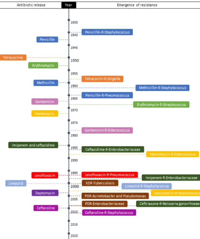
Infectious diseases were the leading cause of death in the world at the beginning of the twentieth century, until the discovery of penicillin by Sir Alexander Fleming in 1928 (2-5). Since then, antibiotics have saved millions of lives and enabled important medical procedures. The “golden era” of antibiotic discovery and clinical development took place in the period between 1940 and 1960, which led to confidence that infectious diseases can be controlled and prevented (2, 5). However, some pathogens soon became resistant to many of those drugs (figure 1) and now antibiotic resistance has been seen to nearly all antibiotics developed (3, 5).
Antibiotic resistance is now a worldwide problem. A study from the Centers of Disease Control and Prevention (CDC, Atlanta) estimated that at least 23000 people died each year in the USA as a direct result of infections caused by antibiotic resistant (AbR) bacteria(6).On this trajectory, as many as 10 million people a year could die from AbR bacteria worldwide by 2050, at a cost of $100 trillion (7). Infectious diseases are the second cause of death in the world, and the third in developed nations (4, 5). In addition to increasing morbidity and mortality, these infections are also a major economic burden to the healthcare systems, as they require significantly longer hospital stays and additional doctor visits (6). As such, in 2013, the CDC described this antimicrobial resistance as one of our most serious health threats and, in 2014, the World Health Organization (WHO) alerted for the real possibility of a post-antibiotic era, in which a common infection could kill (3, 4).
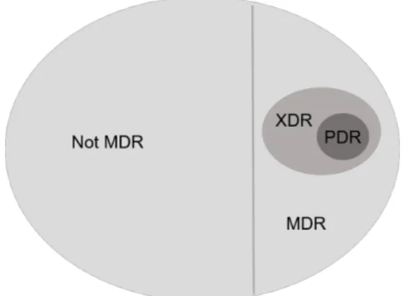
The problem of antimicrobial resistance led to the need to create harmonized definitions with which to classify bacteria that are resistant to numerous antimicrobial agents: multidrug resistant (MDR), extreme drug resistant (XDR) or pandrug resistant (PDR) (figure 2). The MDR category applies with non-susceptibility to at least one agent in three or more antimicrobial categories. XDR implies non-susceptibility to at least one agent in all but two or fewer antimicrobial categories. A PDR strain exhibits non-susceptibility to all agents in all antimicrobial categories (8).
Infections caused by MDR bacteria commonly occur in hospitalized patients, due to the extensive use of antibiotics and the need of invasive procedures(4).However, some MDR bacteria have become quite prevalent in community-acquired infections as well (9). For example, methicillin-resistant Staphylococcus aureus (MRSA) was initially limited to hospital-acquired infections until 1980, when it spread throughout the community (10).
Figure 1- Timeline of antibiotic resistance showing that the introduction of each antibiotic was quickly followed
Antibiotic resistance among Gram-negative bacteria is particularly worrisome because they are becoming resistant to nearly all drug options available (4).The greatest concern is caused by Klebsiella pneumoniae, Acinetobacter baumannii, Pseudomonas aeruginosa, Enterobacter cloacae complex and Escherichia coli pathogenic and MDR strains. These bacteria can cause several infections, ranging from urinary tract to bloodstream infections or severe pneumonias. Currently, K. pneumoniae is one of the most frequently isolated organisms in intensive care unit infections (6).
Figure 2 - Relationship between MDR, XDR and PDR to each other. Adapted from (8).
In face of the rapid emergence of antimicrobial resistant human pathogens there is a global need for new antibiotics. Nevertheless, after the successful discovery of antibiotics in the past, the pipeline is now running dry, as there has been a paucity of new compounds being developed or introduced (3, 7).
1.1.1. CAUSES OF ANTIBIOTIC RESISTANCE CRISIS
The factors that contribute to the emergence and dissemination of AbR bacteria are the following: (i) overuse of antibiotics for human or veterinary infections; (ii) extensive agricultural use; (iii) inefficient infection control practices; (iv) a scarcity of new antimicrobial drugs developed in the last years; and (v) worldwide travel and trade of food and goods (6).
The overuse of antibiotics is the major factor leading to antibiotic resistance around the world and has been reported a direct relationship between antibiotic consumption and the emergence of AbR bacteria(4, 6). Antibiotics are overprescribed by doctors, being among the most commonly prescribed drugs in human medicine (4). However, up to 50% of all the antibiotics prescribed are not needed and in several cases the duration of therapy or the choice of agent is incorrect. Besides that, in many countries antibiotics are underregulated and available to buy over the counter without a prescription, which promotes
increasing the risk of emergence or selection of antimicrobial resistance and further transmission through the food chain (4).
The prevalence of AbR human pathogens is rapidly increasing, but the discovery and development of new antimicrobial drugs have slowed dramatically (figure 3), withmost of pharmaceutical companies abandoning the antibiotic field (4, 7, 10). At this moment, few antibacterial drugs with activity against Gram-negative bacteria are being developed, due to a combination of discovery, clinical, regulatory and economic obstacles (11).
The development of antimicrobial drugs is no longer considered an economically wise investment, as they are short-course therapies and therefore not as profitable as medicines for chronic conditions (7). Furthermore, changes in requirements for clinical trial design have made the approval of new antibiotics more challenging and with higher costs (7, 11).
Figure 3 - Number of Antibacterial Drug Application (NDA) Approval per Years Intervals. Adapted from (5).
1.2. ANTIBIOTIC RESISTANCE THREATS
In 2013, the CDC has prioritized drug-resistant bacteria based on the level of concern into three categories: urgent, serious and concerning. Pathogens categorized as urgent threats to public health are Clostridium difficile, Drug-resistant Neisseria gonorrhoeae and Carbapenem-resistant Enterobacteriaceae (CRE) (2, 4).
Enterobacteriaceae, such as K. pneumoniae, are a family of Gram-negative bacteria responsible for a variety of community and healthcare-associated infections (12). Carbapenems are b-lactam antibiotics typically considered agents of last resort, used for the treatment of severe infections caused by MDR-Enterobacteriaceae. However, over the past ten years CRE have been increasingly reported worldwide. Infections caused by CRE are associated with poor clinical outcomes and high mortality, leading to failure in first line
therapy and to very limited therapeutic options (13). CDC estimated that more than 9000 healthcare-associated infections are caused by CRE each year in the United States, resulting in 600 deaths per year in this country (6).
CRE were initially defined as Enterobacteriaceae that are nonsusceptible to a carbapenem (14). However, resistance to carbapenems in Enterobacteriaceae may result either from expression of carbapenemases or by combined effects of AmpC-type or extended-spectrum β-lactamases (ESBLs) and decreased outer membrane permeability (13). Because of that, in 2015, the CDC revised this definition of CRE to any Enterobacteriaceae which are resistant to any carbapenem (i.e., minimum inhibitory concentrations of > 2 mg/L for doripenem, > 8 mg/L meropenem or imipenem OR > 1 mg/L for ertapenem) or are documented to produce a carbapenemase. In addition, for those Enterobacteriaceae which may have intrinsic reduced susceptibility to imipenem, such as Proteus mirabilis, resistance to a non-imipenem carbapenem is required (14, 15).
Carbapenem-resistant Enterobacteriaceae strains that do not produce carbapenemases (non-CP CRE) are considered to be much less important from a public health perspective than carbapenemase producers (CP-CRE), though occasional outbreaks have been reported (16). The spread of carbapenemase producers is one of the most worrying issues among antibiotic resistance in Gram-negatives, from either a clinical and epidemiological perspective (17).
1.3. MECHANISMS OF RESISTANCE TO CARBAPENEMS
The β-lactam group of antibiotics includes penicillins, cephalosporins, carbapenems, and monobactams (18). They act to inhibit bacterial cell wall synthesis by the following steps: binding of the drug to the penicillin-binding proteins (PBP) located in the bacterial cytoplasmic membrane; inhibition of the transpeptidation reaction that crosslinks the linear peptidoglycan chain constituents of the cell wall; and activation of autolytic enzymes that cause lesions in the bacterial cell wall (19).
Primary mechanisms of carbapenem resistance in Enterobacteriaceae include: (1) enzymatic inactivation of the antibiotic by chromosome or plasmid-encoded enzymes with hydrolytic activity against β-lactam molecules; (2) decreased outer membrane permeability through production of modified porins, loss of porin expression, or a shift in the types of porins found in the outer membrane; and (3) efflux of the antibiotic to the outside of the bacterium through production of an efflux pump (figure 4) (13).
Figure 4 - Primary mechanisms of β-lactam resistance in Enterobacteriaceae. Adapted from (13)
1.3.1. PRODUCTION OF β -LACTAMASES
The most common mechanism of carbapenem resistance in Enterobacteriaceae is the production of β-lactamases (18). These enzymes hydrolyse the β-lactam ring in the periplasmic space, therefore preventing PBP inhibition (figure 5) (20).
Figure 5 - Mechanism of action of beta-lactams. Adapted from (19).
Genes encoding for β-lactamases, bla genes, can be found in the chromosome or in mobile genetic elements, which contributes to their wide dissemination (21).
The destruction of antibiotics by the action of β-lactamases has been a major cause of antibiotic resistance since the discovery of a plasmid-encoded penicillinase, present in penicillin-resistant S. aureus in 1940.From then on, the development of new β-lactams compounds has been followed by the appearance of new enzymes (figure 6).
Figure 6 - Number of β-lactamase enzymes identified since the introduction of the first β-lactam antibiotics.
Adapted from (22). 0 200 400 600 800 1000 1960 1970 1980 1990 2000 2010 2020 Nu m be r o f u ni qu e β-la ct am as es Year
The penicillinases were followed by ESBLs that inactivate oxyimino-cephalosporins (23). In some instances, porin loss in ESBL-producing strains increases resistance to fourth-generation cephalosporins and carbapenems (24). As the prevalence of ESBL and plasmid-borne cephalosporinases increased, so did the consumption of carbapenems, which promoted the emergence and dissemination of carbapenemase-producing Enterobacteriaceae (CPE) through the diffusion of plasmid-borne carbapenemases (20). They comprise a variety of hydrolysing enzymes with the ability to inactivate a broad range of β-lactams, including penicillins, cephalosporins, monobactams, and carbapenems(23).
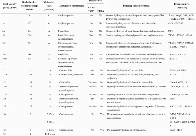
To date, thousands of β-lactamases have been identified (http://www.bldb.eu) and there are two main classification schemes (table 1). The most frequently used is the Ambler classification scheme, which separates β-lactamases into four groups (A, B, C and D) based on amino acid sequence identity. According to this classification, β-lactamases of classes A, C and D have a serine residue in the active site, whereas class B enzymes require zinc for their activity. The other classification is the one proposed by Bush-Jacoby, which relies on substrate specificity and their inactivation by classical lactamase inhibitors to divide β-lactamases into four groups, each with many subgroups (21, 25).
• Group 1 / Ambler Class C: Cephalosporinases that are encoded on the chromosomes of many Enterobacteriaceae and that are not inhibited by the classical β-lactamase inhibitors (clavulanic acid, tazobactam). When produced in large amounts they can provide resistance to carbapenems (25).
• Group 2 / Ambler Classes A and D: This is the largest group of lactamases and is constituted by several subgroups. It comprises β-lactamases with a hydrolytic activity over penicillins, extended-spectrum cephalosporins (ESBLs), monobactams, cloxacillin and carbapenems.
o The subgroup 2f contains the KPC enzymes from the molecular class A (25). Class A enzymes hydrolyse penicillins, cephalosporins, carbapenems and aztreonam. Their hydrolytic activity in vitro is inhibited by clavulanic acid and tazobactam (13). Other class A enzymes with carbapenemase activity are some GES type β-lactamases variants (GES-2, -4, -5, -6). Their hydrolysis spectrum includes penicillins, extended-spectrum cephalosporins and carbapenems. They remain susceptible to classic β-lactam inhibitors. o In the subgroups 2d, 2de and 2df are the OXAs enzymes,
hydrolyse extended-spectrum β-lactams or aztreonam but are active against oxacillin. They also confer high resistance to temocillin, which can be used for phenotypic identification of OXA-48 producers, since the other classes of carbapenemases are susceptible to this drug (26). These enzymes are not inhibited by either clavulanic acid or EDTA, but rather by NaCl (13).
• Group 3 / Ambler Class B: Includes the MBLs, a unique group of β-lactamases that are dependent on zinc ions for activity rather than the active-site serine found in the other classes. They hydrolyse carbapenems but have a poor affinity for monobactams. They are inhibited by chelating agents, such as EDTA, but not by classical β-lactamase inhibitors (25). Despite the lack of activity against monobactams, the presence of additional β-lactamases (e.g. ESBL) tends to compromise the efficacy of these agents (24).
• Group 4: Included enzymes that were incompletely characterized but the most recent review of this classification scheme has omitted this group. (25)
Among the classes of β-lactamases defined by the Ambler classification system, the ones that confer carbapenem resistance in Enterobacteriaceae belong to three of them: Class A, Class B and Class D (table 2) (27).
Table 2- Main carbapenemases in Enterobacteriaceae. Adapted from (13).
Ambler
class Enzyme
Plasmid/ Chromosome
Hydrolysis spectrum Inhibitor
Penicillins 1st generation cephalosporins 2nd generation cephalosporins 3nd generation cephalosporins Aztreonam Carbapenems A SME-1
to -3 Chromosome ++ ++ -- + + + Clavulanate, tazobactam, sulfabactam, NXL-104 NMC-A Chromosome ++ ++ -- + -- ++ IMI-2 Plasmid ++ ++ -- + -- ++ GES-4,-5,-6 Plasmid ++ ++ + + -- + KPC-2 to -12 Plasmid ++ ++ -- ++ + ++ Clavulanate, tazobactam, boronic acid, sulfabactam B IMP-1 to -33 Plasmid ++ ++ ++ ++ -- ++ EDTA VIM-1 to -33 Plasmid ++ ++ ++ ++ -- ++ NDM-1 to -6 Plasmid ++ ++ ++ ++ -- + KHM-1 Plasmid ++ ++ ++ ++ -- ++ D OXA-48 Plasmid ++ ++ +/- +/- -- + NaCl OXA-181 Plasmid ++ ++ +/- +/- -- +
1.3.2. PORIN PERMEABILITY CHANGES
Carbapenem resistance was first described in Enterobacter spp. isolates overexpressing a chromosomal ampC gene encoding an intrinsic cephalosporinase and exhibiting modifications in their outer membrane protein (OmpC or OmpF) porins. This mechanism of carbapenem resistance has also been observed in other species that do not express an intrinsic cephalosporinase, such as E. coli and K. pneumoniae. In those cases, resistance is due to decreased cell membrane permeability owing to modifications in OmpK35 and OmpK36 for K. pneumoniae and in OmpF and OmpC for E.coli, in most cases combined with expression of plasmid-encoded AmpC or ESBL (13).
1.3.3. EFFLUX
Overexpression of AcrA, a component of AcrAB-TolC effux system, was described as responsible for imipenem resistance in Enterobacter aerogenes (13, 24).
1.4. EPIDEMIOLOGY OF CARBAPENEMASES
The first carbapenemase identified in Enterobacteriaceae was the chromosomally encoded NmcA from an E. cloacae clinical isolate in 1993 (13, 28). Nevertheless, carbapenem resistance in Enterobacteriaceae had been a negligible phenomenon until a carbapenem resistant K. pneumoniae strain was identified in 1996, in the United States, harbouring a plasmid-encoded resistance gene (Klebsiella pneumoniae carbapenemase, KPC). Since that time, numerous CPE have been reported and widely disseminated (28).
1.4.1. CLASS A CARBAPENEMASES
Class A carbapenemases include the enzymes KPC, GES and IMI-types, as well as nonmetallocarbapenemase type A (NMC-A) and SME enzymes, which may be found in E. cloacae and Serratia marcescens, respectively. While some carbapenemases are chromosome-encoded (e.g. IMI-1, NMC-A, SME enzymes, SHV-38 and SFC-1), others are plasmid-encoded (e.g. KPC, GES and IMI-2). Among these, KPC enzymes are the most widely spread, whereas the others are more sporadically reported or are confined to certain bacterial species or geographic regions (29).
KPCs have been the most clinically significant class A carbapenemases since their first identification (KPC-2) in 1996 from a K. pneumoniae strain in a North Carolina hospital (USA) (12, 29). Since then, several KPC variants (KPC-2 to KPC-24) have been identified, being KPC-2 and KPC-3 the most widespread variants (30). Within a few years, KPC producers had spread across the United States and all over the world (28).
KPC production have been reported mostly from nosocomial K. pneumoniae isolates and to a much lesser extent from E.coli, Enterobacter spp. and from other enterobacterial species (28). Although community-acquired KPC-producing strains have been reported in the USA and Israel a few years ago their incidence remains low (13).
Figure 7 - Epidemiology of KPC (31). (1) USA; (2) Colombia; (3) Brazil; (4) Argentina; (5) Italy; (6) Greece; (7)
Poland; (8) Israel; (9) China; (10) Taiwan; (11) Canada; (12) Spain; (13) France; (14) Belgium; (15) Netherlands; (16) Germany; (17) UK; (18) Ireland.
The epidemiology of KPC-producing K. pneumoniae varies geographically (figure 7) but they are currently endemic in Israel, Greece, Latin America, Italy, China, Poland, Taiwan and the USA (12, 31). Outbreaks of KPC-producers have been reported in many European countries (KPC-2-producing K. pneumoniae in Greece (32, 33) and KPC-3-producing K. pneumoniae in Italy (34), for example), especially due to patients travelling to endemic areas (31). The rapid and worrisome spread across multiple continents and species has been associated with the dissemination of a specific K. pneumoniae clone (ST258) expressing the genes blaKPC-2 or blaKPC-3 (28, 31). It remains the predominant ST in the USA and other countries, whereas in certain EU countries other (and diverse) K. pneumoniae lineages have been linked to KPC emergence and/or spread (28, 35). On the contrary, ST11 is the predominant clone observed in Asia, and has also been reported in Latin America (31). In Portugal, there was only one report of KPC-2 that was identified in an environmental E. coli isolate in 2010, whereas KPC-3 was first detected in 2009 in a paediatric unit of a central hospital, from 2 strains of K. pneumoniae belonging to ST11 (30, 36). However, some hospital outbreaks revealed the emergence of the carbapenemase KPC-3 in Portugal (35), involving isolates from diverse clones (ST11, ST14, ST15 and ST147) (30).
The blaKPC genes are most frequently associated with a common genetic element (transposon Tn4401) (28). Tn4401 is a Tn3-like transposon (13) which has six isoforms
differing by deletions upstream of the blaKPC gene, leading to different expression levels of the blaKPC gene (figure 8) (30, 31).
Figure 8- The blaKPC-2 containing Tn4401 transposon. Adapted from (31)
The blaKPC has been reported on numerous plasmid types, but the most predominant belongs to the IncF group, and particularly FIIK replicon types in K. pneumoniae. This plasmid type often contains several additional genes responsible for resistance to other antibiotics (31).
1.4.2. CLASS B β-LACTAMASES
Class B carbapenemases were first detected in the 1960s. However, in the 1990s the spread of MGEs harbouring genes encoding metallo-β-lactamases was responsible for increasing reports of clinical infections and nosocomial outbreaks (37). The most important types from a clinical and epidemiological point of view are the IMP-, VIM- and NDM-type enzymes (28, 37). They are usually associated with integrons carrying additional resistance genes that encode other carbapenemases, AmpC cephalosporinases or ESBLs or proteins conferring resistance to other families of antibiotics (24).
The first acquired MBL, IMP-1 (Imipenemase), was detected in S. marcescens strain, in Japan in 1991 (37). The gene blaIMP-1 has since then become a widespread carbapenem resistant determinant with at least 52 variants described worldwide, found in Enterobacteriaceae, and also Pseudomonas spp. and Acinetobacter spp. (12, 21). Nevertheless, IMP-type MBL-containing Enterobacteriaceae are endemic in Japan and Taiwan, with occasionally sporadic outbreaks in other countries (12). Analysis of the blaIMP genetic environments most often revealed features of class 1 integrons, which also contained genes encoding resistance to other antimicrobials (aminoglycosides, class D β-lactamases and chloramphenicol) (12).
The second acquired MBL detected was VIM (Verona integron-encoded metallo-β-lactamase), isolated initially on P. aeruginosa in the late 1990s in Europe (12). VIM-1 was
Marseilles, France (13). Among the 46 variants described, VIM-2 is the most widely distributed MBL (12). The endemic spread of VIM-type enzymes has been reported in Greece (figure 9), where K. pneumoniae and E. coli harbouring blaVIM-1 are the predominant species (12, 28).
Figure 9 - Geographic distribution of VIM-producing Enterobacteriaceae in Europe. Adapted from (38).
The NDM is now one of the most clinically significant carbapenemase (31). NDM-1 was first identified in 2008, in a Swedish patient previously hospitalized in New Delhi, India (15). Since then, there has been a global dissemination of NDM MBLs (figure 10), often in patients with a history of tourism or healthcare travel in the Indian subcontinent (12, 29). NDM-type CPE have been identified on all continents, nevertheless India and Pakistan are the countries with the higher prevalence (27), where NDM producers have been found in the Indian tap and seepage water, suggesting their widespread in the environment (39).
NDM-producing bacteria are occurring both in healthcare settings and the community (12). Since its discovery, 15 NDM variants have been found and 3 more have been assigned accession numbers (40), most of them originated from Asia (31).
Figure 10 - Geographic distribution of NDM-producing Enterobacteriaceae in Europe. Adapted from (38)
The blaNDM-1 gene, unlike other carbapenemases genes, is associated with nonclonally related isolates and species (28). The presence of NDM-1 is usually accompanied by a MDR-phenotype, because plasmids carrying the blaNDM-1 gene are diverse and can harbour a high number of resistance genes encoding additional carbapenemases (21, 28). The blaNDM gene is often observed in the transposon Tn125, flanked by two ISAba125 elements (31, 41).
Of great concern, NDM-1 has been identified in E. coli ST131, recognized as a source of community-associated infections (15, 28). Thus, this may increase the risk of drug-resistant strains being released into the environment and further spread among humans (28).
1.4.3. CLASS D β -LACTAMASES
Only a few class D β-lactamases or oxacillinases (OXA) possess carbapenemase activity. In Enterobacteriaceae, OXA-48 is the most frequent class D carbapenemase, commonly reported from K. pneumoniae and E. coli isolates (31).
OXA-48 was first identified in 2003 in a K. pneumoniae strain, in Turkey, where it has become endemic. Since then, outbreaks have been reported from several other countries (figure 11), including Morocco, Libya, Egypt, Tunisia and India (12, 31).
OXA-48 producers are mostly identified in K. pneumoniae and E. coli, but also in various Enterobacteriaceae (28). A single IncL group plasmid (pOXA-48a) is the main source of the blaOXA-48 gene disseminated in a variety of enterobacterial species (28, 31).
A high related enzyme, OXA-181 (differing from OXA-48 in one aminoacid mutation) was first identified in India (28) and, since then, has been reported in many different countries (31).
The OXA-48-type producers are probably the most difficult carbapenemase producers to identify, because the isolates producing OXA-48 and OXA-181 usually present reduced susceptibility to carbapenems (13, 28). Therefore, the prevalence of these enzymes may be underestimated (12, 29).
Figure 11 - Epidemiology of OXA-48-like producing K.pneumoniae
The prevalence of CPE varies geographically, but currently the most common species of Enterobacteriaceae harbouring transmissible carbapenemase genes is K. pneumoniae (29). CPE is seen predominantly in healthcare associated infections, with the exception of the Indian subcontinent, where community-associated infections linked with NDM-producing bacteria are common
(42).
Furthermore, some studies worldwide have demonstrated the presence of carbapenemase-encoding genes in Enterobacteriaceae from livestock (43), companion animals (44, 45) and wildlife (46).1.5. CPE IN HEALTHCARE SETTINGS
1.5.1. RISK FACTORS FOR CPE INFECTIONS
In healthcare settings, intensive care units (ICUs) are the main reservoirs of Gram-negative multidrug resistant bacteria and K. pneumoniae is one of the most common pathogens found there (47).
The acquisition of CPE by patients can occur through two different modes: horizontal transmission (from another patient, through healthcare staff, from the proximal environment or through shared equipment) or de novo emergence of resistance, in which carbapenem susceptible isolates within a patient are genetically altered (48). Acquisition of CPE via exogenous exposure during hospitalization is the most common pathway. The introduction of CPE into the hospital environment occurs through patients who are colonized or infected, leading to healthcare worker or environmental contamination and subsequent spread to other patients (49). Recent European Society of Clinical Microbiology and Infectious Diseases (ESCMID) Guidelines have identified the contaminated hands of healthcare workers as the interpatient main route of CPE transmission (50). Subsequent dissemination of CPE can result from contamination of the healthcare environment, like medical equipment or hospital surfaces. It is generally accepted that K. pneumoniae exhibits a greater propensity for environmental contamination and persistence than other Enterobacteriaceae (49).Multiple studies have investigated risk factors for infection by CPE, such as KPC-producing Enterobacteriaceae. These factors include previous ICU stay, prolonged hospitalization, critical illness, use of invasive medical devices (catheter, surgical drain), mechanical ventilation, comorbid conditions, recent antibiotic exposure and previous colonization by a CPE strain (12, 50). Important group risks for CPE colonization and persistent carriage includes long-term care facility residence, mediated by cross transmission between patients, and a poor functional status (50). Antibiotic exposure is a risk factor for CPE colonization or infection, as it may drive a selective pressure that enriches pre-existing CPE above the threshold of detection. The antibiotic exposure may increase susceptibility to CPE colonization by disrupting the intestinal microbial ecology, thereby increasing the risk of environmental or personnel contamination and subsequent horizontal transmission to other patients (49). Also, antibiotic exposure may cause bacterial genomic instability, ensuring that if advantageous de novo mutations arise, they become entrenched in the enteric microbiota (49). Some studies have established an association with CRE colonization or infection with previous therapy with certain antibiotics, like carbapenems, fluoroquinolones, vancomycin and metronidazole (12, 49, 50).
1.5.2. PREVENTIVE MEASURES
CPE are a public health threat that requires urgent actions and infection control measures. To avoid the transmission of CPE, the CDC recommends screening of patients, and early identification and isolation of colonized or infected patients, followed by implementation of infection control measures(12, 14, 50).
According to the ESCMID guidelines, the strongest intervention to prevent interpatient transmission seems to be hand hygiene (50-52). But it seems to be insufficient, and hand hygiene adherence should be monitored and communicated to front line staff (14). Based on many studies, other additional measures include isolation of colonized or infected patients, cohorting of patients and medical personnel, educational campaigns, minimizing the use of invasive devices, shortening the duration of mechanical ventilation, promotion of antimicrobial stewardship and daily baths with 2% chlorhexidine impregnated wipes (12, 50, 52). The latter has been used as part of a multifaceted intervention to decrease CPE load on patient’s skin and contamination of health worker’s hands and environmental surfaces, as chlorhexidine gluconate works as an antiseptic agent with broad antimicrobial activity. However, the impact of this measure on KPC transmission is still not very clear (51).
Active screening is an important measure that allows the early identification of patients colonized at hospital admission or during hospitalization and the subsequent cohorting of CPE carriers (12, 50).Rectal swab or stool is the preferred specimen because Enterobacteriaceae form part of the intestinal microbiota. They are typically plated on selective medium, such as chromogenic media, like chromID® CARBA SMART Agar (bioMérieux) or mSuperCARBA™ (CHROMagar™). These media contain a mixture of antibiotics enabling the selective growth of carbapenem resistant strains, and chromogenic substrates that allow for distinction between different Enterobacteriaceae species. Another way of screening is through Polimerase Chain Reaction (PCR)-based technologies, such as GeneXpert® (Cepheid), which has more sensitivity than cultural methods but it is associated with higher costs (29).
In Portugal, the Direção Geral de Saúde (DGS) recommends the screening of high-risk patients for CPE carriage on admission, such as patients with recent hospitalization, in dialysis or chemotherapy treatment, who had been colonized or infected by CPE in the past 12 months, or hospitalized in endemic countries. In high-risk areas (ICU, neonatal unit, transplant unit, oncology service) it also recommends that the screening should be repeated on a weekly basis (53).
The decontamination of patients colonized by KPC-producing K. pneumoniae has been suggested to reduce transmission and prevent future infections. The antibiotic
regimen to eradicate gut carriage of KPC-producing K. pneumoniae uses oral gentamicin or a combination of gentamicin and colistin (50).
The strategy of decolonization has caused some controversy and produced conflicting results (52, 53). Some studies showed that although this therapy can be useful, it also favours secondary resistance to gentamicin and colistin. In that way, this risk should be taken into consideration before starting decolonization and this measure should be reserved for carefully selected patients and special conditions (50, 52).
Since the reservoir for MDR-bacteria colonization is the intestine, approaches to restore a healthy gut microbiota may be proven useful, such as the faecal microbiota transplantation (FMT) (54). FMT consists of the transplantation of the faecal microbiota of a healthy donor into the intestinal tract of a recipient patient. The suspension of stool can be administered via different ways: nasojejunal tube, enemas, colonoscopy or swallowed capsules (55, 56). This strategy has proven to be highly effective against recurrent Clostridium difficile infections (55). Recently, there have been some case reports suggesting that FMT resulted in intestinal decolonization of MDR-bacteria, including CPE (54, 57-59).
1.5.3. TREATMENT OF CPE INFECTIONS
Currently, the most common therapeutic options to treat CRE infections include polymyxins, tigecycline, carbapenems, aminoglycosides and ceftazidime-avibactam (29, 60).
Polymyxins (colistin and polymxin B) were discovered more than 50 years ago and were used for the treatment of Gram-negative bacterial infections. Because of their major adverse effects (neurotoxicity and nephrotoxicity), their use began to decline in the 1980s and they were replaced by the potentially less toxic aminoglycosides. However, in the past few years, due to the increasing resistance of Gram-negative bacteria and to a paucity of new antibiotics, colistin has been brought back to widespread use for the treatment of MDR- organisms, including CPE (29, 50).
Till very recent, resistance to colistin had been linked to mutations in chromosomal genes, and its spread required a de novo emergence or a clonal expansion of the resistant isolates. However, a plasmid-mediated colistin resistance gene, mcr-1, was reported in E. coli from China in 2015 (12) and since then in several Enterobacteriaceae species (e.g. K. pneumoniae, E. aerogenes and E. cloacae) in several countries, including in Portugal (61-63). Although mcr-1 is the predominant one, 7 other variants have been described to date (mcr-2, mcr-3, mcr-4, mcr-5, mcr-6, mcr-7 and mcr-8) (64). The appearance of transmissible
few available antibiotics against multidrug resistant Gram-negative organisms (63, 65). Tigecycline was designed to circumvent efflux-mediated resistance mechanisms and has very broad spectrum of activity against bacteria, including CPE. Due to its large volume of distribution, tigecycline reaches low concentrations in serum, urine and epithelial lining fluid of the lungs, thus it is not approved for the treatment of patients with bacteraemia, severe pneumonia and urinary tract infection. Although tigecycline remains active against the majority of CPE isolates in vitro, resistance is increasing, particularly among patients who had been treated before with this agent. Therefore, tigecycline is mostly used as part of a combination therapy (29, 50, 66).
Paradoxically, carbapenems can be used in the treatment of CPE infections. Several studies showed that prolonged infusion of high-dose carbapenem may have a bactericidal effect when carbapenem MICs of CPE isolates are low (up to 4mg/L). However, treatment with a carbapenem monotherapy is often discouraged (29). On the contrary, dual carbapenem regimens have shown enhanced efficacy over monotherapy and may represent a promising option for infections caused by KPC-producing K. pneumoniae isolates. That occurs because the KPC enzyme may have increased affinity for ertapenem, who would act as a decoy, allowing the second carbapenem to exert its activity (29, 66).
Gentamicin and amikacin belong to the class of aminoglycosides. Their side effects, particularly nephrotoxicity, contributed to aminoglycosides lack of use but the scarcity of treatment options has revived the interest of this drug class (60, 67).The use of these drugs in monotherapy has been discouraged due to their lower efficacy relatively to other antibiotics such as β-lactams or due to its toxicity. However, they are important components when used in combination therapy, often with colistin, a carbapenem or tigecycline (29, 68). Avibactam is a synthetic non β-lactam, β-lactamase inhibitor active against Ambler class A and C β-lactamases and some Ambler class D enzymes, but not MBLs (Ambler class B) (29). The addition of avibactam greatly enhances the activity of ceftazidime against most species of Enterobacteriaceae (69, 70). Thus, the combination ceftazidime-avibactam has been successfully used in the treatment of complicated intra-abdominal and urinary tract infections caused by CPE and other Gram-negative bacilli. However, resistant strains of KPC-3-producing-K. pneumoniae have already been reported in the last few years (70, 71).
1.6. DETECTION OF CARBAPENEMASES
The rapid identification of CPE is important to ensure early beginning of therapy, as well as the prompt implementation of infection control measures, which is associated with a decreased length of stay, lower mortality and reduced costs (52).
Once carbapenem resistance is suspected through antibiotic susceptibility testing results obtained by disk diffusion or an automated system, additional tests can be used to identify carbapenemase producers. There are several approaches to identify the presence of carbapenemases in Enterobacteriaceae, which include molecular, phenotypic and biochemical methods (29, 42).
Non-molecular tests include the modified Hodge test, the carbapenem inactivation method, the Carba NP test or its derivatives and matrix-assisted laser desorption ionization-time of flight (MALDI-TOF MS). All these options target carbapenemase production but none of them are capable of providing the specific carbapenemase type (72).
The Modified Hodge Test is a simple, culture based method that detects the release of carbapenemases into agar media (figure 12). A disk of imipenem is placed on a Mueller-Hinton plate inoculated with a susceptible strain of E. coli and then the suspected strain is streaked from the edge of the disk to the periphery of the plate to form a straight line of thick inoculum. If a carbapenemase is produced the imipenem is hydrolysed allowing the susceptible E. coli to grow towards the disk, which results in a cloverleaf-like appearance. However, the interpretation of results may be subjective and the sensitivity in detecting MBL-strains, especially NDM, is suboptimal (29, 42, 72). Despite these limitations, this method has good sensitivity and specificity in detecting KPC-producing isolates, so it continues to be included as an acceptable method to detect carbapenemase production (48) .
The carbapenem inactivation method (CIM) is a phenotypic test firstly described in 2015. It uses a meropenem disk that is incubated in a suspension of the test strain and then transferred to a Mueller Hinton plate inoculated with a meropenem susceptible E. coli (figure 13). If a carbapenemase is produced, there will be no zone of inhibition around the meropenem disk; in contrast, if no carbapenemase is produced, the meropenem disk will retain its activity and a clear inhibition zone is formed. It is a low cost method, with both sensitivity and specificity reaching 99% for detection of carbapenemases in Enterobacteriaceae strains (42, 74).
Figure 13 – Overview of carbapenem inactivation method. Adapted from (75)
The Carba NP is a colorimetric biochemical test based on the detection of pH changes caused by in vitro hydrolysis of imipenem by carbapenemases. It is highly sensitive in detecting most carbapenemases, except for OXA-48-like and other weak carbapeneamses such as for example GES-5 (42, 72). A variant from this test was afterwards proposed that by using a different pH indicator (bromothymol blue) allowed to reduce the cost per reaction, simplify the protocol by the direct use of bacterial cultures and detect OXA-type carbapenemases. It showed a high specificity (100%) and sensitivity (100%) for Enterobacteriaceae, Pseudomonas spp. and Acinetobacter spp. harbouring carbapenemases (76). A commercial version of this test, Rapid Carb Blue kit (Rosco Diagnostica) is currently available, using tablets containing imipenem and a negative control (77).
MALDI-TOF MS (figure 14) is a rapid and reliable method for microbial identification at a species level by comparing the peptide mass fingerprint (PMF) pattern with a database containing reference spectra (78, 79). It is based on the identification of the mass-to-charge (m/z) ratio of the proteins within the clinical sample. These m/z ratios are generated mostly
from ribosomal proteins, which are highly abundant and unique to bacterial groups or species (78, 80). Detection of carbapenemases is based on the identification of a peak resulting from the hydrolysis of the carbapenem after incubation of the test strain in a carbapenem solution. It has shown high sensitivity (96,7%) and specificity (97,8%) (81).
Figure 14 - Schematic for bacterial identification by MALDI-TOF MS. Adapted from (78)
Molecular assays for detection of carbapenemase producers include PCR, microarrays and whole-genome sequencing (WGS) (42, 72). Their advantages include high sensitivity and specific genetic information (48).However, with these molecular approaches only known genes can be targeted, so those encoding novel carbapenemase will be missed (72). Also, molecular assays are the most expensive method for carbapenemase production detection, due to the high costs of reagents and equipment, and required experienced personnel in molecular biology (48).
PCR allows for rapid identification of specific carbapenemases genes, either by conventional simplex or multiplex PCR. PCR-based commercial assays, such as Xpert Carba-R (Cepheid) have also been proposed (42, 72). This system is a real time PCR assay that detects KPC, NDM, VIM, IMP and OXA-48 from rectal swab specimens or pure colonies, with a sensitivity between 97% and 100% (72, 82). It also allows a rapid identification of carbapenemase-producing organisms, with a turnaround time of approximately one hour (82). Nevertheless, Xpert Carba-R only detects the presence of
Microarray technology can be combined with PCR amplification of target sequences or can be used directly in DNA from bacterial isolates or patients specimens. This method can include higher number of targets than PCR assays and is also capable of distinguish between closely related variants (72). Numerous microarray systems have been developed, including Verigene (Luminex Corporation), which can identify the genus and species of multiple Gram-negative organisms, as well as the presence of several resistance markers (KPC, NDM, CTX-M, VIM, IMP and OXA), directly from positive blood cultures (83). It has an approximate turnaround time of 2 hours and a high sensitivity (about 97%) (84-86). On the other hand, it is a method with substantial material costs (approximately 50-70 euros per test) (85).
Finally, whole-genome sequencing (WGS) can query the entire bacterial genome for all known antibiotic resistant mechanisms, not only carbapenemases (42, 72). Furthermore, it has the ability to provide information on the type of plasmids carrying resistance genes and also on the relatedness of isolates (72). Presently, WGS is still an expensive technology but its application in clinical microbiology laboratories is in sight (42, 72).
CHAPTER 2 - OBJECTIVES OF THE
STUDY
2.1. STATEMENT OF OBJECTIVES
Carbapenems are β-lactam antibiotics considered the treatment of choice for infections caused by MDR-Enterobacteriaceae. However, carbapenemase-producing Enterobacteriaceae (CPE) are increasingly reported worldwide, compromising the effectiveness of these drugs and a return to a pre-antibiotic era. Their rapid dissemination is mainly due to the acquisition of genes located on MGEs (integrons, transposons, plasmids) encoding a high diversity of carbapenemases. These enzymes have been reported all over the world, with significant geographical differences in the prevalence and epidemiology, being those of the KPC-type the most widely distributed, including in Portugal.
In our country, the incidence of CPE in healthcare institutions is increasing and outbreaks caused by KPC-3-producing Enterobacteriaceae have been described. In this context, strict infection control measures for infected patients and active screening of colonized patients are being implemented in different Portuguese hospitals to prevent the spread of these organisms, apparently with a low success. However, the reasons for which their application was not successful in decreasing transmission of CRE strains are poorly understood.
The general aim of the present study was to understand the dynamics of CPE transmission among infected or colonized patients that were hospitalized in one hospital in the North of Portugal (November 2017- February 2018), in order to contribute to improve preventive infection control decisions. To accomplish this aim, molecular characterization of the isolates was performed considering the following specific aims:
1. To evaluate the occurrence and diversity of CPE in the different units of the hospital, including isolates colonizing and infecting patients, and establish CPE evolution throughout time.
2. To analyse epidemiological data of isolates colonizing or infecting patients from the same healthcare institution to assess the risk of developing infection.
3. To propose CPE transmission routes by relating molecular, epidemiological and clinical information available, that would allow identifying at-risk units or patients.
CHAPTER 3 - MATERIALS AND
METHODS
The present study focused on CPE isolates collected from colonized or infected patients. A schematic view of the experimental work developed for the characterization of these isolates is shown in figure 20.
3.1. COLLECTION OF SAMPLES AND SPECIES IDENTIFICATION
A total of one hundred and thirty-two presumptive CPE isolates recovered from hospitalized patients (n = 132) at a hospital from the north of Portugal between November 2017 and February 2018 were collected. They included 97 isolates recovered from faecal samples (February 2018) and 35 isolates recovered from clinical specimens (November 2017 to February 2018).
Figure 15 - Screening protocol adopted by the hospital.
The CPE screenings at this hospital were performed at admission, according to the protocol adopted (figure 15), through a molecular biology technique (PCR, GeneXpert) and
subsequent colonization was confirmed periodically by a cultural method (chromogenic selective medium, chromID Carba Smart from bioMérieux) (figure 16).
The screenings at admission were performed at high-risk patients, that is, with a history of hospitalization in the last 6 months and for a period of time exceeding 72 hours, or patients that were transferred from another healthcare institution. Screenings by inoculation on chromogenic agar were performed after 48h of hospitalization in patients with negative results at admission and every 7 days in hospitalized patients. All patients with positive results were transferred to cohort areas and submitted to infection control measures (hand hygiene, use of personal protective equipment, restriction of visiting hours and reinforcement of environmental control measures).
Identification of clinical isolates and antibiotic susceptibility testing were performed by VITEK (bioMérieux, Marcy l’Etoile, France) at the microbiology laboratory from the hospital. CPE isolates from rectal swabs of colonized patients (i.e. asymptomatic carriage of CPE isolates on the gastrointestinal tract) were presumptively identified by inoculation and growth in selective and chromogenic screening commercial media (chromID CARBA-SMART, bioMérieux) after 18h of incubation at 37ºC. Only colonies with the appearance of E. coli (pink to burgundy colonies) or KESC - Klebsiella, Enterobacter, Serratia, Citrobacter (bluish-green colonies) were selected. Isolates identification was further confirmed by MALDI-TOF MS and species-specific PCR (87). Non-Enterobacteriaceae isolates were discarded.
Figure 16 - Isolates on chromogenic medium. K547, K516, K541: K. pneumoniae; K540: E. coli; K512:
Enterobacter sp; K520A: Citrobacter sp.
For all patients, demographic data including age, sex, pathologies and previous hospitalization data, as well as date of sample collection, was recorded.
3.2. DETECTION AND IDENTIFICATION OF CARBAPENEMASES
Carbapenemase production was initially assessed at the hospital for 29% of the clinical isolates by the immunochromatographic assay Letitest ® (LetiDiagnosis Barcelona, Spain) or real-time PCR Xpert® Carba-R (Sunnyvalle, USA, Cepheid). The presence of carbapenemases was further screened for all isolates by the Blue-Carba test (76) and confirmed by multiplex PCR (88).
The bacterial strains used for Blue-Carba test were obtained from Mueller-Hinton agar. A loop of the bacterial culture was added to both test solution (prepared with Tienam® 500 -equivalent to 3mg of imipenem monohydrate-, bromothymol blue at pH 6 and zinc sulphate 10mM) and to the negative control solution (bromothymol blue at pH 7) in a 96-well flat bottom plate. The results were read after 2 hours of incubation, although some samples had a positive result in a few minutes. According to the protocol, a positive result is obtained when there’s a difference in colour between the test solution and the negative control, for example: yellow/blue, yellow/green, green/blue (figure 17).
Figure 17 - An example of a Blue-Carba test.
The presence and type of carbapenemase was confirmed by PCR directed to the most frequent carbapenemase gene families (blaKPC, blaOXA-48, blaIMP, blaVIM, blaNDM) (88) and further sequencing of amplicons obtained from representative isolates.
3.3. POPULATION STRUCTURE OF CARBAPENEMASE-PRODUCING K.
PNEUMONIAE ISOLATES
The relationship among K. pneumoniae isolates was inferred by Fourier transform infrared (FTIR) spectroscopy, a technique that enables the rapid and non-destructive discrimination of bacterial isolates, providing a fingerprint spectra of the microbial strains, that reflects a specific fingerprint of the biochemical composition of the bacterial cell, that in K. pneumoniae has been correlated with the capsular type (K-type) composition.
The isolates were grown on Mueller-Hinton agar at 37ºC during 18h and then applied directly on the attenuated total reflectance (ATR) crystal since the FTIR was used in the ATR mode. Spectra were acquired at 4000-600 cm-1 with4 cm-1 resolution and 32 scan co-additions. Spectral data was further treated and interpreted by multivariate data analysis using Matlab R2015a version 8.5 (MathWorks, Natick, MA) and PLS Toolbox version 8.5 for Matlab (Eigenvector Research, Manson, WA, USA). The isolates were grouped according to their spectra similarity and were compared with those included in an in-house database that includes representative K. pneumoniae isolates from main multidrug resistant clones and capsular types circulating in healthcare settings in our geographic region and worldwide. This comparison allowed K-type prediction that was subsequently confirmed for representative isolates by sequencing of the wzi gene (89). The wzi gene codes for the outer membrane Wzi, involved in capsule attachment to cell surface, which can be used as a method for K-type prediction. It is one of the six conserved genes (galF, orf2, wzi, wza, wzb and wzc) present in the genomic locus responsible for capsular type production. It is known that its allelic variability can be correlated with K-type (http://bigsdb.pasteur.fr) (89).
3.4. POPULATION STRUCTURE OF CARBAPENEMASE PRODUCING E. COLI
ISOLATES
Identification of E. coli phylogenetic groups was performed by PCR (figure 18), following the quadriplex method described by Clermont et al. (90), where E. coli isolates are assigned into eight phylo-groups (A, B1, B2, C, D, E and clade I). The identification of the phylo-group is based on the presence or absence of four genes (arpA, chuA, yjA and TspE4.C2), which may result in one of fifteen different genotypes. The distinction between some phylo-groups (A/C, D/E, E/clade I) requires additional testing with specific primers (90).
Figure 18 - PCR profiles of E. coli strains.
Considering the high occurrence of B2-ST131 E. coli in Portuguese clinical settings producing ESBL, and its high relevance as a clinical pathogen or human colonizer (91) the presence of E. coli B2-ST131 was screened by PCR among E. coli isolates belonging to the phylogenetic group B2 (92).
3.5. MOLECULAR TECHNIQUES
3.5.1. DNA EXTRACTION
DNA templates were prepared by suspending a freshly grown colony in 20 µL of NaOH + SDS and heating at 100 ºC for 15 minutes. After adding 180 µL of ultrapure sterile water, the extracts were submitted to microcentrifugation at 13000 rpm for 15 minutes. The supernatants were then stored at -4ºC until use.
3.5.2. DNA AMPLIFICATION
PCR is a molecular biology technique that allows the rapid amplification of desired sequences of DNA, generating a large amount of copies (93). PCR is based on the ability of DNA polymerase to assemble new strand of DNA from the existing DNA template and nucleotides (94). Thus, this technique requires six basic components (95-97):
• template DNA that contains the target sequence to be amplified;
• a pair of oligonucleotides (forward and reverse) that flank the target sequence; • di-deoxynucletide triphosphates (dNTPs);
• a thermostable Taq DNA polymerase; • Mg2+ ions (cofactor of the enzyme);
• buffer solution, which supplies the correct pH for the activity of the enzyme.
Bp 200 Bp 500