RESUMO.- [Identificação e resistência antimicrobiana de membros da família Enterobacteriaceae isolados de canários (Serinus canaria).]A família Enterobacteriaceae possui bactérias com potencial zoonótico e a presença des-tas bactérias em canários é relatada na literatura, porém a realidade dos plantéis de criadores de canários é desco-nhecida. Portanto, este trabalho teve como objetivo isolar
enterobactérias de canários belga (Serinus canarius) com o intuito de conhecer os gêneros mais comuns nestas aves e suas respectivas resistências a antimicrobianos. De feverei-ro a junho de 2013 foram coletadas 387 amostras de swabs cloacais de canários de oito propriedades da cidade de For-taleza, Brasil e de 58 necropsias de aves do acervo próprio do Laboratório de Estudos Ornitológicos. As amostras fo-ram submetidas a isolamento microbiológico utilizando-se água peptonada e ágar MacConkey. As colônias foram sele-cionadas de acordo com suas características morfológicas nas placas, submetidas à tipificação bioquímica para iden -tificação e ao teste de sensibilidade a antimicrobianos. Fo
-Identification and antimicrobial resistance of members
from the Enterobacteriaceae family isolated from canaries
(
Serinus canaria
)
1Ruben V. Horn2*, William M. Cardoso2, Elisângela S. Lopes2, Régis S.C. Teixeira2,
Átilla H. Albuquerque2, Roberta C. Rocha-e-Silva2, Débora N. Machado2
and Windleyanne G.A. Bezerra2
ABSTRACT.- Horn R.V., Cardoso W.M., Lopes E.S., Teixeira R.S.C., Albuquerque A.H., Rocha-e-Silva R.C., Machado D.N. & Bezerra W.G.A. 2015. Identification and antimicrobial resis
-tance of members from the Enterobacteriaceae family isolated from canaries ( Seri-nus canaria). Pesquisa Veterinária Brasileira 35(6):552-556. Laboratório de Estudos Orni-tológicos, Faculdade de Veterinária, Universidade Estadual do Ceará, Av. Paranjana 1700, Fortaleza, CE 60740-000, Brazil. E-mail: [email protected]
The Enterobacteriaceae family contains potentially zoonotic bacteria, and their presen-ce in canaries is often reported, though the current status of these in bird flocks is unk -nown. Therefore, this study aimed to identify the most common genera of enterobacteria from canaries (Serinus canaria) and their antimicrobial resistance profiles. From February to June of 2013, a total of 387 cloacal swab samples from eight domiciliary breeding loca-tions of Fortaleza city, Brazil, were collected and 58 necropsies were performed in canaries, which belonged to the Laboratory of Ornithological Studies. The samples were submitted to microbiological procedure using buffered peptone water and MacConkey agar. Colonies were selected according to their morphological characteristics on selective agar and sub-mitted for biochemical identification and antimicrobial susceptibility. A total of 61 isolates were obtained, of which 42 were from cloacal swabs and 19 from necropsies. The most isolated bacteria was Escherichia coli with twenty five strains, followed by fourteen Kleb-siella spp., twelve Enterobacter spp., seven Pantoea agglomerans, two Serratia spp. and one Proteus mirabilis. The antimicrobial to which the strains presented most resistance was sulfonamides with 55.7%, followed by ampicillin with 54.1% and tetracycline with 39.3%. The total of multidrug-resistant bacteria (MDR) was 34 (55.7%). In conclusion, canaries harbor members of the Enterobacteriaceae family and common strains present a high an-timicrobial resistance rate, with a high frequency of MDR bacteria.
INDEX TERMS: Antimicrobial resistance, Enterobacteriaceae, canaries, Serinus canaria, Gram negative bacteria, Passeriformes, Escherichia coli, multidrug-resistance.
1 Received on September 22, 2014.
Accepted for publication on March 10, 2015.
ram isoladas 61 cepas, sendo 42 de suabes cloacais e 19 de necropsias. A bactéria mais isolada foi Escherichia coli com vinte e cinco cepas, seguida por catorze Klebsiella spp., doze Enterobacter spp., sete Pantoea agglomerans, duas Serratia spp. e uma cepa de Proteus mirabilis. As cepas apresenta-ram maior resistência a sulfonamidas com 55,7%, seguidas por ampicilina com 54,1% e tetraciclina com 39,3%. Além disso, o total de cepas resistentes a múltiplas drogas (RMD) foi 34 (55,7%). Portanto, conclui-se que os canários alber -gam enterobactérias e que as cepas apresentam alto índice de resistência a antimicrobianos, com alta frequência de cepas RMD.
TERMOS DE INDEXAÇÃO:Resistência antimicrobiana,Serinus ca-naria, canários, enterobactérias, multirresistência.
INTRODUCTION
Birds of the Passeriformes order usually do not harbor a large quantity of microorganisms in their intestinal tract, however, birds are susceptible to a variety of bacterial in-fections (Joseph 2003), such as those caused by pathogens of the family Enterobacteriaceae. These, however are consi-dered secondary and the presence of predisposing factors are necessary for the infection to occur. Several genera of this family have been reported causing different diseases in passerines, such as: Escherichia coli, Salmonella spp., Citro-bacter spp., Yersinia pseudotuberculosis and Klebsiella spp. (Macwhirter 1994).
The Enterobacteriaceae is a large family of Gram-nega-tive bacteria (Quinn et al. 1994), also called enterobacteria, which do not belong in the normal digestive microbiota of granivorous passerines, and their presence in clinically healthy birds are associated with direct contact with man (Asterino 1996). Similar to all other vertebrates, birds are susceptible and can also transmit enteropathogens to hu-mans and, surprisingly, there are few comprehensive sur-veys done for wild and most domesticated birds (Reed et al. 2003). There are reports of human infections caused by E. coli and S. Typhimurium transmitted indirectly by migra-tory birds belonging to the Passeriformes order (Tsiodras et al. 2008) which, hypothetically, suggest that the trans-mission of these pathogens by passerines when maintai-ned in the home environment can occur. Also, several ou-tbreaks of human salmonellosis have been associated with foodborne contamination by passerines (Kapperud et al. 1998, Alley et al. 2002).
The canary (Serinus canaria) belongs to the Passerifor-mes order and a member of the Fringillidae family. These are small birds (50 to 120mm) with a curve, thick and co-nic beak, often light colored, though some varieties present a thick beak tip (Jackson et al.2003, Gismondi 1995). The canary is common in all continents worldwide, though pri-marily in Europe the bird has become a common compa-nion in the home as well as for competitive purposes, in which breeders compete their birds in three categories: song, color and type (Cattarossi et al. 2013). Due to the ab-sence of studies of the fermentative Gram-negative micro-biota of these birds and their importance in the Brazillian home environment, this study aimed to identify the most
common Enterobacteriaceae genera and to characterize the antimicrobial resistance of the strains isolated from ca-naries (Serinus canaria) of domiciliary breeding locations in Fortaleza city, Brazil.
MATERIALS AND METHODS
From February to June 2013 a total of 387 cloacal swab samples from eight domiciliary breeding locations and 58 necropsy sam-ples, performed as a pool of liver and intestines of each canary, were collected. The cloacal swab samples were collected from each location and submitted to the Laboratory of Ornithological Studies (LABEO) at the State University of Ceará for the microbio-logical procedure. Each bird sampled was visually evaluated for the presence of clinical signs commonly associated with
entero-bacteria infection, such as: diarrhea, ruffled feathers, dyspnea and
weight loss. The dead birds analyzed belonged to the personal collection of LABEO, and the carcasses were maintained frozen at -20°C until sampling was performed. The necropsies were per-formed close to the Bunsen burner, aseptically and using sterile
equipment to prevent contamination.
Cloacal swabs and necropsy samples were placed in tubes containing buffered peptone water broth and incubated at 37°C for 24h. A loopful was collected from each tube, streaked in plates containing MacConkey agar and incubated in the same previous conditions. Colonies were selected according to their morpholo-gical characteristics and from each plate, at least one was selected
for each morphological profile, submitted to Triple-Sugar-Iron
(TSI) agar in order to differentiate from non-fermenter bacteria
following biochemical identification (Quinn et al. 1994) using
conventional tests, such as: motility, indole production, lysine de-carboxylation, ornithine desde-carboxylation, Voges-Proskauer, me-thyl red, citrate, malonate and carbohydrates fermentation. Once
identified, every member of the Enterobacteriaceae family was
stored in nutrient agar for the resistance analysis.
The antimicrobial susceptibility test was performed using the disk diffusion method with the following discs of respective concentrations: ampicillin (10µg), nalidixic acid (30µg),
ceftio-fur (30µg), enrofloxacin (5µg), sulfazotrim (trimethoprim + sul -famethoxazole) (25µg), streptomycin (10µg), neomycin (30µg), polymyxin B (300 U.I.), sulfonamides (300µg), gentamycin (10µg),
chloramphenicol (30µg) and tetracycline (30µg). Briefly, the
strains were reactivated in BHI broth incubated for 24h at 37°C, then streaked in MacConkey agar and after incubation under the same previous conditions a single colony was selected, homogeni-zed in 2mL of saline solution 0.9% until a 0.5 turbidity in
McFar-land scale was achieved. Then, an aliquot was collected, distribu
-ted equally in a plate containing Mueller-Hinton agar (Himedia®) and disks containing the antibiotics were added to the medium. After the incubation period, zone diameters were measured and interpreted following the Clinical Laboratory Standards Institute (CLSI 2012) guidelines, also intermediate zone diameters were interpreted as resistant to the respected antimicrobial.
RESULTS
mira-formed in the sequence and a strain of Escherichia coli was isolated. Of the 384 negative birds, 26 presented diarrhea.
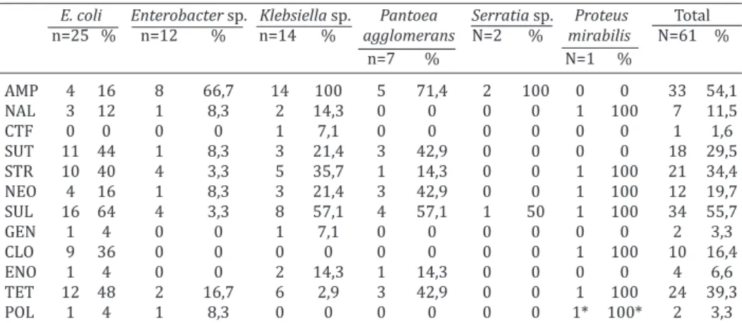
The antimicrobial susceptibility test results are de-monstrated in Table 2. The antimicrobial to which strains presented the most frequent resistance was sulfonami -des with 55.7%, followed by ampicillin with 54.1% and tetracycline with 39.3%. More than 50% of E. coli strains presented resistance to sulfonamides (64%), but a high resistance rate was also observed to tetracycline (48%), sulfazotrim (sulfamethoxazole+trimethoprim) (44%), streptomycin (40%) and chloramphenicol (36%). The ge-nus Enterobacter presented a high resistance rate only to
Table 1. Absolute and relative frequencies of enterobacteria isolated from cloacal swabs and necropsy samples of
canaries (Serinus canaria) from Fortaleza, Brazil
Enterobacteria Swabs Necropsies Total
(n=387) (n=58) (n=445)
n % n % n %
Escherichia coli 14 3,62 11 18,97 25 5,62 Enterobacter spp. 11 2,84 1 1,72 12 2,7 Klebsiella spp. 9 2,32 5 8,62 14 3,15 Pantoea agglomerans 5 1,29 2 3,45 7 1,57 Serratiaspp. 2 0,52 0 - 2 0,45 Proteus mirabilis 1 0,26 0 - 1 0,22 Total 42 10,85 19 32,76 61 13,71
Table 2. Percentage of Enterobacteriaceae strains isolated from canaries (Serinus canaria) resistant to antimicrobials
E. coli Enterobacter sp. Klebsiella sp. Pantoea Serratia sp. Proteus Total n=25 % n=12 % n=14 % agglomerans N=2 % mirabilis N=61 %
n=7 % N=1 %
AMP 4 16 8 66,7 14 100 5 71,4 2 100 0 0 33 54,1 NAL 3 12 1 8,3 2 14,3 0 0 0 0 1 100 7 11,5 CTF 0 0 0 0 1 7,1 0 0 0 0 0 0 1 1,6 SUT 11 44 1 8,3 3 21,4 3 42,9 0 0 0 0 18 29,5 STR 10 40 4 3,3 5 35,7 1 14,3 0 0 1 100 21 34,4 NEO 4 16 1 8,3 3 21,4 3 42,9 0 0 1 100 12 19,7 SUL 16 64 4 3,3 8 57,1 4 57,1 1 50 1 100 34 55,7 GEN 1 4 0 0 1 7,1 0 0 0 0 0 0 2 3,3
CLO 9 36 0 0 0 0 0 0 0 0 1 100 10 16,4
ENO 1 4 0 0 2 14,3 1 14,3 0 0 0 0 4 6,6 TET 12 48 2 16,7 6 2,9 3 42,9 0 0 1 100 24 39,3
POL 1 4 1 8,3 0 0 0 0 0 0 1* 100* 2 3,3
AMP = ampicillin, NAL = Nalidixic acid, CTF = ceftiofur, SUT = Sulfazotrim (Sulfamethoxazole+trimethopr im), STR = streptomycin, NEO = neomycin, SUL = sulfonamides, GEN = gentamycin, CLO = chloramphe-nicol, ENO = enrofloxacin, TET = tetracycline, POL = polymyxin B. *Proteus spp. are naturally resistant to polymyxin B.
Table 3. Multidrug-resistant (MDR) enterobacteria isolated from canaries (Serinus canaria)
No. of antibiotics No. of resistant strains (%)
At least 1 53 (83.88)
>1 34 (55.74)
>2 30 (49.18)
>3 23 (37.70)
>4 14 (22.95)
>5 9 (14.75)
>6 4 (6.56)
>7 1 (1.64)
>8 0 (0)
ampicillin (66.7%), while the genus Klebsiella presented a 100% resistance rate to ampicillin and a high resistan-ce rate to sulfonamides (57.1%). The Pantoea agglome-rans strains presented high resistance rate to ampicillin (71.4%), to sulfonamides (57.1%), to neomycin (42.9%), to sulphametoxazole+trimethoprim (42.9%) and tetracycline (42.9%). The genus Serratia presented resistance only to ampicillin (100%) and sulfonamides (50%), while the only Proteus mirabilis isolated was resistant to nalidixic acid, streptomycin, sulfonamides, chloramphenicol, tetracyline and polymyxin B, to which the genus Proteus is naturally resistant. The total of multidrug-resistant bacteria (MDR) was 34 (55.7%) and the maximum antimicrobials to which a single isolate was resistant was eight, followed by three isolates that were resistant to seven antimicrobials. The Table 3 demonstrates the amount of isolates and antimi-crobials to which resistance was observed.
DISCUSSION
Surveys of Enterobacteriaceae have been performed in se-veral avian species, from which different members of this bacterial family were isolated from birds in captivity belon-ging to 15 orders (Jones & Nisbet 1980). The enterobacteria are widely distributed in the environment, in the microbio-ta of mammals and of some birds (Fudge 2001), however passerines do not possess a functional caeca, or an intesti-bilis (1/387). The most frequent bacteria isolated from
necropsy samples was Escherichia coli (11/58), followed by Klebsiella spp. (5/58), Pantoea agglomerans (2/58) and Enterobacter spp. (1/58). No bird presented more than one enterobacteria in the selective agar plates, though occasio-nally, some presented besides the enterobacteria, a non--fermenter Gram-negative bacteria, which was identified and discarded in the TSI agar.
per-nal microbiota for the digestion of nutrients, which makes the presence of members of the Enterobacteriaceaefamily often related to a disease status (Dorrestein 2003).
In this study, the prevalence of enterobacteria was hi-gher in dead birds, than it was in the live ones, which may be explained by the growth that occurs from the moment of death until the carcass was found and frozen. Also, the te-chnique itself may have favored the necropsy results, since large intestinal samples were collected, when compared to swabs which sample a low amount of bacteria present in the cloaca. Another possibility is due to the pathogenic po-tential that many of these micro-organisms possess, thou-gh with the methodology applied these possible infections cannot be confirmed.
Most mammals and some bird species possess Esche-richia coli in their normal intestinal flora, since they are colonized at birth and remain with this bacterium during the entire life (Fudge 2001). Passerines, in spite of having a permanent microbiota, may host this bacterium with no symptoms, which have been reported (Jones & Nisbet 1980), however other authors have reported this microor-ganisms causing different diseases in passerines (Macwhir-ter 1994, Dorrestein 2003, Cattarossi et al. 2013). In this study, E. coli was the most frequently isolated bacteria from both dead and live canaries, which may suggest a zoonotic potential, since several other birds in this study were pre-sent in home environments and, therefore, in direct contact with the owners. More studies on the virulence of these strains are necessary in order to confirm this hypothesis, since E. coli strains with virulence factors have been pre-viously isolated from passerines (Gibbs et al. 2007).
The genera Enterobacter and Klebsiella may cause pri-mary or secondary infections in passerines, sometimes acting as opportunistic pathogens associated or not with viral, parasitic or fungal infections (Joseph 2003). Bacte-ria from the genus Klebsiella and the species Enterobacter sakazakii have been correlated with the occurrence of high mortality outbreaks in canary flocks, and these microorga -nisms have been considered as important as Salmonella, Escherichia coli, Listeria spp., Staphylococcus spp. and Pseudomonas (Cattarossi et al. 2013). The genus Serratia may be an opportunistic pathogen in passerines, much like Klebsiella and Enterobacter, however it occurs less fre-quently and the most common species are S. marcescens, S. odorifera, S. rubidae and S. liquefaciens (Fudge 2001). In this study, only one strain of S. odorifera and one strain of S. liquefaciens were identified, each present in a different sample.
The most important and most frequently reported bac -terium in the scientific literature isolated from canaries is Salmonella sp. (Harrington et al. 1975, Panigrahy & Gil-more 1983, Asterino 1996, Raidal 1998, Fudge 2001, Dor-restein 2003, Madadgar et al. 2009, Rahmani et al. 2011). However, in this study no bacteria from this genus were isolated, which may be explained by the excessive use of antibiotics by the owners and the absence of outbreaks re-ported in the domiciliary breeding locations visited. The methodology applied may have also hindered the isolation of these bacteria, since it has already been demonstrated
that the polymerase chain reaction (PCR) test present a higher positivity rate when compared to the conventional culture for the survey of Salmonella from the feces of pet birds, however with no significant difference between the two tests (Sareyyüpoğlu et al. 2008). Due to the reports in the literature and the importance that this genus repre-sents, control measures must always be applied in order to guarantee the safety of the birds, owners and environ-ment.
The genus Yersinia possess species that cause zoonotic infections, being Y. pseudotuberculosis the most frequently isolated bacterium from the intestines of wild and captive passerines (Quinn et al. 1994). However, this pathogen is more reported during the winter months, when the cold weather benefits the growth of this pathogen and cause outbreaks of yersiniosis (Fudge 2001). Another predispo-sing factor that favors the occurrence of this infection is the infestation by rodents or wild birds, which may carry this bacterium and disseminate in the environment (Guima-rães 2006). The absence of this genus in the studied birds may be explained by the local weather that do not favor the spread of this bacterium and the absence of problems with rodents or wild birds, as reported by the owners.
According to the literature, several members of the En-terobacteriaceaefamily are known to cause, among other illnesses, intestinal infections, and the main symptom ob-served in these cases of diseased passerines is diarrhea (Macwhirter 1994, Dorrestein 2003, Guimarães 2006). In this study, however, most (26/37) of the canaries that pre-sented diarrhea did not present any enterobacteria, which indicates that different etiologies may be implied, such as gastrointestinal parasites, viral or fungal infections, or food disorders (Joseph 2003).
CONCLUSIONS
Canaries (Serinus canaria) host members of the Ente-robacteriaceaefamily and Escherichia coli is the most fre-quent contaminant.
The enterobacteria strains from these birds present a high resistance rate to antimicrobials, and the emergence of multidrug resistant strains is concerning, since the pro-ximity of these birds and people represents a potential zo-onotic risk.
REFERENCES
Alley M.R., Connolly J.H., Fenwick S.G., Mackereth G.F., Leyland M.J., Rogers L.E., Haycock M., Nicol C. & Reed C.E.M. 2002. An epidemic of salmo-nellosis caused by Salmonella Typhimurium DT160 in wild birds and humans in New Zealand. N. Z. Vet. J. 50:170-176.
Asterino R. 1996. Diseases and care of wild passerines, p.956-980. In: Ros-skopf W.J. & Woerpel R.W. (Eds), Diseases of Cage and Aviary Birds. 3rd ed. Williams and Wilkins, Baltimore.
Cattarossi D., Azzara E. & Catania S. 2013. Clinical and laboratory practice for canaries and true finches. Vet. Clin. North Am., Exotic Anim. Pract. 16:31-46.
CLSI 2012. Performance Standards for Antimicrobial Susceptibility Test-ing. 22nd Informational Supplement. CLSI document M100-22. Clinical and Laboratory Standards Institute, Wayne, PA.
De Champs C., Le Seaux S., Dubost J., Boisgard S., Sauvezie B. & Sirot J. 2000. Isolation of Pantoea agglomerans in two cases of septic monoarthritis af-ter plant thorn and wood sliver injuries. J. Clinic. Microbiol. 38:460-461. Dorrestein G.M. 2003. Diagnostic approaches and management of
disea-ses in captive passerines. Sem. Avian Exotic Pet Medicine 12:11-20. Fudge A.M. 2001. Diagnosis and treatment of avian bacterial diseases.
Sem. Avian Exotic Pet Medicine 10:3-11.
Gaukler S.M., Linz G.M., Sherwood J.S., Dyer N.W., Bleier W.J., Wanne- muehler Y.M., Nolan L.K. & Logue C.M. 2009. Escherichia coli, Salmonella, and Mycobacterium avium subsp. paratuberculosis in Wild European Starlings at a Kansas cattle feedlot. Avian Dis. 53:544-551.
Gibbs P.S., Kasa R., Newbrey J.L., Petermann S.R., Wooley R.E., Vinson H.M. & Reed W. 2007. Identification, Antimicrobial Resistance Profiles, and Virulence of Members from the Family Enterobacteriaceae from the Feces of Yellow-Headed Blackbirds (Xanthocephalus xanthocephalus) in North Dakota. Avian Dis. 51:649-655.
Gismondi E. 1995. Procedencia e historia, p.12-14. In: Ibid. (Eds), Guia Completa de los Canarios de Color. Editorial de Vecchi, Barcelona. Guimarães M.B. 2006. Passeriformes (pássaro, canário, saíra, gralha),
p.324-337. In: Cubas Z.S., Silva J.C.R., Catão-Dias J.L. (Eds), Tratado de Animais Selvagens. Roca, São Paulo.
Harrington Jr R., Blackburn B.O. & Cassidy D.R. 1975. Salmonellosis in ca-naries, Avian Dis. 19:827-829.
Jackson J.A., Bock W.J., Olendorf D., Trumpey J.E. & Hutchins M. 2003. Gr-zimek’s Life Encyclopedia. Vol.11. 2nd ed. Gale, USA. 616p.
Jones D.M. & Nisbet D.J. 1980. The gram negative bacterial flora of the avian gut. Avian Pathol. 9:33-38.
Joseph V. 2003. Infectious and parasitic diseases of captive passerines. Sem. in Avian Exotic Pet Medicine 12:21-28.
Kapperud G., Stenwig H. & Lassen J. 1998. Epidemiology of Salmonella Typhimurium 0:4-12 infection in Norway. Am. J. Epidemiol. 147:774-782.
MacWhirter P. 1994. Passeriformes, p.1172-1199. In: Ritchie B.W., Harri-son G.J. & HarriHarri-son L.R. (Eds), Avian Medicine: principles and applica-tion. Wingers Publishing, Lake Worth, Florida, USA.
Madadgar O., Salehi T.Z., Ghafari M.M., Tamai I.A., Madani S.A. & Yahyareyat R. 2009. Study of an unusual paratyphoid epornitic in canaries (Serinus canaria). Avian Pathol. 38:437-441.
Panigrahy B. & Gilmore W.C. 1983. Systemic salmonellosis in an African gray parrot and salmonella osteomyelitis in canaries. J. Am. Vet. Med. Assoc. 183:699-700.
Quinn P.J., Carter M.E., Markey P.K. & Carter G.R. 1994. Enterobacteriaceae, p.209-236. In: Ibid. (Eds), Clinical Veterinary Microbiology. Wolfe Pu-blishing, London.
Rahmani M., Peighambari S.M., Yazdani A. & Hojjati P. 2011. Salmonella infection in birds kept in parks and pet shops in Tehran, Iran. Int. J. Vet. Res.5:145-148.
Raidal S.R. 1998 Salmonellosis in two canary (Serinus canaria) flocks. Aust. Vet. Pract.28:67-70.
Reed K.D., Meece J.K., Henkel J.S. & Shukla S.K. 2003. Birds, migration and emerging zoonoses: West Nile virus, Lyme disease, influenza A and en -teropathogens. Clin. Med. Res. 1:5-12.
Sareyyüpoğlu B., Çelik ok A., Cantekin Z., Yardimci H., Akan M. & Akçay A. 2008. Polymerase Chain Reaction detection of Salmonella spp. in fecal samples of pet birds. Avian Dis. 52:163-167.