*Corresponding author. Present address: INIAP/IPIMAR, Centro de Investigação Pesqueira do Sul (CRIPSul), Av. 5 de Outubro, 8700-305 Olhão, Portugal. Tel. +351 (289) 700500; Fax: +351 (289) 700535; email: jcurdia@cripsul.ipimar.pt. Presented at the Tenth Congress of the International Society of Invertebrate Reproduction and Development, Newcastle, UK, July 18–July 23, 2004.
0168-8170/05/$05.00 © 2005 Balaban
The reproductive cycle of Patella candei gomesii Drouët, 1858
(Mollusca: Patellogastropoda), an Azorean endemic subspecies
J
.
CÚRDIA1*,
A.
SANTOS RODRIGUES2,3,
A.
M.
F.
MARTINS2,3and
M.
J.
COSTA1,4,51Departamento de Ciências Tecnológicas e Desenvolvimento, 2Departamento de Biologia, 3Research Center in Natural Resources (CIRN), Universidade dos Açores, Rua Mãe de Deus, Apartado 1422, 9501 - 801 Ponta Delgada, Portugal
4Research Center in Biodiversity and Genetic Resources (CIBIO), University of Porto, Porto, Portugal 5Institute of Marine Research (IMAR), Coimbra, Portugal
Received 14 October 2004; Accepted 12 August 2005
Summary
Patella candei gomesii is morphologically plastic comprising two ecomorphs. The “smooth limpet” is characteristic of the eulittoral zone, whereas the “fly limpet” is mainly found higher in the shore, on the splash zone of exposed areas. Their reproductive strategies are poorly understood. This study investigated the reproductive cycles of the ecomorphs using histological techniques. The annual cycles were found to be similar. Both sexes exhibited synchronous patterns and were mature most of the year. In April, significant increases were observed in the relative volume occupied by previtellogenic and vitellogenic cells in females, and by sperma-togonia, spermatids and spermatocytes in males. The maximum values for mature oocytes and spermatozoa were observed in July. It is concluded that the breeding season of P. candei gomesii lasts the whole year peaking in the summer (when reproductive condition is highest and the main spawning event must occur). The implications of these findings for the taxonomy and conservation of the subspecies are further discussed.
Key words: Patella candei gomesii, gametogenesis, Azores, reproductive cycle
Introduction
The limpet Patella candei gomesii Drouët 1858 belongs in the Patella candei complex, which is exclu-sive to Macaro-nesia (Azores, Madeira, Canary Islands, Selvagens and Cabo Verde). The subspecies P. candei gomesii is endemic to the Azores (Titselaar, 1998; Weber and Hawkins, 2002), occurring subtidally
and in mid and high shores, especially on boulders (Côrte-Real et al., 1996; Hawkins, et al. 2000). Within the Azorean archipelago, this subspecies is morpho-logically plastic occurring as two habitat morphs: the “smooth limpet” and the “fly limpet”. The former is characteristic of the eulittoral area, whereas the latter is mainly found higher on the shore, especially the splash
zone in exposed areas (Hawkins et al., 1990). Plasticity has been described in other patellid species and linked to environmental variation (Lewis and Bowman, 1975). Ridgway et al. (1998) emphasized the importance of knowledge on the biogeography of patellid species to understand the evolutionary trends in the NE Atlantic. The determination of the reproductive cycles of P. candei gomesii’s ecomorphs should provide information to clarify pending issues on the sub-species’ taxonomical status. According to Martins et al. (1987), the gonochoristic P. candei gomesii spawns throughout the year, without synchronized resting periods. However, important information might have been left out since the two morphs were not addressed separately in the study. This is relevant not only because different reproductive cycles have been re-ported for separate conspecific populations of molluscs (Sutherland, 1970; Geller, 1990), but also because similar reproductive cycles have been traced in popu-lations under different environmental conditions (Henninger and Hodgson, 2001).
Ultimately, knowing about the reproductive cycles of P. candei gomesii is important for the conservation of this subspecies. Due to its endemic character, there are no populations outside the Azores archipelago to act as reservoirs for recruitment during recovery, which, together with larval loss, makes the subspecies vulnerable (Hawkins et al., 2000). These authors sug-gested that there is a risk of complete extinction, taking into account its endemic nature and the existence of over-exploitation. The enormous impact on rocky shore communities of direct exploitation of resources is well known as targeted species show declining populations (Thompson et al., 2002). In São Miguel, P. candei gomesii is commercially exploited (Martins et al., 1987; Hawkins et al., 2000). Água d’Alto beach has been repeatedly harvested for limpets; these were conspicuously absent in 1992 (Morton et al., 1998), but did recover in 1994 when protection from indis-criminate collecting was enforced by the Azorean Government (Morton et al., 1998). Information regard-ing the reproductive cycles of this subspecies is crucial for an adequate management of limpet harvests.
Several studies have been undertaken concerning the reproductive cycles of Patella species (Orton et al., 1956; Orton and Southward, 1961; Blackmore, 1969; Lewis and Bowman, 1975; Bowman and Lewis, 1986). Most are based on the macroscopic index described by Orton et al. (1956). The present work reports on a preliminary investigation of the reproductive season-ality of the two ecological morphotypes of P. candei gomesii in São Miguel (Azores). A quantitative assess-ment of gametogenesis in P. candei gomesii was carried out applying histological methods.
Materials and Methods
Study area
Limpets were collected from a boulder/cobble beach at Água d’Alto (São Miguel, Azores, Portugal). Specimens of the two ecomorphs were sampled in characteristic habitats, separated horizontally by 100– 150 m: “fly” specimens from a highly exposed area, in the supralittoral zone; “smooth” specimens from rolled stones and cobble, in the intertidal zone. Harvesting took place at four sampling periods: October 2002 (autumn); January 2003 (winter); April 2003 (spring) and July 2003 (summer).
Morphometric analysis
Quantitative measurements of shell length (SL, greatest distance between the anterior and posterior ends of the shell), shell width (SW, greatest distance between margins perpendicular to the anterior/ posterior axis) and shell height (SH, greatest vertical distance from the apex to the base of the shell) were performed to the nearest 0.05 mm using vernier callipers. In addition, total fresh weight (FW) of each individual and shell dry weight (SDW) were measured using a precision scale (0.0001 g). The shell length/ shell height ratio was calculated.
Gonadal maturation state
For each sampling period, 16 specimens (eight males and eight females) were sampled for histological analysis in both habitats. The fresh weight was obtained before excision of the gonad for histological purposes. Testes and ovaries were fixed in 10% for-malin and embedded in paraffin. Serial sections, 7 µm thick, were stained with Mayer’s haemalum and eosin (Martoja and Martoja-Pierson, 1970). The relative volumetric density of gametes was estimated using the M168 Weibel Multipurpose Test System (Weibel, 1979).
Four stages of spermatogenesis were identified based on the classification of Griffond et al. (1991): (1) spermatogonia — medium-sized cells rectangular in shape viewed by light microscopy, with a large nucleus in relation to the quantity of cytoplasm, always located near the acinus epithelium; (2) spermatocytes — smaller than spermatogonia with basophilic cyto-plasm; (3) spermatids — smaller than spermatocytes, spheroid in shape and slightly more basophilic than spermatocytes; (4) spermatozoa — with strong baso-philic head and esosinobaso-philic tail. With light micro-scopy, no differentiation was made between sperma-tocytes II and I.
Following Hill and Bowen (1976), three stages of development were distinguished during oogenesis: (1) previtellogenic oocytes (PV), small, rounded and with strong basophilic cytoplasm; (2) vitellogenic oocytes (V), larger than the previous ones, irregular in shape, sometimes with multiple visible nucleoles, cytoplasm with slight granulations and lightly baso-philic; (3) maturing oocytes (M), larger than the vitellogenic oocytes, round in shape with eosinophilic and granular cytoplasm.
Statistical analysis
In order to identify the gonadal maturation state, scores for volumetric density were summed for each specimen and converted to percentages.
Data were analysed with Statistica V.5.1 (StatSoft). The mean and standard error were calculated. Relative volumetric density of each oogenesis and spermato-genesis stage was compared using a two-way ANOVA (for spermatogonia arcsin transformation was per-formed to comply with requirements for ANOVA) or using the Kruskal–Wallis test (for previttelogenic cells, as data did not comply with ANOVA assumptions). Multivariate analyses were undertaken to assess for seasonal patterns.
Results
This study focused on investigating the repro-ductive cycles of both morphs. The two sexes were analysed. The results for oogenesis and spermato-genesis are presented separately, in sequence, so as to facilitate comparisons between morphs.
Oogenesis
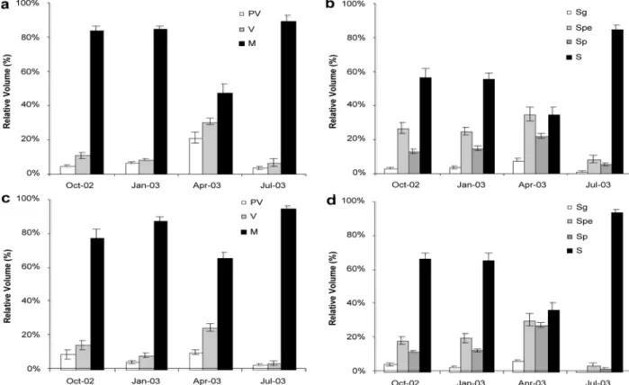
Generally, the two morphotypes followed the same pattern. The differences due to morphotype concerning the three oogenic stages were not statistically signifi-cant (Table 1; Fig. 1A and C). The relative volumetric densities of previttelogenic oocytes were relatively low in October, January and July (fly: 8.6%, 4.2% and 2.2%, respectively; smooth: 4.8%, 6.6% and 3.8%, respectively), with a small peak in April, particularly for the smooth morphotype (21.41%). Medians obtained for April were significantly different from those for the other periods (Kruskal–Wallis, H = 32.7844, p<0.001, Table 1). The percentage volume of vitellogenic cells was low during October, January and July (less than 15%), increasing significantly in April (24.50% fly; 30.71% smooth; ANOVA, p<0.001, Table 1). Maturing oocytes comprised most of the gonadal volume throughout the entire sampling
Fig. 1. Mean values and standard errors of the previtellogenic (PV), vitellogenic (V) and maturing oocytes (M) for “smooth” females (A) and “fly” females (C). Mean values and standard errors of the spermatogonia (Sg), spermatocyte (spe), spermatid (sp) and spermatozoa (S) for “smooth” (B) and “fly” males (D).
Table 1. Two-way ANOVA results for each gametogenic stage
aKruskal–Wallis test was used. bData transformed to arcsine.
N.S., non significant; *Significant at p <0.05; **Significant at p <0.01; ***Significant at p < 0.001.
periods. Maximum values (around 80%) were found for October, January and July. For mature cells, the interaction between the factors Season and Morpho-type was significant (ANOVA, p<0.05, Table 1), meaning that the differences observed in different seasons are not independent of morphotype, even though differences among morphotypes are not significant.
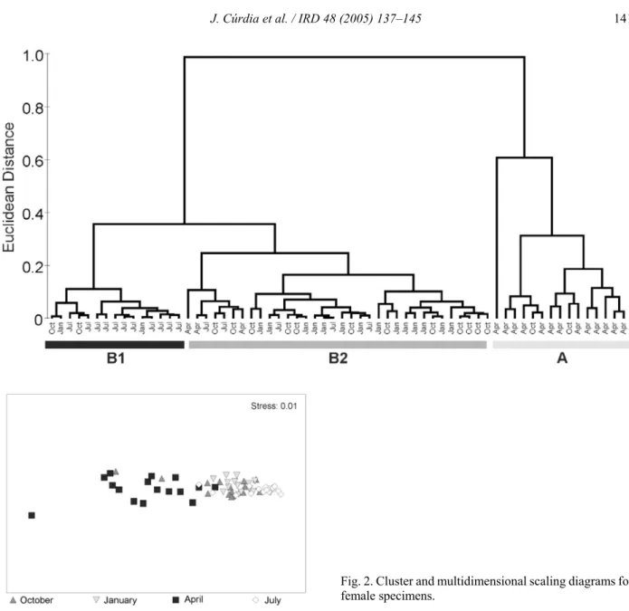
Cluster analysis (Fig. 2A) showed a clear seasonal trend with three main groups. Most April samples cluster together in a separate group (group A, Fig. 2A), with the rest of the samples comprising a major group (group B). Within this group, two subgroups are formed: group B1, mainly July samples; and group B2, most samples from October and January. This seasonal trend is also shown by the Multidimensional Scaling
diagram, Fig. 2B). April and July samples were separated and October and January samples were distributed in the middle. It is relevant that specimens from the ecomorphs have grouped together in terms of the relative volume of each cell type, since it indicates that both follow similar reproductive cycles.
Spermatogenesis
The general pattern was very similar in both morphotypes (Fig. 1B and D). The percentage volume of spermatogonia was lower in October and January, increasing to a maximum in April and decreasing to the lowest values in July. This trend was also observ-able for spermatocytes and spermatids. The differences found between seasons were statistically significant
Fig. 2. Cluster and multidimensional scaling diagrams for female specimens.
(ANOVA, p<0.001, Table 1), with three homogeneous groups (Tukey HSD test, p<0.05), July < October and January < April. In October and January, spermatozoa comprised approximately 60% of the gonadal volume, decreasing to about 35% in April, and increasing to over 85% in July. There were three homogeneous groups, April < October and January < July (Tukey HSD test, p <0.05), reflecting statistically significant differences between seasons (ANOVA, p <0.001).
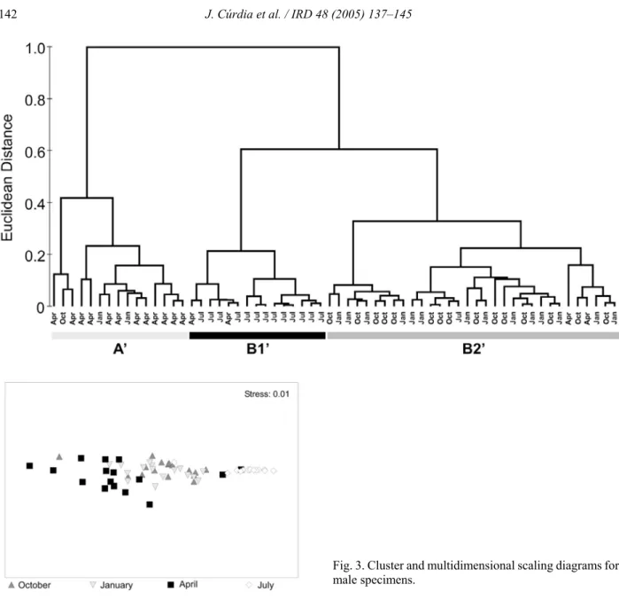
Results from multivariate analysis supported a clear seasonal trend. They did not, however, separate the morphotypes, suggesting that males from both eco-morphs follow similar reproductive cycles. Cluster analysis (Fig. 3A) showed three different groups. The majority of April samples clustered together in a separate group (group AN, Fig. 3A) with the remaining samples forming two subgroups in a larger group: group B1N, mainly July samples; group B2N, most
samples from October and January. Multidimensional Scaling also showed the same seasonal pattern (Fig. 3B). April and July samples were clearly sepa-rated, the latter presenting a higher similarity to October and January samples.
In contrast to oogenesis, differences between morphotypes were statistically significant for sperma-togonia, spermatocyte and spermatozoa (ANOVA, p<0.01, Table 1). The interaction of the two sources of variation was, however, not statistically significant (Season×Morphotype, Table 1), showing that differ-ences between seasons were independent of the differences among morphotypes.
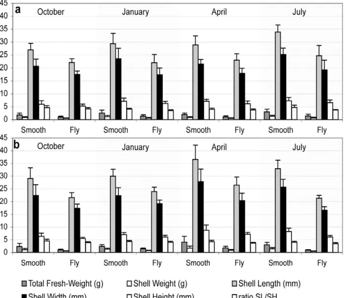
Morphometric parameters
The morphometric parameters had similar values across the time scale (Fig. 4). The two ecomorphs did
Fig. 3. Cluster and multidimensional scaling diagrams for male specimens.
differ in the morphometric variables studied, but the pattern was similar among seasons. The morphometric parameters were concordant with unpublished data concerning larger samples (total of 117 fly limpets and 132 smooth limpets) for the same sites and sampling periods.
Discussion
The main goals of this investigation were the definition and comparison of the reproductive cycles of the two ecomorphs of P. candei gomesii. The results revealed that the morphs’ annual reproductive patterns are similar. This is in contrast to what has been described for Patella depressa from exposed and sheltered sites in the centre of Portugal (Cabo Raso, Cascais). Morais et al. (2003) found that, in the summer, gametogenesis in limpets from the exposed
site was more advanced (mainly stage III and stage IV) than that in limpets from the sheltered site (stage III), according to the maturation scale of Orton et al. (1961). The similar reproductive cycles of the two ecomorphs described in this study may reflect phylo-genetic preservation of reproductive patterns, as re-ferred to by Henninger and Hodgson (2001).
This paper presents findings that challenge the thesis advanced elsewhere of an absence of annual resting periods in this limpet (Martins et al., 1987) by describing quantitative and qualitative seasonal chan-ges in the gonads of P. candei gomesii. Even though females were mature most of the year, the relative volume occupied by previtellogenic and vitellogenic oocytes over mature cells increased significantly in April, but decrease to minimum levels in July, when maximum number of mature cells was registered. The production of new oocytes is the most likely
Fig. 4. Averages and standard errors of fresh weight of the animal (FWG) and weight of the shell (WS), length (SL), width (SW) and height (SH) for males (a) and females (b), morphs are discriminated.
explanation for this pattern, thus suggesting that P. candei gomesii females spawn mainly in the summer. Males present a similar annual pattern: the abundance of spermatogonia, spermatocytes and spermatids increases sharply in April, but decreases to minimum values in July, when maximum values of spermatozoa are observed. The annual cycle suggested by our data, featuring a major spawning event (but with an extended spawning period until January), is consistent with those of most patellid species, which have a marked annual cycle with a single spawning each year (Branch, 1981).
The seasonal changes in the reproduction of P. candei gomesii found in the present work are not unique since reproductive cycles have been described for other, especially European, Patella species.
Sum-mer spawning events have been reported for Patella vulgata in NE England (August: Bowman and Lewis, 1986) and in Portugal (September/October: Guerra and Gaudêncio, 1986). P. depressa has also been identified as a summer breeder in Britain (Orton and Southward, 1961) and in Portugal (early summer and early autumn: Guerra and Gaudêncio, 1986). The fact that in P. candei gomesii the levels of gametes remain high until January is not surprising, as P. depressa in the south of Portugal also presents fat and ripe gonads over the winter season (Guerra and Gaudêncio, 1986).
The annual patterns were found to show consider-able synchrony between sexes, in agreement with what has been described in other limpets (Branch, 1981). According to Yund (2000), free-spawning inverte-brates with synchronous spawning increase the
Fig. 5. Average sea surface temperatures for the period of November 2001 to May 2003 in the geographical area of São Miguel (37E–38EN; 26E–25EW). Data obtained from PODAAC–ESIP (http://podaac-esip.jpl.nasa.gov).
chances of fertilization by reducing the degree of sperm limitation. This is an issue of probable relevance to Patella species in the Azores, since decreases in population density due to human exploitation (Hawkins et al., 2000) are likely to bear a substantial influence on the availability of sperm.
The present research project did not address the factors that possibly trigger the annual changes. In the literature, temperature has been linked to the repro-ductive cycles of Patella species (Orton et al., 1956; Fretter and Graham, 1976). Species with plankto-trophic larvae spawn just prior to the time when plankton is maximal (ensuring food for the larvae) at a time of rising or high sea surface temperatures (Branch, 1981). In fact, spawning in P. depressa seems to coincide with maximum air temperatures (Orton and Southward, 1961; Fretter and Graham, 1976). Such is the case for P. candei gomesii. Sea surface tempera-tures (PODAAC-ESIP; http://podaac-esip.jpl.nasa.gov) for São Miguel present a rather small variation ranging from 15 to 23EC (Fig. 5), with a clear seasonal pattern of higher temperatures during summer and early autumn (July–October) and lower temperatures during late winter and early/mid spring (February–April). This pattern agrees fairly well with our findings, since in April, when temperatures are low, limpets start to develop their gonads; and in July, when temperatures are high, they are fully developed for the postulated major spawning event. Even though temperatures gradually decrease in autumn and early winter, limpets remain capable of spawning until January when tem-peratures attain the lowest values.
Spawning may also be triggered by sea roughness (Orton et al., 1956; Fretter and Graham, 1976; Bowman and Lewis, 1977; Bowman and Lewis, 1986). In the Azores, sea roughness is higher in winter, arguing in favour of the extended period of spawning. This can be extremely important for species (or popu-lations) that live high in the shore, like “fly limpets”. In fact, Notoacmea petterdi, a limpet that lives on the extreme high shore, spawns when storms are frequent, suggesting that these are important for larvae to reach their settlement zones (Branch, 1981). Furthermore, in other gonochoric species such as Patella aspera and P. depressa, spawning is linked to wave action (Fretter and Graham, 1976).
This paper presents clear evidence on the repro-ductive cycles of the two ecomorphs of P. candei gomesii. Highlighting their similarity, the thesis that they are in fact two morphs of the same subspecies is strengthened.
The histological approach provided quantitative data and sharpened the analysis of the reproduction of P. candei gomesii, indicating that the period from April through July is crucial for conservation measures and for further investigations. Extending this approach to other limpets of the Macaronesia should provide interesting cues to understand their biogeography and to foresee adequate conservation strategies.
Acknowledgements
This work was supported by the project “As espécies do género Patella como modelo para o estudo
dos padrões evolutivos e dinâmica de organismos marinhos” (POCTI/BSE/42003/2001), funded by Fun-dação para a Ciência e Tecnologia (Portugal). The authors thank André Amaral for technical advice on the histolological essays and Paulo Melo for field and laboratory support. The authors would also like to thank two anonymous reviewers for their contribution in improving this paper.
References
Blackmore, D.T., Studies of Patella vulgata L. — I. Growth, reproduction and zonal distribution. J. Exp. Mar. Biol. Ecol., 3 (1969) 200–213.
Bowman, R.S. and Lewis, J.R., Annual fluctuations in the recruitment of Patella vulgata L. J. Mar. Biol. Ass. U.K., 57 (1977) 793–815.
Bowman, R.S. and Lewis, J.R., Geographical variation in the breeding cycles and recruitment of Patella spp. Hydrobiologia, 142 (1986) 41–56.
Branch, G.M., The biology of limpets: physical factors, energy flow, and ecological interactions. Oceanogr. Mar. Biol. Ann. Rev., 19 (1981) 235–380.
Côrte-Real, H.B.S.M., Hawkins, S.J. and Thorpe, J.P., Population differentiation and taxonomic status of the exploited limpet Patella candei in the Macaronesian islands (Azores, Madeira, Canaries). Mar. Biol., 125 (1996) 141–152.
Fretter, V. and Graham, A., The Prosobranch molluscs of Britain and Denmark. J. Moll. Studies, Supplement 1 (1976) 1–37.
Geller, J.B., Consequences of a morphological defense: growth, repair and reproduction by thin-shelled morphs of Nucella emarginata (Deshayes) (Gastropoda: Proso-branchia). J. Exp. Mar. Biol. Ecol., 144 (1990) 173–184. Griffond, B., Dadkhan-Teherain, Z., Medina, A. and Bride, M., Ultrastructure of Helix aspersa spermatogenesis: scanning and transmission electron microscopial contri-butions. J. Moll. Studies, 57 (1991) 277–287.
Guerra, M.T. and Gaudêncio, M.J., Aspects of the ecology of Patella spp. on the Portuguese coast. Hydrobiologia, 142 (1986) 57–69.
Hawkins, S.J., Côrte-Real, H.B.S.M., Pannacciulli, F.G., Weber, L.C. and Bishop, J.D.D., Thoughts on the ecology and evolution of the intertidal biota of the Azores and other Atlantic Islands. Hydrobiologia, 440 (2000) 3–17.
Hawkins, S.J., Côrte-Real, H.B.S.M., Martins, H.R., Santos, R.S. and Martins, A.M.F., A note on the identity of Patella in the Azores. In: Proc. First International Workshop of Malacology, Săo Miguel, Azores, Martins, António M. Frias (ed.), Açoreana, Ponta Delgada, 1990, pp. 167–173.
Henninger, T.O. and Hodgson, A.N., The reproductive cycle of Helcion pruinosus (Patellogastropoda) on two South African boulder shores. J. Moll. Studies, 67 (2001) 385– 394.
Hill, R.S. and Bowen, I.D., Studies on the ovotestis of the slug Agriolimax reticulatus (Müller) 1. The Oocyte. Cell Tissue Res., 173 (1976) 465–482.
Lewis, J.R. and Bowman, R.S., Local habitat-induced variations in the population dynamics of Patella vulgata L. J. Exp. Mar. Biol. Ecol., 17 (1975) 165–203. Martins, H.R., Santos, R.S. and Hawkins, S.J., C.M. 1987/
K:53, ICES, 1987.
Martoja, R. and Martoja-Pierson, M., Técnicas de Histologia Animal, Toray-Masson, Barcelona, 1970.
Morais, S., Boaventura, D., Narciso, L., Ré, P. and Hawkins, S.J., Gonad development and fatty acid composition of Patella depressa Pennant (Gastropoda: Prosobranchia) populations with different patterns of spatial distribution, in exposed and sheltered sites. J. Exp. Mar. Biol. Ecol., 294 (2003) 61–80.
Morton, B., Britton, J.C. and Frias Martins, A.M., Coastal Ecology of the Açores, Sociedade Afonso Chaves — Associação de Estudos Açoreanos, Ponta Delgada, 1998. Orton, J.H. and Southward, A.J., Studies on the biology of limpets — IV. The breeding of Patella depressa Pennant on the North Cornish coast. J. Mar. Biol. Ass. U.K., 41 (1961) 653–662.
Orton, J.H., Southward, A.J. and Dodd, J.M., Studies on the biology of limpets — II. The breeding of Patella vulgata L. in Britain. J. Mar. Biol. Ass. U.K., 35 (1956) 149–176. Ridgway, S.A., Reid, D.G., Taylor, J.D., Branch, G.M. and Hodgson, A.N., A cladistic phylogeny of the family Patellidae (Mollusca: Gastropoda). Philos. Trans. Roy. Soc. Lond. B Biol. Sci., 353 (1998) 1645–1671. Sutherland, J.P., Dynamics of high and low populations of
the limpet, Acmaea scabra (Gould). Ecol. Monogr., 40 (1970) 169–188.
Thompson, R.C., Crowe, T.P. and Hawkins, S.J., Rocky intertidal communities: past environmental changes, present status and predictions for the next 25 years. Environ. Conserv., 29 (2002) 168–191.
Titselaar, F.F.L.M., A revision of the recent European Patellidae (Mollusca: Gastropoda) — Part 1. The Patel-lidae of the Azores, Madeira, the Selvagens and the Canary Islands. Vita Marina, 45 (1998) 21–62. Weber, L.I. and Hawkins, S.J., Evolution of the limpet
Patella candei d’Orbigny (Mollusca, Patellidae) in Atlantic archipelagos: human intervention and natural processes. Biol. J. Linn. Soc., 77 (2002) 341–353. Weibel, E.R., Stereological Methods, Academic Press,
London, 1979.
Yund, P.O., How severe is sperm limitation in natural populations of marine free-spawners? Trends Ecol. Evol., 15 (2000) 10–13.