Listeria
monocytogenes
infection on the
host cell cycle
Cláudia Patrícia Fernandes de Brito
Mestrado em Bioquímica
Departamento de Química e Bioquímica 2013
Orientador
Sandra Sousa, Investigador auxiliar, IBMC
Coorientador
I would like to express to all the people who have been involved, and somehow contributed to my Master thesis how thankful I am to have them in my life, specially: To my kind supervisor, Sandra Sousa, who provided me the best scientific orientation and advice. Thanks to her, during last year I evolved in my scientific career since she taught me to be a better researcher, to have a critical thinking and to achieve my goals successfully. She is the most strong and dedicated person that I ever met and that led her to support me during all the good and not so good moments. I am so grateful to have you as my mentor and also as my friend.
To my boss, Didier Cabanes, who gave me the great opportunity to integrate the “Listeria Group”. Thank you for the fruitful discussions that we had and for the wise advices that you gave me.
To my co-supervisor, Elsa Leitão who very professionally taught me to handle different techniques efficiently and motivated me to accurate my expertise skills. I am also thankful for your support and friendship.
To my dear, Ana Costa, who has stand me every single moment in the lab, listening to my doubts, my ideas, fears and dreams. You have been a great and amazing friend.
To all Molecular Microbiology members for the nice environment in the lab and also for their friendship and support. Particularly to Rita, Ana V., Jorge and Rui with whom I spent a lot of funny and relaxing moments.
To my cuties AVV’s, in particular to Maggie, who always cares about me and is constantly present in the most important moments of my life.
To my pretty little girls, Xica and Fabi for their unconditional support. We will always be together.
À Filipa, o meu primeiro e derradeiro “exemplo a seguir”. Agradeço-lhe por fazer de mim uma pessoa empenhada com vontade de aprender sempre mais. Devo-te muito do que sou hoje e orgulho-me de te ter como irmã.
Ao meu maninho Rui por me querer defender de tudo e de todos, pelo orgulho que sempre demonstrou em mim e que me faz querer continuar nesta longa caminhada. Ao meu Pai por ter dado tudo por tudo para que nunca me faltasse nada, por todo o esforço realizado em prol de mim e do resto da minha família. Agradeço-lhe ainda por me ter ensinado que quando nos esforçamos somos sempre recompensados da melhor maneira.
À minha mãe, a pessoa mais importante da minha vida. Agradeço-lhe por ser o meu pilar, o meu porto de abrigo e por sempre ter acreditado em mim e nas minhas capacidades. Obrigada por me fazeres sentir a pessoa mais amada deste mundo. És a mãe que qualquer pessoa desejaria ter.
Listeria monocytogenes (Lm) is a Gram-positive human foodborne pathogen that infects mainly high-risk groups, including elderly, immunocompromized individuals, pregnant women and neonates. This intracellular facultative bacterium is able to invade, survive and multiply inside phagocytic and non-phagocytic cells. To promote cellular infection Lm interferes with and manipulates a number of biological processes. It explores the functions of cellular receptors to induce its internalization, escapes autophagy, controls the expression of the host genome and uses the actin cytoskeleton polymerization machinery to disseminate. However, Lm capacity to interfere with the host cell cycle was never reported, as it was for other human bacterial pathogens. Considering that pathogens often exploit similar pathways to cause infection, we investigated whether Lm interferes with the host cell cycle to create a suitable niche to colonize its host.
Previous studies in our laboratory showed that Lm infection induces DNA strand breaks in colon adenocarcinoma Caco-2 cells leading to the activation of DNA damage/replication checkpoints. As a consequence, infected host cells exhibit an S-phase delay associated with an increase in the overall cell cycle duration, a process favorable to the infection. In this project, we performed infection assays in Caco-2 cells with L. innocua expressing InlA (Li_InlA), which has the capacity to induce its internalization but remains in the phagocytic vacuole. We demonstrate that the effects previously observed on the cell cycle upon Lm infection are not dependent on the bacterium adhesion and invasion steps. In addition, infection assays performed in another cell line (placenta choriocarcinoma Jeg-3 cells) showed that the activation of DNA damage checkpoints and the consequent delay observed in S-phase, are not specific of Caco-2 cells and occur in other cell lines. We further assessed the involvement of ATM and ATR kinases in the Lm-induced activation of DNA damage checkpoints. The depletion of these two DNA damage sensors demonstrated that they are not essential for DNA damage checkpoint activation in response to Lm infection.
Altogether these results suggest that Lm has the capacity to modulate the host cell cycle, probably to insure a beneficial environment that favors its own replication.
Key words: Listeria monocytogenes, cell cycle delay, DNA damage checkpoints, double strand breaks, ATM kinase, ATR
RESUMO
Listeria monocytogenes (Lm) é uma bactéria Gram-positiva que, por ingestão de alimentos contaminados, infecta grupos de alto risco como idosos, indivíduos imunocomprometidos, grávidas e recém-nascidos. Este parasita intracelular facultativo é capaz de invadir, sobreviver e multiplicar-se em células fagocíticas e não-fagocíticas, e ao nível celular, promove a infeção do hospedeiro interferindo e manipulando um grande número de processos biológicos. Para além de explorar as funções de recetores de forma a induzir a sua internalização, é também capaz de escapar à autofagia, controlar a expressão do genoma do hospedeiro e usar a maquinaria necessária à polimerização da actina para disseminar nos tecidos. Ao contrário do que acontece em outras bactérias patogénicas, o papel de Lm no ciclo celular nunca foi descrito. Assim, considerando que muitos patógenos utilizam mecanismos semelhantes para causar infeção, investigou-se se Lm poderá interferir com o ciclo celular do hospedeiro, de forma a criar um ambiente adequado para colonizar o mesmo.
Estudos anteriores realizados no nosso laboratório demostraram que a infeção por Lm provoca lesões nas cadeias de ADN em células Caco-2 (linha celular do adenocarcinoma do cólon) levando à activação de mecanismos de controlo. Consequentemente, as células hospedeiras infetadas apresentam um aumento na duração do ciclo celular associado a um atraso na fase S do mesmo.
Neste projeto, foram realizados ensaios de infeção de células Caco-2 com L. innocua a expressar InlA, uma proteína que promove a internalização da bactéria. A partir destes ensaios foi demonstrado que os efeitos anteriormente observados após infeção por Lm não são dependentes dos passos de adesão e internalização da mesma. Para além disso, ensaios de infeção realizados na linha celular Jeg-3 (células do coriocarcinoma da placenta) mostraram que a ativação dos mecanismos de controlo e o consequente atraso na fase S do ciclo celular, não são processos específicos das células Caco-2 podendo ocorrer noutras linhas celulares. Ainda, foi avaliado o envolvimento das cinases ATM e ATR na ativação dos mecanismos de controlo induzida por Listeria. A depleção destes dois sensores de dano do ADN demonstrou que as mesmas não são essenciais para ativação dos mecanismos de controlo em resposta à infeção.
modular o ciclo celular do hospedeiro, provavelmente, de modo a assegurar um ambiente favorável para a sua replicação.
Palavras-chave: Listeria monocytogenes, atraso no ciclo celular, mecanismos de control do ADN, quebras de cadeia dupla,
CONTENTS
ACKNOWLEDGEMENTS/AGRADECIMENTOS………4 ABSTRACT………6 RESUMO………7 CONTENTS………9 FIGURES INDEX……….11 ABREVIATION LIST………..13 INTRODUCTION……….16 1-Listeria monocytogenes……….17 General features………17 Listeriosis………18Cell biology of infection and virulence factors………..20
a.Adhesion………22
b.Invasion………...22
c.Vacuole lysis………..25
d. Intracellular multiplication………...26
e. Intracellular movement and cell-to-cell spreading………..26
2-Cell cycle……….28
General features………28
Different phases of the cell cycle………28
a.The G0 state………..29
b.Gap-1 phase (G1-phase)……….30
c.DNA Synthesis phase (S-phase)………30
d.Gap-2 phase (G2-phase)……….31
e.Mitotic phase (M-phase)………..31
DNA damage Checkpoints………..32
a.G1/S-phase Checkpoint………..33
b.Intra-S-phase Checkpoint………...34
c.G2/M-phase Checkpoint………..35
3- Modulation of the cell cycle by bacterial pathogens……….36
1- Bacterial strains, cell lies and growth conditions………42
2- Infection assays……….42
3- Flow cytometry analyses..………..43
4- Immunofluorescence analyses……….44
5- Immunoblot analyses………..45
6- Transfection assays……….45
7- Statistical analyses………...46
RESULTS……….47
1- Listeria infection alters the cell cycle stage distribution of host cells…...47
Role of Listeria internalization on the progression of the host cell cycle….47 Listeria-induced effects can be observed in different cell lines……….50
Effect of short time Listeria infections on the host cell cycle………..52
2- Listeria hijacks the machinery of DNA damage checkpoints………55
Lm infection induces activation of DNA damage checkpoints………...55
Role of ATM and ATR kinases on the activation of DNA damage checkpoints upon Listeria infection……….57
DISCUSSION………..61
1- A complete Lm cellular infection is required to alter the cell cycle stage distribution of infected host cells……….61
2- Listeria induces effects on the cell cycle of various human cell lines…...62
3- Listeria hijacks the machinery of DNA damage checkpoints during infection of diverse cell lines……….63
4- ATM and ATR are not essential to activation of the DNA damage checkpoint in response to Lm infection……….64
REFERENCES………66
FIGURES INDEX
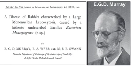
Fig.1 – The original article by E.G.D Murray and portrait of E.G.D Murray………17
Fig.2 – Gram coloration of L. monocytogenes………...18
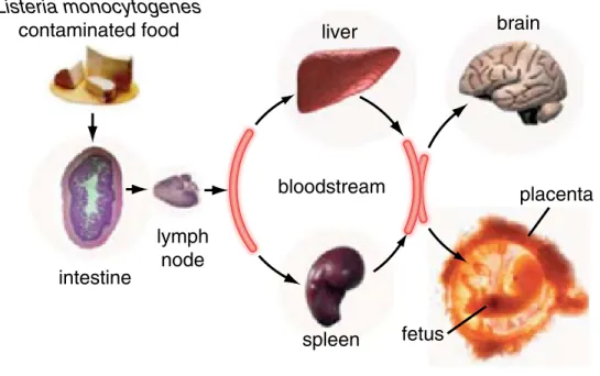
Fig.3 – Sucessive steps of listeriosis………...20
Fig.4 – Schematic representation of the L. monocytogenes cell infection cycle and major virulence factors involved in the successive steps……….….21
Fig.5 – Signaling cascades activated via the InlA-invasion pathway………..23
Fig.6 – Signaling cascades activated via the InlB-invasion pathway………..24
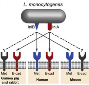
Fig.7– Species specificities of InlA and InlB……….………..25
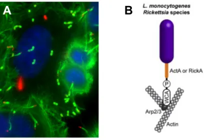
Fig.8 – Polymerization of actin comet tails………. 27
Fig.9 – Schematic representation of the cell cycle and the DNA damage checkpoints…………..….29
Fig.10 – Distribution of cyclin proteins along the cell cycle……….…….32
Fig.11 –The major components of the DNA damage checkpoint activation cascade………..34
Fig.12 – Inter-conversion of ATR- and ATM-activating DNA damage………...….35
Fig.13 – Effect of Escherichia coli CDT on cultured epithelial HeLa cells………..…37
Fig.14 – Assessment of the percentage of infected cells by immunofluorescence microscopy...48
Fig.15 – Cell cycle stage distribution upon Lm and Li_InlA infection of Caco-2 cells………..…49
Fig.18 – Jeg-3 cell cycle stage distribution upon Lm infection – discriminating infected (Inf GFP+) from bystander (Inf GFP-) cells………...…. 52 Fig.19 – Jeg-3 cell cycle stage distribution upon an 11h Lm infection……….…………..53 Fig.20 – Jeg-3 cell cycle stage distribution upon an 11h Lm infection – discriminating infected (Inf GFP+) from bystander (Inf GFP-) cells………...….53 Fig.21 – Jeg-3 cell cycle stage distribution upon a 5h Lm infection………...……….54 Fig.22– Jeg-3 cell cycle stage distribution upon a 5h Lm infection – discriminating infected (Inf GFP+) from bystander (Inf GFP-) cells………55 Fig.23– Caffeine prevents the effects induced by Lm on the host cell cycle of Jeg-3 cells…………57 Fig.24 – Western blot analysis of ATM and ATR silencing (by the INTERFERin reagent) efficiency……….…..58 Fig.25 – ATM and ATR kinases inhibition (by Amaxa system) does not prevent the cell cycle alterations induced by Lm infection………..59 Fig.26 – ATM and ATR kinases inhibition (by IRTERFERin reagent) does not prevent the cell cycle alterations induced by Lm infection………..………...60
ABREVIATIONS LIST
53BP1 – p53-binding protein 1 APC – Anaphase-promoting complex
ATM – Ataxia telangiectasia mutated protein
ATR – Ataxia telangiectasia and Rad3-related protein BHI – Brain Heart Infusion
BSA – Bovine Serum Albumin Cdk – Cyclin dependent kinase CDT – Cytolethal Distending Toxin CFU – Colony forming units
Cif – cycle inhibiting factor CtrD – Control D
DNA – Deoxyribonucleic acid DSB – Double strand breaks
EDTA – Ethylenediaminetetraacetic acid EGF – Epidermal growth factor
ERK – Extracellular-signal-regulated kinases Etop – Etoposide
FBS – Fetal bovine serum G1 – Gap1
G2 – Gap2
GFP – Green fluorescent protein HGF – Hepatocyte growth factor
Hpt – Hexose phosphate transporter protein HRP – Horseradish peroxidase
IGF – Insulin growth factor Inf – Infected
InlA – Internalin A InlB – Internalin B InlC – Internalin C IR – Irradiated
kDa – kilo Dalton
Lap – Listeria adhesion protein
Li-InlA – Listeria innocua expressing InlA Lm – Listeria monocytogenes
LLO – Listeriolysin O LPS – Lipopolysaccharide M-phase – Mitotic-phase
MAP – Microtubule-associated protein MOI – Multiplicity of infection
NER – Nucleotide excision repair NI – Non-infected
NI-NT – Non-infected – Non-transfected OD – Optical density
PBS – Phosphate saline buffer
PC-PLC – Phosphatidylcholine phospholipase C PFA – Paraformaldehyde
PI – Propidium iodide
PI 3-kinase – phosphoinositide-3 kinase-like kinase PlcA – phospholipases PI-PLC
PlcB – phospholipases PC-PLC
PI-PLC – Phosphoinositide phospholipase C RNA – Ribonucleic Acid
RNAse A – Ribonuclease A RPA – Replication protein A S-phase – DNA synthesis-phase SCGE – Single Cell Gel electrophoresis SDS – Sodium dodecyl sulfate
Ser – Serine
siRNA – small interference ribonucleic acid SSB – Single strand breaks
ssDNA – single stranded deoxyribonucleic acid Thr – Threonine
UV – Ultraviolet
WASP – Wiskott-Aldrich syndrome protein
The coevolution of bacterial pathogens and their hosts has contributed to the development of complex and sophisticated functional host-pathogen interfaces (Lara-Tejero and Galan 2002). Therefore, well-adapted pathogens have evolved a variety of strategies to manipulate the host cell functions for their own benefit (Oswald, Nougayrede et al. 2005). Bacteria have acquired an enormous evolutionary potential associated with the fact that they co-inhabit in a variety of environments that impose different life styles, metabolic capacities and ecological niches. Many pathogens, when infecting a host, evolved molecular mechanisms to manipulate host signal transduction pathways promoting their survival and replication. Indeed, with the progress in our understanding of the molecular mechanisms that bacteria use to, enter into, move within and multiply inside host cells, it came to light that microbial pathogens have developed mechanisms to block or subvert normal host-cellular processes, thereby contributing to colonization and pathogenesis (Nougayrede, Taieb et al. 2005). Thus, the elucidation of host-pathogen interaction mechanisms has been one of the major scientific interests in the field of microbiology. Listeria monocytogenes has, in 25 years, become a model widely used in infection biology. Through the analysis of both its saprophytic life and infectious process, new concepts in microbiology, cell biology, and pathogenesis have been discovered (Cossart 2011). This enabled extending the knowledge not only in the host-Listeria interaction field but also in other host-pathogen interaction areas, since pathogens often exploit similar pathways to cause infection.
In this context, it is highly important to continue studying uncovered molecular mechanisms that can help us to provide valuable information for elaborating new therapeutic strategies.
1- Listeria monocytogenes
General features
Listeria monocytogenes is a Gram-positive bacterial species that occurs ubiquitously in nature (Swaminathan and Gerner-Smidt 2007). This pathogen was isolated for the first time by E.G.D. Murray and colleagues in 1926 (Murray, Webb et al. 1926), following an epidemics affecting specially rabbits and guinea pigs in animal care houses in Cambridge (England) (Fig. 1) (Cossart 2007). They named it originally Bacterium monocytogenes since a large number of monocytes were found in the blood of infected animals (Murray, Webb et al. 1926). The following year, Pirie isolated an identical bacterium from the liver of several gerbils (Iatera lobenquiae) (Pirie 1927) to which he named Listerella hepatolytica, in honor of Sir Joseph Lister, a pioneer in the field of bacteriology and antisepsis. In the early 40’s the name Listeria monocytogenes was finally adopted (Camejo, Carvalho et al. 2011).
Fig.1 – The original article by E.G.D Murray and portrait of E.G.D Murray (Cossart 2007).
Along with Staphylococcus, Lactobacillus and Brochothrix, the genus Listeria belongs to the Firmicutes division, characterized by low GC DNA content (38%) (Camejo, Carvalho et al. 2011). Nowadays this genus comprises ten species: L. monocytogenes, L. ivanovii, L. innocua, L. seeligeri, L. welshimeri, L. grayi, L. rocourtiae, L. marthii, L. fleischmannii and L. weihenstephanensis (Vazquez-Boland, Kuhn et al. 2001; Graves, Helsel et al. 2010;
Another key contribution to the field was made in the 1970s
by Racz who used a guinea pig infection model to highlight
for the first time that L. monocytogenes has the unusual
prop-erty to invade non-phagocytic cells such as intestinal epithelial
cells and replicate therein
[8]
. His impressive electron
micro-graphs nearly showed the actin tails that were discovered long
after by Tilney and Portnoy
[9]
(
Fig. 2
).
In the mid-1980s, three groups in the world started to use
the molecular biology tools generated to investigate the basic
properties of Escherichia coli, together with cell biology
ap-proaches to address the virulence of L. monocytogenes.
Liste-ria was known to be hemolytic on blood agar plates, a property
routinely used for its identification. Hemolysis was an easy
phenotype to target. Non-hemolytic mutants were the first to
be generated
[10e12]
. The hemolysin gene, hly was the first
gene of Listeria to be completely sequenced
[13]
. Converging
studies demonstrated that this gene was critical for virulence
and that the encoded protein listeriolysin O (LLO) was
essen-tial for the escape of the internalization vacuole. LLO is an
in-credibly potent signaling molecule and recent studies continue
to highlight that this protein is essential for the virulence of
Listeria by also contributing to many other aspects of the
infectious process
[14]
. Of note, LLO expressed in BCG has
greatly improved the efficacy of a tuberculosis BCG vaccine
which is now under clinical trial
[15]
.
The chromosomal region containing the hemolysin gene is
located in the center of a 10-kb virulence gene cluster which
has been and still is the object of important studies in many
laboratories in the world
[4]
. One of the most fascinating
genes encoded in this region is actA which encodes a protein
polarly distributed on the bacterial surface
[16,17]
. ActA
re-cruits the cellular Arp2/3 complex which in turn polymerizes
actin monomers on the bacterial pole. By doing so, bacteria
propel themselves in the cytosol and can even spread from
cell to cell. The discovery of the actin-based motility of
Liste-ria
[9]
and that of ActA
[16,17]
were instrumental in the
dis-covery of the basic mechanisms allowing mammalian cells to
polymerize actin and move
[18]
. They illustrate more than any
1926
cellular
immunity
bacterial genetics
& cell biology
1962
1987
genomics
& post-genomics
2001
Discovery of
Listeria
E.G.D. Murray
Fig. 1. Key dates in listeriology.
Fig. 2. Actin tails of Listeria inside infected cells (bacteria in blue labeled with
an anti-listeria antibody; actin in green labeled with an anti-actin antibody).
disease in humans and animals, whereas L. ivanovii only affects animals, mainly sheep and cattle. Since the remaining species do not cause disease they are considered non pathogenic (Cossart 2007).
L. monocytogenes are Gram-positive flagellated, non-spore-forming, non-capsulated and facultative anaerobic bacilli (Fig.2) (Parrisius, Bhakdi et al. 1986). These ubiquitous bacteria have the ability to grow in a wide range of temperatures (1-45 ºC, with optimal growth at 30-37 ºC) and resist to relatively extreme pH and high salt concentrations (pH 4.5-9 and 10% NaCl) (Grau and Vanderlinde 1990). The resistance of L. monocytogenes to those adverse conditions reveals its great adaptive capacity and is the main reason why this bacterium is found, and can be isolated, from soil, plants, water and food (Roberts and Wiedmann 2003).
Fig.2 – Gram coloration of L. monocytogenes (http://bacterio.iph.fgov.be/missions/listeria)
Listeriosis
L. monocytogenes is a foodborne human pathogen recognized as the etiological agent of listeriosis, an infectious disease with a mortality rate of 20-30 % in certain risk groups (Camejo, Carvalho et al. 2011). The first human cases were reported in 1929 in Denmark (Nyfeldt 1929) and this potentially fatal disease was long considered as a zoonosis (Cossart 2007). The first human listeriosis outbreak, directly linked to the consumption of
L. monocytogenes contaminated foodstuffs, was only reported in 1983 by Schlech and colleagues, who established for the first time L. monocytogenes as a serious public health problem (Schlech, Lavigne et al. 1983). Since 1986 this bacterium is recognized as a human foodborne pathogen (Cossart 2011). Despite the high rates of contamination with L. monocytogenes, in certain food products, listeriosis is a rare disease compared to other foodborne illnesses, such as salmonellosis. Even thought, listeriosis was the most frequent cause of death due to the consumption of contaminated food in Europe in 2009 (EFSA 2011).
Listeriosis occurs predominantly in well-defined high-risk groups, including pregnant women, neonates, immunocompromised individuals and the elderly. Two forms of listeriosis are caused by L. monocytogenes: a non-invasive form that in immunocompetent individuals develops as a febrile gastroenteritis, and an invasive form that in the high-risk groups can manifest as septicemia, meningitis or meningoencephalitis (Swaminathan and Gerner-Smidt 2007). Perinatal listeriosis increases the probability of abortion, stillbirth or birth of a baby with generalized infection (sepsis), or meningitis, often associated with severe sequels (Allerberger and Wagner 2010). L. monocytogenes is also able to induce a broad variety of uncommon focal infections; cases of endocarditis (Kelesidis, Salhotra et al. 2010), cutaneous infection (Gilchrist 2009), joint infection (Kleemann, Domann et al. 2009; Sendi, Marti et al. 2009), myocarditis and necrotizing fasciitis (Sendi, Marti et al. 2009) have been described. Among the L. monocytogenes strains, those of serovars 1/2a, 1/2b and 4b are responsible for 95% of human infection cases (Swaminathan and Gerner-Smidt 2007).
As mentioned, L. monocytogenes infects humans through the ingestion of contaminated food, via oral route (Lecuit 2007). In the intestine, L. monocytogenes is able to cross the intestinal barrier traversing the epithelial cell layer, and if the immune system does not control the infection, the pathogen disseminates to the bloodstream and mesenteric lymph nodes. L. monocytogenes then reaches the liver and spleen, where it can replicate preferentially inside splenic and hepatic macrophages or epithelial cells (Lecuit 2007). If not controlled properly by the immune system, notably in the liver and spleen, L. monocytogenes infection may cause prolonged and asymptomatic bacteremia where it keep on multiplying (Zenewicz and Shen 2007; Freitag, Port et al. 2009). Host survival is thus dependent on the development of an effective adaptive immune response, which, if
cross the blood-brain or the maternofetal barrier, reach the brain or the fetus, resulting in meningitis or encephalitis mostly in immunocompromised patients, abortions in pregnant women, and generalized infections in infected neonates (granulomatosis infantiseptica) (Fig.3) (Lecuit 2007; Camejo, Carvalho et al. 2011).
Fig.3 – Sucessive steps of listeriosis (Cossart 2011).
To date, there is no drug of choice or therapy to treat L. monocytogenes infections. In vitro, the bacterium is sensitive to penicillin G, chloramphenicol, ampicillin and gentamicin, among others. Even thought, the use of penicillin or ampicillin, or the combination of both with gentamicin, is the choice for many treatments (Swaminathan and Gerner-Smidt 2007; Corr and O'Neill 2009).
Cell biology of infection and virulence factors
Evasion and modulation of the host response is critical for the establishment of a successful infection. For that, Listeria employs a large variety of strategies to evade the host immunity system and to promote its own survival by exploiting the host cells machinery (Corr and O'Neill 2009; Camejo, Carvalho et al. 2011).
responsible for the intracellular life of L. monocytogenes, is
present in L. ivanovii and absent in L. innocua (18). It is also
absent in L. welshimeri but is partially present in L. seeligeri
(19, 20). Several L. monocytogenes genome sequences are now
publically available. Strain differences can be high, i.e., as high as
15%. The genome of L. rocourtiae, together with those of several
other species, has recently been sequenced (21). The genomes of
L. grayi and L. marthii have not been reported yet. Comparative
genomics has proven to be instrumental in identification of new
virulence factors (as detailed later).
A Diversity of Lifestyles: From Planktonic to Biofilms and L Forms.
L.
monocytogenes can adopt a planktonic life or exist as biofilms.
Biofilm formation is in part regulated by PrfA, a major regulator
of virulence genes, suggesting that this requirement may provide
the selective pressure to maintain this regulator when Listeria is in
the environment (22). A third form of life, has been reported, the
L-form. L-forms are peptidoglycan (PG) and cell wall-deficient
derivatives of bacteria. This phenotype was first described in
1935, and L-forms were named (as, of course, was Listeria itself)
in honor of the British surgeon Joseph Lister (1860–1912). A
recent report describes the generation of stable, nonreverting
L-form variants of L. monocytogenes (23). Whether L-L-forms
rep-resent persistent cells that could be involved in chronic infection
represents a fascinating field for future investigations.
Transcriptional Complexity and RNA Regulation
Most virulence factors are regulated by PrfA, a transcriptional
regulator of the CRP family with a consensus binding site in the -35
region of the promoter (24). PrfA is under the control of a
ther-mosensor, a 5
′UTR that adopts alternative secondary structures
depending on the temperature. This results in optimal PrfA
ex-pression at high temperatures and translational reex-pression at low
temperatures, explaining how virulence genes are maximally
expressed at 37 °C (25) (Fig. 4A). The role of sugars in the activity of
PrfA is well established, but the underlying molecular mechanisms
remain elusive (26). Many virulence-associated genes are regulated
by sigma B, one of five sigma factors in Listeria. Knowledge of the
genome sequence allowed determination of complete regulons,
e.g., the partially overlapping PrfA and sigma B regulons and the
VirR regulon (27, 28). VirR, initially identified as a virulence
factor by signature-tagged mutagenesis, is one of the 15
two-com-ponent regulators in L. monocytogenes. It controls cell wall and
membrane modifications and plays a key role in the interaction
with the host. VirR-regulated genes include dltA, involved in
lipotechoic acid modification, and mprf, required for the
lysiny-lation of phospholipids in listerial membranes, and which confers
resistance to cationic antimicrobial peptides (29). The Fur
reg-ulon has also been examined (30). Fur is the regulator of ferric
iron uptake in many bacteria although the situation in Listeria is
not as simple as in Escherichia coli. Fur can bind DNA in absence
of iron, as in Bacillus subtilis or Helicobacter pylori. As iron is
critical for infection, this regulon deserves more investigation.
A regulon similar to the Agr regulon of Staphylococcus aureus
exists in Listeria and, to some extent, is controlling virulence.
However, RNAIII, a key regulator RNA in S. aureus, does not
have a homologue in Listeria (31, 32). Other regulators affecting
virulence include CtsR, HcrA, and codY (reviewed in ref. 3).
An extra layer of complexity in the regulation of gene
expres-sion was unveiled when tiling arrays were used to analyze the
complete transcriptional landscape of L. monocytogenes during
the transition from saprophytic life to virulence and in different
Listeria monocytogenes contaminated food intestine lymph node liver spleen brain fetus placenta bloodstream
Fig. 1. The infection by L. monocytogenes in vivo: bacteria, via
contami-nated food product, reach the intestinal barrier, cross it, and then
dissemi-nate to the brain and placenta (reprinted from ref. 5 with permission from
Elsevier).
NEMO OatA InlB InlA Met E-cadLLO
Formation
Pore
E3 E1 InlC LntA
Histones
modifications
Bacterial
entry
Actin polymerization
Escape from
autophagy
Heterochromatin
targeting
deSUMOylation
PgdA LLO LLO LLOPeptidoglycan
modifications
E2 SUMOylation machinery LntA ActA Arp2/3Mitochondria
fragmentation
InlCEvasion from
innate immune response
B
BAHD1A
LLO PI-PLC ActA ActA LLO PC-PLC InlA, InlB cytoplasm nucleusModulation of
cell to cell
spread
TUBA InlCFig. 2. The infection by L. monocytogenes in vitro. (A) The steps of the
infection are schematically shown together with the bacterial factors
in-volved and the corresponding EM images. (B) Schematic representation of
the roles played by several virulence factors.
L. seeligeri L. marthii L. monocytogenes L. innocua L. ivanovii L. welshimeri 1% I II III L. rocourtiae L. grayi
Fig. 3. Phylogeny of the eight Listeria species. The tree depicted in
contin-uous lines is based on nucleotide variation at 100 core genes, according to
den Bakker et al. (21). The three major L. monocytogenes lineages are
in-dicated with Roman numerals. Dotted lines indicate that the branching order
and distance leading to L. rocourtiae and L. grayi, the two most distant
species, are currently undefined based on this dataset (generated by S. Brisse).
Cossart
PNAS
|
December 6, 2011
|
vol. 108
|
no. 49
|
19485
MIC
ROBIOLOGY
INAUGU
RAL
To escape the innate and adaptive immunity, this facultative intracellular pathogen is able to invade, survive and multiply inside phagocytic (e.g. macrophages and dendritic cells) and non-phagocytic cells (e.g. epithelial cells) (Pizarro-Cerda, Kuhbacher et al. 2012). The mechanisms through which L. monocytogenes enters into non-phagocytic cells and spreads from cell to cell have been investigated with great detail in the last 2 decades (Pizarro-Cerda, Kuhbacher et al. 2012). The L. monocytogenes cell infection cycle (Fig.4) involves several sequential steps and it is completed in about 5 hours. Each step relies on the expression of several bacterial virulence factors being the major ones under control of PrfA (Milohanic, Glaser et al. 2003), the main regulator of virulence gene expression (Scortti, Monzo et al. 2007).
Briefly, following adhesion the bacterium is internalized in a vacuole that is quickly disrupted to allow its escape into the host cell cytoplasm, where it can replicate. In this compartment, L. monocytogenes employs an actin-based process of motility (forming “comet tail” structures) to propel itself within the host cell. During its intracellular movement, L. monocytogenes occasionally encounters the cell membrane and induces the formation of protrusions in the neighboring cells. The bacteria are then internalized into adjacent cells, enwrapped in double membrane vacuoles, which are lysed allowing them to get free in the cytosol therefore initiating a new cycle of infection (Camejo, Carvalho et al. 2011). Detailed description of the different steps of the L. monocytogenes cellular infection cycle is provided below.
Fig.4 – Schematic representation of the L.
monocytogenes cell infection cycle and
major virulence factors involved in the successive steps (adapted from Camejo, Carvalho et al. 2011).
The initial step of the L. monocytogenes cell infection cycle is its adhesion to the surface of host cells. In this process the bacterium interacts with specific cell receptors and triggers the activation of signalling pathways that facilitate cell invasion (Suarez, Gonzalez-Zorn et al. 2001). L. monocytogenes adherence to host cells involves the expression of a number of bacterial surface adhesins, including Lap, LapB and Ami, among others. The Listeria adhesion protein, Lap, interacts with Hsp60, a heat shock protein expressed at the cell surface in certain conditions (Jagadeesan, Koo et al. 2010) promoting bacterial adhesion into intestinal cells (Wampler, Kim et al. 2004; Burkholder and Bhunia 2010). Lap is an alcohol acetaldehyde dehydrogenase essential for full virulence, as confirmed by oral administration of lap-deficient strains to mice (Burkholder, Kim et al. 2009). Described in 2010, LapB is also a surface protein that, via its N-terminal domain, participates in adhesion, invasion and virulence. The gene lapB is only present in Listeria pathogenic strains and its expression is positively regulated by PrfA (Reis, Sousa et al. 2010). The N-acetyLmuramoyl-L-alanine amidase Ami is also an adhesion protein in which the C-terminal domain is responsible for the association of bacteria to the host cell surface (McLaughlan and Foster 1998; Jonquieres, Bierne et al. 1999; Milohanic, Jonquieres et al. 2001; Milohanic, Jonquieres et al. 2004).
b. Invasion
After adhesion, L. monocytogenes is able to drive its internalization into non-phagocytic cells, however, it never reaches the entry rate observed in macrophages (Cossart 2011). Whereas the uptake of L. monocytogenes by phagocytic cells is mostly driven by the cell itself, in non-phagocytic cells the invasion process is controlled by the bacterium. Indeed, the entry into non-professional phagocytes is induced by several Listeria surface proteins that interact with specific host receptors promoting bacterial internalization through a process denominated “zipper mechanism” (Cossart and Toledo-Arana 2008). The two major L. monocytogenes invasion proteins are Internalin A (InlA) and B (InlB), the first proteins identified as mediators of Listeria entry into different non-phagocytic cell types (Gaillard, Berche et al. 1991; Dramsi, Biswas et al. 1995). InlA recognizes and interacts with host E-cadherin (Mengaud, Ohayon et al. 1996), a transmembrane glycoprotein involved in cell-cell adhesion (Smutny and Yap 2010). The interaction InlA/E-cadherin is species-specific and, in humans, it depends on the presence of a proline residue at
position 16 of the E-cadherin molecule. Despite the high homology levels between human and mouse E-cadherin, the mouse protein is not recognized by InlA and does not serve as L. monocytogenes receptor. This was shown to be entirely due to the absence of a proline residue at position 16, that in mouse E-cadherin is replaced by a glutamic acid (Lecuit, Dramsi et al. 1999). The InlA/E-cadherin interaction is critical for epithelial cell invasion and it was showed that this internalin plays a key role in human listeriosis (Jacquet, Doumith et al. 2004) in the crossing of the intestinal (Lecuit, Vandormael-Pournin et al. 2001) and placental barrier (Lecuit, Nelson et al. 2004). Upon InlA/E-cadherin interaction a series of signalling events take place promoting the bacterial internalization. In particular, E-cadherin is phosphorylated by Src kinase and ubiquitinated by the ubiquitin ligase Hakai, leading to the recruitment of clathrin endocytosis machinery to the bacterial attachment sites and providing an initial platform for actin cytoskeleton polymerization (Bonazzi and Cossart 2006; Bonazzi, Veiga et al. 2008). Upon InlA binding, Src and cortactin promote the recruitment and activation of the Arp2/3 complex to the bacterial entry sites favoring dynamic interactions between the E-cadherin cytoplasmic tail and the actin cytoskeleton (Fig.5) (Sousa, Cabanes et al. 2007; Bonazzi, Veiga et al. 2008; Pizarro-Cerda, Kuhbacher et al. 2012).
Fig.5 - Signaling cascades activated via the InlA-invasion pathway (Pizarro-Cerda, Kuhbacher et al. 2012).
The other major internalin InlB, which has a variety of host receptors (Braun, Dramsi et al. 1997; Milohanic, Jonquieres et al. 2001). However, c-Met, a receptor tyrosine kinase,
as the most proeminent InlB receptor (Shen, Naujokas et al. 2000; Zenewicz and Shen 2007). Upon InlB/c-Met interaction a cascade of phosphorylation events is initiated to stimulate the actin polymerization and bacterial internalization (Ireton, Payrastre et al. 1996; Ireton and Cossart 1998). In addition InlB binding induces c-Met ubiquitination by Cbl and bacterial internalization via a clathrin-mediated endocytosis mechanism (Zenewicz and Shen 2007). In response to InlB and c-Met engagement, actin polymerization at the entry site may occurs in two phases: first coordinated by dynamin and cortactin upstream the Arp2/3 complex, and subsequently through a signaling cascade taking place downstream the PI 3-kinase, which involves the small GTPases Rac1 and Cdc42, abi1, WAVE, and N-WASP (Ireton, Payrastre et al. 1996; Ireton and Cossart 1998; Shen, Naujokas et al. 2000; Bierne and Cossart 2007)(Fig.6). LIM-K and cofilin play a critical role in the depolymerization of actin to allow completion of the bacterial internalization process (Bierne, Gouin et al. 2001).
Fig.6 - Signaling cascades activated via the InlB-invasion pathway (Pizarro-Cerda, Kuhbacher et al. 2012)
As described above for InlA, the interaction of InlB with c-Met is also associated to a species specificity. In mice, although InlB is important for bacterial colonization of liver and spleen it is not involved in the intestinal barrier crossing(Disson, Grayo et al. 2008). In
contrast, in guinea pigs and rabbits, there is no virulence attenuation when these animals are infected with a ΔinlB deletion mutant (Khelef, Lecuit et al. 2006), indicating that InlB does not recognize c-Met from guinea pig and rabbit (Fig.7). L. monocytogenes interactions with its host is thus controlled by a double species specificity.
Fig.7– Species specificities of InlA and InlB (Cossart and Toledo-Arana 2008).
c. Vacuole lysis
After invasion and prior to the presence in the host cell cytoplasm, Listeria is temporarily found enclosed in a phagocytic vacuole. The bacterium is able to delay vacuole maturation through inhibition of the host small GTPase Rab5a activity (Alvarez-Dominguez, Madrazo-Toca et al. 2008). Additionally, it later induces the disruption of primary and secondary vacuoles through the activity of a secreted pore-forming toxin Listeriolysin O (LLO) (Portnoy, Jacks et al. 1988; Gedde, Higgins et al. 2000; Zenewicz and Shen 2007), one of the major L. monocytogenes virulence factors. Pore formation is preceded by oligomerization of monomers of toxin to create pore-forming complexes (Cossart 2011). These monomers bind to cholesterol-rich domains in the host cell plasma membrane and are inserted in the membrane lipid bilayer (Schnupf and Portnoy 2007). The LLO-dependent perforation results in transient changes in vacuolar pH and calcium concentration, culminating in membrane disruption, vacuole lysis and delivery of free bacterium in the cytosol (Hamon, Ribet et al. 2012). Vacuole membrane disruption is
previously reported critical position in a loop, of amino acid 16
that confers to internalin a species specificity for human
E-cadherin and other E-cadherins that display a proline at this
position such as guinea pig E-cadherin
[19]
(see below, Section
3.4
). Based on this structure, a ‘‘murinized’’ internalin more
prone to interact with murine E-cadherin has recently been
designed and generated
[27]
.
The first InlB receptor identified was gC1qR/p32
[28]
. This is
an intriguing molecule which is very acidic and can trimerize,
and as such can ‘‘sit’’ on a membrane
[29]
. It is mostly located at
mitochondria and can be detected in the nucleus and on the cell
surface. gC1qR/p32 has been reported to interact with many
viral proteins and recently with Plasmodium falciparum infected
red blood cells interacting with endothelial cells
[30]
.
Probably the most important receptor for InlB is Met,
a tyrosine kinase and the receptor for the hepatocyte growth
factor (HGF)
[31]
. Interestingly HGF and Met have been
proposed to be critical for Plasmodium invasion. Wounding
of hepatocytes upon Plasmodium invasion would induce the
secretion of HGF which would render hepatocytes susceptible
to invasion. Thus Plasmodium would exploit Met as a
medi-ator of signaling
[32]
. Yet this role of Met has not been
confirmed by other authors.
Very recently the co-crystal of InlB and a large fragment of
its receptor Met has also been solved. Again it is the concave
face of the InlB leucine-rich repeat region which interacts
tightly with the first immunoglobulin-like domain of the Met
stalk, a domain which does not bind HGF/SF. A second
contact between InlB and the Met Sema domain locks the
otherwise flexible receptor in a rigid signaling competent
conformation
[5]
(
Fig. 3
A). Full Met activation requires the
additional C-terminal domains of InlB which induce
heparin-mediated receptor clustering and potent signaling. InlB is not
a structural mimic of HGF.
For both internalin and InlB, it is more a functional
mimicry than a structural mimicry that the bacterium uses to
exploit the properties of its receptors.
3.2. Role of lipid microdomains in bacterial entry
The membrane organization is a key component in the
signaling events that follow the interaction between the
bacterium and the host cell. Cholesterol depletion is generally
used to disrupt membrane organization and the integrity of
membrane microdomains. Interestingly, cholesterol depletion
impairs both the internalin- and the InlB-pathways but does
not affect them in a similar way
[33]
. E-cadherin clustering
stringently depends on the presence of membrane
micro-domains while the initial interaction of InlB-expressing
bacteria and Met is not affected by cholesterol depletion.
However, downstream events are affected and dissection of the
InlB pathway using FRET demonstrated that Rac activation
downstream from PI3 kinase was inhibited by cholesterol
depletion although phospholipids were normally produced,
suggesting that the phospholipid distribution in the membrane
is critical for InlB-mediated entry, in particular for the
Rac-mediated actin rearrangements
[34]
(
Fig. 2
A).
3.3. The unsuspected role of the clathrin-mediated
endocytosis in bacterial-induced phagocytosis
For many years, efforts were made to analyze the
mecha-nisms underlying the actin rearrangements taking place during
entry (
Fig. 2
A). An important recent finding concerning the
mechanisms underlying invasion has been the discovery that
clathrin, a molecule known to be involved in internalization of
macromolecules or small objects, was involved in the entry of
Listeria. This discovery followed the observation that the Met
receptor is ubiquitinated by the Cbl ubiquitin ligase. Met
ubiquitination is normally coupled to its internalization during
activation by HGF
[2]
. Additional studies with Listeria
demonstrated that clathrin recruitment during InlB-mediated
entry is necessary for and precedes actin rearrangement
(
Fig. 2
B)
[3]
. The present hypothesis is that clathrin mediates
entry by acting early during the entry process. Clathrin would
recruit dynamin which would then recruit cortactin, which
itself would induce actin polymerization via Arp2/3. How
clathrin is recruited to the bacterium is still elusive.
Never-theless, AP1 is an adaptor protein which can bind PI-4P and is
recruited to the bacterial entry site. Knock-down of AP1
impairs entry. However, detection of AP1 is difficult,
sug-gesting that although PI4-kinase is activated during entry
[35]
,
there may be other clathrin adaptors involved. It is clear that
several pathways orchestrate at the plasma membrane to
Fig. 3. (A) Structure of the leucine-rich repeat domains of InlA, interacting with E-cad, InlB, interacting with Met, and InlJ (see text). The internalins are rep-resented in blue and the corresponding cell receptors in green. (B) Species specificities of InlA and InlB.
(Goldfine and Knob 1992), which cooperate with LLO in the lysis of the primary and secondary vacuoles.
d. Intracellular multiplication
Once it evades from the phagocytic vacuole, L. monocytogenes is able to adapt itself to the host cytosol environment and starts to replicate with a doubling time similar to that of growing in rich medium in pure culture (Cossart, Pizarro-Cerda et al. 2003). Since bacteria require energy to multiply and proliferate, L. monocytogenes uses the cytoplasmic glucose-1-phosphate in a metabolic process dependent on the expression of a bacterial hexose phosphate transporter protein, Hpt (Goetz, Bubert et al. 2001). Hpt is a structural and functional homologue to the eukaryotic glucose-6-phosphate translocase, required to transport glucose-6-phosphate from the cytosol into the endoplasmic reticulum (Chico-Calero, Suarez et al. 2002). Hpt have been shown to be required for L. monocytogenes intracellular multiplication and for virulence in mice (Chico-Calero, Suarez et al. 2002; Zenewicz and Shen 2007).
e. Intracellular movement and cell-to-cell spreading
For the progression of the infection, L. monocytogenes needs to disseminate to the surrounding cells. The polarized expression of the bacterial surface protein ActA, considered one of the major virulence factors of L. monocytogenes, allows the bacterium to move inside the host cytoplasm and spread to neighbor cells. ActA mimics the host cell actin-nucleating factor WASP (Wiskott-Aldrich syndrome protein) (Campellone and Welch 2010) and recruits and activates the host Arp2/3 complex at one pole of the bacteria (Welch, Iwamatsu et al. 1997; Welch, Rosenblatt et al. 1998). The Arp2/3 complex is then able to polymerize actin filaments at one pole of the bacteria, forming a structure resembling a comet tail that enables bacterial propulsion, movement in the host cytosol and invasion of neighboring cells (Fig. 8) (Kocks, Marchand et al. 1995). ActA is sufficient to promote bacterial intracellular motility in the absence of other L. monocytogenes factors (Lasa, David et al. 1995; Skoble, Portnoy et al. 2000; Skoble, Auerbuch et al. 2001). Internalin C (InlC) contributes to the formation of protrusions through inhibition of Tuba and N-WASP activity, probably by hindering the interaction between N-WASP and Tuba, which makes tense apical junctions become slack (Rajabian, Gavicherla et al. 2009).
Fig. 8 - Polymerization of actin comet tails. A) Immunofluorescence showing the actin cytoskeleton of HeLa cells and the
polymerization of actin comet tails (in green) by L. monocytogenes (in red). B) Representation of the actin comet tail polymerization complex (Pizarro-Cerda and Cossart 2006).
The bacterial movement within a cell is random. When, during its intracellular movement, L. monocytogenes reaches the cell plasma membrane, it induces the formation of cell protrusions and internalization into a recipient cell. This leads to the formation of a secondary vacuole with a double membrane that, as mentioned before, requires the concerted activity of LLO and PC-PLC to be lysed.
The successive intra- and intercellular cycles of infection allow tissue dissemination, without the need of bacteria to pass through the extracellular environment, thus being protected from host immune defenses (Vazquez-Boland, Kuhn et al. 2001; Dussurget, Pizarro-Cerda et al. 2004; Cossart and Toledo-Arana 2008).
General features
The cell cycle or cell division cycle is the orderly sequence of events by which a cell duplicates its genetic material and divides into two identical daughter cells. This process is divided in four phases, G1 (Gap1), S (synthesis of DNA), G2 (Gap 2), and M (mitosis) that were discovered and defined in 1953 by Swift and by Howard and Pelc (Howard and Pelc 1953). Subsequent pioneering work on cell physiology provided more details concerning the cell cycle and rapidly established that the duration between cell divisions takes about 1 day (G1 - 8 h, S - 8 h, G2 - 2 h, M - 1 h). Biochemistry and molecular biology approaches allowed the identification of cyclins, a family of closely related proteins that appear and disappear during the cell cycle phases in a strictly controlled “cyclic” pattern (Sherr 1996). The abundance of these proteins varies throughout the cell cycle and their activity is controlled at different levels: protein synthesis, activation status mainly via phosphorylation events, and protein degradation (Minshull, Pines et al. 1989). Nurse and colleagues identified the Cyclin dependent kinases (Cdks) (Norbury and Nurse 1992), a family of serine/threonine protein kinases necessary for the cell cycle progression. Cdks are activated by cyclins and phosphorylate specific substrates at specific points in the cell cycle. The transition from one cell cycle phase to the next is controlled by different Cdks that are activated in a cell cycle and stage-specific manner, being the main engines that drive the cell cycle forward (Morgan 1997; Malumbres and Barbacid 2001; Vermeulen, Berneman et al. 2003).
Different phases of the cell cycle
As mentioned above, the cell cycle is divided into four distinct phases. During the G1 phase, the cell needs to grow before proliferation and needs to be prepared for DNA replication that will occur in the following phase. Thus, important regulatory mechanisms act during G1: if a cell does not reach its homeostatic size, it will not receive the necessary signal for proliferation and is protected by a specific anti-mitogenic signal (Tessema,
Lehmann et al. 2004). S-phase is the second stage of the cell cycle during which the duplication of the genetic material of a cell occurs (Malumbres and Barbacid 2001). During G2 phase, the intermediate stage between DNA duplication and cell division (Molinari 2000), cell prepares to enter in the M phase, in which the duplicated chromosomes are distributed equally into the two daughter cells. Since this replication is strikingly important, regulatory mechanisms need to be present ensuring the fidelity of the process. Responding to signals from the extra- or intracellular environment, cells decide either to start a new round of cell division or withdraw from the cell cycle to become quiescent or terminally differentiated in a resting stage termed as G0 (Fig. 9)(Ravitz and Wenner 1997; Douglas and Haddad 2003). Detailed description on each cell cycle stage is provided below.
Fig.9 – Schematic representation of the cell cycle and the DNA damage checkpoints.
a. The G0 State
In vivo, adult organisms cells are quiescent and only few are actively dividing at any time (Molinari 2000). In vitro, some cell lines can be made quiescent by cell-cell contacts at high density or by serum or nutrient deprivation. They require anchorage to a solid surface for growth, and in suspension are arrested in G0. Quiescent cells generally have unduplicated DNA, but they differ from G1 cells, which are actively expressing growing and proliferation factors (Ford and Pardee 1999). However, cells in G0 can re-enter the cycle once stimulated by extracellular proliferative factors.
Besides G0-phase, G1-phase is the only stage where cells respond to extracellular proliferative stimulation. Therefore, cells in G1-phase are the most common target for mitogenic signals (e.g. epidermal growth factor, EGF, and insulin-like growth factor, IGF), responsible for cell-cycle entry or progression, and anti-proliferative signals, responsible for cell-cycle arrest or exit (Tessema, Lehmann et al. 2004). Growth factors act by binding extracellularly to their specific transmembrane receptor proteins. The engagement of these receptors, often receptor tyrosine kinases, initiates a multistep signal transduction cascade that involves products of many genes including, Ras, MAP and PI-3 kinases (Kerkhoff and Rapp 1998). Consequetly, these signalling pathways lead to the accumulation of D- and E-type cyclins (Molinari 2000) (Fig. 10) that allow the cells to overcome the inhibition of Cdks activity, thus initiating events necessary for the progression into S-phase. The final commitment to proceed with the cell cycle is made near the end of the G1-phase and is named the G1/S transition checkpoint (see DNA damage Checkpoints section below). This represents a “point of no return” because, beyond this checkpoint, cells no longer respond to external signals and proceed with the cycle until completion (Sherr 2000).
c. DNA Synthesis phase (S-phase)
S-phase is the stage of the cell cycle in which accurate duplication of chromosome occur (Dalton 1998). It starts when proteins required for DNA replication reach a sufficient level. Importantly, chromosomes are replicated only once during this stage, successive rounds of duplication cannot occur (Kelly and Brown 2000). The re-replication of DNA before proper completion of cell division is prevented by the so-called replication licensing system (Chong and Blow 1996). Like in G1, the licensing process and progression during S-phase is strictly regulated by Cdk activities (Kelly and Brown 2000; Nishitani and Lygerou 2002). Early in S-phase, cyclins D and E are targeted by ubiquitination to be degraded by proteasomes (King, Glotzer et al. 1996; Elledge and Harper 1998). At this time, cyclin A levels rise, which activates Cdk2 and enables DNA replication and thus, S-phase progression (Fig. 10). After complete duplication of all the chromosomes, the cell cycle enters the second gap phase.
d. Gap-2 phase (G2-phase)
Cells at G2 stage contain replicated chromosomes consisting of two sister chromatids. During this phase, cells check if all the genetic material and cellular structures are properly duplicated before starting the process of cell division itself. Damaged DNA and/or incomplete duplication during the S-phase triggers checkpoint pathways that initiate cell-cycle arrest in the G2-phase (see DNA damage Checkpoint section below). During this phase, an increase in the levels of cyclin A (mostly involved in S-phase events, but also essential for cells to enter mitosis) and cyclin B are detected (Fig. 10). Cyclin B is believed to be the main mitotic cyclin and, although forming a complex with Cdk1 in G2-phase, this complex is only activated early in mitosis, when it translocates from the cytoplasm to the nucleus (Jackman, Firth et al. 1995).
e. Mitotic phase (M-phase)
The M-phase can be sub-divided in mitosis where the segregation of the cellular components occurs, and cytokinesis, the final division process where the cell is divided into two daughter cells. As already mentioned, entry into mitosis is induced by increased activity of Cdk1/Cyclin B complex (Jackman, Firth et al. 1995; Lee and Yang 2001). This activity is tightly controlled by both complex localization and phosphorylation (Jackman and Pines 1997). Once Cdk1/Cyclin B complex is fully activated, occurs a large increase in the phospho-protein content of the cell, believed to be important for the dramatic morphological changes accompanied with mitosis. These changes include nuclear envelope breakdown, chromosome condensation, disassembly of the microtubule network and rearrangement into mitotic spindles and reorganization of the cytoskeleton. These events prepare the cell for division and make the M-phase the most dynamic stage of the cell cycle.
Mitotic progression, sister chromatid separation and exit from mitosis are controlled by the anaphase-promoting complex (“APC”), an ubiquitin-protein ligase that targets key proteins for proteolysis allowing the cell coming to the end of the cycle (Scholey, Brust-Mascher et al. 2003). When this complex is activated, mitotic cyclins are degraded in a defined order: the degradation of Cyclin A precedes that of Cyclin B (Fig.10). Soon after the Cyclin B degradation, cells can exit mitosis (Koepp, Harper et al. 1999).
Fig.10 Distribution of cyclin proteins along the cell cycle (Bardin and Amon 2001).
DNA damage checkpoints
During the cell cycle, the progression from one stage to another is thoroughly controlled. Checkpoints control the order and timing of cell-cycle transition and ensure that critical events, such as DNA replication and chromosome segregation, are completed accurately. They serve as a brake to pause the cycle in case of DNA damage (e.g. UV light exposure, ionizing irradiation, chemical exposure) or errors made in the replicative process (e.g. cellular metabolism and replication errors) (Hartwell and Weinert 1989; Elledge 1996; Malumbres and Barbacid 2001; Nyberg, Michelson et al. 2002). When the DNA is damaged, eukaryotic cells respond to this injury with a multifaceted response, ensuring survival and propagation of accurate copies of the genome to subsequent generations. This response coordinates cell cycle progression with DNA repair, chromatin remodelling, some metabolic adjustments or cell death. The arrest or delay of cell cycle progression, that provides time for DNA repair, is mediated by a network of signalling cascades (Lukas and Bartek 2004). These biochemical cascades include sensor proteins (e.g. ATM (Ataxia telangiectasia mutated protein) and ATR (ataxia telangiectasia and Rad3-related protein)) that sense the damage and help to generate the proper signals to activate the DNA damage checkpoints. These signals are amplified and propagated by adaptors/mediators (e.g. γH2A.x and p53-binding protein 1 (53BP1)) that activate signal transducers (e.g. Chk1 and Chk2), which in turn activate the downstream checkpoint effectors (e.g. p53 and Cdc25). The connection between checkpoint sensors and the core cell cycle machinery is
Cyclin A
made by these effector proteins (Fig. 11) (Sancar, Lindsey-Boltz et al. 2004). Checkpoint signalling components are commonly shared and used to generate responses to diverse stimuli. Thus, despite the general hierarchical arrangement, cell cycle checkpoint mechanisms do not operate as simple and linear pathways. Instead, the terms upstream and downstream, in the strict biochemical sense, are difficult to discriminate. Further, the DNA damage checkpoints are not only activated by DNA damage, but rather are biochemical pathways operative under normal growth conditions that can be amplified upon an increase in DNA injury (Sancar, Lindsey-Boltz et al. 2004).
These signalling cascades are defined by the transition between phases: G1/S- and G2/M-phase checkpoints; or within the S G2/M-phase: Intra-S checkpoint (Fig.9). Although these checkpoints are distinct, they all respond to DNA damage and share many proteins. The intra-S-phase checkpoint differs from the G1/S and G2/M checkpoints since it also has to recognize and deal with replication intermediates and stalled replication forks. In conclusion, the DNA damage response, during any phase of the cell cycle, has the same pattern: after detection by sensor proteins, signal transducers are activated by mediator proteins transducing the signal to effectors. These effector proteins launch a cascade of events that cause cell cycle arrest, apoptosis, DNA repair, and/or activation of damage-induced transcription programs (Houtgraaf, Versmissen et al. 2006).
a. G1/S checkpoint
The G1/S checkpoint prevents cells from entering in the S-phase, inhibiting the initiation of replication in the presence of DNA damage (Pardee 2002). After DNA injury, the ATM and/or ATR transducers are activated and phosphorylate many target molecules, notably the transducer proteins Chk2 and Chk1, respectively, and also p53. These phosphorylations result in the activation of two signal transduction pathways, one to initiate and one to maintain the G1/S arrest (Bartek and Lukas 2001). The phosphorylation of Chk2 and also Chk1, in turn leads to the subsequent phosphorylation of Cdc25A phosphatase, causing its inactivation by nuclear exclusion and ubiquitin-mediated proteolytic degradation (Molinari 2000; Falck, Lukas et al. 2001) and leading to the arrest of cells between G1 and S phases of the cell cycle (Fig.11).
Fig.11-The major components of the DNA damage checkpoint activation cascade. Black arrows represent activation and the magenta ones inhibition.
b. Intra-S-Phase Checkpoint
The intra-S-phase checkpoint is activated by damage encountered during the S-phase or by unrepaired damages that escape the G1/S checkpoint and leads to a blockage in replication (Paulovich and Hartwell 1995; Nasmyth 1996). When the damage is a double-strand break resulting from replication of a nicked or gapped DNA, ATM is required for the activation of the checkpoint (Howlett, Taniguchi et al. 2002)In contrast, when DNA is damaged by UV or chemicals that make bulky base lesions, the damage is sensed by ATR protein (Abraham 2001; Cortez, Guntuku et al. 2001). ATR binds to RPA-coated single-stranded DNA (Zou and Elledge 2003), generated from the repair or replication of these lesions, and becomes activated. Activated ATR phosphorylates Chk1, which in turn
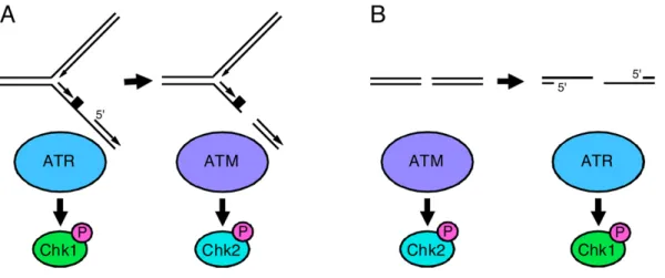
phosphorylates and down-regulates Cdc25A inhibiting the DNA replication and progression in the S-phase (Heffernan, Simpson et al. 2002). Although ATM and ATR are recruited by different types of lesion in the DNA their signalling cascades are interconnected. Indeed, DSBs can originate ssDNA (single-stranded-DNA) by DNA end resection or nucleotide excision repair (NER) and single strand breaks (SSBs) can originate DSBs by nuclease cleavage (Fig.12).
Fig.12 – Interconversion of ATR- and ATM-activating DNA damage. (A) ssDNA activates ATR. Nucleases can cleave
this structure causing DSBs to forms which activate ATM. (B) DSBs activate ATM but will also activate ATR as a consequence of DNA end resection or nucleotide excision repair (Cimprich and Cortez 2008).
c. G2/M checkpoint
The G2/M checkpoint prevents cells from undergoing mitosis in the presence of DNA damage. Depending on the type of DNA damage, the ATM-Chk2 signal transduction pathway and/or the ATR-Chk1 pathway is activated to arrest the cell cycle (Brown and Baltimore 2003). As in other checkpoints, with certain types of DNA lesions, such as those created by UV light, ATR-Chk1 signalling initiates cell-cycle arrest, but the maintenance of the arrest is then performed by ATM-Chk2 signalling (Abraham 2001). With other types of lesions, such as ionizing radiation-induced double-strand breaks, the order of action is reversed (Brown and Baltimore 2003). In any event, checkpoint kinases inhibit the entry into mitosis by down-regulating Cdc25C, which leads to inhibition of Cdk1/CyclinB activity (Yarden, Pardo-Reoyo et al. 2002).
Figure 4. Inter-conversion of ATR- and ATM-activating DNA lesions
(A) Stalled replication forks activate ATR. Nucleases can cleave stalled forks causing DSBs to form which activate ATM. The rate at which DSBs form at stalled forks is greatly increased in cells with defective ATR signaling. (B) DSBs activate ATM but will also activate ATR as a consequence of DNA end resection. This process is ATM- and cell cycle-dependent such that most ATR activation by double-strand breaks occurs in S and G2 phase cells. CHK1 and CHK2 are primarily ATR and ATM substrates respectively.
Cimprich and Cortez Page 28
Nat Rev Mol Cell Biol. Author manuscript; available in PMC 2009 March 31.
NIH-PA Author Manuscript
NIH-PA Author Manuscript