Universidade Nova de Lisboa
Instituto de Higiene e Medicina Tropical
Generation and characterisation of monoclonal antibodies against
cell cycle and cytokinesis regulators in Trypanosoma brucei
Cristina Isabel Correia de Almeida Costa
DISSERTAÇÃO PARA A OBTENÇÃO DO GRAU DE DOUTOR EM CIÊNCIAS BIOMÉDICAS, ESPECIALIDADE DE PARASITOLOGIA
iii
Universidade Nova de Lisboa
Instituto de Higiene e Medicina Tropical
Thesis: Generation and characterisation of monoclonal antibodies against cell cycle and cytokinesis regulators in Trypanosoma brucei
Author: Cristina Isabel Correia de Almeida Costa
Supervisor: Doctor Carlos Novo, IHMT, UNL
Co-supervisor: Professor Tansy C. Hammarton (University of Glasgow)
Tutorial Committee:
Doctor Carlos Novo (IHMT, UNL) Doctor Luís Távora Tavira (IHMT, UNL)
Professor Virgílio Estólio do Rosário (IHMT, UNL)
v
Universidade Nova de Lisboa
Instituto de Higiene e Medicina Tropical
Título da tese: Produção e caracterização de anticorpos monoclonais contra reguladores do ciclo celular e da citocinese em Trypanosoma brucei
Autor: Cristina Isabel Correia de Almeida Costa
Orientador: Investigador Doutor Carlos Novo, IHMT, UNL
Co-orientador: Professora Doutora Tansy C. Hammarton (University of Glasgow)
Comissão Tutorial:
Investigador Doutor Carlos Novo (IHMT, UNL) Investigador Doutor Luís Távora Tavira (IHMT, UNL) Professor Doutor Virgílio Estólio do Rosário (IHMT, UNL)
vii
Dedico esta tese,
À minha irmã Ana Isabel que comigo construiu um castelo À minha mãe, minha estrela-guia, que me tornou na mulher que sou hoje Ao meu querido avô, que tanta saudade deixa, que me ensinou a ser “Guerreira”
“…Ser feliz é reconhecer que vale a pena viver Apesar de todos os desafios, incompreensões e períodos de crise.
Ser feliz é deixar de ser vítima dos problemas e
Se tornar um autor da própria história.
É atravessar desertos fora de si, mas ser capaz de encontrar
Um oásis no recôndito da sua alma...
É agradecer a Deus a cada manhã pelo milagre da vida.
Ser feliz é não ter medo dos próprios sentimentos.
É saber falar de si mesmo.
É ter coragem para ouvir um 'não'.
É ter segurança para receber uma crítica, mesmo que injusta.
Pedras no caminho?
Guardo todas, um dia vou construir um castelo...”
ix
ACKNOWLEDGEMENTS
A lot of people have been by my side on this amazing adventure.
First of all, I would like to thank my supervisor, Dr Carlos Novo, for all the trust he put on me, for the guidance and support, and especially for always having my back and helping me when I was in need.
I would also like to thank my co-supervisor Prof. Tansy Hammarton, who allowed me to work in her lab and taught me so many things I would not know where to start. Thank you for the support, the confidance and for helping me “grow” in the lab.
This project allowed me to meet amazing people, whether in former UTPAM: Tiago, Bé, Ângela, Maria, Carla, Sofia, Ana Armada, Ana Domingos and Fernando; and recently in IHMT: Pimpolho, Cátia, Isabel, Pedro, Ana, Idalécia and so many other people. To all of you, thanks for the support and for accompanying me on this journey.
During my PhD I travelled regularly to Glasgow and a part of me will always stay there. First of all I would like to thank my “brother” Dave (one of my Musketeers). He turned Glasgow into a second home, his friendship helped me through the bad moments, but most important: he always had a HUG and a SMILE to share. To my two other Musketeers: Will and Nath, our adventures shall be immortal. To my sweet Craig, for caring and giving me love; to Elaine, for the fun and friendship; to Cat, for being my shoulder on the last year; to Jimbo, for the fun and the boozy lunches. To the brasilian gang: Herbert (my dear friend Safado), Tatiana, Dani, thank you for the the crazy times. To the Hammarton group: Corinna, Sophie, Glynn, Elizabeth, thanks for the help around the lab and for making work days such a blast. To everyone else (Jeremy, Elmarie, Esther, Amy, Ben, Fiona, Kirsty, Eileen…), it has been a pleasure to share such great moments and unforgettable experiences. I hope to see you all one day again, on a Friday afternoon at 4 p.m. in the usual place: Tennents!!
x
Raquel, this would not have been possible without you. You became my “sister”, and I shall never forget that you always saw the best in me. Thank you for always being there, for supporting me through the Westerns of Doom, for the crazy adventures, and for letting me be a part of your life outside the lab where I met Zé and Mini-Tarzan. Still today, when I am sad, I grab a glass of wine, start cooking and a voice starts singing…”You are the dancing queen….”.
To my family: thank you for believing in me. To my mother and father: without your faith in me I would not have successfully finished this project. To my guiding-star, my mum, for teaching me how to be loyal to myself and helping me become who I am today. Life has given me a lot of lessons, but it was you who helped me to interpret them and gave me the strength and values to stand up for myself. To my sister, we have been through a lot together… But we are two strong branches of the same tree, and we stay united and true. My admiration for you has no end, and no matter what life brings you, you will always have me to lean on. To my brother-in-law, for being so unique and special. To my second mum, Neia, for giving me the greatest gift of all: unconditional love. To my “little sister” Dani, for showing me the poem that will accompany me for the rest of my life and for guiding me so many times. To my family in Lisbon, thank you for giving me so many reasons to smile, especially my small princesses that bring me so much joy.
E porque a amizade é o maior ingrediente na receita da vida, finalmente um agradecimento a todos os meus amigos. Ao pessoal de Viseu, em especial à Suzy, Joaninha, Rafa, Mafaldinha e Vitinho, obrigada pela força. Um agradecimento especial à Marta e à Xanó, que ao longo dos últimos 13 anos têm sido as minhas “soul sisters”. Ao pessoal de Lisboa, entre muitos: o Afilhado, o Zica, a Marta e Fáfá (a dupla que tanto me aguentou…), o Duarte e Inês, foi um prazer partilhar estes anos com vocês e receber tanto.
xiii
Generation and characterisation of monoclonal antibodies against cell cycle and cytokinesis regulators in Trypanosoma brucei
Cristina Isabel Correia de Almeida Costa
Keywords: Trypanosoma brucei, cell cycle regulation, CRK12:CYC9, endocytosis, cytokinesis, synchronization, monoclonal antibodies, cytoskeleton
Trypanosoma brucei is the causative agent of African Trypanosomiasis, being transmitted by the bite of a blood-feeding invertebrate vector into the mammalian host. It affects 36 sub-Saharan African countries and the lack of efficient diagnostic methods and safe and effective drugs has led to the need for new control measures and novel treatment strategies.
Cell cycle progression in T. brucei is quite unique. Being so distinct from the mammalian cell cycle, regulatory proteins are believed to constitute good drug targets. However, many of these proteins remain to be identified, and their roles in specific pathways are still unknown. The cdc-2 related kinases (CRKs) and their cyclin partners are among the different cell cycle regulators that are vital for an accurate progression through the cell cycle. In T. brucei, several CRKs (CRK1-4 and CRK6-12) and cyclins (CYC2-11) have been identified, although a role in cell cycle regulation has not been established for all of them and only two active CRK:cyclin complexes have been identified in vivo. During this project, CRK12 and CYC9 have been proved to constitute a novel CRK:cyclin complex in vivo, in both the bloodstream (BSF) and the procyclic (PCF) stages. Interestingly, each protein regulates different biological processes in BSF trypanosomes. In fact, while CYC9 is involved in cell cycle regulation, blocking cytokinesis once depleted, CRK12 has a critical role in the endocytic pathway. It is the first time a CRK is connected with regulation of endocytosis, causing enlargement of the flagellar pocket if depleted. The interaction of CRK12 and CYC9 with additional proteins and the presence of functional redundancy between kinases/cyclins might explain the different functions.
The use of monoclonal antibodies (MAbs) and immunisation strategies as therapeutic techniques has been the subject of several studies. With that aim, α-CRK12 MAbs were efficiently generated, recognising the protein specifically in different T. brucei cell extracts. However, this MAb might not be suitable for immunolocalisation studies. Time constraints did not allow neutralization/protection studies.
xvii
Produção e caracterização de anticorpos monoclonais contra reguladores do ciclo celular e da citocinese em Trypanosoma brucei
Cristina Isabel Correia de Almeida Costa
Palavras-chave: Trypanosoma brucei, regulação ciclo celular, CRK12:CYC9, endocitose, citocinese, sincronização, anticorpos monoclonais, citoesqueleto
Trypanosoma brucei é o parasita que causa Trypanossomose Africana, sendo transmitido pela picada de um inseto vetor para a corrente sanguínea do hospedeiro mamífero. Afeta atualmente 36 países sub-saharianos e a falta de métodos de diagnóstico eficazes e de tratamentos seguros e eficazes, levou à necessidade de se desenvolverem novas medidas de controlo e novas estratégias terapêuticas.
O ciclo celular do parasita T. brucei é invulgar. Uma vez que é tão distinto do ciclo celular dos mamíferos, as proteínas que o regulam têm sido consideradas como possíveis alvos terapêuticos. No entanto, muitas proteínas reguladoras ainda não foram identificadas e a função específica de algumas proteínas envolvidas na progressão do ciclo celular é desconhecida. De entre os diversos reguladores do ciclo celular, estão as CRKs e as ciclinas que as ativam. Em T. brucei, foram identificadas 11 CRKs (CRK1-4 and CRK6-12) e 10 ciclinas. No entanto, ainda nem todas têm uma função conhecida e apenas dois complexos CRK:ciclina foram identificados in vivo. Durante este projeto, provou-se a existência de um novo complexo: CRK12:CYC9, que interage in vivo tanto na forma sanguínea como na forma procíclica. Um resultado interessante é que, apesar de interagirem, cada proteína regula diferentes processos biológicos na forma sanguínea do parasita. Na realidade, enquanto a CYC9 está envolvida na regulação do ciclo celular, levando a um bloqueio da citocinese em células em que não é expressa, a cinase CRK12 assume um papel essencial na regulação da endocitose. Esta consiste na primeira vez que uma CRK foi relacionada com o processo de endocitose de T. brucei, levando na sua ausência a um alargamento da bolsa flagelar. De forma a garantir estas diferentes funções, tanto a cinase CRK12 como a ciclina 9 podem interagir com outras proteínas, ou ser substituídas funcionalmente por outras cinases/ciclinas.
O uso de anticorpos monoclonais e ensaios de proteção têm sido estudados frequentemente como alternativas terapêuticas. Com esse objetivo, anticorpos monoclonais foram produzidos contra CRK12. Apesar de se ter produzido um anticorpo que reconhece especificamente esta cinase em diferentes extratos celulares de T. brucei, não foi possível usar o mesmo em estudos de localisação por fluorescência e os ensaios de neutralização/proteção não foram feitos devido a limitações de tempo.
Um projeto alternativo desenvolvido tinha como objetivo identificar possíveis proteínas reguladoras. Como tal, extratos de citoesqueleto foram obtidos em células sincronizadas em mitose e citocinese, e usados para geração de anticorpos monoclonais. Um total de 28 anticorpos foram selecionados. Apesar de não mostrarem especificidade contra uma fase específica do ciclo celular, produziram-se anticorpos com imunolocalisações muito interessantes. Além disso, estes anticorpos poderiam num futuro ser usados como ferramentas valiosas para estudar diferentes processos biológicas em T. brucei.
xix
ABREVIATIONS
4-NPP 4-nitrophenyl phosphate
AE attached epimastigote
AP alkaline phosphatase
ApoAI apolipoprotein AI ApoLI apolipoprotein L-I
AUK aurora kinase
BB basal body
BiP binding protein
bp basepair
BSA bovine serum albumin
BSD blasticidin
BSF bloodstream form Trypanosoma brucei
CBP calmodulin binding protein
CDK cyclin-dependent kinase
CIP calf intestinal alkaline phosphatase
CLH clathrin
CRK cdc-2 related kinase
CYC cyclin
DABCO 4-diazabicyclo[2.2.2]octane DAPI 4,6-diamidino-2-phenylindole
DE asymmetrically dividing epimastigote DMEM Dulbecco modified Eagle’s medium
DNA deoxyribonucleic acid
dNTP deoxyribonucleotide triphosphate
DTT dithiothreitol
EDTA ethylene diamine tetra acetic acid EGTA ethylene glycol tetraacetic acid ELISA enzyme linked immunosorbent assay
xx
ER endoplasmic reticulum
ES expression site
ESAG expression site associated gene
FAZ flagellar attachment zone
FBS foetal bovine serum
FITC fluorescein isothiocyanate
FP flagellar pocket
FPC flagellar pocket collar FTZ flagellum transition zone
FTZC flagellum transition zone component GFP green fluorescent protein
GPI glycosylphosphatidylinositol
Hb haemoglobin
HDL high-density lipoprotein
HGPRT hypoxanthine guanine phosphoribosyltransferase HMW high molecular weight (protein ladder)
Hpr haptoglobin related protein
HRP horse radish peroxidase
HSLS high salt lysis solution
HU hydroxyurea
HYG hygromycin
IFA immunofluorescence
IFT intraflagellar transport
IP immunoprecipitation
IPTG isopropyl β-D-1-thiogalactopyranoside
kDNA kinetoplast DNA
LB Luria bertani medium
LE long epimastigote
LMW low molecular weight (protein ladder) LSG lysis solution with glycerol
LSGI lysis solution with glycerol and inhibitors
xxi
MAPs microtubule associated proteins
MBP myelin basic protein
MOPS 3-(N-morpholino)propanesulfonic acid
MS mesocyclic trypomastigote
MT metacyclic epimastigote
MTOC microtubule-organizing center
MtQ microtubule quartet
M-VATs metacyclic variable antigen types
N/K nucleus/kinetoplast
NEO neomycin
NHS normal human serum
NLS nuclear localisation signal NP40 octylphenyl-polyethylene glycol
NSR normal rabbit serum
OD optical density
ORF open reading frame
PAP peroxidase anti-peroxidase
PBS phosphate buffered saline
PCF procyclic form Trypanosoma brucei
PDEB phosphodiesterase
PEG-DMSO polyethylene glycol–dimethyl sulfoxide
PFA paraformaldehyde
PFR paraflagellar rod
PLK polo-like kinase
PMSF phenylmethanesulfonylfluoride PNPP p-nitrophenyl phosphate
proBB pro-basal body
RAB ras-related proteins in brain
RNA ribonucleic acid
RNAi ribonucleic acid interference
SAXO stop axonemal protein
xxii
SE short epimastigote
SL slender trypomastigote
SRA serum-resistance associated
ST stumpy trypomastigote
TAC tripartite attachment complex TAP tandem affinity purification
TAX trypanosome axonemal protein
TbHPHbR haptoglobin-Hemoglobin receptor TBS tris buffered saline
TDB trypanosome dilution buffer
TEM transmission electron microscopy
Tet tetracycline
Tf transferrin
TfR transferrin receptor
TLF trypanosome lytic factor
TOR target of rapamycin
UTR untranslated region
v/v volume to volume
vPBS Voorheis’s modified PBS VSG variant surface glycoprotein
w/v weight to volume
WB western blotting
WT wildtype
xxiii
TABLE OF CONTENTS
Acknowledgements ... ix
Abstract ... xi
Resumo ... xv
Abreviations... xix
Table of Contents ... xxiii
Index of Figures ... xxix
Index of Tables ... xxxiii
1. Introduction ... 1
1.1. African Trypanosomiasis... 3
1.1.1. Epidemiology ... 4 1.1.2. Origin of human infectivity ... 7 1.1.3. AAT ... 9 1.1.4. HAT ... 10 1.1.5. Diagnosis ... 12 1.1.6. Treatment and control ... 14
1.2. Life cycle ... 18
1.2.1. T. brucei development in the mammalian host ... 18 1.2.2. T. brucei development in the tsetse fly host ... 22
1.3. Human natural immunity and Trypanosome immune evasion ... 25
1.3.1. The trypanosome lytic factor ... 25 1.3.2. The endocytic apparatus and immune evasion ... 30 1.3.3. Surface receptors ... 34
xxiv
1.4.1. Cytoskeleton ... 39 1.4.2. Subpellicular microtubules ... 40 1.4.3. Core cytoskeleton components: actin and tubulin ... 41 1.4.4. FAZ ... 43 1.4.5. PFR ... 46 1.4.6. Basal body ... 51 1.4.7. Flagellum ... 55 1.4.8. Microtubule Associating Proteins ... 59
1.5. The cell cycle ... 60
1.5.1. Cell cycle regulators: CRKs and cyclins ... 65
2. Materials and Methods ... 69
2.1. Culture, Transformation and storage of bacterial cells ... 71
2.1.1. Bacterial strains ... 71 2.1.2. Bacterial culture and storage ... 71 2.1.3. Production of competent cells ... 72 2.1.4. Bacterial transformation ... 72
2.2. Culture, Transfection, storage and analysis of T. brucei cells ... 73
xxv
2.3. Molecular Biology ... 80
2.3.1. Amplification and cloning of DNA ... 80 2.3.2. DNA gel electrophoresis ... 83 2.3.3. Restriction endonuclease analysis ... 83 2.3.4. Sub-cloning of DNA fragment into vectors of interest ... 84 2.3.5. Site-directed mutagenesis ... 85 2.3.6. Sequencing ... 85 2.3.7. Preparation of DNA for transfection into parasites ... 85 2.3.8. Plasmid generation ... 86
2.4. Protein biochemistry ... 87
2.4.1. Recombinant protein expression ... 87 2.4.2. Purification of His-tagged proteins ... 88 2.4.3. Dialysis of proteins ... 89 2.4.4. Determination of protein concentration ... 89 2.4.5. Protein electrophoresis ... 90 2.4.6. Coomassie staining ... 91 2.4.7. Western Blotting ... 91 2.4.8. Dot-blot ... 94 2.4.9. Immunoprecipitaton using protein G agarose beads ... 94 2.4.10. IP using Dynabeads® ... 95 2.4.11. Kinase assays ... 96
2.5. Uptake assays ... 96
2.5.1. FM4-64 uptake assay ... 96 2.5.2. Transferrin uptake assay ... 97
2.6. Microscopy techniques... 97
xxvi
2.6.2. Visualising direct-cell fluorescence ... 98 2.6.3. IFA microscopy using whole cells ... 99 2.6.4. IFA microscopy using cytoskeletons ... 101 2.6.5. Transmission Electron Microscopy ... 101
2.7. Antibody production ... 101
2.7.1. Immunisation of Balb/c mice ... 101 2.7.2. Antibody titre monitoring of mouse serum using ELISA ... 102 2.7.3. Production of hybridoma cell lines ... 103 2.7.4. Hybridoma cell culture and storage ... 105 2.7.5. Cloning by limiting dilution ... 106 2.7.6. Screening of hybridoma supernatants by ELISA ... 106 2.7.7. Screening of hybridoma supernatants by WB ... 107 2.7.8. Antibody classification ... 107 2.7.9. Antibody purification ... 108
3. Results and Discussion ... 111
3.1. T. brucei CRK12: Project aims ... 113
3.1.1. Generation of plasmids for production of recombinant CRK12 ... 114 3.1.2. Purification of recombinant His:CRK12 ... 118 3.1.3. Production of MAbs against CRK12 ... 121 3.1.4. Characterisation of 4D7: anti-CRK12 MAb ... 125 3.1.5. Ability of 4D7 to pull-down CRK12 from T.brucei cell lysates ... 128 3.1.6. Localisation of CRK12 by IFA using 4D7 ... 129
3.2. Characterisation of CRK12:CYC9 – a novel CRK:CYC complex
identified in T. brucei ... 131
xxvii
3.2.3. CRK12 and CYC9 function in BSF ... 142 3.2.4. The effect of CRK12 and CYC9 knockdown on cell growth and DNA content 142
3.2.5. CYC9 knockdown causes defects in cytokinesis ... 147 3.2.6. CRK12 knockdown causes defects in endocytosis ... 151
3.3. Mitosis and cytokinesis antibodies: Project aims ... 161
3.3.1. Cell cycle synchronization ... 161 3.3.2. Production of MAbs against cytoskeleton proteins expressed during mitosis and cytokinesis in BSF trypanosomes ... 166 3.3.3. Classification of the cell cycle specificity of the generated antibodies .. 171 3.3.4. Characterisation of MAbs recognising proteins with cytoplasmic distribution or with inconclusive results ... 176 3.3.5. Characterisation of MAbs recognising the cell body and the flagellum . 189 3.3.6. Characterisation of MAbs recognising the BB/FP region ... 194 3.3.7. Characterisation of MAbs with flagellar distribution ... 197 3.3.7.1. Characterisation of MAbs recognising the whole flagellar structure
197
3.3.7.2. Characterisation of MAbs recognising specific points in the flagellum 213
3.3.8. Characterisation of MAbs with unique localisation ... 221
4. Conclusions ... 225
4.1. CRK12 and CYC9 function in distinct biological processes but comprise a
novel CRK:CYC complex in T. brucei ... 229
4.2. Mitosis and cytokinesis antibodies ... 237
4.3. Final conclusions ... 244
xxix
INDEX OF FIGURES
Figure 1. 1 - Distribution of Human African trypanosomiasis and prevalence of each human-infective species. ... 7 Figure 1. 2 - Life cycle of Trypanosomes, showing the role of the insect vector and the human and animal reservoirs in the T. brucei cycle. ... 10 Figure 1. 3 – The life cycle and parasiteamia profile of T. brucei in the mammalian host. ... 20 Figure 1. 4 – Transition from slender to stumpy forms in the mammalian host. ... 21
Figure 1. 5 – Development of T. brucei in the tsetse fly. ... 23
Figure 1. 6 – Characteristics of the two trypanolytic particles, TLF1 and TLF2. ... 26
Figure 1. 7 – Model of the lytic mechanism. ... 29
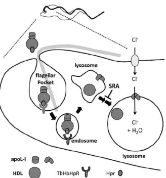
Figure 1. 8 – Schematic representation of the T. brucei endocytic system. ... 31
Figure 1. 9 – Flow diagram of fluid-phase and VSG through the endocytic apparatus. .... 33 Figure 1. 10 – Schematic representation on Tf uptake in BSF trypanosomes. ... 35
Figure 1. 11 – Schematic representation of a T. brucei BSF cell. ... 37 Figure 1. 12 – Negatively stained PCF T. brucei cytoskeleton after detergent extraction. ... 39 Figure 1. 13 – Longitudinal section of the FAZ (A) and the PFR (B). ... 44
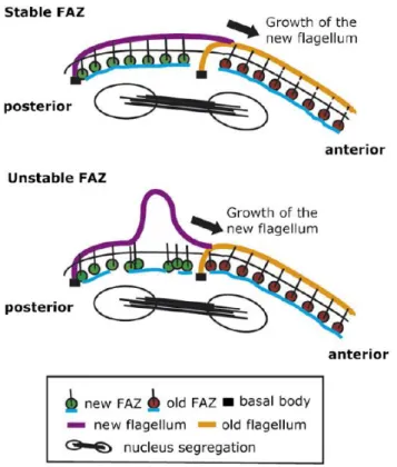
Figure 1. 14 – Schematic representation of a stable vs unstable FAZ in T. brucei cells following knockdown of FAZ1. ... 46 Figure 1. 15 – The T. brucei flagellum and the PFR.. ... 47
Figure 1. 16 – Transverse sections of uninduced and induced PFR2 RNAi cells. ... 49
Figure 1. 17 – BB architecture. ... 51
Figure 1. 18 – Model of the kinetoplast duplication cycle. ... 52
Figure 1. 19 – Repositioning of the BB during cell division, shown by a series of negatively stained cytoskeleton extracts. ... 54 Figure 1. 20 – Architecture of the FP. ... 57
Figure 1. 21 – Schematic representation of T. brucei cell cycle in PCF cells. ... 61
xxx
Figure 3. 1 - DNA gel electrophoresis of CRK12 fragments amplified from T. brucei 427 genomic DNA. ... 115 Figure 3. 2 - DNA gel electrophoresis of restriction endonuclease reactions performed for CRK12 constructs to confirm the presence of the correct insert. ... 116 Figure 3. 3 - DNA gel electrophoresis of restriction endonuclease digestion of pHG97, pHG98, pHG110 and expression vectors pET28a+ and pGEX5X1. ... 117 Figure 3. 4 - Expression of 6xHis:CRK12 under different conditions. ... 119 Figure 3. 5 - Solubilization of 6xHis:CRK12... 120 Figure 3. 6 - Purification of 6xHis:CRK12 by affinity chromatography. ... 121 Figure 3. 7 - ELISA results for supernatants from 24 well plates. ... 123
Figure 3. 8 - Screening of supernatants from hybridoma cultures by WB against recombinant 6xHis:CRK12 and a negative control – 6xHis:PLK. ... 124 Figure 3. 9 - Screening of anti-CRK12 supernatants against tet induced ty:CRK12 expression. ... 125 Figure 3. 10 - Purification of MAb anti-CRK12 4D7 by affinity chromatography, using a protein G HP purification column. ... 126 Figure 3. 11 - WB analysis of specificity of 4D7 against T. brucei CRK12. ... 127
Figure 3. 12 - Ability of 4D7 anti-CRK12 antibody to pull-down ty:CRK12 from BSF T. brucei. ... 128 Figure 3. 13 - Localisation of CRK12 in PCF and BSF parasites. ... 129
Figure 3. 14 – Specificity of anti-CRK12 4D7 antibody for IFA. ... 130 Figure 3. 15 - WB analysis to confirm expression of ty:GFP:CRK12 in transfected cell lines. ... 132 Figure 3. 16 -Analysis of CYC9:TAP expression in BSF transfectants. ... 133
Figure 3. 17 –Analysis of ty:CRK12 IP from BSF parasites co-expressing CYC9:TAP. 134 Figure 3. 18 –Localisation of CYC9:TAP in BSF T. brucei parasites. ... 136 Figure 3. 19 - Localisation of CYC9:TAP in PCF T. brucei parasites. ... 137 Figure 3. 20 - Localisation of ty:CRK12 in uninduced over-expression BSF cell lines.. . 139 Figure 3. 21 - Localisation of ty:CRK12 in induced over-expression BSF cell lines. ... 140
Figure 3. 22 – Cumulative growth curves of BSF CRK12 RNAi cell lines. ... 143
xxxi
Figure 3. 25 - Flow cytometry profiles of BSF CYC9 RNAi cell line. ... 146
Figure 3. 26 – DAPI staining of BSF CYC9 RNAi cell lines. ... 147 Figure 3. 27 - Analysis of the cytokinesis stage of 2N2K cells following depletion of CYC9 in BSF T. brucei. ... 148 Figure 3. 28 - DAPI staining of typical 2N2K cells at different cytokinesis stages. ... 149
Figure 3. 29 – DAPI staining of multinucleate/kinetoplast cells visible at 18 hours post CYC9 RNAi induction in the BSF. ... 150 Figure 3. 30 - DAPI staining of BSF CRK12 RNAi cell lines. ... 152 Figure 3. 31 - Analysis of the cytokinesis stage of 2N2K cells following depletion of CRK12 in BSF T. brucei. ... 153 Figure 3. 32 - DAPI staining of BSF cells with enlarged FPs following CRK12 RNAi induction. ... 154 Figure 3. 33 – Quantification of abnormal kinetoplast positioning in 1N2K and 2N2K cells following induction of CRK12 RNAi. ... 155 Figure 3. 34 – TEM images of FPs for uninduced ( -tet) and induced (+tet) CRK12 BSF RNAi cells.. ... 156 Figure 3. 35 – FM4-64 uptake assay using CRK12 BSF RNAi cells. ... 157
Figure 3. 36 – AF594-transferrin uptake assay using CRK12 BSF RNAi cells. ... 158 Figure 3. 37 - Flow cytometry profiles of PCF WT cell line, grown in the presence of HU for 12 hours. ... 162 Figure 3. 38 - N/K configuration analysis of PCF WT cells treated with HU. ... 163 Figure 3. 39 - Flow cytometry profiles of BSF WT cell line, grown in the presence of HU for 6 hours. ... 165 Figure 3. 40 - N/K configuration analysis of BSF WT cells treated with HU. ... 166
Figure 3. 41 – Cytoskeleton protein samples of BSF WT. ... 167
xxxii
xxxiii
INDEX OF TABLES
Table 1. 1 - Localisation of each stage during the parasite life cycle. ... 24 Table 2. 1 - T. brucei strains used in this study. ... 74 Table 2. 2 - T. brucei strains generated in this study. ... 74 Table 2. 3 - Final antibiotic concentrations for T. brucei culture. ... 75 Table 2. 4 - Protein molecular weight characteristics of the different polyacrylamide gel slices obtained for synchronized BSF samples. ... 80 Table 2. 5 - Oligonucleotides used during this study. ... 81 Table 2. 6 - Composition of the PCR reaction mixes used in this study... 82
Table 2. 7 – Plasmids generated in this study. ... 86 Table 2. 8 - Plasmids used in this study. ... 86
Table 2. 9 – Reagents used for preparation of the separating gel for SDS-PAGE at different concentrations. ... 90 Table 2. 10 - Reagents used for preparation of the stacking gel for SDS-PAGE. ... 90 Table 2. 11 - Antibodies used for WB. ... 93 Table 2. 12 - Antibodies used for IFA microscopy. ... 100
Table 2. 13 - Schematic representation of the distribution of reagents in the ELISA plate used for antibody classification. ... 108 Table 3. 1 - Expected sizes of fragments obtained after restriction endonuclease digestion of CRK12 constructs. . ... 115 Table 3. 2- Nucleotide point mutations observed by DNA sequencing and corresponding changes at the protein level. ... 116 Table 3. 3 - Results from supernatant screening against recombinant 6xHis:CRK12. . .... 122
1
3
1.1.
AFRICAN TRYPANOSOMIASIS
Trypanosomiasis is an infectious disease caused by unicellular parasites from the Protozoa kingdom and genus Trypanosoma. Trypanosoma species infect a variety of hosts, and the genus includes two human-infective species: T. cruzi which causes Chagas’ disease in South America and accounts for 13,000 deaths per year (Barrett et al., 2003) (also known as American Trypanosomiasis); and T. brucei that causes Human African Trypanosomiasis (HAT, also known as Sleeping Sickness) in sub-Saharan Africa. These species differ considerably in terms of their biology and host-parasite interactions, causing diverse symptoms and there is a need for several approaches in terms of control and treatment (Barrett et al., 2003). Besides human infective hosts, several Trypanosoma species can affect animals: T. vivax infects ruminants, horses and camels; T. equiperdum causes venereal equine disease and can be spread through coitus; T. congolense causes acute fever, anaemia and neurological signs in dogs and cats; T. simiae leads to pyrexia in pigs; while T. evansi affects all domestic mammals. Recent studies (Lai et al., 2008) have shown that T. equiperdum and T. evansi are actually strains of T. brucei that have lost part or all of their kinetoplastid DNA (kDNA), and in 2005 it was confirmed the first report of human infection due to T. evansi, transmitted probably by the blood of an infected animal (Joshi et al., 2005).
4
Human sleeping sickness occurs in 36 sub-Saharan African countries and takes two forms according to the subspecies of parasite causing the infection: T. b. gambiense causes a chronic infection and is found in west and central Africa; while an acute disease arises from infection with T. b. rhodesiense, which mainly affects both eastern and southern Africa. T. b. gambiense causes more than 95% of all cases reported. Both species are fatal if left untreated, although recent data have shown that infection with T. b. gambiense is not 100% fatal and can naturally progress to an asymptomatic infection or to a spontaneous resolution (Jamonneau et al., 2012). Epidemiological history and actual data are reported mainly by the World Health Organization (WHO –
www.who.int), which has described a decrease of the total number of HAT cases in 2009 to below 10,000, for the first time in 50 years. However, an approximate estimate suggests that 10% of cases are left unreported, and for those reported, the commencement of treatment is delayed due to time taken to visit the health centres, inappropriate diagnosis and lack of diagnostic facilities. According to WHO, elimination of HAT is feasible if new diagnosis and staging methods are created, if safe and effective drugs are generated for both stages of the disease, if mobile teams are used to find new active cases and vector control is employed.
1.1.1. Epidemiology
African Trypanosomiasis has been identified and characterised in several reservoirs in the African continent since the 14th century (Cox, 2004). By 1903, Scottish pathologist and microbiologist David Bruce had identified the causative agent and the vector, and concluded that the bite of the tsetse fly would transmit the disease (Cox, 2004, Fevre et al., 2006). In 1910, the differentiation between subspecies of the protozoa was achieved, a time at which the first treatment was introduced, an arsenic-based drug which caused blindness as a side effect. T. b. gambiense was described by Forde in 1902 (Forde, 1902), while T. b. rhodesiense was only identified in 1910 in Zambia (Stephens and Fantham, 1910).
5
was dependent on understanding the life cycle and adaptive changes undergone by the parasite, involving activation or repression of metabolic pathways and ultrastructural changes (Vickerman, 1985, Vickerman, 1994). Phylogenetic trees were created using the gene encoding for the nuclear small subunit (SSU) rRNA, which underwent an evolutionary change in history and as such allowed an estimation of the relationship between kinetoplastids. The Kinetoplastida taxon was created 40 years ago, consisting of protozoan flagellates with a disk-shaped mass of mitochondrial DNA (kDNA), condensed in a structure termed the kinetoplast, located at the base of the flagellum and associated with the basal body (BB). Molecular phylogenetic studies soon included other informative markers, such as heat-shock proteins, which meant that our understanding of the evolutionary relationship between different protozoans evolved over the years (Cavalier-Smith, 1993, Fernandes et al., 1993, Stevens et al., 2001, Simpson et al., 2006). In the first studies, T. brucei appeared on a long deep branch of the phylogenetic trees, considered then to be the most ancient divergent within the trypanosomatids and having a distant relationship with the Leishmania genera. In 2001 (Stevens et al., 2001) with the study of the molecular evolution of trypanosomatids, the idea of T. brucei and T. cruzi clades appeared, having a close relationship between themselves and with Leishmania. Later on (Simpson et al., 2006), the phylogenetic trees showed that Leishmania and Trypanosoma were two of the branches inside the Trypanosomatida group, the second one further divided into the T. brucei and T. cruzi clades.
6
a solution to this problem, involving the collaboration of several agencies, organizations and research institutes. Between 1995 and 2006, the total number of cases showed a 68% decrease, stimulated mainly by an increase of awareness and surveillance, and from the effort of public institutions and private pharmaceutical companies, such as Aventis, that in 2001 agreed to supply eflornithine to WHO for free (Barrett, 2006).
Currently, the number of cases reported to the WHO has decreased to <10,000, although it is believed that a lot of cases are left unreported or undiagnosed, and care should be taken as negligence could lead to resurgence of HAT. However, the prevalence of the disease differs from one country to another, and the numbers differ between the two different species. The reduction in cases observed until 2006 was mostly visible for T. b. gambiense, while T. b. rhodesiense cases did not show a similar decrease due to lack of control measures. A 69% decrease in new cases was observed for infections due to T. b. gambiense, responding to the implementation of control activities and active surveillance. On the contrary, for T. b. rhodesiense (causing 3% of the total number of cases) the containment measures were insufficient, probably due to the part played by the animals as reservoirs for transmission (MacLeod et al., 2001b, MacLeod et al., 2001a, Welburn et al., 2001, Hide and Tait, 2009). In the years between 1997 and 2006, 24 of the 36 endemic countries for HAT suffered from T. b. gambiense transmission, while in 2006, 11 out of these 24 reported no cases (Simarro et al., 2008). In that same period, 13 countries endemic for T. b. rhodesiense showed a 21% reduction in the number of new cases reported, even though only 4 countries implemented control measures (Simarro et al., 2008).
7
Figure 1. 1 - Distribution of Human African trypanosomiasis and prevalence of each human-infective species. The black line divides the area between the distribution of each species. (Brun et al.,
2010)
1.1.2. Origin of human infectivity
Despite the widespread distribution of tsetse vectors and animal trypanosomiasis, the human disease is only found in discrete foci, which periodically give rise to epidemics followed by periods of endemicity. Nearly 300 separate active foci are recognised. Over the years, several biochemical and molecular tools have been designed and used to better understand parasite genetic diversity, increasing our knowledge on the role of reservoir hosts and population structure in the epidemic events (MacLeod et al., 2001b, MacLeod et al., 2001a, Hide and Tait, 2009).
8
2001a). Considering that mixed infections occur at a significant frequency, and a significant proportion of tsetse flies carry more than one genotype, the possibility of genetic exchange occurring in the field arises.
The extent of genomic exchange in T. brucei spp. is unclear, and has been debated over the years using different molecular epidemiological tools (Sternberg and Tait, 1990, Tait and Turner, 1990, Gibson and Stevens, 1999). Enzyme-based and DNA-based methods (Hide and Tait, 2009) have shown that the presence of significant genetic exchange could explain the emergence of new epidemics. Using these tools and considering the major epidemics observed during the years, Hide and Tait (Hide and Tait, 2009) concluded that new outbreaks could be generated by the importation of reservoir species with a human strain into an area previously occupied only by animal strains (Welburn et al., 2001). The emergent human strain spreads quickly into the naive population, establishing a new site of human-infective species, masking the occurrence of genetic exchange.
Human infectivity had multiple origins (MacLeod et al., 2001a, Barrett et al., 2003) and generated independently in different geographical regions. Using different marker systems, researchers have concluded that evolution of T. brucei seemed to have occurred in four separate occasions, giving rise to T. b. rhodesiense Uganda; T. b. rhodesiense Zambia; T. b. gambiense Type 1 and T. b. gambiense type 2 (MacLeod et al., 2001a). Type 1 gambiense trypanosomiasis is genetically highly homogeneous and distinguishable from the other human-infective strain and T. b. brucei, while Type 2 is closely related to West African T. b brucei (Barrett et al., 2003). T. b. rhodesiense expresses a serum-resistance associated (SRA) gene (section 1.3), as opposed to the animal-infective species and to T. b. gambiense, a gene that has been extensively used in epidemiological studies (MacLeod et al., 2001a, Barrett et al., 2003, Hide and Tait, 2009). These studies have shown that human-infective trypanosomes are present in animal populations, and analysis of T. b. rhodesiense isolates lead to the conclusion that these strains are host-range variants of local T. b. brucei populations, that have become genetically isolated.
9
of new drug targets and pathways, and provided a starting point for functional studies. Being a model for the human-infective species, the animal-infective parasite has been used for a long time in research to increase the understanding of parasite development and biological characteristics.
1.1.3. AAT
Trypanosomiasis affects human health, both directly through sleeping sickness but also indirectly by the destruction of livestock. Parasite diseases that affect livestock cause serious economical problems, leading to food shortages in the communities and making impossible the use of animals for transport and traction. AAT can be caused by several species, the most important being: T. congolense, T. vivax and T. b. brucei, but concurrent infection can be observed with more than one species. AAT caused by the species T. b. brucei affects a large range of animals, from cattle and sheep, to dogs and cats. However, the major obstacle to economic development of affected rural areas comes from the presence of the disease in cattle (Nagana) which causes an acute or chronic disease characterised by fever, anaemia, diarrhoea, and often resulting in animal death.
10
acting indirectly in human health, animals can also host human pathogen parasites, and act as an important reservoir for human infection (Welburn et al., 2001).
1.1.4. HAT
As mentioned previously, inside the Trypanosomatidae, two different species can lead to HAT, having different rates of progression and clinical features, but being both spread by the bite of tsetse flies (Figure 1. 2). The parasite can be found in the peripheral circulation in the first stage, in what is known as a haemolymphatic phase (Barrett et al., 2003, Brun et al., 2010). The leading signs of the first stage are chronic and intermittent fever, headache, general malaise, anaemia, and parasites can be found in the blood or lymph, although usually they are below detection levels. The intermittent fever is related to the mounting of an immune system response by the host (Brun et al., 2010), which the parasite can evade by antigenic variation.
In the second stage, the so-called neurological phase, the nervous system is disturbed and the more obvious signs appear: changes in behaviour, confusion, sensory disturbance, and disruption of the sleep cycle (Barrett et al., 2003, Brun et al., 2010). Brains function deteriorates, culminating in coma and then death. Such disorders are rarely seen during the first stage, and their frequency increases with the duration of the infection.
11
Over the years, the clinical signs have been followed to increase our knowledge of disease progression and distribution (Smith et al., 1998, Fevre et al., 2006). Patient follow-up has proved that the symptoms of HAT differ between African patients and travellers, with normally the latter presenting greater abnormalities (Brun et al., 2010). The presence of a chancre at the site of the bite is more common, patients suffer from acute fevers whatever the causative agent, and other severe symptoms like loss of kidney function have been described in travellers.
The clinical presentations of disease caused by the human-infective species – T. b. gambiense and T. b. rhodesiense – are remarkably different. The former causes a chronic illness that lasts for years, while the latter usually appears as an acute febrile illness that leads to patient death in weeks/months if left untreated.
T. b. gambiense affects populations in west and central Africa, being transmitted by flies of the G. palpalis or G. fuscipes groups (Chappuis et al., 2005). Infection occurs mainly in forested rivers, shores or near plantations, causing a chronic infection in which people can be infected for several months with an asymptomatic disease (Barrett et al., 2003, Brun et al., 2010). When symptoms appear, the patients are normally in an advanced stage, where the CNS is affected. The disease progresses slowly, starting with mild symptoms such as intermittent fever, headaches, muscle and joint aches, and possible itching, swollen lymph nodes and weight loss. Normally, after 1-2 years, neurological signs appear due to CNS invasion, such as personality changes, daytime sleepiness and progressive confusion, together with hormonal imbalances and balance or walking problems. If untreated, the infection often kills in 3 years and rarely lasts longer than 6-7 years (Brun et al., 2010), and patients suffer from dysfunction of the immune system, enter a deep coma and often show parallel bacterial infections (Chappuis et al., 2005).
12
(Chappuis et al., 2005). The first symptoms appear within 1-2 weeks of the infective bite, being indistinguishable from other tropical fevers. As opposed to T. b. gambiense infection, even at the early stage, the patient can show severe symptoms, such as heart failure or pulmonary oedema (Barrett et al., 2003), which can cause patient death even before the CNS is invaded by the parasite (Chappuis et al., 2005). Staging of infection for this specie is not easy, although normally the second stage of the disease starts a few weeks after infection and most deaths (80%) occur within months (Chappuis et al., 2005, Brun et al., 2010).
1.1.5. Diagnosis
Although clinical symptoms can point to HAT, they cannot be taken into consideration alone, as other diseases such as malaria, enteric fever and HIV can mimic the signs and lead to misdiagnosis (Chappuis et al., 2005). Laboratory methods are needed to confirm infection with trypanosomes, and complete the insufficient data given by the clinical features. Diagnosis is performed in three steps: screening for a potential infection; diagnostic confirmation by examination of blood, lymph node or Cerebral Spinal Fluid (CSF) aspirates; and finally staging to determine disease progression and course of treatment, by examining the CSF. It is necessary to take into account that material resources and human investment are scarce in Africa, and consequently many infected individuals might perish before being diagnosed.
13
and as such an alternative test was created: LATEX/T. b. gambiense (Büscher et al., 1999), which is similar to CATT but uses a combination of three VATs (LiTat 1.3, 1.5 and 1.6). When compared to CATT, this assay has a higher specificity (96%-99%) but a lower or similar sensitivity (Chappuis et al., 2005).
Some serological tests such as immunofluorescence (IFA), enzyme-linked immunosorbent assays (ELISA) (Olaho-Mukani et al., 1994, Lejon et al., 1998) and PCR (Kanmogne et al., 1996, Truc et al., 1999, Kabiri et al., 1999, Kyambadde et al., 2000) can be performed, although their use is limited in remote areas and conditions need to be standardized (Barrett et al., 2003, Fevre et al., 2006). Some assays use antibody levels as infection markers, as IgG and IgM antibodies are present in high concentrations during infection, although this technique greatly depends on the antigen used (Barrett et al., 2003, Chappuis et al., 2005, Fevre et al., 2006). New technologies have also been reviewed, such as detection of trypanosome specific antibodies in the saliva (Lejon et al., 2003), or the use of proteomic signatures and mass spectrometry analysis (Papadopoulos et al., 2004), although these assays would be impossible to perform in the field.
Parasitological confirmation is made by microscopic examination of lymph node aspirates, blood, or CSF (Cattand et al., 1988, Barrett et al., 2003, Chappuis et al., 2005, Fevre et al., 2006, Brun et al., 2010), which gives a definite diagnosis. This method of direct detection is only accurate at parasite concentrations above 104.ml-1 (Fevre et al., 2006), which makes it easier to detect T. b. rhodesiense infections, due to the higher density of circulating parasites. Because there is no serological test for T. b. rhodesiense, detection of infection due to this agent relies primarily on clinical symptoms and signs, which can be quite non-specific, followed by molecular methods such as PCR analysis for the SRA gene (Welburn et al., 2001) and microscopic confirmation (Chappuis et al., 2005, Brun et al., 2010).
14
more of the following: trypanosomes; raised white blood cells (more than 5 cells.µl-1); increased protein content (370 mg.l-1). As the second stage causes sleeping disorders, sleep-wake recordings can be used to access the CNS involvement (Buguet et al., 2005, Simarro et al., 2008).
Over the years, advances have been made in diagnostic tools for HAT. However, care should be taken that these are suitable for African rural centres, which need the most basic and cost-effective techniques.
1.1.6. Treatment and control
There are no vaccines against sleeping sickness, and preventive measures consist of minimizing contact with tsetse flies. The principles of control consist of treating the infected individuals, finding and treating new cases promptly, and controlling the vector. In terms of treatment, few drugs are available for treatment of HAT and selection is dependent on the parasite species involved and on the stage of the disease. Drugs need to be of low toxicity and easy to administer, which is not the case for current first stage and second stage drugs; while drugs for the late stage need to be able to cross the blood-brain barrier in order to clear trypanosomes in the CSF. Along with the toxicity of the drugs used, other difficulties arise from lack of supply and the development of parasite resistance (Matovu et al., 2001b, Geerts et al., 2001).
15
used at both stages of the disease although being preferably used for late stage infection (Van Nieuwenhove, 1999).
Suramin, indicated for early-stage treatment, consists of a highly charged molecule that is believed to be taken up by the trypanosomes by receptor-mediated endocytosis, when bound to serum proteins (Burchmore et al., 2002). It was discovered in 1921, is administered intravenously, but along with its high toxicity has been reported to fail in 25%-35% of the cases (Burchmore et al., 2002). Its dosage begins with 5 mg.kg-1 weight on day 1 and treatment continues for 30 days using 20 mg.kg-1 (Burchmore et al., 2002, Barrett et al., 2003), provoking, however, certain undesirable side effects in the urinary tract and allergic reactions (May and Allolio, 1991).
The second drug used only for early stage disease is pentamidine, which is generally well tolerated by patients as it is not as toxic as other HAT drugs (Fevre et al., 2006, Brun et al., 2010), although it is less effective against early stage T. b. rhodesiense (Apted, 1980). Pentamidine has been used for over 50 years for sleeping sickness treatment (Pépin and Milord, 1994), and appears to work directly on the parasite, although the way it induces parasite clearance has not been established yet (Burchmore et al., 2002). It is given intramuscularly daily, for a total of 7 to 10 injections of 4 mg.kg-1 weight (Burchmore et al., 2002, Barrett et al., 2003).
16
Keiser, 2001, Matovu et al., 2001a), with increasing reports of drug failures concentrated mainly in central Africa, reaching averages of 30% in some foci.
In the case of HAT caused by T. b. gambiense, when patients reach the second stage of the disease, their other possible treatment relies on eflornithine injections (Burri and Brun, 2003), given for a total of 14 days with several daily infusions due to the short half-life of the drug (Burchmore et al., 2002, Simarro et al., 2008, Brun et al., 2010). This anti-trypanocidal drug was first developed as an anti-cancer agent, being uptaken from the plasma membrane in a passive mode and acting as an inhibitor to a decarboxylase enzyme. Its high efficiency, low mortality rate and reduced adverse reactions make eflornithine the first line treatment for sleeping sickness caused by this specie. As side effects, patients can suffer from anaemia, diarrhoea and vomiting, amongst others; but the major drawback of this treatment is its high price. A drug used for treatment of Chagas disease – Nifurtimox – has been recently introduced as a combined treatment with eflornithine (Priotto et al., 2009), although being effective only for infections with T. b. gambiense.
To overcome the adverse effects of trypanocidal drugs, combination treatments have been studied, which would lessen the dose-related side effects (Priotto et al., 2006, Priotto et al., 2007, Priotto et al., 2009). On the other hand, a new orally available drug – DB289 - is at the final clinical trial phase but seems to be effective only in first stage disease (Barrett et al., 2003, Simarro et al., 2008).
cross-17
resistance occurs more often when drugs target related metabolic pathways (Matovu et al., 2001b), raising the possibility that by affecting different metabolic processes in combination, one can increase the efficiency of treatment.
The development of new drugs should focus on pathways that are present in the parasite but absent from their hosts (Barrett et al., 1999), using technologies such as comparative biochemistry and comparative genomics to identify ideal targets. The trypanosome genome project (Berriman et al., 2005) was essential to detect new potential drug targets and establish them as new treatment pathways. Target validation has been based widely on gene knockout and ribonucleic acid interference (RNAi) approaches (Ngô et al., 1998, Wirtz et al., 1999, Barrett et al., 1999, Alsford and Horn, 2008, Monnerat et al., 2009), although lately, high-throughput screening approaches have emerged (Alsford et al., 2011, Alsford et al., 2012). By stipulating that a gene is essential for parasite viability, one cannot assume that it consists of a good target; and caution should be taken when using gene knockout experiments as the sole criterion. Several metabolic pathways and signalling pathways have been studied over the years as credible targets (Burchmore et al., 2002, Barrett et al., 2003); and key cell cycle regulators have been established as good drug targets (Hammarton et al., 2003b).
18
decrease in reservoir infection is more difficult due to the variety of animal hosts (section 1.1.3). As such, vector control is the primary strategy used.
1.2.
LIFE CYCLE
Elucidation of the life cycle of African trypanosomes started in 1894, when British Officer Bruce went to South Africa to investigate the outbreak of a disease in cattle. Although expecting to find a bacterial source for the disease, he discovered trypanosomes in the blood of the infected animals, and demonstrated that transfer of parasites could be achieved from one animal to the other (Cox, 2004). The role of the tsetse flies as carriers of the disease was also identified by Bruce, allowing later on the establishment of the overall life cycle of T. brucei (Figure 1. 2).
Over the years, the life cycle of T. brucei has been the subject of several studies (Vickerman, 1985, Matthews, 1999, Matthews et al., 2004, Van Den Abbeele et al., 2010, Peacock et al., 2011), with significant advances in our knowledge of the different morphologies, gene expression rates and proliferation statuses at the different stages, and allowing the identification of the regulatory machinery. Being transmitted by the bite of the tsetse fly, T. brucei has a biphasic life cycle, linked strongly with the cell cycle as the parasite passes through replicative and cell cycle-arrested forms (Matthews and Gull, 1994, Hammarton et al., 2003b), and undergoes several pre-adaptive morphological changes (Matthews, 1999, Matthews et al., 2004, Matthews, 2005, Macgregor et al., 2012).
1.2.1. T. brucei development in the mammalian host
19
the peak of parasitaemia (Matthews, 1999, Matthews et al., 2004, Matthews, 2005, Macgregor et al., 2012). Infections can be sustained due to the capacity of trypanosomes for antigenic variation (Ziegelbauer and Overath, 1990), allowing them to periodically change their variant surface glycoproteins (VSG) which constitute their surface coat (McCulloch, 2004, Pays et al., 2004), outpace the immune system and maintain an infection. Together with antigenic variation, immune evasion is enabled by endocytic recycling of membrane bound antibodies (Overath and Engstler, 2004, Vanhollebeke et al., 2010).
20
Figure 1. 3 – The life cycle and parasiteamia profile of T. brucei in the mammalian host. An
undulating parasiteamia profile is maintained by the transition between the two BSF: proliferative slender and cell cycle arrested stumpy forms; the first being responsible for the increase in parasite numbers. Transformation into ST forms is induced by the release of SIF, leading to developmental changes in order to generated transmissible T. brucei forms, adapted for uptake by a tsetse fly during a blood meal. Once in the vector host, they differentiate to midgut procyclic forms, after which they proliferate and migrate to the salivary glands, where they attach and develop to metacyclic forms, which are infective to mammals.
(Macgregor et al., 2012)
SL form parasites are able to take advantage of the nutrients present in the bloodstream, degrading them in spherical glycosomes, and components of the Krebs cycle and oxidative phosphorylation are mainly absent. The replacement of proliferative with non-proliferative forms ensures the morphological changes needed for survival in the invertebrate host. A few days after the infection, the parasite population consists uniformly of proliferative SL form trypanosomes. After 4 days of infection, differentiation into ST forms begins and cells become arrested in G1 phase (Macgregor
21
modification of the kinetoplast position by growth of the posterior end of the cell. An essential change upon differentiation into ST form is the mitochondrial elaboration, which is estimated to occur after 15 minutes of the start of differentiation (Bakker et al., 1995). SL form parasites are adapted to the rich glucose environment of the blood, and as ST forms quickly need to adapt to loss of glucose when in the tsetse fly, a fully active mitochondrial respiratory chain is essential. All these changes are essential as they pre-adapt the parasites to the fly midgut (Tetley and Vickerman, 1985). Additionally, transition from SL to ST form is accompanied by changes in the endocytic machinery, as this pathway is mainly absent from procyclic forms (PCF) (Vanhollebeke et al., 2010). Among these changes, a relocalisation of the lysosome is observed, with it acquiring an anterior position to the nucleus (Vanhollebeke et al., 2010), and internalisation of several ligands is reduced. In fact, in ST form trypanosomes, the haptoglobin-hemoglobin receptor (TbHpHbR) is downregulated (Vanhollebeke et al., 2010), causing a block in uptake of the complex, making this form more resistant to the trypanolytic particles in human serum.
Figure 1. 4 – Transition from slender to stumpy forms in the mammalian host. Release of SIF stimulates slender forms to begin the transition to stumpy forms. Before cell cycle arrest in G1 phase, a
committed slender form can still undergo 3 rounds of cell division, after which SIF production is lost. By this time, intermediate forms no longer express the TbHpHbR, but express PAD1 and ESAG9 (expression
site associated gene-9) proteins. Several morphological changes then occur upon transformation into stumpy forms, and cells that are not uptaken by the tsetse fly are eventually removed from the
22
Initiation and coordination of differentiation is controlled by different signalling pathways (Hammarton et al., 2003b, Matthews et al., 2004), which consequently ensure a correct organelle positioning and establish metabolic pathways that are characteristic of a PCF form.
1.2.2. T. brucei development in the tsetse fly host
When the tsetse fly feeds on an infected mammal the resulting bloodmeal contains both SL and ST parasites. The latter are arrested in the G0/G1 position of the
cell cycle and possess a pre-adapted metabolism, making them ideal candidates to start the differentiation process and colonize the tsetse fly midgut. Once within the tsetse fly, parasites undergoes several complex cycles of differentiation and multiplication in distinct compartments of the tsetse fly (Figure 1. 5 and Table 1. 1). In the first two to three days after feeding, approximately 99% of the ingested parasites and cleared by the tsetse fly, and the remaining are fully differentiated into PCF cells (Van Den Abbeele et al., 1999). One of the first detailed studies focusing on the life cycle in the invertebrate host was published in 1913 (Robertson, 1913), and subsequently added to by various labs (Lewis and Langridge, 1947, Tetley and Vickerman, 1985, Vickerman et al., 1988, Van Den Abbeele et al., 1999, Van Den Abbeele et al., 2010, Gibson and Bailey, 2003, Macleod et al., 2007).
23
epimastigotes attach to the wall and can proliferate even while attached (Vickerman et al., 1988), can undergo meiosis and engage in sexual changes (Gibson and Bailey, 1994, Gibson et al., 2008, Peacock et al., 2011), expressing meiosis-specific proteins in the nucleus. Short epimastigotes further develop into metacyclic epimastigotes (MT), a cell cycle arrested form ready for retransmission into a mammalian host (Van Den Abbeele et al., 1999) and expressing metacyclic-specific VSG (Tetley et al., 1987).
Figure 1. 5 – Development of T. brucei in the tsetse fly. Light microscopy images showing different
morphological stages of T. brucei in the different compartments of the tsetse fly. Scale bar – 10 µm. (Van Den Abbeele et al., 1999)
24
of 2 weeks, and in the end, the fly has the ability to produce metacyclics incessantly. In nature, there are a high amount of refractory tsetse flies (Welburn and Maudlin, 1999), which means that the high prevalence of mixed infections is concentrated in a small proportion of flies.
Analysis of the differentiation of the ST form into the PCF in vitro is possible by stimulating the cells with citrate and cis-aconitate and by reducing the temperature from 37ºC to 27ºC (Ziegelbauer et al., 1990). Cis-aconitate addition to the medium is able to induce the differentiation events, even when the incubation temperature is not lowered.
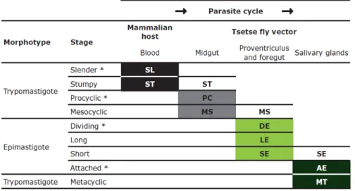
Table 1. 1 - Localisation of each stage during the parasite life cycle. * - indicates proliferative stages.
SL – slender trypomastigote; ST – stumpy trypomastigote; PC – PCF trypomastigote; MS – mesocyclic trypomastigote; DE – asymmetrically dividing epimastigote; LE – long epimastigote; SE – short epimastigote; AE – attached epimastigote; MT – metacyclic epimastigote. (Rotureau et al., 2011)
25
length is accompanied by kinetoplast migration and metabolic changes, mainly related to the fact that the PCF displays a relatively conventional Krebs cycle, linked to electron transport chain-mediated ATP generation, and as such, expression of mitochondrial enzymes and proteins increases. A huge difference between BSF and PCF trypanosomes is the reduced endocytic activity in the latter, and one of the most characterised events in PCF differentiation is the gain of the insect stage-specific procyclin coat and loss of the BSF VSG coat.
1.3.
HUMAN NATURAL IMMUNITY AND TRYPANOSOME
IMMUNE EVASION
1.3.1. The trypanosome lytic factor
Humans have generated a particular innate immunity system against African trypanosomes, which is the reason why T .b. brucei is non-infective to humans. In fact, only T. b. gambiense and T. b. rhodesiense can resist trypanolysis caused by this defense and cause sleeping sickness. The host proteins involved in this process and the resistance mechanisms created by the parasites have been reviewed by several authors (Raper et al., 2001, Vanhamme, 2010, Vanhollebeke and Pays, 2010).
26
Later in 1989, Hadjuk and colleagues demonstrated that resistance to lysis is due to resistance to a component present in normal human serum (NHS), a specific subclass of a high-density lipoprotein (HDL) (Hajduk et al., 1989). The components responsible for lysis of the trypanosomes are called trypanosome lytic factor (TLF), and several authors reported the presence of two independent trypanolytic particles (Smith et al., 1995, Tomlinson et al., 1995, Hager and Hajduk, 1997, Raper et al., 1999) (Figure 1. 6): TLF1 contains apolipoprotein AI (ApoAI), apolipoprotein AII, paraoxonase and haptoglobin related protein (Hpr); while TLF2 consists of a complex made of ApoAI, IgM and Hpr (Tomlinson et al., 1995, Raper et al., 1999).
Figure 1. 6 – Characteristics of the two trypanolytic particles, TLF1 and TLF2. (Raper et al., 2001)
The animal-infective specie T. b. brucei does not infect humans due to killing by the TLF. Mice infected with T. b. brucei and treated with TLF1 show a decrease in parasite numbers, and pre-treatment with the same factor results in protection against T. b. brucei (Barker et al., 2001).
27
b. gambiense group 1 resists lysis in a constitutive way, T. b. gambiense group 2 shows variable resistance (Mehlitz et al., 1982, Tait et al., 1984, Zillmann et al., 1984, Agbo et al., 2001, Capewell et al., 2011). As T. b. gambiense does not have the SRA gene, which is responsible for serum resistance in T. b. rhodesiense, they are thought to have evolved an independent mechanism to avoid killing by trypanolytic human serum factors (Kieft et al., 2010, Capewell et al., 2011, Symula et al., 2012, Bullard et al., 2012). Initial studies on this subject indicate that T. b. gambiense group 1 parasites evade human innate immunity by decreasing expression of the TbHpHbR (section 1.3.3) and by presenting sequence changes to this receptor (Kieft et al., 2010, Symula et al., 2012). On the contrary, T. b. gambiense group 2 serum resistance seems to be independent of TbHpHbR (Capewell et al., 2011, Symula et al., 2012), suggesting differences in the mechanism of serum resistance between the two groups.
These results have also elucidated the role of the TbHpHbR in serum resistance. In fact, early analyses proved that this receptor’s function in humans changed in order to elicit innate host immunity against the parasite (Vanhollebeke et al., 2008). Although entry of TLF1 and TLF2 can be performed via this receptor, parasites have also evolved alternative pathways for uptake of these factors, either by the use of other receptors or by fluid-phase uptake (Bullard et al., 2012).
As opposed to T. b. gambiense, T. b. rhodesiense resistance is found to depend on expression of the SRA gene, which is not always expressed as it is associated with antigenic variation and expressed as a VSG-expression site (ES) (Xong et al., 1998). Later studies showed the homology between SRA and VSG, and confirmed that resistance to TLF is related to high level of expression of SRA in T. b. rhodesiense resistant forms (Milner and Hajduk, 1999). In TLF-resistant parasites, SRA mRNA is present in 1000-fold higher amounts than in T. b. rhodesiense sensitive trypanosomes.