Normal HC11 and ras-transformed
mouse mammary cells are resistant
to the antiproliferative effects of
retinoic acid
1Laboratório de Oncologia Experimental, LIM-24, Hospital das Clínicas,
and 2Disciplina de Oncologia, Departamento de Radiologia,
Faculdade de Medicina, Universidade de São Paulo, São Paulo, SP, Brasil I. Snitcovsky1,
M.L.H. Katayama2,
M.A.A.K. Folgueira2
and M.M. Brentani2
Abstract
The objective of the present study was to determine the effects of retinoic acid on the growth of the mouse mammary cells HC11 and HC11ras, which are a model for in vitro breast cancer progression. The expression of the two classes (RARs and RXRs) of retinoic acid receptor mRNAs was determined by Northern blot analysis. Receptor functional integrity was determined by testing whether RAR ß mRNA could be induced by retinoic acid. The effects of a 72-h exposure to 50 µM 13-cis retinoic acid on HC11 and HC11ras cell proliferation and HC11 cell differentiation were investigated by flow cytometric cell cycle analysis, and by determination of ß-casein mRNA expression, respectively. The possibility that retinoic acid would induce the ex-pression of the vitamin D receptor and synergize with vitamin D, a known inhibitor of HC11 cell growth, was also investigated. HC11 cells expressed higher mRNA levels of both RAR α and RAR γ when compared to HC11ras cells. In contrast, RAR ß, as well as RXR α, ß and γ expression was low in both HC11 and HC11ras cells. In addition, RAR ß mRNA was induced by retinoic acid treatment in both cells. In spite of these observations, no effects were seen on cell proliferation or differentiation upon exposure to retinoic acid. Neither vitamin D receptor induction nor synergy with vitamin D on growth inhibition was observed. We conclude that the RAR expression profile could be related to the transformed state in HC11ras cells and that the retinoic acid resistance observed merits further investigation. Correspondence
M.M. Brentani Disciplina de Oncologia Departamento de Radiologia FMUSP
05508-900 São Paulo, SP Brasil
Presented at the I Symposium on Advances in Medical Research, Institute of Medical Investigation Laboratories, HC-FMUSP, São Paulo, SP, Brazil, March 21-22, 2003. Publication supported by FAPESP.
Received June 16, 2003 Accepted July 31, 2003
Key words
•Retinoic acid receptors •Mouse mammary cells •HC11ras cells •Carcinogenesis
Introduction
Murine carcinogenesis in several organs, including the breast, is inhibited by retinoids (1). Data showing the growth inhibiting ef-fect of retinoids on murine breast tumors induced by N-nitromethylurea (2) have
in-spired clinical protocols like the human breast cancer chemoprevention trial using the retin-oid fenretinide (3). It has recently been shown that the retinoid LGD 1096 suppresses estro-gen receptor- (ER) negative tumor develop-ment in virus Erb-b2 transgenic mice (4).
which there are two classes, RARs and RXRs, with three subtypes (α, ß, γ) in each class. RARs form heterodimers with RXRs, which control gene transcription, binding to specif-ic DNA regions known as retinoid respon-sive elements (RAREs and RXREs) or inter-fere with the function of the AP-1 transcrip-tional factor, which binds to other DNA sites (5). RAR ß expression tends to be very low or absent in breast cancer cells when com-pared to senescing cells (6). ER-positive breast cancer cell lines express higher levels of RAR α, compared to ER-negative lines (7).
The exact molecular mechanisms under-lying the chemopreventive effects of retinoids are unknown, but probably involve growth regulation and induction of differentiation. These compounds are capable of inhibiting the proliferation of several breast cancer cell lines (8) by inhibiting G1 transition in the cell cycle (9).
An interesting in vitro model of breast cancer progression, in which the action of retinoid can be investigated, is the mouse mammary cell line HC11 (10), and HC11ras, obtained by the stable transfection of a mu-tated oncogene Ha-ras into HC11 cells (11). HC11 cells isolated from the normal mam-mary glands of a midpregnant mouse retain important normal features like the capacity to differentiate and express as a marker the milk protein ß-casein, after lactogenic hor-mone induction. HC-11 cells present muta-tions in both alleles of the p53 tumor sup-pressor gene (12), which could explain the immortalized phenotype of these cells. HC11ras cells, in contrast, do not differenti-ate upon lactogenic hormone exposure and are tumorigenic when injected into immuno-suppressed mice (13). A previous study by our group has shown that vitamin D inhibits the proliferation of parental HC11 cells, but not of Ha-ras-transformed HC11 cells (14). The objective of the present investiga-tion was to determine the effects of retinoic acid on the growth of the mouse mammary
cells HC11 and HC11ras. The expression of the two classes (RARs and RXRs) of reti-noic acid receptor mRNAs was measured by Northern blot analysis. Receptor functional integrity was studied by testing whether RAR ß mRNA could be induced by retinoic acid, since RAR ß itself is a retinoid transcrip-tional target (5). The effects of a 72-h expo-sure to 50 µM 13-cis retinoic acid on prolif-eration of HC11 and HC11ras cells and dif-ferentiation of HC11 cells were investigated by flow cytometric cell cycle analysis and by determination of ß-casein mRNA expres-sion, respectively. The possibility that reti-noic acid would induce the expression of the vitamin D receptor (VDR) and synergize with vitamin D, a known inhibitor of HC11 cell growth, was investigated, since the VDR promoter contains a candidate retinoic acid-responsive element (15).
Material and Methods
Cell culture
HC11 cells (donated by Dr. Nancy Hynes, Friedrich Meischer Institute, Basel, Switzer-land) were seeded at an initial cell density of 2 x 10⁴ cells/cm2 and cultured in RPMI 1640
medium supplemented with 10% fetal calf serum (FCS), 5 µg/ml insulin and 2 mM glutamine. When exposed to 100 nM vita-min D (Biomol Research Laboratories, Inc., Plymouth Meeting, PA, USA), 50 µM 13-cis
retinoic acid or 10 µM 9-cis retinoic acid (Sigma, St. Louis, MO, USA), cells were maintained under the same conditions, ex-cept for a 24-h preculture in 10% charcoal-adsorbed FCS.
Flow cytometric DNA content determination
percent of cells in the G0/G1, S and G2/M phases was determined by the ModFit soft-ware (Becton Dickinson).
Vitamin D receptor evaluation using a specific monoclonal antibody
HC11 cells were grown for 48 h with or without 10 µM 9-cis retinoic acid. The latter was chosen due to its action on both RXRs and RARs, which could possibly imply a greater capacity to trans-activate the target VDR (5). VDR expression was evaluated in indirect immunofluorescence assays using a specific murine monoclobal antibody. Cells were fixed in 70% cold ethanol and main-tained at -20ºC for at least 12 h, washed twice in PBS and incubated with 13 µg/ml anti-VDR (VD2F12) (17) for 60 min. Posi-tivity for the marker was identified by enu-merating the fraction of cells located above the channel where 1% positivity was ob-tained for the background stain. Fluores-cence intensity, which reflects the number of antigen molecules/cell, was evaluated on the basis of the mean fluorescence channel.
RNA isolation and Northern blot assays
Total RNA from HC11 cells was isolated using the TRIZOL reagent (Gibco-BRL, Rockville, MD, USA). Twenty-microgram samples were electrophoresed on 1% aga-rose-3% formaldehyde gels and the RNA was transferred to Hybond N nylon filters (Amersham Pharmacia Biotech, Little Chalfont, Buckinghamshire, UK) which were hybridized in 50% formamide, 5X SSPE, 0.2% SDS, 5% dextran sulfate, 5X Denhardt’s solution containing 100 µg/ml salmon sperm DNA, and a 3 x 10⁶ cpm/ml [
α32P]-dCTP
(Amersham) oligo-labeled specific probe using the random primer labeling technique (Klenow fragment of E. coli DNA poly-merase; Gibco-BRL, Gaithersburg, MD, USA) for 20 h at 42ºC. The following frag-ments were used as probes: a 1.9-kb EcoRI,
1.4-kb SacI/BamHI and 1.5 kb EcoRI frag-ments of human RAR α, ß and γ, respec-tively (5), and 4.8-kb EcoRI, 1.7-kb EcoRI/
Pst1, 1.67-kb Asp718/BamHI fragments for RXR α, ß and γ, respectively (18), and a
2.1-kb fragment of human VDR cloned at the
EcoRI site of pGEM (19). Membranes were washed for 15 min, twice at room
tempera-RAR ß
RAR ß 18S
18S
3.4 kb
3.4 kb 1.9 kb
1.9 kb
Con 2 h 4 h 6 h 15 h 72 h
A
Con 2 h 4 h 6 h 15 h 72 h
B
Figure 2. RAR ß receptor mRNA expression in retinoic acid-in-duced HC11 (A) and HC11ras (B) cells. Cells were exposed or not (Con) to 50 µM 13-cis retinoic acid for different times. Total RNA was extracted and sub-jected to Northern blot analysis and filters were sequentially hy-bridized with [32P]-labeled probes for RAR ß and 18S rRNA as a control for RNA loading. RAR α
RAR ß
18S
3.2 kb
RAR γ
2.4 kb
3.4 kb
3.1 kb
1.9 kb
HC11 HC11ras
A
RXR α
RXR ß
18S 1.9 kb
3.0 kb 5.6 kb
HC11 HC11ras
B
ture in 2X SSPE, 0.1% SDS, once in 1X SSPE, 0.1% SDS, once in 0.2X SSPE, 0.1% SDS, and finally for 30 min at 52ºC in 0.1X SSPE and 0.1% SDS. Hybridization with the 18S ribosomal RNA probe, a 1.9-kb frag-ment cloned at the SalI/EcoRI site of plas-mid pBR322 (20), was subsequently per-formed to check for equivalence of RNA loading. Band intensities in autoradiograms were quantified by densitometric scanning (UltroScan XL, Pharmacia LKB Biotech-nology, Uppsala, Sweden) and data are re-ported as the ratio of specific mRNA to 18S rRNA. All solutions were prepared as de-scribed in Ref. 21.
Differentiation assay
HC11 cells were induced to differentiate and synthesize the milk protein ß-casein by growing and maintaining the cultures with
10 ng/ml murine EGF (Sigma). After 3-4 days, EGF was removed and the competent cultures were treated for 3 days with RPMI medium supplemented with 1 µM dexameth-asone, 5 µg/ml insulin and 5 µg/ml prolactin (DIP).
Results
Retinoic acid receptor expression
We started by determining the mRNA of retinoic acid receptors in HC11 and HC11ras cells in order to obtain evidence for a pos-sible responsiveness to retinoids. HC11 cells express approximately two times more mRNA of both RAR α and RAR γ, when
compared to HC11ras cells. RAR ß expres-sion, in contrast, was low in both cells (Fig-ure 1A). RXR α, ß and γ expression was low
in both HC11 and HC11ras cells (Figure
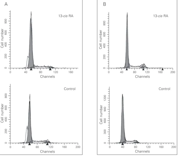
Figure 3. Effect of retinoic acid on cell cycle distribution. HC11 (A) and HC11ras (B) cells were grown without (control) or with 50 µM 13-cis retinoic acid (RA) for 72 h, harvested, permeabil-ized and labeled with propidium iodide. DNA content was evalu-ated by fluorescence intensity, and appears on the x-axis as channel numbers. Cell number is depicted on the y-axis of the histograms.
B
Channels 160
Cell number
800
600
400
200
0
120 80 40
0 200
13-cis RA
Channels 160
Cell number
1200
900
600
300
0
120 80 40
0 200
Control
A
Channels
160
Cell number
800
600
400
200
0
120 80 40 0
13-cis RA
Cell number
800
600
400
200
0
160 120 80 40
0 200
Control
1B). In addition, RAR ß mRNA was induced after a 72-h treatment with 50 µM 13-cis
retinoic acid, in both HC11 (Figure 2A) and HC11ras (Figure 2B) cells, by 50- and 2-fold, respectively.
S-phase assessment
We determined the effect of retinoic acid on cell proliferation, on the basis of the cell cycle distribution measured by flow cyto-metric analysis. Exposure to 50 µM 13-cis
retinoic acid for 72 h failed to affect the S-phase fraction in both HC11 and HC11ras cells. Representative histograms show an S-phase fraction of 21 and 23% in control and induced HC11 cells, respectively. Similarly, other representative histograms show an S-phase fraction of 19 and 20% in control and induced HC11ras cells, respectively. These results indicate that both HC11 (Figure 3A) and HC11ras (Figure 3B) cells are resistant to the growth-inhibiting effects of high lev-els of 13-cis retinoic acid.
Differentiation assessment
Expression of the milk protein ß-casein mRNA was used as a marker of the differen-tiation of HC11 cells. Exposure to 50 µM
13-cis retinoic acid for 72 h failed to induce ß-casein mRNA expression (Figure 4), indi-cating that retinoic acid had no effect on cell differentiation, in contrast to cells differenti-ated by exposure to DIP.
Investigation of the combined effect of retinoic acid and vitamin D
Retinoic acid receptors may be functional in these cells, since there was an induction, albeit late (72 h) of de RAR ß mRNA after treatment with 13-cis retinoic acid. Thus, we wondered whether there could be a sensiti-zation to the growth-inhibiting effect of vita-min D on HC11 cells by a possible induction of the VDR after exposure to retinoic acid.
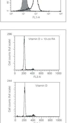
Therefore, we studied the expression of VDR protein by monoclonal antibodies and flow cytometric analysis. There was no increase in the expression level of the VDR protein after a 48-h induction with 10 µM 9-cis
retinoic acid. VDR expression was detected in more than 80% of both control and reti-noic acid-induced cells (Figure 5). Accord-ingly, retinoic acid failed to potentiate the antiproliferative effect of vitamin D. The S-phase fraction was 12.2 and 10.0% after a 72-h induction with 100 nM vitamin D alone or 100 nM vitamin D plus 50 µM 13-cis
retinoic acid, respectively (Figure 6).
Discussion
The expression of retinoic acid receptor mRNAs was initially determined in parental and ras-transformed HC11 cells. We found that HC11 cells expressed higher mRNA levels of both RAR α and RAR γ as
com-pared to HC11ras cells. In contrast, RAR ß, as well as RXR α, ß and γ expression, was
low in both HC11 and HC11ras cells. RAR ß expression was shown to be very low in the breast cancer cell lines MCF-7, T-47D, MDA-MB-361 and BT-474, as determined at the mRNA level, in contrast to RAR α and RAR γ expression, which was variable (6,7). In
breast cancer samples, RAR ß expression, as determined by in situ hybridization, was lower compared to normal adjacent tissue, in contrast to RAR α, RAR γ and RXR α which were expressed equally by tumor and normal tissues (22,23). Our results showing a low
Figure 4. ß-Casein mRNA ex-pression in HC11 cells induced with 50 µM 13-cis retinoic acid for different times and exposed to 1 µM dexamethasone, 5 µg/ ml insulin and 5 µg/ml prolactin (DIP). The control (Con) was HC11 cells which were not in-duced. Total RNA was subjected to Northern blot analysis and fil-ters were sequentially hybrid-ized with [32P]-labeled probes for ß-casein and 18S rRNA as a control for RNA loading. Only DIP treatment induced ß-casein expression, in contrast to reti-noic acid, which failed to differ-entiate the cells.
ß-Casein
18S
1.4 kb
1.9 kb
RAR ß expression in parental HC11 cells, which present several normal features, con-trast with those of another study in which RAR ß was highly expressed in benign breast lesions (24). We would suggest that the con-comitant loss of p53 and RAR ß might be a marker of progression in the process of car-cinogenesis. The RAR ß induction seen in HC11 and HC11ras cells after retinoic acid exposure suggests the presence of functional retinoic receptors, since one of the transcrip-tional targets of RAR ß is the RAR ß gene itself (5). The modest level of induction (2 times) observed in HC11ras cells compared to that seen in HC11 cells (50 times) could be due to the lower basal expression of RAR
α and RAR γ of HC11ras cells. Although
capable of expressing apparently functional retinoid receptors, both HC11 cells and HC11ras cells were resistant to the antipro-liferative effects of retinoic acid. A possible explanation for these observations is the low RAR ß basal expression, since this specific receptor could be critical to mediate the growth-inhibiting effects, as previously sug-gested (6). There are, however, conflicting reports concerning the relative importance of these receptors, showing the critical role played either by RAR α (25,26) or by RAR γ
(27) as mediators of the biological effects of retinoic acid. Another factor possibly asso-ciated with resistance to the antiproliferative effect of retinoic acid is the lack of expres-sion of ER, both in HC11 and HC11ras cells. The ER-negative breast mammary cell line MDA-MB-231, which is retinoic acid resis-tant, becomes sensitive when stably trans-fected with ER, although the underlying molecular mechanism remains to be eluci-dated (28).
We also investigated the effect of retin-oid acid on HC11 cell differentiation using ß-casein milk protein mRNA expression as a marker. Our results showed no induction of ß-casein upon long term exposure to retinoic acid. Our findings are in contrast to another study, in which retinoids were found to be Figure 6. Effect of retinoic acid
plus vitamin D on the cell cycle distribution. Cells were exposed to 100 nM vitamin D alone or 100 nM vitamin D plus 50 µM 13-cis retinoic acid (RA) for 72 h, harvested, permeabilized and la-beled with propidium iodide. DNA content was evaluated by fluorescence intensity, and ap-pears on the x-axis as channel numbers. Cell number is indi-cated on the y-axis of the histo-grams. Two assays were per-formed with similar results. Figure 5. Effect of retinoic acid on vitamin D receptor (VDR) pro-tein expression in HC11 cells. VDR content was evaluated with a monoclonal antibody by flow cytometry analysis. In the histo-grams, the cell number is shown on the y-axis and the fluores-cence channel number on the x-axis. The open area represents the nonspecific staining and the filled area represents cells spe-cifically labeled with anti-VDR an-tibody. HC11 cells were grown for 48 h without (control) or with 10 µM 9-cis retinoic acid (RA).
FL2-A
Cell counts (full scale)
296
1000 800 600 400 200 0
Vitamin D + 13-cis RA
Cell counts (full scale)
244
1000 FL2-A
800 600 400 200 0
Vitamin D
Cell counts
200
100
FL1-H
160
120
80
40
0
101 102 103 104 9-cis RA
FL1-H
Cell counts
200
100
160
120
80
40
0
capable of inducing differentiation in the breast cancer cell line SKBR3 by regulation of the cadherin adhesion molecule expres-sion and function (29). We cannot rule out the possibility of partial differentiation since ß-casein is a marker of milk production and thus indicates a final stage of differentiation of HC11 cells.
Our own data (14) had shown that vita-min D inhibits the proliferation of HC11 but not of HC11ras cells. Since vitamin D in-duces hypercalcemia, it would be of poten-tial clinical interest to use this compound at lower doses. Thus, we determined whether the addition of retinoic acid to vitamin D could have a synergistic effect on HC11 cells. The S-phase inhibition was similar in both the combination and vitamin D only treatment. These findings are in contrast to those of others, in which this combination was found to be synergistic in MCF-7 and T-47D mammary cells (30,31). The coop-erative effects of vitamin D and retinoic acid could be explained by the formation of VDR/ RXR heterodimers and by the enhancement of the trans-activating capacity (32). An-other possible mechanism of synergy could be the induction of VDR by retinoic acid (15). The lack of synergy, alternatively, could be explained by the competition of RAR and
VDR for the partner RXR (33). Thus, there could be either synergy or antagonism with the combination of retinoic acid and vitamin D depending on the cellular context.
We conclude that the RAR expression profile could be related to the transformed state in HC11ras cells. A key unknown com-ponent in the retinoic pathway may be al-tered in both parental and ras-transformed HC11 cells. A possible candidate could be the recently identified retinoid target tran-scriptional factor SOX9, which seems to mediate growth inhibition in breast cancer cell lines (34). The resistance to retinoic acid described here merits further investigation.
Acknowledgments
We are grateful to Dr. Nancy Hynes (Friedrich Meischer Institute, Basel, Swit-zerland) for the generous donation of the HC11 and HC11ras cells, to Dr. N. Arnheim (Department of Biochemistry, State Univer-sity of New York), to Dr. Pierre Chambom (Institut de Chimie Biologique, Faculté de Medecine, Strasbourg, France), and to Dr. Ronald M. Evans (Howard Hughes Medical Institute, San Diego, CA, USA) who kindly donated probes for 18S rRNA, RAR (α, ß, γ) and RXR (α, ß, γ), respectively.
References
1. Moon RC, Mehta RG & Destriac CJ (1992). Retinoids as chemopre-ventive agents for breast cancer. Cancer Detection and Prevention, 16: 73-79.
2. Gandilhon P, Melancon R, Djiane J & Kelly PA (1982). Comparison of ovariectomy and retinyl acetate on the growth of established 7,12-dimethylbenz(a)anthracene-induced mammary tumors in the rat.
Journal of the National Cancer Institute, 69: 447-451.
3. Costa A, Formelli F, Chiesa F, Decensi A, De Palo G & Veronesi U (1994). Prospects of chemoprevention of human cancers with the synthetic retinoid fenretinide. Cancer Research, 54: 2032s-2037s. 4. Wu K, Zhang Y, Xu XC et al. (2002). The retinoid X receptor-selective
retinoid, LGD1069, prevents the development of estrogen receptor-negative mammary tumors in transgenic mice. Cancer Research, 62: 6376-6380.
5. Chambon P (1996). A decade of molecular biology of retinoic acid receptors. FASEB Journal, 10: 940-954.
6. Swisshelm K, Ryan K, Lee X, Tsou HC, Peacocke M & Sager R
(1994). Down-regulation of retinoic acid receptor beta in mammary carcinoma cell lines and its up-regulation in senescing normal mam-mary epithelial cells. Cell Growth and Differentiation, 5: 133-141. 7. Roman SD, Clarke CL, Hall RE, Alexander IE & Sutherland RL
(1992). Expression and regulation of retinoic acid receptors in hu-man breast cancer cells. Cancer Research, 52: 2236-2242. 8. Sheikh MS, Shao ZM, Li XS, Dawson M, Jetten AM, Wu S, Conley
BA, Garcia M, Rochefort H & Fontana JA (1994). Retinoid-resistant estrogen receptor-negative human breast carcinoma cells trans-fected with retinoic acid receptor-alpha acquire sensitivity to growth inhibition by retinoids. Journal of Biological Chemistry, 269: 21440-21447.
9. Marth C, Bock G & Daxenbichler G (1985). Effect of 4-hydroxy-phenylretinamide and retinoic acid on proliferation and cell cycle of cultured human breast cancer cells. Journal of the National Cancer Institute, 75: 871-875.
Epithelial mouse mammary cell line exhibiting normal morphogen-esis in vivo and functional differentiation in vitro. Proceedings of the National Academy of Sciences, USA, 81: 3756-3760.
11. Happ B, Hynes NE & Groner B (1993). Ha-ras and v-raf oncogenes, but not int-2 and c-myc, interfere with the lactogenic hormone dependent activation of the mammary gland specific transcription factor. Cell Growth and Differentiation, 4: 9-15.
12. Merlo GR, Venesio T, Taverna D, Marte BM, Callahan R & Hynes NE (1994). Growth suppression of normal mammary epithelial cells by wild-type p53. Oncogene, 9: 443-453.
13. Hynes NE, Taverna D, Harwerth IM, Ciardiello F, Salomon DS, Yamamoto T & Groner B (1990). Epidermal growth factor receptor, but not c-erbB-2, activation prevents lactogenic hormone induction of the beta-casein gene in mouse mammary epithelial cells. Molec-ular and CellMolec-ular Biology, 10: 4027-4034.
14. Escaleira MT & Brentani MM (1999). Vitamin D3 receptor (VDR) expression in HC-11 mammary cells: regulation by growth-modula-tory agents, differentiation, and Ha-ras transformation. Breast Can-cer Research and Treatment, 54: 123-133.
15. Miyamoto K, Kesterson RA, Yamamoto H, Taketani Y, Nishiwaki E, Tatsumi S, Inoue Y, Morita K, Takeda E & Pike JW (1997). Structural organization of the human vitamin D receptor chromosomal gene and its promoter. Molecular Endocrinology, 11: 1165-1179. 16. Vindelov LL & Christensen IJ (1990). A review of techniques and
results obtained in one laboratory by an integrated system of meth-ods designed for routine clinical flow cytometric DNA analysis.
Cytometry, 11: 753-770.
17. Dame MC, Pierce EA, Prahl JM, Hayes CE & DeLuca HF (1986). Monoclonal antibodies to the porcine intestinal receptor for 1,25-dihydroxyvitamin D3: interaction with distinct receptor domains.
Biochemistry, 25: 4523-4534.
18. Mangelsdorf DJ, Borgmeyer U, Heyman RA, Zhou JY, Ong ES, Oro AE, Kakizuka A & Evans RM (1992). Characterization of three RXR genes that mediate the action of 9-cis retinoic acid. Genes and Development, 6: 329-344.
19. Faraco JH, Morrison NA, Baker A, Shine J & Frossard PM (1989). ApaI dimorphism at the human vitamin D receptor gene locus.
Nucleic Acids Research, 17: 2150.
20. Arnheim N (1979). Characterization of mouse ribosomal gene frag-ments purified by molecular cloning. Gene, 7: 83-96.
21. Maniatis T, Fritsch EF & Sambrook J (1989). Molecular Cloning: A Laboratory Manual. 2nd edn. Cold Spring Harbor Laboratory, New York.
22. Xu XC, Clifford JL, Hong WK & Lotan R (1994). Detection of nuclear retinoic acid receptor mRNA in histological tissue sections using nonradioactive in situ hybridization histochemistry. Diagnostic Mo-lecular Pathology, 3: 122-131.
23. Widschwendter M, Berger J, Daxenbichler G, Muller-Holzner E, Widschwendter A, Mayr A, Marth C & Zeimet AG (1997). Loss of
retinoic acid receptor beta expression in breast cancer and morpho-logically normal adjacent tissue but not in the normal breast tissue distant from the cancer. Cancer Research, 57: 4158-4161. 24. Pasquali D, Bellastella A, Valente A, Botti G, Capasso I, del Vecchio
S, Salvatore M, Colantuoni V & Sinisi AA (1997). Retinoic acid receptors alpha, beta and gamma, and cellular retinol binding pro-tein-I expression in breast fibrocystic disease and cancer. European Journal of Endocrinology, 137: 410-414.
25. Fitzgerald P, Teng M, Chandraratna RA, Heyman RA & Allegretto EA (1997). Retinoic acid receptor alpha expression correlates with ret-inoid-induced growth inhibition of human breast cancer cells regard-less of estrogen receptor status. Cancer Research, 57: 2642-2650. 26. Farias EF, Arapshian A, Bleiweiss IJ, Waxman S, Zelent A & Mira-Y-Lopez R (2002). Retinoic acid receptor alpha2 is a growth suppres-sor epigenetically silenced in MCF-7 human breast cancer cells. Cell Growth and Differentiation, 13: 335-341.
27. Widschwendter M, Daxenbichler G, Culig Z, Michel S, Zeimet AG, Mortl MG, Widschwendter A & Marth C (1997). Activity of retinoic acid receptor-gamma selectively binding retinoids alone and in com-bination with interferon-gamma in breast cancer cell lines. Interna-tional Journal of Cancer, 71: 497-504.
28. Rosenauer A, Nervi C, Davison K, Lamph WW, Mader S & Miller Jr WH (1998). Estrogen receptor expression activates the transcrip-tional and growth-inhibitory response to retinoids without enhanced retinoic acid receptor alpha expression. Cancer Research, 58: 5110-5116.
29. Shah S, Pishvaian MJ, Easwaran V, Brown PH & Byers SW (2002). The role of cadherin, beta-catenin, and AP-1 in retinoid-regulated carcinoma cell differentiation and proliferation. Journal of Biological Chemistry, 277: 25313-25322.
30. Saunders DE, Christensen C, Williams JR, Wappler NL, Lawrence WD, Malone JM, Malviya VK & Deppe G (1995). Inhibition of breast and ovarian carcinoma cell growth by 1,25-dihydroxyvitamin D3 combined with retinoic acid or dexamethasone. Anti-cancer Drugs, 6: 562-569.
31. Koga M & Sutherland RL (1991). Retinoic acid acts synergistically with 1,25-dihydroxyvitamin D3 or antioestrogen to inhibit T-47D human breast cancer cell proliferation. Journal of Steroid Biochem-istry and Molecular Biology, 39: 455-460.
32. Kliewer SA, Umesono K, Mangelsdorf DJ & Evans RM (1992). Retinoid X receptor interacts with nuclear receptors in retinoic acid, thyroid hormone and vitamin D3 signalling. Nature, 355: 446-449. 33. Evans TR & Kaye SB (1999). Retinoids: present role and future
potential. British Journal of Cancer, 80: 1-8.
![Figure 1. Expression of RAR and RXR in HC11 and HC11ras cells. Total RNA was subjected to Northern blot analysis and filters were sequentially hybridized with [ 32 P]-labeled probes for RAR α, ß and γ (A) and RXR α, ß and γ (B) and 18S rRNA as a control fo](https://thumb-eu.123doks.com/thumbv2/123dok_br/15815408.652278/3.918.380.654.733.1078/figure-expression-subjected-northern-analysis-filters-sequentially-hybridized.webp)