Efficacy of Chlorine-based, Enzymatic and
Combined Chlorine-enzyme Treatments on Biofilm
Removal
Integrated Masters in Bioengineering
Master Thesis in Biological Engineering
Manuel Matias Lopes de Lemos
bio10076@fe.up.pt
i
Abstract
Bacterial spoilage is a major concern in drinking water and in the food industry implying both economic and public health consequences. It has become increasingly clear that bacteria, including foodborne pathogens such as Escherichia coli, grow predominantly as biofilms on surfaces, in most of its habitats. Due to resistance to the commonly used antimicrobial agents, the use of enzyme to break down the extracellular polymeric matrix in biofilms is a possible alternative and this strategy is considered eco-friendly. The purpose of this study was to investigate and also to compare the efficacy of a chlorine-based treatment with sodium hypochlorite, an enzymatic treatment and a treatment based on the combination of enzyme and chlorine (sodium hypochlorite) on biofilm removal. E. coli biofilms adhered to 3 different contact surfaces (stainless steel 316 [SS 316], polyvinyl chloride [PVC], high-density polyethylene [HDPE]) and also to biofilms of different stages of maturation (1, 2 and 3 days of age). For the chlorine-based and enzyme-chlorine treatments were used concentrations of 0,6 % (v/v) and 0,5 % (v/v), respectively, based on minimum inhibitory concentration (MIC) and minimum bactericidal concentration (MBC) values. The results demonstrate that the chlorine-based treatment showed the greater potential as an antimicrobial agent (biofilm inactivation) regardless the adhesion surface (SS 316, PVC, HDPE). HDPE showed to be the contact surface on which the biofilm inactivation was easier. The enzymatic treatment showed the higher potential for biofilm mass removal. The combined chlorine-enzyme treatment demonstrated modest biofilm control activity. The chlorine-enzyme demonstrates an intermediate efficiency among the three different strategies, which elevates its importance. This overall study clarifies the potential of a selected commercial enzymatic solution (BIOREM A1® + BIOREM 10® by REALCO) on biofilm inactivation and removal, when used alone and combined with sodium hypochlorite.
Key-words: Sodium hypochlorite, enzyme, chlorine-enzyme, antimicrobial, biofilm, removal, inactivation, surfaces.
ii
Acknowledgements
Firstly, I would like to thank professor Manuel Simões for his guidance and availability during the entire development of this study. Also thank the staff from laboratory E007 of the Chemical Engineering department, especially to Inês Gomes, Ana Meireles and Rita Fulgêncio for sharing their wealth of knowledge and for the willingness to help me in any situation.
I would like to thank to my friends of Metal&Bio for filling my academic course with uncountable good memories and extra motivation.
Finally, I would like to give a special thanks to my family, my father Manuel Matias Rodrigues de Lemos, my mother Maria da Conceição Gomes Lopes and my brother Pedro Matias Lopes Lemos for all the solicitude, support, sacrifice, advice, commitment and strong education. Thanks to my family, my life values are very well structured and keep getting stronger and stronger over time.
“It pays to be a winner” “The only easy day was yesterday” - United States Navy SEALs
iii
Table of contents
Abstract --- i
Acknowledgements --- ii
Table of contents --- iii
List of figures --- v
List of tables --- vii
1. Introduction --- 1
1.1 Biofilm characteristics and implications --- 1
1.1.1 Bacterial attachment and biofilm formation in processing environments --- 2
1.1.2 Bacterial interactions in biofilm communities and its implications --- 4
1.1.3 Detachment of cells from the contact surfaces --- 6
1.2 Techniques of biofilm control --- 7
1.2.1 Biocide treatment --- 7
1.2.2 Enzymatic treatment --- 10
1.2.3 Chlorine treatment --- 12
1.2.4 Novel concepts for biofilm control --- 14
2. Materials and methods --- 16
2.1 Bacterial strain and growth media --- 16
2.2 Antimicrobial agent --- 16
2.3 Enzyme --- 16
2.4 Determination of minimum inhibitory and minimum bactericidal concentrations --- 16
2.5 Rinsing of coupons --- 17
2.6 Determination of bacterial adhesion formation on selected surfaces --- 17
iv 2.7 Biofilm formation --- 18 2.8 Biofilm control --- 18 2.9 Biomass quantification --- 19 2.10 Statistical analysis --- 19 3. Results --- 20
3.1 Minimum inhibitory and minimum bactericidal concentrations --- 20
3.2 Control of adhered cells --- 23
3.3 Removal of biofilms --- 32
4. Discussion --- 34
5. Conclusions --- 45
6. Suggestions for future work --- 46
7. References --- 47
v
List of Figures
Figure 1 – Processes involved in biofilm formation (Simões et al., 2010).
(Page 3)
Figure 2 – Determination of minimum concentration for the chlorine-based
treatment (a). Reduction of CFU with the different tested concentrations (b). (Page 21)
Figure 3 – Determination of the minimum concentration of the combined
chlorine-enzyme treatment (a). Reduction of CFU the different tested concentrations (b).
(Page 22)
Figure 4 – Adhesion of microorganisms to stainless steel 316 after treatment with the different strategies under static conditions. * means 0 CFU (a). Decrease in adhesion of microorganisms to stainless steel 316 after treatment with the different strategies under static conditions (b). The mean ± standard deviation of, at least, three replicates is illustrated.
(Page 24)
Figure 5 – Adhesion of microorganisms to stainless steel 316 after treatment
with the different strategies with agitation of 150 rpm. *means 0 CFU (a). Decrease in adhesion of microorganisms to stainless steel 316 after treatment with the different strategies with agitation of 150 rpm (b). The mean ± standard deviation of, at least, three replicates is illustrated.
(Page 25)
Figure 6 – E. coli adhered on HDPE after treatment with the different strategies without agitation. *means 0 CFU (a). Decrease in adhesion of microorganisms to HDPE after treatment of the different strategies without agitation (b). The mean ± standard deviation of, at least, three replicates is illustrated.
(Page 27)
Figure 7 – E. coli adhered on HDPE after treatment with the different strategies with agitation of 150 rpm. *means CFU (a). Decrease in adhesion of microorganisms to HDPE after treatment with the different strategies with agitation of 150 rpm (b). The mean ± standard deviation of, at least, three replicates is illustrated.
(Page 28)
Figure 8 – E. coli adhered on PVC after treatment with the different strategies without agitation. *means 0 CFU (a). Decrease in adhesion of microorganisms
vi on PVC after treatment with the different strategies without agitation (b). The mean ± standard deviation of, at least, three replicates is illustrated.
(Page 30)
Figure 9 – E. coli adhered on PVC after treatment with the different strategies with agitation of 150 rpm. *minus 0 CFU (a). Decrease in adhesion of microorganisms on PVC after treatment with the different strategies with agitation of 150 rpm (b). The mean ± standard deviation of, at least, three replicates is illustrated.
(Page 31)
Figure 10 – Removal of E. coli biofilms formed in the microtiter plate, with three different ages, after the treatment with the selected products. The mean ± standard deviation of, at least, three replicates is illustrated.
vii
List of tables
Table 1 – General overview on the reduction/killing of E. coli for each contact surface material and for each treatment. The mean ± standard deviation of, at least, three replicates is illustrated.
1
1. Introduction
1.1 Biofilm characteristics and implications
Bacterial spoilage is a major concern in the food industry implying both economic and public health consequences (Lequette et al., 2010). During the last decades, it has become increasingly clear that bacteria, including foodborne pathogens such as Salmonella enterica, Listeria monocytogenes and Escherichia coli, grow predominantly as biofilms on surfaces, in most of their habitats, rather than in the planktonic mode (Lindsay and von Holy, 2006). However, it has been observed that the resistance of biofilm cells to antimicrobials is significantly increased compared with what is normally seen with the same cells being planktonic (Gilbert et al., 2002). Thus, it is believed that biofilm formation enhances the capacity of foodborne bacteria to survive stresses that are commonly encountered within food processing, such as refrigeration, acidity, salinity or disinfection (Giaouris et al., 2012). Regarding the meat industry, biofilms formed by pathogenic and spoilage bacteria may create a persistent source of product contamination, leading to serious hygienic problems and also economic losses due to food spoilage (Sofos and Geornaras, 2010). Consequently, food spoilage and deterioration may result in huge economic losses, food safety is a major priority in globalizing market nowadays, with worldwide transportation and consumption of raw, fresh and minimally processed foods (Shi and Zhu, 2009).
Furthermore, in real food processing environments, biofilm communities may be inhabited by numerous different species in close proximity. Hence, spatial and metabolic interactions between species may contribute to the organization of multispecies biofilms, and the production of dynamic local environment. Thus, biofilms containing mixed species are usually more stable than the biofilms containing a single species, with the cell-to-cell interactions demonstrating a key role in biofilm formation, structure, as well as in the
2 resistance of biofilm community members against antimicrobial treatments (Habimana et al., 2010; Nadell et al., 2009; Kostaki et al., 2012).
Biofilms on drinking water distribution system pipes may lead to a number of unwanted effects on the quality of the distributed water. Thus, bacterial growth may affect the turbidity, taste, odor and color of the water (Ndiongue et al., 2005). Escherichia coli is one of the most frequently isolated bacteria in this context. Moreover, the emergence and large diffusion of resistance to many antibiotics in this ubiquitous species are a particular reason of concern (Crémet et al., 2013). Therefore, total coliforms and Escherichia coli are routinely monitored by drinking water companies and their detection is often an indication of (1) inadequate treatment, (2) breech in distribution system integrity and (3) regrowth. Hence, once introduced into distribution systems, the presence of E. coli, to an extent, total coliforms can become a concern of drinking water safety and public health (Murphy et al., 2008). Nevertheless, water-borne infectious diseases not only cause loss of life and illness but also have negative effects on the economy related to medical expenses and productivity losses (Helbling and VanBriesen, 2007).
1.1.1 Bacterial attachment and biofilm formation in processing environments
Microbial attachment and biofilm formation depends on the interaction between three main components: the bacterial cells, the attachment surface and the surrounding medium (Van Houdt and Michiels, 2010). Furthermore, hydrophobic interactions between the cell surface and the substratum may enable the microorganism to overcome repulsive forces and attach (Donlan, 2002). Hence, the properties of the attachment surface, such as roughness, physico-chemical properties, resistance to corrosion, are also important factors that affect biofilm formation potential and thus determine the hygienic status of the material (Tang et al., 2011). Therefore, environmental factors such as pH, temperature, osmolarity, O2 levels, nutrient composition and the presence of
other bacteria also play important roles in the process of biofilm formation (Stepanovic et al., 2003; Habimana et al., 2010).
3 The formation of biofilms includes a number of sequential processes (Figure 1), that involve movement of microorganisms to surfaces followed by initial microbial attachment, then formation of microcolonies, and extracellular polymeric substances (EPS) production and biofilm maturation (Simões et al., 2008). The biofilm formation is a relatively slow process, and the several layers of bacterial cells that are entrapped within the EPS containing matrix, are the responsible ones for a few millimeters of biofilm formation (Kumar and Anand, 1998). The EPS matrix is also responsible for promoting the interaction and consequent adhesion to the surface, since it acts like glue (Louiselle and Anderson, 2003). The biofilm composition is not made with only one kind of bacterial cells, since other different microorganisms are able to colonize. Thus, biofilm composition can be heterogeneous (Kumar and Anand, 1998). Microorganisms in biofilms show an increased resistance to antimicrobial agents due to (1) a restricted penetration of antimicrobials into the biofilms, (2) decreased growth rates, and (3) expression of resistance genes, which makes treatments for infections related with biofilms very challenging (Peeters et al., 2008; Buckingham-Meyer et al., 2007).
4 The undesirable biofilm formation can result in serious operation and maintenance costs since it causes biofouling of heat exchange systems and marine structures, corrosion of metal surfaces, deterioration of dental surfaces, contamination of household products, including food and pharmaceuticals and the infection of short or long-term biomedical implants, as well (Simões et al., 2011).
The surfaces of most bacterial cells are negatively charged, and this net negative charge of the cell surface is adverse to bacterial adhesion, due to the electrostatic repulsive force. However, the bacterial cell surface possesses hydrophobicity due to fimbriae, flagella and lipopolysaccharide (LPS) (Takahashi et al., 2010). The adhesion of bacteria is affected by some cell surface characteristics like hydrophobicity and relative surface charge, in addition to the presence of particular surface structures such as flagella, pili and EPS (Peng et al., 2001).
The existence of various factors such as fluid dynamics and shear effects of the bulk fluid can lead to the detachment and dispersal of biofilms. Nevertheless, the attached bacteria are also able to detach and disperse from the biofilm as it ages, in order to survive and colonize new niches (Kumar and Anand, 1998). Furthermore, physical forces acting on the biofilm can influence the biofilm structure as well, once the velocity field of the fluid in contact with the microbial layer, can affect biofilm structure and behavior. Hence, this represents an important factor in the removal and control of biofilms since their mechanical stability plays a key role (Simões et al., 2005).
1.1.2 Bacterial interactions in biofilm communities and its implications
Regarding mixed culture biofilms, cooperative interactions include co-aggregation and metabolic interactions. Thus, the production of extracellular matrix constituents may also be observed as cooperation, as different bacteria may contribute to the matrix, which may protect the members of the community against certain stresses, such as disinfectants and mechanical shear forces
5 (Strassman et al., 2011; Mitri et al., 2011). According to Popat et al. (2012), the matrix can be seen as an example of “public goods” based on terminology from human societies recently introduced to the biofilm field. On the other hand, while existing in multispecies biofilms, the non-producing EPS bacteria can then be described as “cheaters”, as they benefit from the protection of the matrix without being involved in its production (Popat et al., 2012).
Nevertheless, some bacteria secrete signaling compounds that can be recognized by themselves and also by other bacteria, and these compounds can then be regarded as “public goods” as well. Thus, this mode of communication between bacteria is known as quorum sensing (QS) and involves production and sensing of signaling molecules such as autoinducers (Miller and Bassler, 2001). Therefore, intra- and interspecies cell-to-cell signaling can modulate bacterial behavior and be involved in regulations of a variety of physiological activities including growth, pathogenecity, sporulation, genetic competence and biofilm formation (Annous et al., 2009; Moons et al., 2006; Yang et al., 2011).
The mixed-species biofilms can protect the biofilm embedded bacteria from antimicrobials including disinfectants, once formed. Whereas, surface attached bacteria surviving sanitation regimes, or other different type of treatment, can modulate surface attachment ability and biofilm growth of other bacteria, including pathogens (Pan et al., 2009; Van Houdt and Michiels, 2010). In other studies on antibiotic susceptibility of biofilms it is presented a higher resistance in mixed-species biofilms compared to single-species biofilms (Burmølle et al., 2006; Elias and Banin, 2012). Thus, the matrix composition of mixed-species biofilms may reduce the permeation and diffusion of antibacterial compounds. Therefore, the interactions leading to specific spatial distribution of cells having different resistance to disinfectant in mixed-species biofilms can also explain resistance (Leriche et al., 2003). Moreover, several studies have shown that bacterial interactions affect mixed-species biofilm structure (Habimana et al., 2010; Rieu et al., 2008).
Based on Elias and Banin, (2012), a drastic change in certain factors such as nutritional conditions, bacterial co-aggregation, metabolic requirements,
6 exposure to antimicrobial agents and other environmental factors ( e.g. shear forces, temperatures, atmosphere, etc), can deeply impact the structure, dynamics and thus the properties of the biofilm community.
1.1.3 Detachment of cells from the contact surfaces
In the last years either due to resistance to the commonly used antimicrobial agents or by the difficulty to eradicate biofilms due to its intrinsic resistance, new biofilm control techniques have been developed. Thus, because of governmental pressure, most of them are thought to provide minimal environmental impacts. These techniques imply the use of green biocides (less harmful for the environment and biodegradable) and aids to the common biocides that could be used in lower concentrations and still inactivate the harmful bacteria (Sokunrotanak et al., 2012). Other techniques imply the use of other alternatives such as the application of electric currents, on which Hong et al., (2008) reported that the application of a cathodic current is known to promote the detachment of bacteria from the electrode surface as a result of the electrostatic and electrophoretic repulsive forces generated. However, the very small remaining population of bacteria binding strongly to the solid surface presents this behavior due to uneven distribution of the magnitude of adhesion forces between the bacteria and the surface. This population can regrowth and reseed a biofilm.
Nevertheless, the use of enzymatic treatments to break down EPS in biofilms is a possible alternative when standard cleaning agents do not give satisfactory results in removing and/or inactivating biofilms. Several commercial biocides used in antifouling coatings have been recently banned, consequently the screening for alternative eco-friendly biocides appears to be urgent (Lequette et al., 2010; Camps et al., 2011). Moreover, there are other ways to induce significant biofilm detachment by using several physical treatments such as ultrasound treatments, thermal shocks, or mechanical treatments using pigs or shear stress induced by the fluid hydrodynamics (Rediske et al., 2000; Eguía et al., 2008). Pechaud et al. (2012), proved that, for well-established biofilms,
7 the combination of chemicals (oxidizing biocides, non-oxidizing biocides and surfactants) and hydrodynamic treatments (increase of the Reynolds number), would improve significantly the biofilm removal compared to the biochemical or chemical treatment alone.
1.2 Techniques of biofilm control
Several approaches to inhibit biofilm development have been used for many years due to the demand to eradicate harmful biofilms. The focus has mostly been concentrated on the prevention of bacterial contamination by both physical and chemical interventions. However, concerns have been raised over both the effectiveness and safety of these approaches, which has resulted in the search, development and application of novel means for removing and/or inhibiting biofilm formation. Therefore, alternative biocides must be safe for the consumers and also harmless to the environment.
1.2.1 Biocide treatment
Biocides represent chemical substances or microorganisms which can exert a controlling effect on any harmful organism by chemical or biological means. Biocidal substances and products can be employed as antifouling agents or disinfectants. Furthermore, biocides can be added to other materials to protect them against biological infestation and growth (Paulus, 2006).
Biocides are part of a chemical treatment, which are applied in order to reduce the potential for the development of biofilms on surfaces. Its goal is to eliminate microorganisms and, commonly, biocides are used with biodispersants that impose an electric charge either to the substrate or the individual cells or clusters to reduce the possibility of attachment (Bott, 1998). Biocides and disinfectants have been the main weapons utilized to control undesired biofilms. Hence, these agents work by killing microorganisms. Thus, in many systems where problematic biofilm fouling occurs, the desired end
8 result is a clean surface rather than an inactive but physically intact biofilm (Chen and Stewart, 2000). In general, halogen biocides, particularly hypochlorites, are frequently used for biological control of water systems. However, the mechanism of biofilm disinfection by halogen biocides is not well clarified (Tachikawa et al., 2005).
The mode of action of disinfectants depends on the type of biocide employed. Hence, the potential target sites either in positive or Gram-negative bacteria, are the cell wall or outer membrane, the cytoplasmatic membrane, functional and structural proteins, DNA, RNA and other cytosolic components (Bridier et al., 2011). The cells inserted in the biofilms matrix are known to express phenotypes that differ from those of their planktonic counterparts, and to exhibit specific properties including an increased resistance to biocide treatments (Wong et al., 2010). Furthermore, bacterial resistance to biocides may be intrinsic, genetically acquired or phenotypic, which sometimes is considered to be a tolerance than a real resistance, since it is specially induced by a physiological adaptation to the biofilm mode of life (Langsrud et al., 2003).
The multiple layers of cells and EPS constitute a complex and compact structure within which biocides find it difficult to penetrate and reach internal layers. Jang et al. (2006) reported that chlorine, at a certain concentration, did not penetrate beyond a depth of 100 µm into a complex dairy biofilm that was 150-200 µm thick. In fact, because biocides are often highly chemically reactive molecules, the presence of organic matter such as proteins, nucleic acids or carbohydrates can significantly impair their efficacy and potential interaction between antimicrobials and biofilm components seem more likely to explain the limitations of penetration into the biofilm (Bridier et al., 2011).
Moreover, some authors like Ganeshnarayan et al. (2009), demonstrated that in the absence of any electrostatic interaction, the majority of particles tested could penetrate and diffuse into a biofilm, suggesting that nothing prevented the diffusion of antimicrobial agents as a function of their size from a steric standpoint. However, the diffusion of positively charged particles within
9 negatively charged biofilms was detracted because of electrostatic interactions (Ganeshnarayan et al., 2009).
Although biocides represent the more significant countermeasure to control biofilm formation, these chemical substances may kill the attached microorganisms but may not be effective in biofilm removal, leaving biomass on the surface that may contribute to microbial recovery and consequently, to biofilm regrowth. In order to improve biofilm control, the use of surfactants was applied in industry, presenting more biodegradable and less toxic properties (Simões et al., 2006). Surfactants are classified according to the ionic nature of their hydrophilic group, namely, as anionic, cationic, non-ionic and zwitterionic. The chemical nature of surfactants causes modifications on the surface properties of the submerged surfaces by decreasing their surface tension, preventing attachment of microorganism with potential to form biofilm and promoting the detachment of these adhered microorganisms from the surface (MacDonald et al., 2000).
Simões et al. (2006) used the aliphatic cationic surfactant cetyl trimethyl ammonium bromide (CTAB) and the anionic surfactant sodium dodecyl sulfate (SDS) for biofilm control and stated that inactivation and removal are distinct processes. In this study, the ability of CTAB and SDS to inactivate biofilms was higher than that required to remove them. Furthermore, residual biofilms were not completely inactivated, since the biofilms left on the surface after surfactant treatment recovered their metabolic acitivity.
Jaramillo et al. (2012) utilized benzalkonium chloride (BAC) as surface coating, obtaining a very high biofilm-reducing capacity, showing that BAC has a biofilm-reducing potential. BAC is a cationic detergent expressing a high affinity to membrane proteins. The antibacterial potential of BAC relies on the changes caused on the ionic resistance of the cell membranes (Pozarowska and Pozarowski, 2011).
Moreover, Lebert et al. (2007) reported that thymol, mainly present in the essential oil of thyme, had inhibitory and biocidal effects on a range of bacteria including E. coli, Staphylococcus aureus, but not against Pseudomonas aeruginosa. Thus, they observed that this sensitivity was species dependent.
10 They also proposed to use of compounds in combination such as monolaurin, eugenol and sodium citrate, using hurdle technology.
1.2.2 Enzymatic treatment
Enzymes are of supreme importance in biology, and life. The metabolism depends on a complex network of chemical reactions brought by specific enzymes, and any modification of the enzyme pattern may have consequences for any living organism. On the other hand, enzymes can act as catalysts, which are receiving an increasing attention from physical chemists. One of the most fascinating fields of scientific investigation, which became much pursued, is the mechanism of action of enzymes (Dixon and Webb, 1964).
The application of an enzymatic treatment for the cleaning and disinfection was proved efficient by degrading the key components of the biofilm matrix. The specific required enzymes typically vary according to the type of biofilm community (Kumar and Anand, 1998). Enzymes can be used for degradation of biofilm but due to the heterogeneity of the EPS in the biofilm, a mixture of enzyme activities may be necessary for a sufficient degradation. According to the different EPS compositions a cocktail of enzymes should be applied, and the concentration and enzyme type should be well determined. Specifically, proteases are mainly used in pipelines and for removal of protein from contact lenses. Therefore, the lack of techniques for quantitative evaluation of the effect of enzymes limits their usage. The monocomponent enzymes can be used for biofilm removal, although, the heterogeneity of the biofilm matrix limits the potential of monocompound enzymes (Augustin et al., 2004).
The mechanism of the EPS physical integrity degradation is through weakening proteins, polysaccharides, carbohydrates and lipids which make up the structures of the EPS. In order to obtain an efficient removal, it is important that the structural components of EPS should be known before application of the relevant enzymes (Molobela et al., 2010). The multi structural components of the EPS may be derived from proteins, glycoproteins, nucleic acid, glycolipid,
11 phospholipids including humic substances which are non cellular substances (Liu et al., 2004). Therefore, enzymes degrade the proteins in EPS through binding and hydrolysis of the protein molecules and converting them into smaller units that can be transported through the cell membranes and then be metabolized. Thus, the enzymatic action will determine its efficacy since it depends on the specific protein structure (Molobela et al., 2010).
Hence, two major types of soluble enzymes have been used for biofilm removal, which are proteinases or polysaccharide-degrading enzymes (Johansen et al., 1997). From this last group, the most important are the enzymes which degrade plant cell wall materials (cellulose, hemicellulose, pectin, etc) sometimes commercialized for food industry applications such as fruit juice extraction, to single purified enzymes, including hydrolases (glycosidases, amino-glycosidases, esterases) and lyases The combination of both polysaccharide-degrading enzymes and proteinases provide more efficient enzyme-based biofilm removal yields (Johansen et al., 1997; Yamasaki et al., 2005; Orgaz et al., 2006;). However, some studies like Zhang et al. (2001) state that carbohydrates are the main constituents of the EPS while other studies particularly that of Orgaz et al. (2006) reported the domination of proteins. Nevertheless, the EPS components of the biofilms differ in quantity, structure or nature depending on the microorganisms within the biofilm (Liu et al., 2004).
Concerning Molobela et al. (2010) and regarding the proteins domination in EPS structure, it was stated that amylase enzymes were less effective than proteases, in biofilm degradation. This is related to the dominance of proteins in the EPS and in most cases these are found mostly at the outer layer of the biofilms. Therefore, regarding this study, it is unlikely that amylase enzymes can degrade in EPS proteins, which explains why the amylase enzymes were less efficient for biofilm degradation.
The proteinases have either to be used independently, or in those applications involving combinations of enzymes. Orgaz et al. (2007), proposed as an alternative solution to the second step of a combination treatment, the use of a delayed release encapsulated proteinase. Thus, this temporary barrier
12 would protect enzymes in solution from digestion, allowing them enough time to develop their own activity before the release of proteinase.
Based on Wiatr (1991), a blend of enzyme mixture consisting of protease, α-amilase and β-glucanase was found effective in cleaning a simulated industrial biofilm formed during paper pulp manufacture. Enzymatic treatment can be efficient in decreasing the biofilm cohesion by destroying the physical integrity of the matrix while having no identified negative impact on the environment (Lequette et al., 2010). Recently, the use of hydrolytic enzymes was proposed, in order to act on EPS components as an environmental friendly strategy to prevent mainly marine biofouling. A number of several enzymes such as proteases and carbohydrases have been studied for the prevention of adhesion of marine microorganisms to solid surfaces, whereas proteases like subtilisin have shown to inhibit biofilm formation by cultures of Pseudomonas fluorescens (Zanaroli et al., 2011).
Furthermore, Loiselle and Anderson (2003) reported that the enzyme cellulase inhibits biofilm formation. The effect of cellulase in breaking down EPS was supported by the decrease in apparent molecular weight and the increase in production of reducing sugars when EPS was exposed to cellulase.
1.2.3 Chlorine treatment
Chlorination is one of the most widely used processes for microbial control in both drinking water and industrial water processing. Chlorine is a powerful antimicrobial substance due to its potential oxidizing capacity (Sisti et al., 1998). In addition to drinking water disinfection there are a number of other uses for chlorine in the food industry, including reduction of microbial populations on the surfaces of raw foods, such as fruits and vegetable, and sanitation of surfaces in food processing environments (Virto et al., 2005). In order to attain a rapid rate of killing, generally, disinfectants such as chlorine are used at very high concentrations. At these very high concentrations, it is very difficult for microorganisms to survive. However, the use of such high concentrations increases the risk of formation of potentially hazardous
by-13 products or the production of off-tastes and odors, which are the main disadvantage of chlorination (Richardson, 2003). Although at low chlorine levels microorganisms survive the treatment, the cells may be injured rather than inactivated. Consequently, under suitable conditions injured cells might repair cellular damage and recover (Richardson, 2003).
Thus, strong oxidizing biocides are usually reliably effective against planktonic cells, sometimes weak oxidants or nonoxidants are superior for controlling biofilms. Hence, planktonic and biofilm cells also exhibit different susceptibilities to a certain antimicrobial concentration. Therefore, bacterial adaptive responses play a role in the design of control strategies (Kim et al., 2008).
The use of chlorine as a strategy to remove the EPS has been discussed by Samrakandi et al. (1997). Furthermore, Kumar and Anand (1998) list chlorine as one of the chemicals that depolymerizes the EPS. Sodium hypochlorite is the oldest and the most widely used of the chlorine compounds employed in chemical disinfection. Thus, upon dissolution in water, ionization takes place, and the hypochlorite ion establishes equilibrium with HOCl, as shown in Eq.1 and Eq. 2 (Lomander et al., 2004):
Eq. 1
Eq. 2
However, it has been shown experimentally that the bactericidal action of chlorine releasing agents results from an oxidative interaction with the sulfydryl groups on certain enzymes in the cell membrane or protoplasts. Therefore, due to the high oxidizing reactivity of chlorine, the activity of cellular proteins is destroyed. In addition, it is believed that chlorine induces irreversible decarboxylation reactions. Moreover, it is necessary to take care in order to make sure that sufficient free chlorine is available in the sanitizing solution, since chlorine reacts competitively with organic material to reduce the concentration of sanitizer that will reach the bacteria (Lomander, et al., 2004).
14 Nevertheless, sodium hypochlorite is the best example of a chlorine compound used as a disinfectant and its bactericidal effect is based on the penetration of the chemical and its oxidative action on essential enzymes in the cell. Thus, sodium hypochlorite is known to be very active in killing most bacteria, fungi and viruses and it is also known as a strong oxidizing agent (Byun et al., 2007).Therefore, it is important to devise chlorination strategies and develop combination treatments with synergistic actions against the target microorganisms (Virto et al., 2005).
1.2.4 Novel concepts for biofilm control on contact surfaces
The search for new substances for biofilm disinfection is an important area of focus. The growing negative perception of the consumers against artificial synthetic chemicals has been supporting the effort toward the development of environmental friendly disinfectants (Giaouris et al., 2013).
Recently, several authors have been stated the antimicrobial action of crude essential oils and/or their active components against biofilm embedded bacteria. These essential oils are active volatile compounds that are produced as secondary metabolites by many herbs and spices (aromatic plant essences), playing an important role in plant defense (Giaouris et al., 2013). Knowles and Roller (2001) presented the biocidal properties of carvacrol, which is a major component of the essential oils of oregano and thyme, against microbial biofilms.
The discovery that many bacteria use quorum sensing (QS) circuits to develop biofilms makes it an attractive target for their control (Lazar, 2011). Hence, quorum sensing involves a density-dependent recognition of signaling molecules, namely autoinducers, resulting in modulation of gene expression (Miller and Bassler, 2001; Skandamis and Nychas, 2012). Thus, as biofilms typically contain high concentration of cells, the autoinducers activity and quorum sensing regulation of gene expression have been proposed as crucial components of biofilm physiology (Parsek and Greenberg, 2005). Therefore,
15 there is a reason to believe that quorum sensing inhibition may represent a natural, widespread, antibiofilm strategy (Chorianopoulos et al., 2010). However, the practical application of such products in real food processing environments may encounter non-manageable difficulties, such as the inability quorum sensing inhibitors to be effective against food relevant biofilms, which may incorporate a high amount of food residues and mineral components (Giaouris et al., 2013).
During the last few years, various other novel promising methods have also been successfully evaluated for the control of biofilm formation. Hence, these include the use of bacteriophages as antimicrobial agents, technological safe bacteria, like lactic acid bacteria, as biosanitizers, bacteriocins, TiO2
photocatalysts, ionizing and UV radiation, ultrasonic treatment, ozone, electrolyzed water, microemulsions and nanoemulsions, natural products, such as honey at 0,5% (v/v) and use of biosurfactants, mainly as anti-adhesion and detachment agents (Ayebah et al., 2006; Bae and Lee, 2012;Baumann et al., 2009; Chorianopoulos et al., 2011; Gómez et al., 2012; Lee et al., 2011; Ndahetuye et al., 2012; Oulahal-Lagsir et al., 2000 Simões et al., 2008; Soni and Nannapaneni, 2010; Teixeira et al., 2007). Therefore, there are several methods that may represent advantageous alternatives for the control of biofilm formation in the near future.
The purpose of this study was to investigate and also compare the efficacy of biofilm removal using a chlorine-based treatment with sodium hypochlorite, an enzymatic treatment and a treatment based on the combination of enzyme and chlorine (sodium hypochlorite). In this study three different materials (Steel 316, polyvinyl chloride, high-density polyethylene) were selected to test the adhesion of microorganisms and their removal with the previously referred treatments either under static and dynamic conditions. In order to observe the effect of the different treatments with the age of the biofilms, this study involved 1 day- , 2 days- and 3 days-aged biofilm formation with periodic tests to analyze the killing and removal of biofilm bacteria.
16
2. Materials and Methods
2.1 Bacterial strain and growth medium
The strain used in this study was E. coli CECT 434. This strain was already used as model microorganism for antimicrobial tests (Borges et al., 2012). This microbial strain was stored at -80 ºC in cryovial and 30% (v/v) glycerol, and it was subcultured in Muller-Hinton Agar (MHA) before testing (Borges et al., 2013).
2.2 Antimicrobial agent
The sodium hypochlorite (Sigma) was used in this study.
2.3 Enzyme
The enzymatic treatment involved a combination of BIOREM A1® (sequestrant, dispersing agent, surfactants) and BIOREM 10® (stabilizing agent, enzymes) provided by REALCO(Belgium). This solution is based on enzymatic detergents. The concentration used was already pre-defined by REALCOand accordingly to BIOREM A1® and BIOREM 10® dosage instructions, it was used a dosage of 0,25 % (v/v) of BIOREM A1® and 0,05 % (v/v) of BIOREM 10®.
2.4 Determination of minimum inhibitory and minimum bactericidal concentrations
E. coli CECT 434 was inoculated into 100 mL of Muller-Hinton (MH) medium and cultivated overnight in an incubator (Shake series I26, Eppendorf, Germany) at 30 ºC with constant shaking at 120 rpm. The minimum inhibitory concentration (MIC) was determined using a plate-based assay method (Casey et al., 2004). Overnight grown cultures were diluted with fresh sterile growth medium, in order to set the OD600 to approximately 0,1 and aliquots of 180 µL
17 were added to the wells of polystyrene 96-well plates (Orange Scientific, USA) supplemented with 20 µL of disinfectant solutions at different concentrations. Sterile fresh growth medium and bacterial suspension controls were also included. The initial OD600 (ODi) was determined with an absorbance microplate
reader (Spectramax M2e, Molecular devices, Inc., USA) and the plates were incubated for 24 h at 30 ºC with shaking at 120 rpm. The final OD600 (ODf) was
determined and the MIC was obtained as the lowest concentration of antimicrobial that achieved the minimum difference between ODf and ODi.. In
order to determine the minimum bactericidal concentration (MBC), 10 µL aliquots were taken from each well of the previously performed MIC plates and spot plated onto Plate Count Agar (PCA) plates (Kowser and Fatema, 2009). The plates were incubated at 30 ºC and the MBC was determined as the lowest concentration of biocide at which no growth occurred after 1 day of incubation.
2.5 Rinsing of coupons
The tested materials were ASI 316 stainless steel (SS 316), polyvinyl chloride (PVC) and high-density polypropylene (HDPE). In order to prepare the materials for further analysis, they went through a process of rinsing starting on the immersion in a solution of commercial detergent (Cif, Unilever) and ultrapure water for 30 minutes. In order to remove any remaining detergent, the materials were rinsed in ultrapure water followed by an immersion in ethanol at 96% (v/v) for 30 minutes. After being rinsed three times with ultrapure water, the materials were dried (Simões et al., 2007).
2.6 Determination of bacterial adhesion formation on selected surfaces
E. coli CECT 434 was incubated for 24 h at 30 ºC with constant shaking at 120 rpm. The overnight bacteria were centrifuged (12 min, 4ºC, 4000 rpm) in 2 cycles discarding the supernatant and the pellet was resuspended in saline solution (0.9% NaCl, v/v). Overnight bacterial culture were grown to an OD610 of
18 ethanol, sterile water), were placed in polystyrene 12 well-plates and 2 mL of cell suspension were added to each well containing a coupon and then incubated for 2 h at 30ºC with constant shaking of 150 rpm. The coupons were taken from cell suspension and placed in another 12 well-plates containing 2 mL of each biocide and also a negative control of saline, separately, for 30 minutes. The coupons were then washed with saline solution and the adhering cells were resuspended to eppendorf tubes with 1,5 mL of saline and triplicate dilutions were made. The drop-plate method was performed in PCA plates in order to count the colony-forming units (CFUs). The plates were incubated for 24 h at 30 ºC and the CFUs were counted. The same procedure was applied when tests did not involve shaking (static conditions).
2.7 Biofilm formation
Biofilms were developed according to a modified microtiter plate test proposed previously (Stepanovic et al., 2000). Biofilms were grown in sterile polystyrene 96 well-plates (Orange Scientific, USA). Overnight grown cultures were diluted with sterile fresh growth medium in order to set the OD620 to
approximately 0,04. The microtiter plates were inoculated with fresh sterile growth medium and bacterial suspension per well and other wells were inoculated with growth medium without adding any bacteria as negative controls. The microtiter plates were incubated for 24 h in an incubator (30 ºC, 150 rpm). All experiments were performed in triplicate with three repeats. Taking into account the age of the biofilms (1 day, 2 days and 3 days-old), for each day that passed, the growth medium was removed and changed to a new one in order to maintain the development of biofilm formation.
2.8 Biofilm control
The biofilms formed were used to assess the effects of the different disinfectants used. Different microplates were used for the different aged-biofilms. In order to test the effects of the age of biofilms with the different disinfectants, for each day, a microplate with biofilm was taken and it was
19 added 200 µL of each disinfectant followed by incubation at 30 ºC for 30 minutes at a shaking of 150 rpm, repeating this last process 3 times.
2.9 Biomass quantification
The biofilm mass was quantified using crystal violet (Merck, Portugal) staining method according to Borges et al., (2012). The absorbance was measured at 570 nm using an absorbance microplate reader (Spectramax M2e, Molecular Devices, Inc., USA). The percentage of reduction was assessed based on the following equation:
Eq. 3
Where C denotes the absorbance for control wells (absence of disinfectants), and T is the absorbance for biofilms exposed to chlorine-based, enzymatic and chlorine-enzyme treatments (Lemos et al., 2013).
2.10 Statistical analysis
The results were analyzed through t-students function on Microsoft Office Excel 2007 and by One Way ANOVA function in software SPSS 20.0 (Statistical Package for the Social Sciences) with Tukey comparison test, assuming a significance level for the separation set at (P < 0,05).
20
3. Results
3.1 Minimum inhibitory and minimum bactericidal concentrations
.
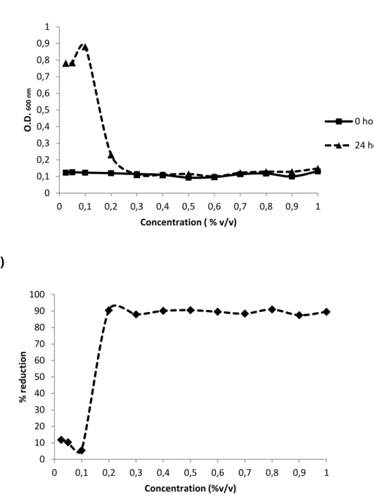
The sensitivities for chlorine and chlorine-enzyme of E. coli CECT 434 were investigated by using concentrations from 0,025 % (v/v) to 1% (v/v). For the chlorine-based and enzyme-chlorine treatments concentrations of 0,6 % (v/v) and 0,5 % (v/v), respectively, were used based on MIC and MBC. The values of MIC for chlorine-based treatment and chlorine-enzyme treatment were very approximate and, the same happened with MBC values regarding each of these two treatments. The reduction of cell microorganisms for the different concentrations as well as the determination of the minimum concentration to apply can be seen in Figure 2 and Figure 3.
21
(a)
(b)
Figure 2 – Determination of minimum concentration for the chlorine-based treatment (a). Reduction of CFU with the different tested concentrations (b).
0 0,1 0,2 0,3 0,4 0,5 0,6 0,7 0,8 0,9 1 0 0,1 0,2 0,3 0,4 0,5 0,6 0,7 0,8 0,9 1 O. D . 60 0 n m Concentration ( % v/v) 0 hours 24 hours 0 10 20 30 40 50 60 70 80 90 100 0 0,1 0,2 0,3 0,4 0,5 0,6 0,7 0,8 0,9 1 % r e d u ction Concentration (%v/v)
22
(a)
(b)
Figure 3 – Determination of the minimum concentration of the combined chlorine-enzyme treatment (a). Reduction of CFU the different tested concentrations (b).
0 0,1 0,2 0,3 0,4 0,5 0,6 0,7 0,8 0,9 1 0 0,1 0,2 0,3 0,4 0,5 0,6 0,7 0,8 0,9 1 O. D . 60 0 n m Concentration (% v/v) 0 hours 24 hours 0 10 20 30 40 50 60 70 80 90 100 0 0,1 0,2 0,3 0,4 0,5 0,6 0,7 0,8 0,9 1 % R e d u ction Concentration (%v/v)
23
3.2 Control of adhered cells
In this study three different materials were selected (stainless steel 316, polyvinyl chloride, high density polyethylene) to test the adhesion of microorganisms and also the percentage of reduction after exposure to the previously referred treatments, at the concentrations determined by MIC and MBC, either subjected to agitation or not (static and dynamic conditions). Regarding the treatment related with the different strategies, different results were obtained for each material, either in the presence of agitation or in the absence of it.
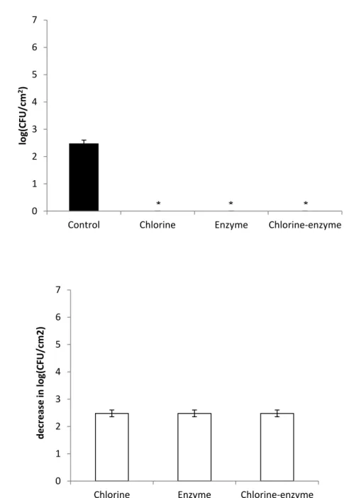
In the case of stainless steel 316, in the absence of agitation for an obtained control treated with saline solution of 5,35 log (CFU/cm2), it was observed a decrease of 5,35 log (CFU/cm2) for based and chlorine-enzyme strategies, showing a total killing of the adhered cells. For the enzymatic treatment, involving only the REALCO enzymes, there was a decrease in 4,31 log (CFU/cm2) after the biofilm enzymatic treatment for 30 min, as it is possible to observe in Figure 4. In terms of killing percentages, after the chlorine-based and combined treatments, the bacteria adhered on the coupons of stainless steel 316 were totally (100%) killed (P < 0,05). For the single enzymatic treatment a cell killing percentage of 99,7 ± 0,5% (P < 0,05) was obtained. Considering the treatments applied in the presence of agitation of 150 rpm, only the enzymatic treatment seemed to be less effective than the other treatment strategies. It was measured a control of 5,8 log (CFU/cm2) and after enzymatic treatment it was obtained a decrease of 1.02 log (CFU/cm2), as demonstrated in Figure 5. This corresponds to percentage inactivation of 91,8 % ± 2,1% (P < 0,05). The cell killing efficiency for based and chlorine-enzyme treatment was 100%.
24
(a)
(b)
Figure 4 – Adhesion of microorganisms to stainless steel 316 after treatment with the different strategies under static conditions. * means 0 CFU (a). Decrease in adhesion of microorganisms to stainless steel 316 after treatment with the different strategies under static conditions (b). The mean ± standard deviation of, at least, three replicates is illustrated. 0 1 2 3 4 5 6 7
Control Chlorine Enzyme Chlorine-enzyme
lo g(CFU/c m 2) 0 1 2 3 4 5 6 7
chlorine enzyme chlorine-enzyme
d e cr e ase in log(CFU /c m ^2) * *
25
(a)
(b)
Figure 5 – Adhesion of microorganisms to stainless steel 316 after treatment with the different strategies with agitation of 150 rpm. *means 0 CFU (a). Decrease in adhesion of microorganisms to stainless steel 316 after treatment with the different strategies with agitation of 150 rpm (b). The mean ± standard deviation of, at least, three replicates is illustrated. 0 1 2 3 4 5 6 7
Control Chlorine Enzyme Chlorine-Enzyme
lo g(CFU/c m 2) 0 1 2 3 4 5 6 7
Chorine Enzyme Chlorine-enzyme
d e cr e ase in log(CFU /c m ^2) * *
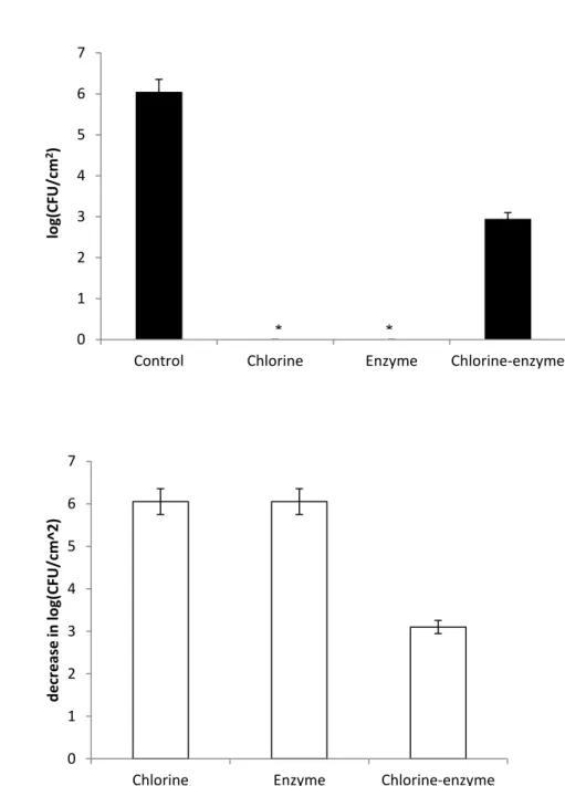
26 A value of 2,48 log (CFU/cm2) of E.coli adhered on HDPE for the control with saline solution was obtained. After the exposure to the different strategies of treatment it showed a total cell killing (Figure 6), i.e. 100% CFU reduction (P > 0,05) considering the absence of agitation.
However, when subjected to an agitation of 150 rpm, it seems that only the use of chlorine-enzyme treatment was not able to perform total cell killing inactivation. With a control of 6,06 log (CFU/cm2), this strategy promoted a decrease of 5,2 log (CFU/cm2), as it is observed in Figure 7. Despite the reduction is not able to reach 100%, based on the values of CFU/cm2 the reduction is still extremely high, 99,9 ± 0,1% (P > 0,05).
27
(a)
(b)
Figure 6 – E. coli adhered on HDPE after treatment with the different strategies without agitation. *means 0 CFU (a). Decrease in adhesion of microorganisms to HDPE after treatment of the different strategies without agitation (b). The mean ± standard deviation of, at least, three replicates is illustrated.
0 1 2 3 4 5 6 7
Control Chlorine Enzyme Chlorine-enzyme
lo g(CFU/c m 2) 0 1 2 3 4 5 6 7
Chlorine Enzyme Chlorine-enzyme
d ec rease in log(CF U /c m 2) * * *
28
(a)
(b)
Figure 7 – E. coli adhered on HDPE after treatment with the different strategies with agitation of 150 rpm. *means CFU (a). Decrease in adhesion of microorganisms to HDPE after treatment with the different strategies with agitation of 150 rpm (b). The mean ± standard deviation of, at least, three replicates is illustrated.
0 1 2 3 4 5 6 7
Control Chlorine Enzyme Chlorine-enzyme
lo g(CFU/c m 2) 0 1 2 3 4 5 6 7
Chlorine Enzyme Chlorine-enzyme
d ec rease in log(CF U /c m ^2) * *
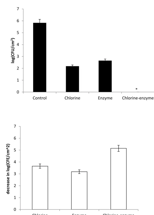
29 Regarding the PVC surfaces, while not submitted to agitation, the control with saline solution had a value of 5,42 log (CFU/cm2). The chlorine-enzyme treatment promoted a reduction of 4,68 log (CFU/cm2), as it can be seen in Figure 8. This value corresponds to 96,7 ± 6,7% (P > 0,05) in the CFU/cm2. The other treatments showed a decrease of 5,42 log (CFU/cm2), or 100% cell killing (P > 0,05).
The treatments performed under agitation of 150 rpm and, the total inactivation (P < 0,05) was only achieved by chlorine-enzyme treatment. A control (untreated adhered cells) of 5,15 log (CFU/cm2) was obtained. The chlorine-based and enzymatic treatments promoted a decrease of 4,6 log (CFU/cm2) and 4,5 log (CFU/cm2), respectively, as it can be seen in Figure 9. Moreover, despite these values are very proximate, the enzymatic treatment still shows a minor decrease comparing to chlorine-based treatment which is reflected on the CFU/cm2 values where it seems that the chlorine-based treatment reduction was around 97 ± 6,3% while the enzymatic treatment reduction was about 91 ± 18%. Even though both reductions are significant there is still a significant statistical difference (P < 0,05).
30
(a)
(b)
Figure 8 – E. coli adhered on PVC after treatment with the different strategies without agitation. *means 0 CFU (a). Decrease in adhesion of microorganisms on PVC after treatment with the different strategies without agitation (b). The mean ± standard deviation of, at least, three replicates is illustrated.
0 1 2 3 4 5 6 7
Control Chlorine Enzyme Chlorine-enzyme
lo g(CFU/c m 2) 0 1 2 3 4 5 6 7
Chlorine Enzyme Chlorine-enzyme
d e cr e ase in log(CFU /c m ^2) * *
31
(a)
(b)
Figure 9 – E. coli adhered on PVC after treatment with the different strategies with agitation of 150 rpm. *minus 0 CFU (a). Decrease in adhesion of microorganisms on PVC after treatment with the different strategies with agitation of 150 rpm (b). The mean ± standard deviation of, at least, three replicates is illustrated.
0 1 2 3 4 5 6 7
Control Chlorine Enzyme Chlorine-enzyme
lo g(CFU/c m 2) 0 1 2 3 4 5 6 7
Chlorine Enzyme Chlorine-enzyme
d e cr e ase in log(CFE /c m ^2) *
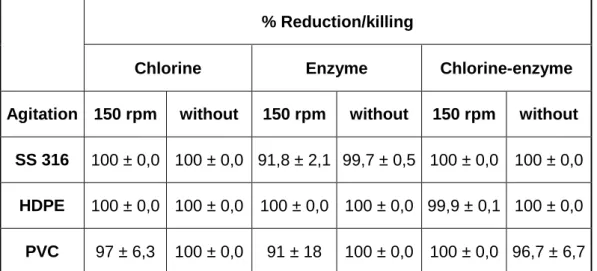
32 Table 1 presents a general overview on the reduction of E. coli from each contact surface material and respective treatments.
Table 1 – General overview on the reduction/killing of E. coli for each contact surface material and for each treatment. The mean ± standard deviation of, at least, three replicates is illustrated.
% Reduction/killing
Chlorine Enzyme Chlorine-enzyme
Agitation 150 rpm without 150 rpm without 150 rpm without SS 316 100 ± 0,0 100 ± 0,0 91,8 ± 2,1 99,7 ± 0,5 100 ± 0,0 100 ± 0,0
HDPE 100 ± 0,0 100 ± 0,0 100 ± 0,0 100 ± 0,0 99,9 ± 0,1 100 ± 0,0
PVC 97 ± 6,3 100 ± 0,0 91 ± 18 100 ± 0,0 100 ± 0,0 96,7 ± 6,7
3.3 Removal and inactivation of biofilms
In order to observe the effect of the different treatments with the age of the biofilms, this study involved a 3 days-aged biofilm formation with periodic tests to analyze the removal of microorganisms through crystal violet staining method applied for each day of biofilm formation. The biofilms formed were treated with the different strategies considering that after each period of biofilm formation the biocides were added to the biofilm in 3 cycles of 30 min of incubation, at 150 rpm. This condition was selected based on the adhesion assays. The worst cases of adhesion control (killing) were those where E. coli adhered under dynamic conditions.
Regarding the 1 day-aged biofilms the enzymatic treatment caused the highest removal with 70 ± 4% (P < 0,05), while the lowest was promoted with
33 the chlorine-based treatment with 53 ± 9%. For the chlorine-enzyme treatment it was obtained a removal of 62 ± 9% (P < 0,05).
The 2 days-aged were treated with the same strategy and it was found that chlorine and chlorine-enzyme treatments promoted biofilm removals of 33 ± 8% and 29 ± 4%, respectively. Those removal percentages were not statistically different (P > 0,05). The enzymatic treatment caused the highest biofilm removal, 68 ± 7%. However, not significantly different from the previous treatment of 1 day-aged biofilms (P > 0,05).
The 3 days-aged biofilms were less affected by the treatments than the younger biofilms. The chlorine-enzyme treatment presents again a higher value of removal of19% ± 5 % than the value for the chlorine-based treatment of9% ± 5%, and in this case the difference is statistically significant (P < 0,05). Conversely, the removal for enzymatic treatment presented an accentuated decrease comparing to the other biofilm age periods with a removal of 24 ± 17%, however, it still presents the higher removal value compared with the other strategies (P < 0,05).
The different removal percentages related to each treatment and biofilm age can be seen in Figure 10.
Figure 10 – Removal of E. coli biofilms formed in the microtiter plate, with three different ages, after the treatment with the selected products. The mean ± standard deviation of, at least, three replicates is illustrated.
0 10 20 30 40 50 60 70 80 90 100
1 day 2 days 3 days
% R e m o val Chlorine Enzyme Chlorine-enzyme
34
4. Discussion
Microorganisms are implicated in industrial biofouling, contamination of drinking water distribution system, infections, and numerous other costly and life-threatening problems. Consequently, the control of bacteria in biofilms is of extreme importance and the chlorine-based, enzymatic and chlorine-enzyme treatments proved to have effects on killing and removing biofilm cells from stainless steel 316, HDPE and PVC surfaces commonly used in food processing facilities.
During the investigation it was possible to realize that there was a difference in the adhesion and killing of E. coli either when submitted to agitation at 150 rpm and either when not subjected to shear stress. Also, the type of adhesion surface influenced microbial attachment and the further susceptibility to the treatments.
Although the adhesion was higher on stainless steel 316 and HDPE under agitation rather than without, the adhesion for PVC did not present the same result since adhesion was only superior, even with a small difference, when not subjected to shear stress forces. However, it has been demonstrated that shear stress can induce cell adhesion, influence cell proliferation and orientation, and induce other physiological responses (Donlan and Costerton, 2002; Liu et al., 2006; Dardik et al., 2005; Thomas et al., 2002).
Other studies stated that differences in the shear stress field can induce heterogeneity within a biofilm and that sometimes this heterogeneity is correlated to different antimicrobial susceptibilities (Sakamoto et al., 2010; Salek et al., 2011). Hence, microbial adhesion depends on the species involved and on environmental factors, particularly the hydrodynamic conditions, the type of surface and the fluid nutrient composition. Thus, understanding the factors affecting the adhesion process is the key to control biofilm formation (Lorite et al., 2011).
Several studies suggested that surface defects such as cracks and crevices are more likely to reflect the degree of soiling and microbial attachment
35 on a surface (Hilbert et al., 2003). Joseph et al. (2001) noted that efficiency on biofilm formation as well as resistance to treatment with sanitizers varies depending on the type of surface. Based on Silva et al. (2008), the surface roughness influences bacterial adhesion, and higher the surface roughness, the higher the significant effect on cell retention. Thus, the high porosity of rough surfaces provides a larger surface area for bacterial attachment than smooth surfaces, and so, biofilm maturation might be faster on rough compared to smooth surfaces. Hence, surface properties such as hydrophobicity, electrical charge, roughness and porosity are determinant in the adhesion process. Moreover, the surface roughness impedes hygiene and cleaning procedures (Silva et al. 2008).As for the effect of roughness of stainless steel surface to microbial adhesion or removal, contrasting observations have been reported in literature. Hilbert et al. (2003) stated that surface roughness did not significantly affect the attachment to and removal from stainless steel surface for Pseudomonas sp., Listeria monocytogenes and Candida lipolytica. Additionally, Boulangé-Petermann et al. (1997) found no clear relationship between the roughness parameter and the number of viable Streptococcus thermophillus adherent to stainless steel surfaces. Moreover, Flint et al. (2000) also showed that the adhesion of thermo-resistant streptococci was almost independent from surface roughness. On the other hand Ortega et al. (2008) presented increased adhesion and decreased removability of Staphylococcus epidermis for a rough stainless steel surface compared with a smoother surface. Furthermore, some earlier works demonstrated a positive correlation between cleanability and increased surface smoothness in the removal of biofilms. Thus, the effect of surface roughness might depend on the microbial species, possibly due to difference in adhesion manner and/or cell surface characteristics (Ortega et al., 2008). Based on Ortega et al. (2010), the surface roughness was found to affect the removal of adherent cells.
Parikh (2011), reported that biofilm survival of Listeria monocytogenes was found to be greater on rough rather than smooth HDPE surfaces and so cutting boards with a smooth surface should be considered due to delay in biofilm maturation. Moretro and Langsrud (2004) showed that biofilm adheres to rough surfaces more strongly than smooth surfaces. Also, the high rate of
36 evaporation on smooth surfaces may have resulted in more injured cells and thus lower bacterial survival on the smooth surfaces (Moretro and Langsrud, 2004). According to Wong (1998), biofilm survival is affected by temperature, relative humidity and attachment surface, and one or multiple factors may have played an important role in reduced survival.
Therefore, previous research demonstrated that cell attachment and biofilm formation are influenced by several factors, including the characteristics of strains, physical and chemical properties of the substrate for attachment, growth phase of the bacteria, temperature, growth media and the presence of other microorganism (Wong 1998). Frank (2000), reported that stainless steel is moderately hydrophilic with a negative surface charge, while PVC is hydrophobic. The different hydrophobic characteristics of PVC, HDPE and stainless steel affect bacterial attachment and detachment to surfaces. Thus, if surface tension of the microorganism is higher than that of the surrounding medium, cells tend to attach to hydrophilic (high surface tension) surfaces. In general, bacterial surface tension is lower than that of surrounding medium and more typically adherence to hydrophobic surfaces is observed. Beresford et al. (2001), found that L. monocytogenes adhesion was greater on PVC than on stainless steel after a short exposure time of 2 h incubation. However, this difference was not significant. In the present study, there was no significant difference in the initial E. coli population on stainless steel and on PVC coupon surfaces. However, for HDPE it was registered significant differences in the values of cell attachment under different conditions. Hence, the factors in addition to surface conditioning, roughness, and micro-topography and hydrophobic interaction, such as electrostatic and exopolymer interaction, seem to affect the attachment of bacteria to various materials (Palmer et al., 2007). Although these other factors are important, it appears that biofilm cells on PVC are more difficult to detach and inactivate than those on stainless steel and HDPE primarily because of stronger hydrophobic interactions between bacteria and PVC surfaces.
The difference of action for the different disinfection strategies is noticeable for each one of the contact surfaces studied. Generally, if adhesion occurs or not under dynamic conditions, the stainless steel 316 surfaces