Faculdade de Farmácia
Research Institute for Medicines and Pharmaceutical Sciences
(iMed.UL)
Neuron Glia Biology in Health and Disease Group
-
ROLE OF S100B ON CENTRAL NERVOUS SYSTEM
DEMYELINATION AND REMYELINATION
Vera Alexandra Padrela Martins Afonso
Dissertação de Mestrado
MESTRADO EM CIÊNCIAS BIOFARMACÊUTICAS
Faculdade de Farmácia
Research Institute for Medicines and Pharmaceutical Sciences
(iMed.UL)
Neuron Glia Biology in Health and Disease Group
ROLE OF S100B ON CENTRAL NERVOUS SYSTEM
DEMYELINATION AND REMYELINATION
Vera Alexandra Padrela Martins Afonso
Dissertação de Mestrado orientada pela Prof.ª Doutora Adelaide Maria Afonso
Fernandes Borralho e pela Doutora Andreia Pereira Barateiro
MESTRADO EM CIÊNCIAS BIOFARMACÊUTICAS
Gostaria de começar por agradecer à Professora Catedrática Dora Brites,
investigadora principal do grupo Neuron Glia Biology in Health and Disease, a
prontidão com que me acolheu e a oportunidade de realizar o meu projecto de
mestrado. Apesar de não estar envolvida directamente neste trabalho, não posso
deixar de agradecer pelo seu espirito crítico e conhecimento infindáveis, essenciais
para o desenvolvimento desta dissertação.
À Professora Doutora Adelaide Fernandes, um grande obrigada não chega. Não
consigo expressar a minha gratidão e, acima de tudo, admiração! Obrigada por todo o
conhecimento que me passaste ao longo deste ano e meio, pela tua dedicação e por
teres sido sempre incansável, mesmo quando o panorama não era o melhor.
À Doutora Andreia Barateiro, um enorme obrigada por me teres orientado durante
o tempo em que a Adelaide não esteve presente. Por toda a paciência e
disponibilidade para me esclarecer todas as dúvidas. Foi uma nova experiência para
as duas e acredito que o resultado foi positivo.
O meu maior agradecimento é dirigido aos meus pais e à minha irmã, Diana, a
principal força motriz de todo este trabalho. Obrigada por todo o apoio incondicional.
Aos meus amigos. Aos que me acompanham há anos, e aos que fui ganhando ao
longo deste período. Peço desculpa muitas vezes pela minha ausência, e agradeço a
sua paciência e persistência. Agradeço ainda todo o seu apoio e compreensão, foram
cruciais para me dar força todos os dias e sem eles não teria sido capaz de chegar até
aqui.
Deixo ainda um agradecimento a todos os que passaram na minha vida ao longo
destes dois anos e meio e que, ainda que indirecta e inconscientemente, contribuíram
Table of Contents
Resumo ... ix Abstract ... vii Abbreviations ... v Introduction ... 1 S100B ... 1 1.1. S100B double life ... 11.1.1. Intracellular functions of S100B in the Central Nervous System ... 5
1.1.2. Extracellular functions of S100B in the Central Nervous System ... 7
1.2. S100B as a biomarker of brain damage ... 11
Demyelinating Disorders ... 12
2.1. Multiple Sclerosis as the most common demyelinating disease ... 13
2.1.1. MS clinical course ... 14
2.1.2. MS pathophysiology ... 15
2.2. S100B in Multiple Sclerosis ... 18
Experimental Demyelinating Models ... 19
3.1. In vivo Animal Models... 19
3.2. Ex vivo Organotypic Slice Cultures ... 21
Aims ... 23
. Material and Methods ... 25
Animals ... 25
Organotypic Cerebellar Slice Cultures and Treatment ... 25
Total RNA Extraction, Reverse Transcription and Semi-quantitative Real-Time Polymerase Chain Reaction ... 27
Immunostaining procedure ... 28
S100B assay ... 30
Statistical Analysis ... 30
Results ... 31
S100B is overexpressed and released from central nervous system cells following demyelination insult ... 31
S100B is mainly released by astrocytes during demyelination ... 32
Blockade of S100B partially attenuates demyelination induced by LPC ... 32
Abrogation of S100B decreases glial reactivity provoked by demyelation ... 36
Inflammatory response triggered by demyelination is attenuated by S100B blockade .. 42
Discussion ... 45
Figure Index
I.
Introduction
Figure I. 1 – Schematic representation of S100B intracellular effects in central nervous system cells. ... 5 Figure I. 2 – Schematic representation of S100B and other markers expression along OL lineage development ... 7 Figure I. 3 – Schematic representation of S100B extracellular effects on central nervous system cells ... 9 Figure I. 4 – Extracellular effects of different S100B concentrations upon oligodendrocyte precursor cells (OPC) development ... 11 Figure I. 5 – Schematic illustration of how of disability evolves through time in MS. ... 15 Figure I. 6 – Schematic representation of MS pathophysiology mechanisms ... 17
II.
Material and Methods
Figure II. 1 – Schematic representation of COSC protocol ... 26 Figure II. 2 – Schematic representation of culture treatment ... 27
III. Results
Figure III. 2 - S100B is mainly released from astrocytes upon demyelination ... 33 Figure III. 3 – Antibody directed blockade of S100B partially attenuates demyelination caused by LPC ... 35 Figure III. 4 – Blocking S100B decreases the expression of myelin genes following demyelination ... 36 Figure III. 5 – S100B abrogation causes an increase in OPC activation ... 37 Figure III. 6 – S100B blocking prevents astrocytic activation triggered by demyelination ... 39 Figure III. 7 –S100B blocking partially attenuates microglia activation induced by LPC demyelination ... 40 Figure III. 8 – Blocking S100B apparently induces clearance of debris by microglia following demyelination. ... 42 Figure III. 9 – S100B abrogation significantly decreases expression of pro-inflammatory cytokines HMGB1 and IL-18 elicited by demyelination ... 42
IV.
Discussion
iv
Index of Tables
I.
Introduction
Table I. 1 – S100B intracellular main target proteins and respective functions ... 4
II. Materials and Methods
Table II. 1 – List of pairs of primers used for qRT-PCR assays ... 27 Table II. 2 – List of primary antibodies used for immunoshistochemistry assays. ... 29 Table II. 3 – List of secondary antibodies used for immunoshistochemistry assays. ... 29
Abbreviations
ADEM Acute disseminated encephalomyelitis
AHL Acute hemorrhagic leukoencephalitis
bFGF Basic fibroblast growth factor
Ca2+ Calcium
CD Cluster of Differentiation
cDNA Complementar deoxyribonucleic acid
CNS Central nervous system
COSC Cerebellar organotypic slice cultures
CSF Cerebrospinal fluid
DIV Days in vitro
DNA Deoxyribonucleic acid
EAE Experimental autoimmune encephalomyelitis
ELISA Enzyme-linked immunosorbent assay
ERK Extracellular signal-regulated kinases
FGF Fibroblast growth factor
FGFR Fibroblast growth factor receptor
GFAP Glial fibrillary acidic protein
HLA Human leukocyte antigen
HMGB1 High mobility group box chromosomal protein 1
Iba-1 Ionized calcium-binding adapter molecule 1
IFN- Interferon-
IL Interleukin
iNOS Inducible nitric oxide synthase
LPC Lysophosphatidylcholine or Lysolecithin
MBP Myelin basic protein
MEM Minimal essential media
mGluR3 Metabotropic glutamate receptor 3
MHC II Major histocompatibility complex class II
MOG Myelin oligodendroglial glycoprotein
mRNA Messenger ribonucleic acid
MS Multiple sclerosis
NF-κB Nuclear factor kappa-light-chain-enhancer of activated B cell
NG2 Neural/glial antigen 2
vi nM Nanomolar NMO Neuromyelitisoptica NO Nitric oxide NTC Non-template control OL Oligodendrocyte
OPC Oligodendrocyte precursor cell
PBS Phosphate buffered saline
PCR Polymerase chain reaction
PFA Paraformaldehyde
PLP Proteolipid protein
PNS Peripheral nervous system
PPMS Primary progressive multiple sclerosis
PRMS Progressive remitting multiple sclerosis
qRT-PCR Quantitative real-time polymerase chain reaction
RAGE Receptor for advanced glycation end products
RNA Ribonucleic acid
ROS Reactive species of oxygen
RRMS Relapsing remitting multiple sclerosis
SPMS Secondary remitting multiple sclerosis
TNF Tumour necrosis factor
WT Wild-type
Abstract
S100B is a small Ca2+ binding protein member of S100 family. Within the central
nervous system, S100B is mostly expressed by astrocytes, though it has been shown to be also expressed by oligodendrocytes, microglia and some neural populations. S100B exerts both intracellular and extracellular functions. Within cells S100B acts as a signaling molecule, being involved in the regulation of energy metabolism and in the modulation of both proliferation and differentiation of neurons and glia. When secreted to the extracellular space, S100B exerts beneficial or detrimental effects in a concentration-dependent manner: (i) nanomolar concentrations have been reported to exert trophic effects whereas (ii) micromolar concentrations have shown to produce neurodegenerative, neuroinflammatory and/or apoptotic outcomes. Multiple sclerosis (MS) is an autoimmune chronic inflammatory disease with a neurodegenerative component, characterized by the occurrence of focal areas of inflammatory demyelination, variable gliosis and relative axonal loss, with limited remyelination. Moreover, data from previous research of other authors and from ongoing work of our group have shown augmented S100B in both CSF, serum and post-mortem plaques of MS patients.
With this work we intended to explore the role of S100B in MS main mechanisms. For this end we used mouse cerebellar organotypic slice cultures, a previously described model of demyelination. Therefore, we first evaluated the expression and secretion of S100B in the course of a demyelinating insult. Our results reveal that S100B is released and overexpressed upon demyelination mainly by astrocytes. Next, we also evaluated in what way S100B might affect demyelination, reactivity of glial cells and inflammatory response in consequence of demyelination, by blocking extracellular S100B with a specific antibody. Upon the demyelination insult, we verified an upregulation of myelin proteins and pro-inflammatory cytokines gene expression, as well as activation of astrocytes and microglia. In turn, blockade of S100B appeared to prevent demyelination and reduce both reactive gliosis and expression of pro-inflammatory factors. Altogether, these data strongly suggest that high levels of S100B may exacerbate demyelination and/or delay remyelination.
Resumo
A S100B é uma proteína de ligação ao Ca2+ da super-família de proteínas S100.
No Sistema Nervoso Central a S100B é maioritariamente expressa pelos astrócitos, apesar de haver evidência da sua expressão em oligodendrocitos, microglia e certas populações de neurónios. Sabe-se que o S100B pode ter diferentes funções, consoante a sua localização: intracelular ou extracelular. Quando expresso intracelularmente actua como uma molécula sinalizadora, estando envolvido em processos de regulação do metabolismo energético, bem como na proliferação e diferenciação neuronal e das células gliais. Uma vez secretado para o espaço extracelular, o S100B pode exercer efeitos benéficos ou prejudiciais nas células cerebrais, dependendo da sua concentração: (i) enquanto que concentrações fisiológicas na ordem dos nanomolar tem efeitos tróficos, (ii) concentrações mais elevadas, da ordem dos micromolar, induzem a neurodegeneração, neuroinflamação e apoptose destas células. A Esclerose Múltipla (EM) é uma doença autoimune crónica inflamatória com uma grande componente neurodegenerativa. Patologicamente, a EM é caracterizada pela ocorrência de focos de desmielinização e inflamação, reactividade das células gliais e disfunção neuronal, podendo haver alguma remielinização. Dados de pesquisa publicados por outros autores e dados preliminares do nosso grupo demonstram a presença de níveis anormais de S100B em amostras de líquido cefalo-raquidiano, soro e placas escleróticas de doentes de EM.
Com este trabalho pretendemos investigar o papel da proteína S100B nos principais mecanismos que levam ao desenvolvimento da EM. Para tal, recorremos a um modelo de culturas organotipicas de cerebelo de ratinho, um modelo já usado para estudos de mielinização e desmielinização. Começámos por avaliar os níveis de expressão e secreção de S100B no decurso da desmielinização. De acordo com os resultados obtidos, verificou-se uma sobre-expressão e aumento de secreção de S100B, em resposta ao insulto desmielinizante, mediada maioritariamente pelos astrócitos. Seguidamente decidimos também verificar de que modo o S100B poderá estar a afectar a desmielinização e a reactividade das células gliais e a resposta inflamatória, através do bloqueio extracelular de S100B com um anticorpo especifico para a proteína. Após a desmielinização, observou-se uma marcada desmielinização associada a uma sobre-expressão de genes que regulam as proteínas da mielina e de certas citocinas pro-inflamatórias, bem como a activação dos astrócitos e micróglia. Por sua vez, o bloqueio do S100B extracelular mostrou ter um efeito preventivo da desmielinização bem como atenuante da reactividade glial e da expressão de citocinas
x
pró-inflamatórias. Estes resultados permitem-nos inferir que níveis elevados de S100B podem favorecer a desmielinização e/ou retardar a remielinização.
Introduction
S100B
S100 proteins consist in one of the largest family of small proteins involved in
calcium ion (Ca2+) homeostasis. Members of the S100 protein family are known to
contain two EF-hand helix-loop-helix Ca2+ binding domains: a canonical C-terminal and
a non-canonical N-terminal (Kawasaki et al., 1998). Ca2+ binds to these domains
causing conformational changes, exposing a hydrophobic cleft, which is required for
interaction with target proteins and their modulation (Yap et al., 1999; Zimmer and
Weber, 2010).
S100A1 and S100B were the first members of S100 family to be identified as
components of the S100 protein fraction that was extracted from bovine brain, this
fraction presented homodimers and heterodimers of both proteins (Moore, 1965; Moore
et al., 1968). S100B is expressed by a restrict number of cell types from different
organs and tissues, although it appears to be abundantly expressed by central and
peripheral nervous system glial cells (Donato, 2003; Rambotti et al., 1989). In these
cells S100B may be observed both in a soluble form diffusely in the cytoplasm or
associated with intracellular membranes, centrosomes, microtubules and type III
intermediate filaments (Donato, 2003, 2007).
1.1.
S100B double life
As most S100 proteins, S100B exists within cells as antiparallel homodimers, with
2
Heizmann, 2002) or as an heterocomplex in association with S100A1 monomer (Isobe
et al., 1983).
Recent studies have shown that S100B monomers have the ability to assemble
into more complex oligomers, such as tetramers, hexamers and octamers, and that
both the degree of oligomerization and the type of bond that holds the monomers might
be related to its function (Donato, 2007). Dimeric forms of S100B are mainly associated
with intracellular roles, such as modulation of microtubule assembly and regulation of
cell cycle by interaction with transcription factors in a Ca2+-dependent manner (Davey
et al., 2001). Therefore, changes in intracellular Ca2+ concentrations cause S100B
secretion to the extracellular space where it is known to exert cytokine-like functions
(Davey et al., 2001). Such functions require specific binding to cell receptors such as
the receptor for advanced glycation end products (RAGE), to which S100B binds
preferentially when in tetrameric/octameric forms promoting RAGE dimerization
(Ostendorp et al., 2007).
RAGE receptor is a member of the immunoglobulin-like cell surface receptor super
family that is primarily involved in inflammation, nephropathy, neurodegeneration, and
cancer. It is composed by an extracellular moiety, with one N-terminal V-type and two
C-type Ig domains, a single transmembrane spanning helix and a short cytosolic
domain, which is necessary for signal transduction (Neeper et al., 1992; Rauvala and
Rouhiainen, 2010; Soroka et al., 2003). The outcome of S100B binding to RAGE
depends on the extracellular levels of the ligand: nanomolar (nM) concentrations of
S100B were shown to exert neurotrophic and neuroprotective functions at neuronal
level, promoting neurite growth and neuronal survival (Barger et al., 1995; Kögel et al.,
2004); and micromolar (µM) concentrations have been shown to accomplish opposite
effects, activating a pro-apoptotic pathways (Van Eldik and Wainwright, 2003; Huttunen
et al., 2000). Recent studies have shown that extracellular S100B does not operate
solely via RAGE binding. It was suggested that in proliferating myoblasts S100B binds
to the cell fibroblast growth factor (FGF) receptor 1 (FGFR1) trough interaction with
basic FGF (bFGF) (Riuzzi et al., 2011). This engagement results in the recruitment of
mitogenic signaling pathway activated by bFGF-FGFR1 assembling and inactivate
RAGE signaling, promoting skeletal muscle regeneration (Riuzzi et al., 2012; Sorci et
al., 2013).
As previously mentioned, within cells S100B might act as an intracellular regulator.
Indeed, upon Ca2+ binding S100B dimmers suffer a conformational change exposing
the hydrophobic residues, and consequently creating a hydrophobic cleft through which
S100B can bind to target proteins (Iuvone et al., 2007). Many target proteins of S100B
have been identified, involving S100B in diverse intracellular events as regulation of
Ca2+ homeostasis, cell proliferation and differentiation, protein phosphorylation,
enzyme and channel activities, protein degradation, assembly of several cytoskeleton
components, cell locomotion, dark adaptation of photoreceptors and the innate
inflammatory response (Table I. 1). It is quite clearly that S100B plays a protective role
as long as it is kept within the cells at physiological levels.
Within the Central Nervous System (CNS), S100B is known to be actively secreted
by astrocytes, which is believed to occur through activation of metabotropic glutamate
receptor 3 (mGluR3) in a neural and synaptic activity dependent manner (Sakatani et
al., 2008; Shashoua et al., 1984). Once in the extracellular space S100B exerts
beneficial or detrimental effects on neurons, microglia and astrocytes depending on the
concentration (Van Eldik and Wainwright, 2003). At physiological concentrations
around nM levels S100B behaves as a signaling trophic protein promoting neuronal
survival, astrocytic proliferation and microglia quiescence (Gonçalves et al., 2000; Reali
et al., 2005; Selinfreund et al., 1991; Zhang et al., 2011b).
When S100B levels rise to the range of µM the whole scenario is reversed with
production of reactive species of oxygen (ROS) leading to neuronal death. Moreover,
inflammatory activities of astrocytes are potentiated and microglia is activated as
evidenced by the expression and release of pro-inflammatory cytokines (Adami et al.,
2001; Businaro et al., 2006; Petrova et al., 2000). Several studies have shown that
4
RAGE and might depend on the intensity and extent of activation of the receptor, as
well ason its level of expression in CNS cells (Donato, 2007).
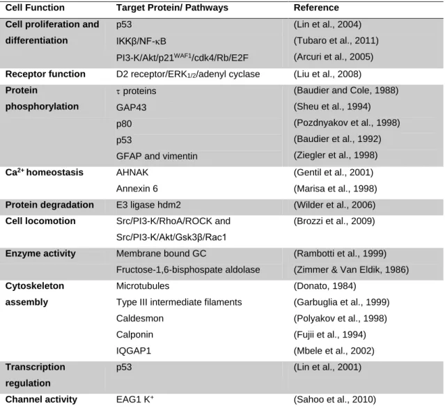
Table I. 1 – S100B intracellular main target proteins and respective functions.
Cell Function Target Protein/ Pathways Reference Cell proliferation and
differentiation
p53 IKKβ/NF-B
PI3-K/Akt/p21WAF1/cdk4/Rb/E2F
(Lin et al., 2004) (Tubaro et al., 2011) (Arcuri et al., 2005) Receptor function D2 receptor/ERK1/2/adenyl cyclase (Liu et al., 2008)
Protein phosphorylation proteins GAP43 p80 p53
GFAP and vimentin
(Baudier and Cole, 1988) (Sheu et al., 1994) (Pozdnyakov et al., 1998) (Baudier et al., 1992) (Ziegler et al., 1998) Ca2+ homeostasis AHNAK Annexin 6 (Gentil et al., 2001) (Marisa et al., 1998) Protein degradation E3 ligase hdm2 (Wilder et al., 2006) Cell locomotion Src/PI3-K/RhoA/ROCK and
Src/PI3-K/Akt/Gsk3β/Rac1
(Brozzi et al., 2009)
Enzyme activity Membrane bound GC
Fructose-1,6-bisphospate aldolase
(Rambotti et al., 1999) (Zimmer & Van Eldik, 1986) Cytoskeleton
assembly
Microtubules
Type III intermediate filaments Caldesmon Calponin IQGAP1 (Donato, 1984) (Garbuglia et al., 1999) (Polyakov et al., 1998) (Fujii et al., 1994) (Mbele et al., 2002) Transcription regulation p53 (Lin et al., 2001)
Channel activity EAG1 K+ (Sahoo et al., 2010)
Nevertheless, although having been extensively studied over the past five
decades, there is no evidence of accordance between intracellular regulatory activities
of S100B and its extracellular effects; that is, no unitary theory of intracellular and
1.1.1. Intracellular functions of S100B in the Central Nervous System
It is now known that within CNS S100B is mostly expressed by astrocytes and
some oligodendrocytes (OL) of hippocampus and brain cortex, although it can also be
present in certain neuronal subpopulations (Gerlach et al., 2006; Hachem et al., 2005;
Steiner et al., 2007). The greatest amount of S100B is located within the cytoplasm,
with only about 7% bound to the membranes. S100B involvement in cellular events has
been extensively studied and includes the regulation of cell proliferation and
differentiation, as well as the control of the assembly of cytoskeleton components,
intracellular Ca2+ homeostasis and proteins activities, interactions and modifications
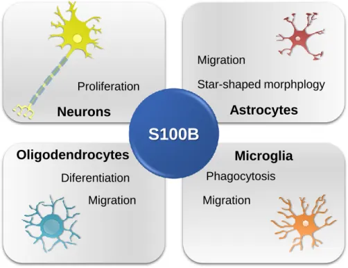
(Figure I. 1) (Donato et al., 2009).
Figure I. 1 – Schematic representation of S100B intracellular effects in central nervous system cells.
It is known that S100B interacts with cytoskeleton components, modulating their
assembly and disassembly (Garbuglia et al., 1998; Sorci et al., 1998). Indeed, recent
data have shown that S100B is involved in astrocytic migratory ability and star-shaped
morphology by regulating the molecular organization of actin filaments (F-actin) in a
PI3-K dependent manner. PI3-K regulates then Akt(PKB)/GSK3β/Rac1 and small GTPase RhoA/ROCK pathways, both involved in the formation of stress fibers
S100B
Neurons
Astrocytes
Microglia
Oligodendrocytes
Star-shaped morphplogy Migration Proliferation Diferentiation Migration Migration Phagocytosis6
promoting the creation of cytoplasmic extensions and favoyring cell locomotion (Brozzi et al., 2009; Watanabe et al., 2004). In addition, S100B was found to be associated
with microtubule-like structures and centrosomes on microglia, suggesting its
involvement on activation and phagocytic ability characteristics of these glial cells
(Adami et al., 2001).
S100B was shown to play an important role in the regulation of neuronal
proliferation and differentiation during early development stages. The overexpression of
S100B in the neuronal cell line pheochromacytoma PC12 exposed to the neurotrophin
nerve growth factor (NGF) has been demonstrated to enhance cell proliferation and
reduce differentiation through activation of Akt/p21WAF1/cyclin D1-cdk4/Rb/E2F pathway
(Arcuri et al., 2005). It has also been shown that S100B is involved in the regulation of
astrocytes proliferation and differentiation, trough activation of PI3-K in a Src
kinase-dependent manner (Brozzi et al., 2009). Experiments in mouse cerebellar cultures
have demonstrated that S100B plays a role in the maintenance of Ca2+ homeostasis in
astrocytes which in turn are important in the regulation of neurophysiology and in the
modulation of neuronal activity (Agulhon et al., 2008; Xiong et al., 2000).
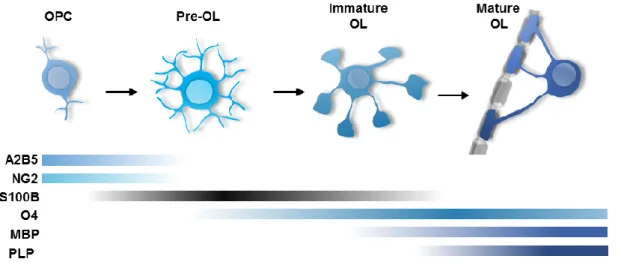
Moreover, recent data regarding oligodendroglial cells revealed the presence of an
expression pattern of S100B along their differentiation and maturation (Figure I. 2)
(Deloulme et al., 2004). Mature myelinating OL are known to descend from a pool of
multipotent precursor cells maintained in specific regions of the CNS. These precursor
cells undergo several changes at morphological and antigenic markers expression
levels, until becoming myelinating OL, being described four transitional cell stages: OL
precursor cells (OPC), preoligodendrocytes (or late OPC), immature (or
pre-myelinating) OL and mature (or pre-myelinating) OL (Dawson et al., 2000; Levine et al.,
2001).
Particularly, S100B is mostly expressed in these intermediate cell stages,
suggesting its enrolment in OPC proliferation and differentiation into mature OL. Also,
S100B expression was shown to greatly increase during the transition between
cytoplasmic S100B is associated with cytoskeleton components, such as microtubules,
present in the extensions and membranous sheaths developed by maturing OL
(Deloulme et al., 2004; Zhang, 2001).
Figure I. 2 – Schematic representation of S100B and other markers expression along OL lineage development. OPC, oligodendrocyte precursor cells; OL, oligodendrocyte; NG2, chondroitin sulphate proteoglycan; MBP, myelin basic protein; PLP, proteolipid protein, (Adapted from Deloulme et al., 2004; S. C. Zhang, 2001)
All this regulation events carried out by S100B makes evident its importance along
the CNS development, as well as in the course of brain insults when astrocytes and
microglia become activated and, together with OPC, migrate to areas of insult in an
effort to restore its proper performance (Zlokovic, 2008)
.
1.1.2. Extracellular functions of S100B in the Central Nervous System
As previously mentioned, within CNS S100B is mainly secreted by astrocytes in a
constitutive manner (Shashoua et al., 1984). Secretion of S100B has been shown to be
regulated by a number of external factors and condition, some of them augment it
others repress it. Indeed, tumor necrosis factor- (TNF-α), interleukin-1β (IL-1β), serotonin, lysophosphatidic acid, low levels of glutamate, low extracellular Ca2+ levels,
metabolic stress and serum deprivation are known to enhance S100B release. On the
8
activity, cell confluence, Ca2+ channel blockade and gap junction inhibition are known
to reduce S100B secretion (Sorci et al., 2010).
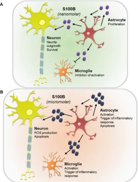
Interestingly, depending on the concentration present in the extracellular space,
S100B is known to act as a neurotrophic or neurotoxic molecule (Figure I. 3). Indeed,
under physiological conditions, S100B levels in the brain extracellular space range
around a few nM concentrations. At this concentration S100B is known to exert trophic
effects on neurons, stimulating neurite outgrowth and increasing neuronal survival
during development and following injury via stimulation of extracellular signal-regulated
kinases (ERK) 1 and 2 ad of the nuclear factor-B (NF-B) as well as up-regulation of the anti-apoptotic protein Bcl-2 (Barger et al., 1995; Businaro et al., 2006; Huttunen et
al., 2000). Still, under stress conditions S100B tends to be secreted in higher amounts,
reaching concentrations on the order of M, at which S100B exerts neurotoxic effects via overproduction of reactive oxygen species and activation of apoptotic pathways
(Donato et al., 2009).
As described above, both trophic and toxic effects of extracellular S100B are
mediated in the brain by binding to the receptor RAGE (Donato, 2007; Leclerc et al.,
2009). RAGE is expressed in several cell types, including neurons, and its ligation by
S100B on these cells has been shown to be someway responsible for the protein dual roles (Sorci et al., 2013). Low S100B doses protect neuronal cells from β-amyloid toxicity and also activates anti-apoptotic pathways via RAGE engagement. In contrast,
an excessive stimulation of RAGE as consequence of S100B binding results in the
hyper-activation of the pro-apoptotic Ras/MEK/ERK pathway and subsequent
overproduction of reactive oxygen species (Businaro et al., 2006; Huttunen et al.,
2000). Nevertheless, opposite outcomes resultant from S100B/RAGE interaction might
depend on the number of RAGE molecules available on the cell surface. Moreover,
since only high levels of S100B are able to promote RAGE dimerization consequent
activation of pro-inflammatory cells, it is possible that, under low levels of S100B,
RAGE may recruit other factors to different intracellular signaling pathways (Sorci et al.,
Figure I. 3 – Schematic representation of S100B extracellular effects on central nervous system cells. At nanomolar concentrations S100B exerts a beneficial effect on CNS cells (A), whether higher concentrations are detrimental, favoring a pro-inflammatory scenery (B).
Besides neurons, extracellular S100B also affects surrounding glial cells, mainly
astrocytes and microglia, in a concentration-dependent manner (Nardin et al., 2007).
S100B acts on astrocytes in an autocrine way, and at nM levels stimulates their
proliferation by phosphorylation of ERK1/2 (Gonçalves et al., 2000; Selinfreund et al.,
1991). This astrogliosis is counteracted by elevated concentrations of S100B, that stimulate inducible nitric oxide synthase (iNOS) activity, via activation of NF-κB and nitric oxide (NO), once synthesized leads to astrocyte apoptosis (Lam et al., 2001; Petrova et al., 2000). Similarly, high S100B stimulates IL-1β, interleukin-6 (IL-6) and TNF-α release from astrocytes, meaning that S100B is likely to take part on activation of brain inflammatory response (Hu and Van Eldik, 1999). Controversially, high S100B
10
levels might be beneficial in the course of brain inflammatory processes, once by
inducing apoptosis might contribute to the reduction of activated astrocytes which in
turn might attenuate inflammatory response.
Extracellular S100B also affect microglia, the brain resident macrophages (Adami
et al., 2001). At physiological levels, S100B has the effect to prevent microglia
activation via STAT3 pathway (Zhang et al., 2011b). By contrast, µM concentrations of S100B synergistically with cofactors like bacterial endotoxin or interferon-γ (IFN- γ) are known to mediate microglia activation. This activation occurs via stimulation of iNOS
and consequent enhance of NO release, being crucial to trigger brain inflammatory
response (Adami et al., 2001; Petrova et al., 2000). Some effects of S100B on both
astrocytes and microglia also seem to be mediated by RAGE ligation (Ponath et al.,
2007; Sorci et al., 2013). Thus, extracellular S100B is now being used as a parameter
of glial activation or commitment in several situations of brain injury.
S100B expression and release was thought to be restricted to astrocytes for
decades. However recent data has demonstrated that mature OL from the OL-93 cell
line also secrete S100B at a higher level than astrocytes under serum and glucose
deprivation conditions (Steiner et al., 2008a). Consequently, such high concentrations
of S100B might be detrimental for both OL proper function and OPC differentiation into
myelinating OL. In this regard, our group observed that exposure of OPC to
pathological concentrations of S100B (1µM) decrease OPC differentiation when
compared to OPC treated with control or S100B physiological levels (10 nM), impairing
therefore their ability to became myelinating cells (Figure I. 4; unpublished data). These
Figure I. 4 – Extracellular effects of different S100B concentrations upon oligodendrocyte precursor cells (OPC) development. Only low doses of S100B reduce the number of myelin
basic protein (MBP)+ cells (red), slightly increasing the number of NG2+ immature ones (green). (Adapted from Santos G PhD work).
1.2.
S100B as a biomarker of brain damage
There are several studies that reveal an association between elevated levels of
S100B and CNS disorders, based both on S100B detection on body fluids for
post-mortem specimens. Augmented S100B levels were first detected in cerebrospinal fluid
(CSF) of multiple sclerosis (MS) patients in the acute phase (Michetti et al., 1979).
Since then, studies of S100B detrimental effects were extended to other diseases of
the CNS. High levels of S100B have been detected in a number of brain-related
diseases such as Alzheimer disease, Down syndrome, amyotrophic lateral sclerosis
and Parkinson disease (Harpio and Einarsson, 2004; Shakeri et al., 2012; Steiner et
12
Actually, due to high levels of S100B expression in the brain and because it is
released from injured astroglial and/or OL cells in the course of an insult, S100B is
increasingly being considered as a biomarker for brain damage (Shakeri et al., 2012;
Sun and Feng, 2013). Indeed, S100B has already been identified as a potential
biomarker in cases of premature and traumatic brain injury, once it has been reported
to be augmented in the CSF and serum of patients following such traumas (Beharier et
al., 2013; Kleindienst and Ross Bullock, 2006).
Moreover, the fact that some of the detrimental effects resulting from
overexpression of S100B are mediated trough RAGE engagement and that both
astrocytes and microglia express this receptor, suggest a possible involvement of
S100B/RAGE interaction in the course of some neurodegenerative disorders (Bianchi
et al., 2011; Donato et al., 2009; Ponath et al., 2007). In fact, recent data from a human
neuroblastoma cell line (LAN-5) suggest that while nM levels of S100B counteract amyloid-β (Aβ) peptide neurotoxic effects in a RAGE dependent manner, µM levels S100B exacerbates Aβ toxicity (Businaro et al., 2006).
Overall, given the importance of S100B in the regulation of several events within
CNS cells as well as its implication in some neurodegenerative diseases, it is of great
interest to explore its possible role in other brain damage related pathologies.
Demyelinating Disorders
A demyelinating disease consists of total or partial loss of the myelin sheath that
surrounds axons, compromising their proper function and integrity. This loss occurs in
response to intrinsic or external factors, such as inflammatory processes, metabolic
derangements, hypoxia-ischemia and viral infections, that might target either the own myelin or the cells that synthesize it – OL in the CNS or Schwann cells in the peripheral nervous system (PNS) (Love, 2006; Mayo et al., 2012). Usually, following
demyelination spontaneous remyelination might occur allowing a partial recovery, and
it is the balance between demyelination and remyelination that defines the outcome of
CNS demyelinating disorders include MS, Marburg disease, neuromyelitisoptica (NMO), Balo’s concentric sclerosis, acute disseminated encephalomyelitis (ADEM) and acute hemorrhagic leukoencephalitis (AHL) (Bunyan et al., 2012; Popescu and
Lucchinetti, 2012). Since MS is the most prevalent demyelinating disorder among
young adults we focused our study is this disorder.
2.1.
Multiple Sclerosis as the most common demyelinating disease
MS is the major demyelinating disease of the CNS, occurring in an inflammatory
background. MS is considered to be the most common cause of non-traumatic
disability in young adults, with a great socio-economic impact worldwide, with each individual costing about €2 million during his lifetime. Most recent data show that there are about 2.5 million people with MS worldwide, with an incidence of 2.5 per 100 000,
and a higher prevalence in developed regions (e.g. Europe with 80/100 000) than in
underdeveloped countries (e.g. Africa with 0,3/100 000) (WHO, 2008). In Portugal,
accordingly to the most recent studies there is an estimated prevalence of 56,2 per
100 000, i.e., there are 5620 MS patients per 10 million inhabitants, of which only about
3500 to 4000 are known to receive directed therapy for the disease (de Sá et al.,
2012).
MS is characterized by the occurrence of areas of acute focal inflammatory
demyelination, variable gliosis and relative axonal loss with limited remyelination,
culminating in the formation of chronic multifocal sclerotic plaques. These lesions occur
preferentially in optic nerves, subpial spinal cord, brainstem, cerebellum, and
periventricular white matter regions (Compston and Coles, 2008; Love, 2006). MS
pathogenesis is not fully understood and even though it is subject of controversy, there
are strong evidence for an autoimmune pattern with auto-reactive immune cells
crossing the blood-brain barrier to attack myelin and axons (Brassat, 2012; Corthals,
2011).
The onset of MS occurs between 15 and 55 years and is two to three times more
14
past few years have shown that it results from the interaction of diverse risk factors
such as: genetic susceptibility (MHC class II-associated HLA-DRB1*15, ethnic origin
and sex), viral infection (EpsteinBarr virus), behavior (smoking) and environment
(latitude and lack of vitamin D) (Ramagopalan et al., 2010). Symptomatically, MS is
characterized by the occurrence of motor weakness, vision loss, diplopia, ataxia,
cognitive impairment, bowel and bladder dysfunction and cortical dysfunctions such as
seizures (Compston and Coles, 2008; Milo and Miller, 2014).
2.1.1. MS clinical course
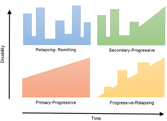
MS clinical course has been categorized in four types, accordingly to the
emergence of disabilities (Figure I. 5). Most frequently, MS disease starts with a course
of demyelination insults followed by periods of neurological recovery, being designated
relapsing-remitting MS (RRMS). This revisable disability is characterized by focal areas
of inflammatory demyelination in which myelin, OL and axons become damaged
(Hanafy and Sloane, 2011; Peterson and Fujinami, 2007). Relapses are associated
with the reactivation of old lesions or even with the appearance of new ones, while
remittance periods are the result of the resolution of inflammation and remyelination
(Chandran et al., 2008; Compston and Coles, 2008). The time elapsed between
relapses is variable, with the latent phase between the first manifestation of MS and the
first relapse going from little months to several years. With the recurrence of relapsing
and remittance episodes, recovery from each episode tends to be incomplete with
consequent accumulation of persistent symptoms (Compston and Coles, 2008; Trapp
and Nave, 2008).
RRMS stage is normally followed by a phase of uninterrupted disease progression,
determined as secondary progressive MS (SPMS). This phase is characterized by a
continuous and irreversible neurological degeneration, as there is no remyelination and
subsequently no axonal regeneration (Vukusic and Confavreux, 2003). A small percentage of MS patients (10-15%) doesn’t pass by the relapsing-remitting phase, but instead experiment an uninterrupted disease progression from the beginning, being
referred as primary progressive MS (PPMS). The rarest type of MS is
progressive-relapsing MS (PRMS) characterized by a progressive onset interspersed by acute
relapses, with or without recovery, and a continuous progression in the period between
the relapses. PRMS may represent a rare subtype of PPMS since both present a
similar history (Lim et al., 2004; Wolinsky, 2003).
Figure I. 5 – Schematic illustration of how of disability evolves through time in MS.
Progressive forms of MS are the most troublesome, not only because patients
become disabled due to fail of remyelination but also because there is no successful
therapeutic at the time to combat physical, cognitive, and quality of life deterioration
that these MS patients have to face. Thus, it is imperative to research and develop
novel therapies in this direction.
2.1.2. MS pathophysiology
The main hallmark of MS is the loss of myelin. The main function of myelin sheaths
16
maintenance of axonal homeostasis and integrity (Compston and Coles, 2008;
Griffiths, 1998). Within the CNS, myelin is synthesized by mature OL that emit
processes from their own cytoplasmic membranes and continuously envelop axons,
forming a multilamellar compacted membrane (Deber and Reynolds, 1991). As
extensions of cytoplasmic membranes, myelin sheaths consist on a proteolipidic
foundation and are constituted by two major proteins: proteolipid protein (PLP) and
myelin basic protein (MBP). Unlike PNS, where each myelin sheath is produced by a
single Schwann cell, in the CNS OL are able to furnish about 40 or more adjacent
axons, influencing axonal caliber and improving their integrity and stability (Tzakos et
al., 2005; Witt and Brady, 2000).
Although immune-mediate theory of MS pathology is not the only under
discussion, it is the one that is most described and has been target of extensive
research (Corthals, 2011; Nakahara et al., 2012). In this context, the loss of myelin
sheaths occurs as consequence of the migration across the blood brain barrier of
auto-reactive T-lymphocytes against myelin components. These activated T-cells will be
induced to differentiate in lymphocytes T helper CD4+ (ThCD4+) which in turn will be
reactivated by MHC-II-expressing macrophages or dendritic cells that present myelin
elements (Figure I. 6) (Mayo et al., 2012).
In turn, reactivated T-cells circulate within the CNS and together with activated
macrophages, microglia and astrocytes secrete soluble factors like pro-inflammatory
cytokines and chemokines, such as IFN-, TNF-, high motility group box 1 (HMGB1) and matrix metalloproteinases (MMPs) (Amor et al., 2010; Van der Walt et al., 2010).
The accumulation of these pro-inflammatory factors as well as the cells that produce
them leads to the recruitment of naïve microglia and consequent activation, amplifying
the inflammatory and immune response that culminates with demyelination (Lassmann
and van Horssen, 2011; Nakahara et al., 2012).
A growing body of evidence strongly suggests the involvement of inflammasomes
in the regulation of inflammatory response that leads to demyelination. NLRP3, a
is pyrin-domain containing 3, is a core component of the inflamasome complex
(Lequerré et al., 2007; Sutterwala et al., 2006). Moreover, NLRP3 has been suggested
to be implicated in neuroinflammation and demyelination events in an in vivo model of
MS trough interleukin-18 (IL-18). The same study has revealed that IL-18, one of the
end products of the inflammasome, exacerbates demyelination and inflammatory
outcomes, such as astrocytic and microglia activation (Jha et al., 2010).
Figure I. 6 – Schematic representation of MS pathophysiology mechanisms. Autoreactive
T CD4+ cells are activated in the periphery, transmigrate trough the blood-brain barrier (BBB) into the central nervous system (CNS), and are locally reactivated by antigen presenting cells (APS). Activated T CD4+ cells secrete pro-inflammatory cytokines and, together with other successively recruited and activated immune cells, such as T CD8+ and B cells, create a pro-inflammatory environment, leading to myelin, oligodendrocyte (OL) and axon damage, with consequent neurologic dysfunction. MBP, myelin basic protein; PLP, proteolipid protein; Tc, T cytotoxic lymphocytes; Th, T helper lymphocytes; APC, antigen presenting cell; IL-18, interleukin-18; IFN-, interferon-; TNF-, tumor necrosis factor ; HMGB1, high motility group box 1; MMP, matrix metalloproteinase.
Demyelination Blood Brain Blood Brain Barrier INF- TNF- MMPs Th CD4+ APC Tc CD8+ Oligodendrocyte Neuron Myelin Sheaths (MBP, PLP) Microglia Astrocyte IL-18 INF- TNF- HMGB1 MMPs B cell Migration Migration Activation
18
Another important factor in the potentiation of inflammation is HMGB1, which
within the CNS can be secreted by astrocytes, microglia and neurons (Andersson et
al., 2000). HMGB1 triggers inflammatory response mainly through binding to RAGE,
the same receptor by which extracellular S100B operates (Kokkola et al., 2005).
Recent studies have shown an exacerbated expression of both HMGB1 its receptor
RAGE and in MS lesions, suggesting a potential interaction of these molecules in the
inflammatory process involved in MS pathogenesis (Andersson et al., 2008).
Following demyelination myelin debris are phagocytized by local macrophages,
differentiated from monocytes that cross the blood-brain barrier (BBB), and from
resident microglia. Additionally, astrocytes and microglia secrete neurotrophic
molecules that mediate the recruitment of OPC to the freshly demyelinated focuses.
Once in MS lesions, OPC are induced to proliferate and differentiate into OL,
pre-myelinating OL, and then mature pre-myelinating OL. These cells will then regenerate
myelin sheaths, restoring axonal conduction in some way (Miron et al., 2011). This,
remyelination, is as effective as sooner migration of OPC occurs, i.e., it has been
shown that remyelination might not succeed in case of a late migration of OPC or
failure to differentiate. With the progression of the disease and the exposure to
repeatedly demyelinating insults, OPC pool undergoes exhausted and remyelination
also tends to fail (Chandran et al., 2008; Franklin and Ffrench-Constant, 2008; Patel
and Klein, 2011).
2.2.
S100B in Multiple Sclerosis
As mentioned above, MS pursues a path of neuroinflammation, glial reactivity and
oligodendropathy. Similarly, S100B has been shown to be involved in the previous
events in some way. In fact, it has been shown the presence of S100B in acute lesions
of post-mortem brain tissue of MS patients in the relapsing remitting stage, allowing to
distinguish between this and the progressive phases (Petzold et al., 2002). In line with
these results, our group, in collaboration with Jack van Horssen (VU University,
and chronic lesions mainly by astrocytes. While in active lesions S100B surrounds the
demyelinated area, in chronic ones S100B is diffusely expressed within the
demyelinated areas. Expression of S100B receptor RAGE was also shown to be
exacerbated in active MS lesions, being expressed by macrophages/microglia.
S100B was also shown to be increased in CSF and serum of MS patients in both
RRMS and progressive stages, levels that significantly decreased after therapy with an
immunosuppressive agent (Bartosik-Psujek et al., 2011; Petzold et al., 2002).
Accordingly, recent data from ongoing work of our group in collaboration with João
Cerqueira (ICVS, Univ. Minho) demonstrated that in CSF samples from RRMS patients
there is a significant increase of S100B production at the time of diagnosis. These
results suggest that S100B might be a potential biomarker for MS diagnosis and
prognosis allowing the distinction of different MS stages. On this background we
hypothesize that S100B might play a role in the course of demyelination and
remyelination events regarding MS.
Experimental Demyelinating Models
The lack of an effective therapy for the treatment of MS to date as well as the
unknown of its correct pathophysiology led to the development of experimental models
that mimic both the symptoms and the hallmarks of the disease. Rodents, such as rat
and mice, have been a preferential target to develop human disease models, as they
are easy to manipulate but also because of their relative genetically closeness to
humans (Craig et al., 2003). The heterogeneity of MS causes it to be difficult to
represent, leading to the development new models that allow the study of mechanisms
of the disease towards its treatment (Murta and Ferrari, 2013).
3.1.
In vivo Animal Models
One of the most widely applied models in MS research is experimental
20
effort to understand neuroparalytic accidents, a complication of vaccination against
rabies virus. Animals, initially non-human primates, were injected with brain tissue
homogenates containing adjuvants, similarly to what was being used for rabies
vaccination, and it was observed the occurrence of allergic encephalitis in result of
such inoculations. Experiments were extended to other animals such as pigs, dogs,
rats and mice and soon it was found a resemblance between EAE and MS
histopathology (Croxford et al., 2011; Jervis, 1954; Waksman and Adams, 1962).
Currently, EAE is mostly produced in C57BL/6 mice with the disease being induced by:
(i) inoculation with myelin oligodendroglial glycoprotein (MOG) emulsified in an immunopotentiator solution (Freund’s adjuvant supplemented with Mycobacterium
tuberculosis); and (ii) and injection of pertussis toxin on the day of immunization and 2
days thereafter, which will stimulate cell-mediate immune response (Paterson, 1979).
Although it has been demonstrated that EAE model plays a key role in the
understanding of autoimmunity, neuroinflammation, cytokine biology and
immunogenetics of MS, it presents some limitations (Waksman, 1999). In fact, EAE
does not reproduce relapses which difficults the study of remyelination. There are still
some differences between EAE and MS, such as the regions of the CNS that are
mostly affected (spinal cord white matter and cerebral for EAE and cerebellar cortex for
MS) and the cells of the innate immune response that are mostly activated (CD4+ for
EAE and CD8+ T cells for MS) (Ransohoff, 2012; Saxena et al., 2011).
In order to fulfill some gaps presented by EAE model such as the lack of
remyelination, an important hallmark of RRMS, there was a need to develop other
models. An alternative in vivo model that showed to reproduce MS pathology was the
administration of neurotoxins (Rodriguez, 2007). One of the most common toxin used
is the copper chelator cuprizone that is fed with chow preferentially to vulnerable
strains of animals (mice, rats) during 4-6 weeks, after which extensive remyelination
ensues. Pathologically, cuprizone has demonstrated to cause mitochondrial
dysfunction and to be selectively toxic to mature OL (Komoly et al., 1987; Venturini,
1973). One of the main advantages of cuprizone toxic model is the resemblance of
closer insight into the disease itself and the expansion of treatment options for the
patients (Denic et al., 2011; Ransohoff, 2012).
3.2.
Ex vivo Organotypic Slice Cultures
In vivo models represent an expensive cost, both economically and ethically; thus,
alternatives should be preferentially used whenever possible. Organotypic slice
cultures have been used for decades in diverse CNS research fields, including
electrophysiology and drug screening, mainly due to the maintenance of the
three-dimensional architecture that held the tissues together allowing cell-cell interactions,
and to their long-term survival in culture (Gähwiler, 1988; Stoppini et al., 1991).
Regarding MS, cerebellar organotypic slice cultures (COSC) have been shown to
mimic demyelination and inflammatory processes when exposed to the toxin
lysolecithin or lysophosphatidylcholine (LPC) (Allt et al., 1988). This toxin was primarily
used to induce demyelination in a mice in vivo model by sub-perineurial injection on
mature myelinated peripheral nerve fibers. Demyelination capacity was proposed to
occur due to toxic detergent effects of LPC on myelin sheath (Hall and Gregson, 1971).
Also, LPC-induced demyelination is thought to occur through the recruitment of
macrophages and microglia which phagocyte the nearby myelin (Munder and Modolell,
1979).
The strength of this model when compared to in vitro models (i.e. cultures of OL
and OPC), is the presence of the other glial cells, microglia and astrocytes, and
neurons. This presence is a key factor as axons are needed for myelin ensheathment
by OL, and also because astrocytes and microglia secrete factors that might promote
or prevent myelination, accordingly to the surrounding environment (Dean et al., 2011;
Schnädelbach et al., 2001). Moreover, the fact that the three-dimensional cell structure
is relatively preserved is an essential aid for the occurrence of these events, as
relationships among the different cell types are maintained (Avossa et al., 2003;
Ghoumari et al., 2003). Furthermore, cerebellum is preferentially used in research
22
brain stem, due to the abundance of white matter. Also, when submitted to LPC toxic
stimulus, cerebellum requires less 2 hours of incubation than spinal cord and brain
stem, which together allow a clearer perception of the lesions and to obtain faster
Aims
The main purpose of this work is to assess whether S100B is expressed in the
course of a demyelinating insult and in what way its expression affects demyelination
and remyelination, as well as reactivity of glial cells. For this, we will use a COSC from
wild-type (WT) CD1 mice as model and induce demyelination insults using LPC.
In a first approach we will keep COSC for 7 days in culture, at which demyelination
will be induced. COSC will then be maintained in culture for more 48 hours after the
insult. At this point, we will determine S100B gene expression and release to the
extracellular space, and which cells are producing it.
At a second approach we will incubate COSC with an anti-S100B antibody that will
block S100B protein, in order to assess whether S100B is affecting demyelination and
in what extent. We will also evaluate the reactivity of glial cells in response to the
. Material and Methods
Animals
Pregnant CD1 mice were acquired from Instituto de Higiene e Medicina Tropical
(IHMT, Lisboa, Portugal). Animals were supplied with standard laboratory chow and
water ad libitum. Animal care followed the recommendations of European Convention
for the Protection of Vertebrate Animals Used for Experimental and Other Scientific
Purposes (Council Directive 86/609/EEC) and National Law 1005/92 (rules for
protection of experimental animals). All animal procedures were approved by the
Institutional Animal Care and Use Committee. All efforts were made to minimize animal
suffering, to reduce the number of animals used, and to use alternatives to in vivo
techniques.
Organotypic Cerebellar Slice Cultures and Treatment
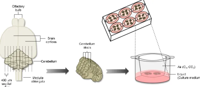
Parasagittal slices were obtained from cerebellum of CD1 mouse pups at postnatal
day 10 (Figure II. 1). In brief, brains were removed, cerebellum and attached hindbrain
were isolated in phosphate buffered saline (PBS) and 400 µm slices were obtained
using a Mcllwain tissue chopper and kept in an air-liquid interface system. Separated
slices were placed in the upper chamber of a 0.4 μm pore cell culture (BD Falcon, #353493, Lincoln Park, NJ, USA) in a number of 4 slices per insert. Cell culture inserts
were maintained in 6-well cell culture plates containing 1 mL of medium in the plate
well at a 37ºC and 5% CO2 conditioned atmosphere. Slice culture media consisted of
26
USA), 25% heat-inactivated horse serum (Gibco), 25% Earl’s balanced salt solution (Gibco), 6,5 mg/mL glucose, 25mM HEPES (Biochrom AG, Berlin, Germany) and 1%
of both L-glutamine (Sigma-Aldrich, St. Louis, MO, USA) and penicillin/streptomycin
(Sigma-Aldrich). After 3 DIV, slice culture media was totally replaced by a serum-free
media consisting of 98% Neurobasal-A (Gibco) and 2% B-27 (Gibco), supplemented
with 2 mM L-glutamine, 36 mM glucose, 1% U/mL penicillin/streptomycin and 25 mM
HEPES. Half media was replaced every day and slices were maintained for 7 days in
vitro (DIV) before treatment, to allow myelination and the clearance of debris
(Birgbauer et al., 2004).
Figure II. 1 – Schematic representation of COSC protocol. Cerebellum, separated from the brain of CD1 mouse pups at postnatal day 10, are sectioned into 400 µm thick slices. Separated slices are placed into the upper chamber of a cell culture insert and kept in an air-liquid interface system.
Following the 7 days, COSC were exposed to a demyelinating insult with LPC (0,5
mg/mL in serum-free culture media (Sigma-Aldrich). Following 16h LPC, COSC were
transferred to serum-free media in which cultures were maintained up to 48h
(Birgbauer et al., 2004; Miron et al., 2010). In parallel experiments, to ascertain S100B
role on demyelination and glial reactivity, COSC were incubated with LPC in the in the
presence or absence of anti-S100B antibody (Figure II.2) (1:1000, #Z0311, Dako,
Glostrup, Denmark). Supernatants were collected before and after LPC treatment.
Slices were collected at 9 DIV (48h post-LPC), and either stored in TRIzol® reagent at
in PBS for 10 min twice and stored in PBS at 4ºC for immunohistochemistry assays.
Figure II. 2 – Schematic representation of culture treatment. 0h, 16h and 48h correspond to 7 DIV, 8DIV and 9 DIV, respectively. LPC, lysophosphatidylcholine.
Total RNA Extraction, Reverse Transcription and Semi-quantitative
Real-Time Polymerase Chain Reaction
In order to determine expression of S100B and other genes of interest, total
cytoplasmic RNA was isolated from 9 DIV slices using the TRIzol® reagent method according to the manufacturer’s instructions (Invitrogen, Carlsbad, CA, USA). RNA concentration was quantified using Nanodrop ND-100 Spectrophotometer (NanoDrop
Technologies, Wilmington, DE, USA). Aliquots of 800 ng of total RNA were reversely
transcribed using the Rivertaid H Minus First Strand cDNA Synthesis Kit (Thermo
Fisher Scientific, MA, USA), under recommended conditions. qRT-PCR was performed
on a real-time PCR detection system (Applied Biosystems 7300 Fast Real-time PCR
System, Applied Biosystem, Madrid, Spain) using a SYBR Green qRT-PCR Master Mix
(Thermo Fisher Scientific, MA, USA). The PCR was performed in 8-well strips with
each sample performed in duplicate, and a non-template control (NTC) was included
for each amplificate. The sequences used as primers are listed in the Table II. 1
Table II. 1 – List of pairs of primers used for qRT-PCR assays. All primers were purchased from Thermo Fisher Scientific, MA, USA.
Gene Sense Anti-Sense
-actin gctccggcatgtgcaa aggatcttcatgaggtagt S100B gagagagggtgacaagcacaa ggccataaactcctggaagtc MBP ccatccaagaagaccccaca cccctgtcaccgctaaagaa
PLP tggcgactacaagaccacca gacacacccgctccaaagaa
HMGB1 ctcagagaggtggaagaccatgt gggatgtaggttttcatttctctttc IL-18 tggttccatgctttctggactcct ttcctgggccaagaggaagtg
28
qRT-PCR was performed under optimized conditions: 50ºC for 2 min, 95ºC for 10
min followed by 40 cycles at 95ºC for 15 s and 62ºC for 1min. In order to verify the
specificity of the amplification, a melt-curve analysis was performed, immediately after
the amplification protocol (95ºC for 15 s, followed by 60ºC for 30s and 95ºC for 15s).
Relative mRNA concentrations were calculated using the Pfaffl modification of the ΔΔCT equation, where CT is the cycle number at which fluorescence passes the
threshold level of detection, taking into account the efficiencies of individual genes. The
results were normalized to the housekeeping gene -actin in the same sample and the initial amount of the template of each trial was determined as relative expression by the
formula 2-ΔΔCT. ΔC
T is the value obtained for each sample by performing the difference
between the mean CT value of each gene of interest and the mean CT value of -actin.
ΔΔCT of one sample is the difference between its ΔCT value and the ΔCT of the sample
chosen as reference.
Immunostaining procedure
In order to determine the location of S100B expression as well myelination
impairment, membranes containing the fixed slices were cut out from cell culture
inserts, placed onto a slide and blocked in 1nM HEPES (Sigma-Aldrich), 2%
heat-inactivated horse serum, 10% heat-heat-inactivated goat serum, 1% BSA (Sigma-Aldrich)
and 0,25% Triton X100 (Roche Diagnostics, Indianapolis, USA) in Hank’s Balanced Salt Solution (HBSS, Gibco) for three hours, at room temperature. Slices were then
incubated with (Table II. 2) primary antibodies diluted in the blocking solution, for 24h at
4ºC. Following this, slices were washed three times for 15 min each with PBS with
0.01% Triton X100 (PBS-T) before incubation with secondary antibodies (Table II. 3) in
blocking solution for another 24 h at 4ºC). Slices were then washed three times for 15
min each with PBS-T, incubated with DAPI (1:1000, 3 min), washed three times for 15
min each with PBS-T and mounted using Fluoromount-G (Southern Biotech,
Percentage of the area immunoreactive for each antibody was measured from 8
bit.lsm files of 512x512 pixel resolution images captured using a 20x/1.2(zoom) lens on
a Confocal Point Scanning Microscope Zeiss LSM 510 META. Approximately, 4-5
images were captured per slice per condition, thus reducing any variations in image
acquisition. Binary masks were defined using a cut-off intensity threshold value,
defined as the minimum intensity due to specific staining above background values.
Then, the percentage of the area occupied by NF200, S100B, MBP, GFAP, NG2 and
Iba-1 staining was measured automatically for each separated stack of every acquired
image using ImageJ software. Regarding myelination, percentage of myelinated fibers
was obtained by the ratio between the area of co-localization of NF-200 and MBP and
the total area occupied by NF-200. Moreover, percentage of total area occupied by
myelin was calculated by the average of MBP area of each co-staining (with NF-200,
NG2, GFAP and Iba-1). Results are given by averaging values determined in the
separate microscopic fields from slices of different animals. Values are expressed as
mean ± SEM.
Table II. 2 – List of primary antibodies used for immunoshistochemistry assays.
Table II. 3 – List of secondary antibodies used for immunoshistochemistry assays. Antibody Host Brand Category Number Diluiton
S100 Rabbit Dako Z0311 1:400
NF-200 Mouse Novocastra NF200 1:200
MBP Rat Serotec MCA409S 1:50
GFAP Mouse Novocastra GFAP-GA5 1:100
Iba-1 Rabbit Wako 019-19741 1:250
NG2 Rabbit Milipore AB5320 1:50
Antibody G Brand Category Number Diluiton
Alexa 488 anti-mouse Goat Invitrogen A10680 1:1000 Alexa 488 anti-rabbit Goat Invitrogen A11008 1:1000 Alexa 594 anti-rat Donkey Invitrogen A21209 1:1000 Alexa 594 anti-rabbit Goat Invitrogen A11001 1:1000