FACULTY OF PHARMACY
UNIVERSIDADE DE LISBOA
Screening of necroptosis modulators with
potential therapeutic application
Lídia Mafalda Lopes Franco
Supervisor: Prof. Dr. Rui E. Castro
Co-supervisor: Prof. Dr. Cecília M. P. Rodrigues
Dissertation
MASTER DEGREE IN BIOPHARMACEUTICAL SCIENCES
2016
FACULTY OF PHARMACY
UNIVERSIDADE DE LISBOA
Screening of necroptosis modulators with
potential therapeutic application
Lídia Mafalda Lopes Franco
Supervisor: Prof. Dr. Rui E. Castro
Co-supervisor: Prof. Dr. Cecília M. P. Rodrigues
Dissertation
MASTER DEGREE IN BIOPHARMACEUTICAL SCIENCES
2016
The studies presented in this thesis were performed within the Cellular Function and Therapeutic Targeting research group, at the Research Institute for Medicines (iMed.ULisboa), Faculty of Pharmacy, Universidade de Lisboa, under the supervision of Rui E. Castro, PhD. and Cecília M. P. Rodrigues, PhD.
v
Abstract
Programmed cell death is a cellular death process in which a cell is eliminated in response to an inherent and genetically defined set of molecular events. In contrast, non-programmed cell death or classical necrosis is distinguished by its non-physiological or pathological triggering, coupled with lack of requirement for specific internal cellular signaling pathways. For a long time, apoptosis was considered as the sole form of programmed cell death. However, recent evidence suggests that necrosis can also be highly regulated, incorporating features of both necrosis and apoptosis in a process also referred to as necroptosis or programmed necrosis.
The establishment of necroptosis as an alternative form of regulated cell death resulted in the publication of several studies implicating it as the main contributor in the etiology and/or progression of human diseases, such as pancreatitis, ischemic injury and neurodegenerative diseases, among others. Additionally, accumulating evidence indicates that necroptosis is involved in the regulation of cancer. In fact, it is widely accepted that evasion of cell death is one of the hallmarks of cancer. Many lines of clinical and experimental evidence have demonstrated that failure in apoptosis is the most frequent cause of therapeutic resistance; thus, exploring other mechanisms of cancer cell death might lead to the development of strategies to overcome therapeutic resistance.
Ongoing progress in translational research in necroptosis has highlighted the increasing need for the identification of biomarkers of diagnosis, monitoring, and drug development. In addition, only a few small molecules, inhibiting molecular mediators of necroptosis, like Necrostatin-1, have been described. However, and thus far, no inhibitors of necroptosis are available for clinical use.
Targeting regulated necrosis may provide an unprecedented opportunity to develop novel therapeutic strategies. Screening assays of compound libraries will explore biological pathways that can be targeted with small molecules. The subsequent identification of target proteins of newly discovered hits is crucial to understand the cellular mechanism by which the small molecule acts. This project focused on the identification and characterization of
vi
novel modulators of necroptosis, which may ultimately translate into therapeutic strategies for the treatment of diseases associated with deregulated levels of necroptosis.
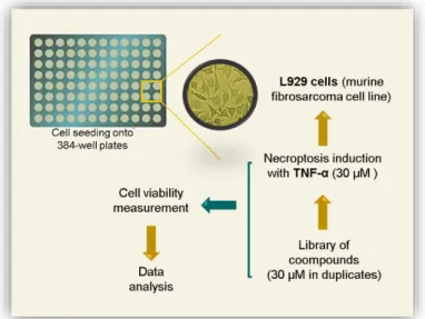
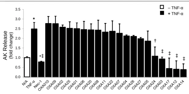
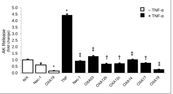
In order to identify novel modulators of necroptosis, we performed a biological screening using a library of twenty-one small molecules and identified three compounds with the ability to inhibit TNF-α-induced L929 cells necroptosis by more than 80%. To further characterize the activities of selected compounds, the half maximal effective concentration (EC50) for inhibiting necroptosis and the half maximal inhibitory concentration (IC50) for assessing drug toxicity were determined in the same cell model. Our results show that the selected hits inhibit necroptosis in L929 cells at micromolar EC50 concentrations and were not cytotoxic at the chosen range of concentrations. In addition, the mechanisms by which the hits inhibit necroptosis appear to involve, at least in part, inhibition of necroptosis signaling proteins RIP1 and MLKL.
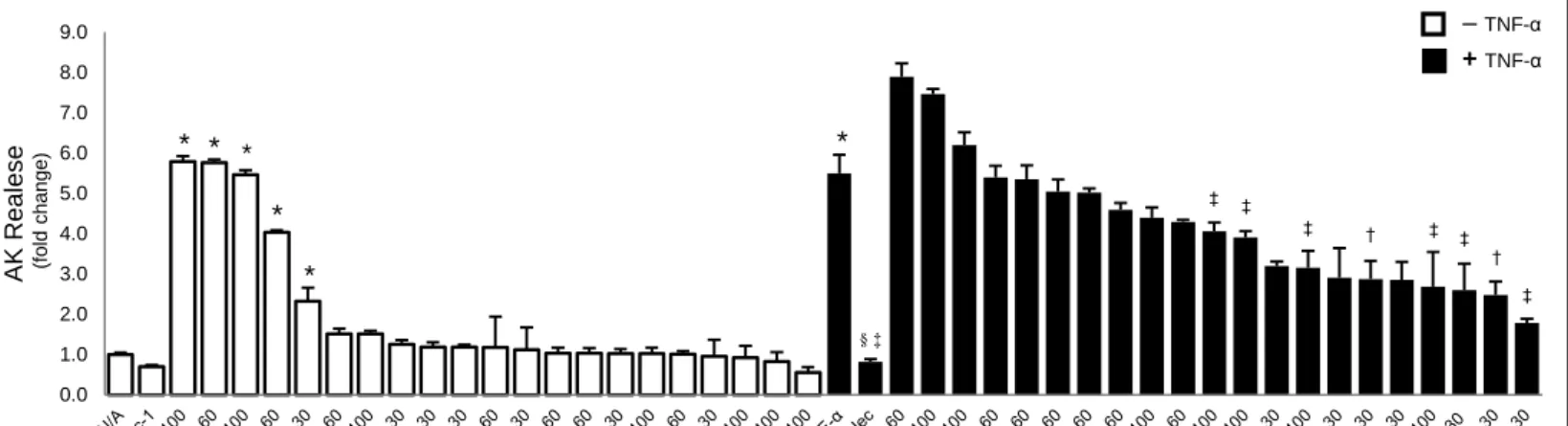
In parallel, we evaluated the ability of different bile acids, well-established modulators of apoptosis, in inhibiting necroptosis. Six bile acids and four newly synthesized derivatives were studied; one molecule inhibited TNF-α-induced necroptosis by more than 60% in L929 cells
In conclusion, the work presented here describes the discovery of four potential inhibitors of necroptosis, their confirmation/validation and possible mechanism(s) of action. A better understanding of the mechanisms of action of these compounds, as well as the study of their pharmacokinetic properties should help to determine their potential utility in disease treatment.
vii
Resumo
A morte celular programada é um processo através do qual uma célula é eliminada em resposta a um conjunto de eventos moleculares bem definidos. Por outro lado, a morte celular não programada, ou necrose clássica, distingue-se por ser provocada de forma não fisiológica ou patológica, não necessitando de maquinaria celular interna específica. Durante muito tempo, a apoptose foi considerada como a única forma de morte celular programada. Contudo, estudos recentes sugerem que a necrose pode ser altamente regulada, num processo designado por necroptose, a qual incorpora aspetos característicos da necrose e da apoptose.
Após o estabelecimento da necroptose como uma forma alternativa de morte celular, alguns estudos já a evidenciaram como o principal contribuinte na etiologia e/ou progressão de doenças humanas, nomeadamente, em pancreatite, doença isquémica e doenças neurodegenerativas, entre outras. Adicionalmente, a necroptose está envolvida na regulação do cancro. Em particular, a evasão da morte celular é considerada uma das principais características do cancro e diversos estudos clínicos e experimentais têm demonstrado que defeitos na apoptose das células cancerígenas são das causas mais frequentes de resistência à terapêutica. Assim, a investigação de outros mecanismos de morte das células cancerígenas poderá permitir o desenvolvimento de estratégias que visem ultrapassar esta resistência.
O progresso contínuo na investigação da necroptose realçou a necessidade, cada vez maior, de identificar biomarcadores para utilização em diagnóstico, monitorização e desenvolvimento de fármacos. Já foram descritos compostos que inibem elementos da via de sinalização da morte celular por necroptose, como a Necrostatina-1; contudo, ainda não estão disponíveis inibidores da necroptose na prática clínica.
Estratégias que tenham como alvo a necroptose constituem oportunidades únicas para desenvolver novas terapias para o tratamento das doenças anteriormente referidas. Os ensaios de screening de bibliotecas de compostos permitem identificar e, posteriormente, explorar vias biológicas que podem ser atingidas por pequenas moléculas, nomeadamente a
viii
via da necroptose. A subsequente identificação das proteínas-alvo dos compostos bioativos é crucial, uma vez que esta interação é a chave para compreender o mecanismo de ação dos mesmos. Deste modo, este projeto teve como objetivo a identificação e caracterização de novos moduladores da necroptose, os quais podem posteriormente traduzir-se em novas estratégias terapêuticas para o tratamento de patologias que resultem de níveis desregulados de necroptose.
De modo a identificar novos moduladores da necroptose, realizou-se um screening com uma biblioteca de vinte e uma pequenas moléculas e identificaram-se três compostos com capacidade de inibir a necroptose induzida pelo TNF-α em células L929, em mais de 80%. Para caracterizar as atividades dos compostos selecionados, determinaram-se os seus EC50 (half maximal effective concentration) e IC50 (half maximal inhibitory concentration) no mesmo modelo celular. Os resultados indicaram que os hits selecionados bloqueiam a necroptose a concentrações na ordem dos micromolar, não apresentando citotoxicidade no intervalo de concentrações utilizado. Além disso, o mecanismo pelo qual os hits inibem a necroptose parece envolver, pelo menos em parte, a inibição das proteínas de sinalização RIP1 e MLKL.
Em paralelo, testou-se a capacidade de diferentes ácidos biliares, moduladores estabelecidos da apoptose, em inibir a via da necroptose. Foram estudados seis ácidos biliares e quatro derivados; um mostrou ser capaz de inibir a necroptose induzida pelo TNF-α em células L929, em mais de 60%.
Em suma, o trabalho apresentado descreve a descoberta de quatro potenciais inibidores da necroptose, a sua confirmação/validação e possível mecanismo de ação. Uma maior compreensão dos mecanismos de ação destes compostos, assim como o estudo das suas propriedades farmacocinéticas irá ajudar a determinar a sua utilidade na clínica.
Palavras chave: ácidos biliares; MLKL; necroptose; pequenas moléculas; RIP1;
ix
Aknowledgements/Agradecimentos
A todas aqueles que direta ou indiretamente me ajudaram a terminar a presente tese, especialmente:
Ao Prof. Dr. Rui Eduardo Castro pela orientação prestada durante, principalmente, este segundo ano. Senti que, sem a sua ajuda, não seria possível, em primeiro lugar, ter alcançado os resultados agora descritos e, posteriormente, em terminar esta tese. Foi muito importante a sua orientação e revisão da mesma (mesmo com pouco tempo disponível) e agradeço-lhe muito por todas as sugestões, comentários e correções.
À Prof.ª Dr.ª Cecília Rodrigues, pela oportunidade de integrar o seu grupo de investigação no iMed.ULisboa, por toda a orientação ao longo do mestrado e, finalmente, pela revisão da tese.
Ao estudante de Doutoramento Hugo Brito, graças a ti foi possível conciliar o segundo ano de mestrado com o trabalho. Acredito verdadeiramente que, sem a tua ajuda, poderia ter sido impossível. Nunca te poderei agradecer todas as vezes que me ajudaste na leitura das placas, na recolha de células ou nos Western Blot (já para não falar a trabalhar no GraphPad!). Muito obrigada pela paciência, transmissão de conhecimento e disponibilidade, sempre que foi necessário.
Gostaria também de agradecer a todos os alunos de doutoramento que fazem parte do grupo de investigação liderado pela Prof.ª Dr.ª Cecília. Apesar do trabalho constante e das muitas horas já passadas no CPM, havia sempre tempo para responderem às minhas dúvidas existenciais ou ajudarem nas técnicas mais elaboradas.
A todos os professores do Mestrado em Ciências Biofarmacêuticas da Faculdade de Farmácia da Universidade de Lisboa por todos os conhecimentos transmitidos ao longo destes dois anos.
À Prof.ª Rita Guedes, pela colaboração nesta tese, nomeadamente, pela realização dos estudos de docking molecular in silico com um dos compostos estudados.
x
Ao Prof. Carlos Afonso da Faculdade de Farmácia e ao Prof. Jorge Salvador da Faculdade de Coimbra pela disponibilização dos compostos testados neste trabalho.
Aos meus colegas e amigos, particularmente, à minha equipa no trabalho; pela amizade, companheirismo e bons momentos passados tanto na ANF como fora dela. E às minhas amigas de Faculdade, principalmente à Cristina Bonito e à Patrícia Peixoto; apesar da distância e menos tempo em conjunto, a nossa amizade perdura.
A todos os meus familiares, especialmente pais e irmãs, pelo apoio, motivação e compreensão fornecidos.
Ao meu namorado, por todo o amor, dedicação, paciência e presença constantes que demonstrou ao longo da realização deste trabalho... e sempre.
xi
Table of contents
Abstract ... v Resumo ... vii Acknowledgments ... ix Abbreviations ... xivChapter1: Introduction and Objectives ... 1
Introduction ... 2
1. Molecular mechanisms of necroptosis ... 3
2. Interplay between metabolism, oxidative stress and necroptosis ... 6
3. Necroptosis-associated pathologies ... 9
3.1. Ischemia-reperfusion (IR) injury ...9
3.2. Neurodegenerative diseases ... 10
3.3. Pancreatitis ... 12
3.4. Sepsis and systemic inflammatory syndromes ... 12
3.5. Acute kidney injury (AKI) ... 14
3.6. Pathogen invasion... 14
3.6.1. Viral infections ... 14
3.6.2. Bacterial and parasites infections ... 15
3.7. Liver diseases ... 16
4. Currently available tools for targeting necroptosis ... 19
4.1. Necrostatins (NECs) ... 19
4.2. Vorinostat ... 20
4.3. Ponatinib and Pazopanib ... 21
4.4. 1-Benzyl-1H-pyrazole Derivatives ... 22
4.5. Aminoisoquinolines, pyrrolo[2,3-b]pyridines, and furo[2,3-d]pyrimidines ... 22
4.6. Necrosulfonamide (NSA) ... 23
4.7. IM-54 ... 23
4.8. NecroX analogs ... 24
4.9. GSK-843 and GSK-872 ... 24
4.10. Necroptosis inhibitors from natural products and isolated compounds ... 25
5. Identification and validation of protein targets of bioactive small molecules ... 27
6. Bile acids and cell death ... 29
Objectives ... 32
xii
Cell culture, reagents and antibodies ... 34
Chemical screening ... 34
Viability assay ... 35
Microscopy ... 35
Total Protein Isolation ... 36
Western Blot Analysis ... 36
Statistical Analysis ... 37
Chapter 3: Results and Discussion ... 39
(A) Evaluation of BAs as potential modulators of necroptosis ... 40
(B) Evaluation of a group of novel small molecules as potential modulators of necroptosis ... 44
Chapter 4: Conclusion and Future Perspectives ... 53
References ... 59
xiii
Index of Figures and Tables
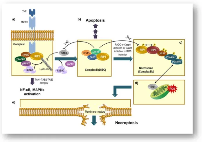
Figure 1: TNF-induced formation of apoptotic and necroptotic signaling complexes ... 5
Figure 2: Schematic overview of the screening workflow ... 40
Figure 3: CA, DCA and CDCA fail to modulate TNF-α-induced L929 cell necroptosis ... 41
Figure 4: OCA, UDCA, TUDCA as well as SB16 and SB22 inhibit TNF-α-induced L929 cell necroptosis ... 42
Figure 5: Dose response curve showing EC50 of OCA, UDCA and TUDCA on inhibition of L929 cell viability ... 43
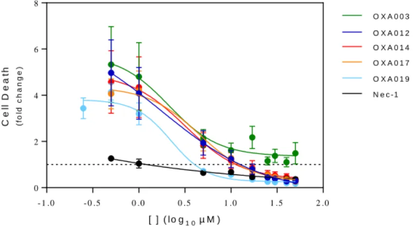
Figure 6: OXA003, 012, A014 and 017 inhibit TNF-α-induced L929 cell necroptosis ... 44
Figure 7: Different lots of OXA003, OXA012, OXA014 and OXA017, as well as a newly synthesized OXA - OXA019 - completely prevent TNF-α-induced L929 cell necroptosis ... 45
Figure 8: Dose response curve showing EC50 of OXA003, OXA012, OXA014, OXA017 and OXA019 on inhibition of TNF-α-induced L929 cell death ... 46
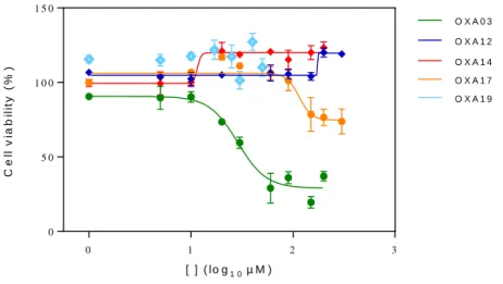
Figure 9: OXA012, OXA014 and OXA019 are not toxic to L929 cells in a wide range of concentrations ... 47
Figure 10: All OXA compounds display reduced protection from TNF-α-induced necroptosis with time ... 48
Figure 11: Evaluation of L929 cells morphology by phase-contrast microscopy showing protection from TNF-α-induced L929 cell death by OXA012, OXA014, OXA017 and OXA019 ... 49
Figure 12: OXA012 and OXA019 inhibit TNF-α induced RIP1 and MLKL phosphorylation .. 50
Figure 13: In silico molecular docking calculations for OXA12 ... 51
Supplementary Figure 1 (larger view of Figure 4): Hydrophilic BAs OCA, UDCA, TUDCA as well as SB16 and SB22 inhibit TNF-α-induced L929 cell necroptosis. ... 75
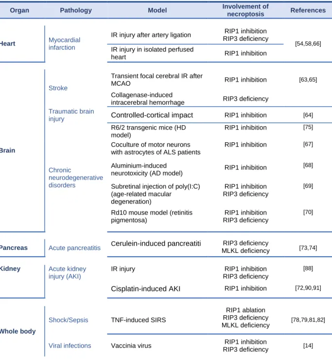
Table 1: In vivo pathologies associated with necroptosis ... 18
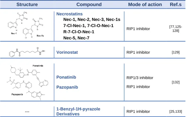
Table 2: Summary of necroptosis inhibitors ... 26
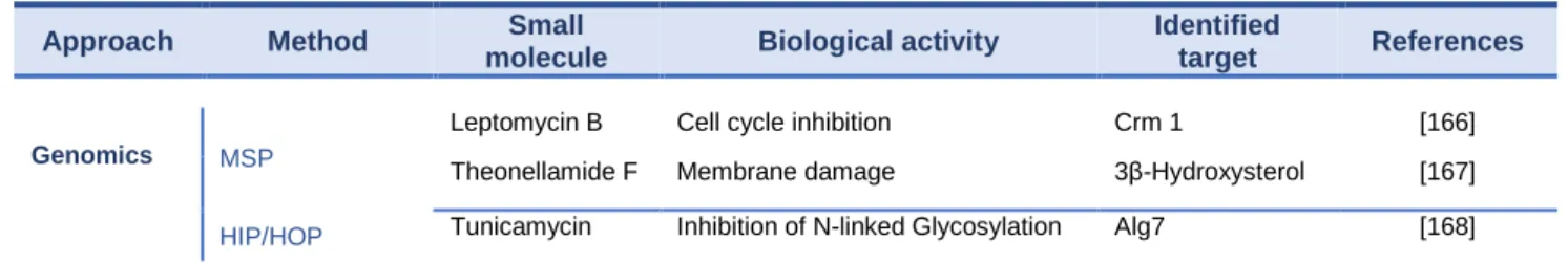
Table 3: Approaches for small-molecule target identification... 28
xiv
Abbreviations
AA, arachidonic acid
ALD, alcoholic liver disease ASH, alcoholic steatohepatitis ATP, adenosine 5’-triphosphate AIF, apoptotic inducible factor ANT, adenine nucleotide translocator CA, cholic acid
CDCA, chenodeoxycolic acid
cIAP, cellular inhibitor of apoptosis protein ConA, concanavalin A
cPLA2, cytosolic phospholipase A2 CYLD, cylindromatosis
DAMP, danger associated molecular pattern DCA, deoxycholic acid
DISC, Death-inducing signaling complex Drp1, dynamin-related protein
EC50, half maximum effect concentrations
FADD, FAS-associated death domain FLIP, FLICE-like inhibitory proteins GLUD1, glutamate dehydrogenase 1 GLUL, glutamate-ammonia ligase HCC, hepatocellular carcinoma HDAC, histone deacetylase HIM, homotypic interaction motif HMGB1, high mobility group box 1 IC50, half maximal inhibitory concentration
IDO, indoleamine-pyrrole 2,3-dioxygenase IKK, inhibitor of NF-κB kinase
IR, ischemia-reperfusion JNK, c-Jun N-terminal kinase L929, murine fibrosarcoma cell line LPS, lipopolysaccharide
LUBAC, linear ubiquitin chain assembly complex MLKL, mixed lineage kinase domain-like protein NADPH, nicotinamide adenine dinucleotide phosphate-oxidase
NAFLD, non-alcoholic fatty liver disease NASH, non-alcoholic steatohepatitis Nec-1, necrostatin-1
NEMO, NF-kB essential modulator NF-κB, nuclear factor‑κB
NOX, NADPH oxidase
NOXO1, NADPH oxidase organizer 1 NSA, necrosulfonamide
OCA, obeticholic acid
PARP1, poly(ADP-ribose)polymerase 1
PDGFRβ, platelet-derived growth factor receptor β PGAM5(L/S), phosphoglycerate mutase family
member 5 (long/short)
PTPC, permeability-transition pore complex PYGL, phosphorylase glycogen liver RIP, receptor interacting protein RN, regulated necrosis
RNS, reactive nitrogen species ROS, reactive oxygen species SMase, sphingomyelinase
STAT3, signal transducer and activator of
transcription 3
TAB, TAK1 binding protein
TAK1, transforming growth factor‑β-activated kinase 1
TNF-α, tumor necrosis factor alpha TNFR1, tumor necrosis factor receptor 1
TRADD, TNF receptor-associated death domain TRAF 2/5, TNF receptor-associated factor 2/5 TUDCA, tauroursodeoxycholic acid
UDCA, ursodeoxycholic acid
VEGFR, vascular endothelial growth factor receptor
WT, wild type
zVADfmk, arbobenzoxy-valyl-alanyl-aspartyl-[O-methyl]- fluoromethylketone
2
Introduction and Objectives
2
Introduction
Programmed cell death plays an important role in regulating the fate of individual cells during development and adult life in multi-cellular organisms [1]. While apoptosis was once considered as one of the most relevant forms of programmed cell death, this view has changed in recent years in light of the discoveries of additional modes of mammalian programmed cell death [2].
Apoptosis is a form of programmed cell death mediated by caspases [3,4]. The execution of apoptosis by caspases leads to distinguishable morphological features, such as exposure of phosphatidylserine (PtdSer) on the outer leaflet of the plasma membrane, chromatin condensation, intranucleosomal DNA cleavage and cytoplasmic membrane blebbing without disrupting its integrity [5,6]. The exposure of PtdSer on apoptotic cells provides a signal for their removal through engulfment by macrophages and other professional phagocytic cells, leading to clearance of dead cell bodies without eliciting inflammatory responses [7]. On the other hand, necrosis is characterized by early plasma membrane permeabilization and organelle swelling. Necrosis frequently occurs when cells are challenged with excessive external stress, such as heat, ischemia, and pathogen infection. The loss of plasma membrane integrity during necrosis leads to leakage of intracellular contents, some of which – such as high mobility group box 1 (HMGB1) – are known as potent pro-inflammatory factors [8]. Necrosis is considered to be accidental and occurs independently of signal transduction pathways. Interestingly, contrary to the traditional belief, recent studies have led to the revelation that regulated forms of necrosis also exist [9]. Among these, necroptosis is probably the most well understood form of regulated necrosis at the moment, at least in molecular terms [10]. In parallel with necroptosis, additional caspase-independent regulated forms of necrosis have been described, for example parthanatos, characterized by overactivation of the DNA damage-responsive enzyme poly(ADP-ribose)polymerase 1 (PARP1), adenosine 5’-triphosphate (ATP) depletion and apoptotic inducible factor (AIF)
3 nuclear translocation, as well as ferroptosis, which relies on dysfunctional intracellular iron mechanisms.
Compared to the rapidly growing knowledge on the molecular mechanisms that regulate necroptosis, less is known about the relevance of this type of regulated necrosis under physiological conditions or in the pathogenesis and progression of human pathologies. Still, it appears that, defective or excessive cell death by necroptosis can lead to diseases, in a similar way to dysregulation of apoptosis as an underlying mechanism of various pathophysiological states. In cancer for example, there is mounting evidence showing that too little necroptosis can contribute to tumor formation as well as to resistance of established tumors to current cancer therapies.
Necroptosis is increasingly recognized as an emergent clinically relevant therapeutic target. However, important concerns have been raised regarding applicability of the few existing necroptosis modulators. As such, novel specific and potent pharmacological molecules are urgently needed.
1. Molecular mechanisms of necroptosis
Necroptosis is best characterized in the setting of tumor necrosis factor alpha (TNF-α)-induced cell death. In addition to activation by members of the TNF family, necroptosis can also be activated by the Fas ligand, toll-like receptors, lipopolysaccharides (LPS), and genotoxic stress [11,12]. Different kinds of physical-chemical stress stimuli can initiate necroptosis, including anticancer drugs, ionizing radiation, photodynamic therapy, glutamate, calcium overload [13], and virus-mediated activation of DNA dependent activator of IFN-regulatory factors (DAI) [14,15].
The decision on whether death receptor activation results in apoptosis or necroptosis mainly depends on two kinases, receptor interacting protein (RIP) 1 and 3 [1]. RIPs are a group of threonine/serine protein kinases with a relatively conserved kinase domain but distinct non-kinase regions. A number of different domain structures were found in different
4 RIP family members, and these domains appear to be key factors in determining the specific function of each RIP kinase. RIPs have been demonstrated to play an important role in different biological processes, including those in innate immunity, but also in death-inducing processes [16]. RIP1 and RIP3 interact with each other through the homotypic interaction motif (HIM) at their C terminus region [16]. RIP1 is thought be a crucial kinase making the decision between cell survival or death [14]; its ubiquitination promotes cell survival while de-ubiquitination promotes kinase-dependent cell death [18]. Necrostatin-1 (Nec-1), a small molecule, blocks RIP1 kinase activity and, thus, death receptor-induced necroptosis [19]. Cho et al. reported that RIP3 controls programmed necrosis and augments RIP1 recruitment to the necrosome, a platform allowing engagement of necroptosis [14]. However, in some circumstances, increased RIP3 expression, or the absence of caspase-8, can bypass the requirement for RIP1 in tumor necrosis factor receptor 1 (TNFR1)-induced necroptotic signaling [20,21]. Viral infection and ethanol induced liver injury reflect two physiological circumstances where RIP3 may be activated independently of RIP1.
Under conditions insufficient to trigger apoptosis, TNFα activates TNFR1 which, in turn, recruits RIP1 kinase and other proteins, namely TNF receptor-associated death domain (TRADD), cellular inhibitor of apoptosis protein 1 (cIAP1), cIAP2, TNF receptor-associated factor 2 (TRAF2) and TRAF5 to form complex I (Fig. 1a). RIP1 is then subject to Lys63‑linked polyubiquitylation by cIAP ligases, which allows docking of transforming growth factor‑β-activated kinase 1 (TAK1) in complex with TAK1 binding protein 2 (TAB2) or TAB3, as well as of the inhibitor of nuclear factor‑κB (NF-κB) kinase (IKK) complex. The assembly of the IKK complex activates the NF‑κB pathway, enhanced by recruitment of the linear ubiquitin chain assembly complex (LUBAC) through the linear ubiquitin chains on RIP1. Subsequently, cylindromatosis (CYLD) removes Lys63‑linked polyubiquitins (Lys63-Ub) from RIP1, rendering complex I unstable and allowing RIP1 to dissociate from the plasma membrane and interact with TRADD, FAS-associated death domain (FADD), pro-caspase 8 and FLICE-like inhibitory proteins (FLIPs) to form the pro-death complex II (Fig. 1b), which can initiate apoptosis. The long isoform of FLIP (cFLIPL) and pro-caspase 8 form a
5 and inactivates RIP1 and RIP3, as well as CYLD,to prevent necroptosis. In the absence of caspase-8 activation, RIP3 can also be recruited to complex II to form complex IIb or the necrosome (Fig. 1c) [22].
Increased RIP3 expression is thought to result, at least in part, from increased expression of proinflammatory cytokines and activation of the inflammasome [23]. Mixed lineage kinase domain-like protein (MLKL) represents one of the RIP3 substrates identified so far. RIP3 phosphorylates MLKL at both threonine 357 and serine 358, and these phosphorylation events are required for cell death [25]. Further, MLKL serves as an adaptor to bring the RIP1-RIP3 complex into proximity with other RIP3 substrates. For instance, it increases mitochondrial ROS production by targeting specific mitochondrial proteins [26]. Recent studies have also demonstrated that MLKL is able to translocate to the plasma membrane and trigger cytotoxic influx of either calcium or sodium ions and, hence, disrupt
Figure 1: TNF-induced formation of apoptotic and necroptotic signaling complexes (see text for
6 osmotic homeostasis [27,28]. Nevertheless, a direct pore forming mechanism of MLKL has also been suggested [29,30]. The drug necrosulfonamide (NSA) was identified as an inhibitor of necrotic cell death through a mechanism that prevents MLKL binding to other proteins downstream of the RIP1-RIP3 complex [31]. PGAM5 is one of such proteins and has two splice variants, PGAM5L and PGAM5S [23]. RIP1, RIP3, MLKL and PGAM5L form a dynamic and transient complex that is recruited to PGAM5S on the mitochondrial membrane and then activates dynamin-related protein 1 (Drp1) [32] (Fig 1c, d) inducing reactive oxygen species (ROS) production and membrane permeability, ultimately leading to membrane rupture [23] (Fig 1e). Moreover, the RIP1/RIP3 kinase cascade has also been reported to regulate mitochondrial oxidative stress through c-Jun N-terminal kinase (JNK) [33].
2. Interplay between metabolism, oxidative stress and necroptosis
Among the different stimuli that can induce necroptotic cell death, several metabolism-related molecules and pathways have been identified, including ROS, ischemia-reperfusion injury, calcium overload or ATP depletion [35]. This list of metabolic stimuli points to a connection between metabolism and the necroptosis signal transduction machinery. Metabolic events can regulate the intracellular signal transduction cascade leading to necroptotic cell death, although little is known about the molecular details of this regulation.
Still, accumulating evidence points to redox processes as playing an important role in the regulation of necroptosis. For example, ROS production is rapidly increased during the early stages of necroptosis [36,37], while its inhibition significantly reduces induction of necroptosis [36,38]. These results suggest that ROS are actively involved in mediating necroptosis.
There are several potential intracellular sites that can induce ROS production in the course of necroptosis. Within mitochondria, respiratory chain complexes I and III are considered the main sites for ROS production during programmed cell death [38]. In addition, the mitochondrial adenine nucleotide translocator (ANT) can also contribute to ROS production in the course of cell death. ANT is localized in the inner mitochondrial membrane
7 and is responsible for the exchange of ADP against ATP [39]. Inhibition of ANT leads to decreased ADP with concomitant increases in ATP levels in the mitochondrial matrix, which in turn reduce the activity of ATP synthase and induce hyperpolarization of the mitochondrial membrane potential, thereby favoring production of ROS [40]. Because RIP1 has been described to negatively regulate ANT activity, it is tempting to speculate that elevated RIP1 activity during necroptosis may inactivate ANT, thereby favoring the production of ROS. In addition, RIP1 is responsible for phosphorylation–dependent interaction between signal transducer and activator of transcription 3 (STAT3) and the mitochondrial electron transport chain complex I subunit GRIM-19, leading to enhanced mitochondrial respiration and ROS production [41].
Nicotinamide adenine dinucleotide phosphate-oxidase (NADPH) oxidases (NOX) constitute another source of ROS [42]. Recently, TNFα has been reported to increase activity of the NOX1 complex via a mechanism involving RIP1 [43]. TNFR1 complex I has also been reported to serve as platform enabling the docking of NOX1 at the plasma membrane, thereby promoting the generation of ROS [43]. This process involves TNFα-dependent association of NADPH oxidase organizer 1 (NOXO1) subunit with RIP1, TNFR-associated death domain protein (TRADD) and riboflavin kinase, which in turn results in ROS production via NOX1 [43]. ROS generation by NOX1 may result not only in lipid peroxidation and membrane damage but also engage a feed-forward amplification loop to trigger further ROS production via the mitochondrial respiratory chain. Another amplification loop may involve the lysosomal compartment, where hydrogen peroxide can interact with ferrous ions to produce hydroxyl radical, a highly reactive ROS species [44]. Such amplification loops can lead to overproduction of ROS at the mitochondrial respiratory chain. This bears the danger of a lethal vicious cycle eventually resulting in the generation of reactive nitrogen species (RNS). RNS species can function as oxidants to produce protein or lipid oxidation, thereby altering protein function and causing membrane damage [45]. Downstream of the RIP1/RIP3 necrosome complex, different metabolic pathways, including glycogenolysis and glutaminolysis, have also been suggested to mediate signaling events during necroptosis.
8 RIP3 has been shown to promote the activity of several metabolic enzymes including glycogen phosphorylase (PYGL), glutamate-ammonia ligase (GLUL) and glutamate dehydrogenase 1 (GLUD1) [37]. PYGL is a key enzyme in the catabolic metabolism of glycogen, mediating the conversion of glycogen into glucose-1-phosphate, which in turn can be modified into glucose-6-phosphate, a substrate for glycolysis that can promote the generation of ROS through a mitochondrial metabolic burst. In addition, RIP3 was shown to activate GLUL and GLUD1 [37], two glutaminolysis enzymes. Enhanced glutaminolysis can engage the Krebs cycle, eventually contributing to increased ROS generation. Thus, RIP3-dependent changes in glycogenolysis and glutaminolysis can result in enhanced energy metabolism and increased ROS production.
Intracellular ATP content constitutes a central regulator in the decision on the mode of cell death. Upon depletion of ATP, human T-cells have been reported to switch from apoptosis towards necrosis. In this model, the generation or addition of ATP results in restored ability of T cells to undergo apoptotic cell death [46]. Apoptosis is an ATP-dependent cell death mechanism and different steps in the apoptotic signaling cascade are dependent on an adequate supply of bioenergetic substrates and ATP consumption. These include the activity of the translational machinery, protein degradation via the ubiquitin proteasome system, and activity of DNA repair enzymes such as Poly (ADP-ribose) polymerase 1 (PARP1) [47-49].
PARP1 has been described to play an important role in the metabolic regulation of cell death. PARP1 is localized in the nucleus and becomes activated upon extensive DNA damage [50,51]. PARP1 overactivation leads to NAD, ATP and an overall acute bioenergetic depletion, which promotes the release of apoptosis-inducing factor (AIF) from the interspace of mitochondria and its translocation to the nuclear compartment [52]. Within the nucleus, AIF is supposed to be required for large-scale DNA fragmentation in a caspase-independent manner [52].
Other molecular mechanisms that contribute to the execution of necroptosis upon assembly of the RIP1/RIP3 necrosome are: 1) activation of JNK-mediated degradation of
9 ferritin, thus increasing the labile iron pool and, consequently, ROS formation and 2) sphingomyelinase (SMase)-mediated generation of ceramide, which is converted into sphingosine by ceramidase and promotes a cytosolic Ca2+ wave that activates calpains and cytosolic phospholipase A2 (cPLA2). cPLA2 triggers lipid peroxidation by mobilizing the lipoxygenase substrate arachidonic acid (AA). AA is converted by lipoxygenase into membrane-damaging lipid hydroperoxides [52]. Finally, sphingosine, calpains and lipid hydroperoxides induce lysosome membrane permeabilization (LMP), resulting in the leakage of cytotoxic hydrolases into the cytosol. The production of ROS from all these sources initiate vicious cycles of damage by exacerbating mitochondrial uncoupling and lipid peroxidation and favoring the opening of the permeability-transition pore complex (PTPC). This results in the permeabilization of mitochondrial membranes and the translocation of cytotoxic proteins, including AIF, from the mitochondrial intermembrane space to the cytosol.
3. Necroptosis-associated pathologies
Cell death is a common feature in several human diseases such as ischemia-reperfusion injury and neurodegeneration. Although inhibition of apoptosis in many animal models of human diseases demonstrated the benefits of blocking cell death, the lack of “druggable” targets in the apoptosis pathway has so far prevented the development of apoptosis inhibitors for the treatment of human diseases. The discovery of necroptosis and RIP1 kinase as a “druggable” target in the necroptotic pathway elicited significant interest in studying the implication of necroptosis in human diseases, as well as the potential benefits of its inhibition.
3.1. Ischemia-reperfusion (IR) injury
Ischemia is caused by obstruction of blood flow to a tissue, resulting in limited supply of oxygen and nutrients and, if prolonged, in an impairment of energy metabolism and cell death. Restoration of the blood flow (reperfusion) results in oxygen reintroduction, neutrophil infiltration, cytokine production and generation of ROS, leading to cell death associated with
10 inflammation [44,53]. Interestingly, Nec-1 was shown to reduce inflammation and the infarct size caused by experimental myocardial IR [54-56], and reduced tissue injury upon cerebral [19,57], retinal [100] or renal IR [58,59] and neonatal hypoxia-ischemia in the brain [60,61], by preventing oxidative stress, necrosis and inflammation. Moreover, Nec-1 inhibited RIP3 upregulation, RIP1-RIP3 complex formation and phosphorylation, and MLKL recruitment to RIP1, in neonatal hypoxia-ischemia and myocardial IR [54]. RIP3-deficient mice also show reduced injury upon renal IR [58], as well as decreased cardiac hypertrophy and inflammation after myocardial infarction [62].
Noteworthy, both preocclusion and postocclusion delivery of Nec-1 exerts protective effects [19,56], suggesting that Nec-1 effectively mitigates not only ischemic but also reperfusion injury. An active role for necroptosis has also been demonstrated in animal models of traumatic brain injury [63,65], traumatic spinal cord injury [64], and intracerebral hemorrhage [63], which may share a part of the disease mechanism with IR. In addition, kidney transplantation is inevitably accompanied by IR injury, and RIP3 deficiency reduces injury and improves function of donor kidneys [66].
Taken together, these data indicate that necroptosis is a crucial component of IR injury. However, simultaneous inhibition of both apoptosis and necroptosis had a synergistic effect on cerebral IR injury [57]. Moreover, another study showed that both RIP1/RIP3- and CypD-dependent regulated necrosis participates in renal IR injury by two inCypD-dependent pathways [58]. Different forms of regulated necrosis may thus contribute to IR.
3.2. Neurodegenerative diseases
Huntington’s disease (HD) is an autosomal dominant disease characterized by motor and cognitive deficits, as well as psychiatric symptoms. The pathology is mainly due to the aggregation of mutant polyQ huntingtin protein, leading to neuronal dysfunction and cell death. Nec-1 reduces cell death in an immortalized striatal neuronal line expressing mutant HTT, and delays the onset and progression of disease in the mutant HTT-expressing R6/2
11 transgenic mouse model [75]. Furthermore, increased expression of RIP1 was observed at the onset of disease symptoms [75]. However, the survival benefit remained modest, indicating that necroptosis is probably not the only cell death mechanism contributing to neuronal cell death in this model.
Amytrophic lateral sclerosis (ALS) is a neurodegenerative condition with loss of motor neurons. Using a coculture system of primary motor neurons with astrocytes from ALS patients, it has been shown that both familial and sporadic ALS astrocytes compromise neuronal survival in a RIP1 kinase-, MLKL-, and Bax-dependent manner, while motor neurons death can be rescued by Nec-1 [67]. Therefore, RIP1 inhibition could potentially benefit both familial and sporadic ALS patients. The search for a common neurotoxic factor in ALS astrocytes is underway, which may shed light on the mechanistics of ALS pathogenesis.
Alzheimer’s disease (AD) is characterized by a progressive loss of memory and motor functions. Histologically, the disease is due to both the formation of extracellular β-amyloid plaques and neuronal cell death in the cerebral cortex and other subcortical regions. It was recently shown that Nec-1 administration decreases aluminum-induced neuronal cell death, inhibits expression of AD-related proteins and improves learning and memory retention in a mouse model of AD [68].
Retinal degeneration accounts for vision loss in the aged population and in patients with genetic disorders such as retinitis pigmentosa. In the rd10 mouse model of retinitis pigmentosa, the expression of RIP1 and RIP3 is augmented. Blockade of RIP1 or deletion of RIP3 preserves the survival and function of cone cells which are majorly responsible for daylight vision [69]. In dsRNA-induced retinal degeneration, a mouse model of age-related macular degeneration, RIP1 and RIP3 are also required for cell death and inflammation of retinal pigment epithelium and photoreceptors, and the degeneration of retinal pigment epithelial cells [70]. Given the sensitivity of retina to necroptosis, RIP kinases might be targeted for therapeutic intervention to treat degenerative vision loss.
12
3.3. Pancreatitis
Both apoptotic and necrotic cell death mechanisms have been observed during acinar cell death [71]. Of note, the severity of pancreatitis directly correlates with the extent of necrosis. Switching cell death to apoptosis, by pharmacological caspase activation, results in a milder form of pancreatitis, illustrating the harmful nature of in vivo necrosis [71,72]. Importantly, RIP3-deficient mice are protected from cerulein-induced pancreatitis, demonstrated by the absence of pancreatic acinar cell loss and necrosis, as well as lower levels of serum amylase [73]. RIP3 expression has been shown to correlate with the sensitivity of pancreatic acinar cells to undergo necroptosis, as RIP3 expression was dramatically increased in WT mice pancreas, which most probably primes the cells for subsequent necroptosis [73]. Interestingly, Mlkl-deficient mice were also reported to be partially protected from cerulein-induced pancreatitis [74], thus confirming necroptosis involvement in the pathogenesis of this disease. Surprisingly, a recent study showed that Nec-1 administration does not elicit any protective effects in this context [90]. Still, it is possible that these negative results are reflecting issues of Nec-1 pharmacokinetics [76,77].
3.4. Sepsis and systemic inflammatory syndromes
In recent years, several research laboratories have shown that regulated necrosis appears to be involved in sterile inflammation and septic shock. Duprez et al. were the first to demonstrate that targeting RIP1 (by Nec-1) or RIP3 (RIP3 deficiency) can substantially improve survival in a model of TNF-induced systemic inflammatory response syndrome, and in cecal ligation and puncture as a model for sepsis [78]. The authors showed that, although RIP3 deletion was protective in the cecal ligation and puncture model of sepsis, it did not affect the levels of bacteremia itself. This work indicates that RIP1/RIP3-mediated necroptosis most probably acts downstream of bacterial infection in the inflammatory phase of sepsis [79]. Because RIP1-deficient mice die shortly after birth [80], a new strategy to reveal the function of RIP1 kinase activity in vivo was recently developed by generating RIP1
13 kinase dead knock-in mice. Like RIP3-/- mice, these RIP1 kinase dead knock-in mice are completely protected against a lethal dose of TNF [81]. It was shown that the drop of the body temperature, as a result of TNF-induced systemic inflammatory syndrome, is prevented in MLKL-/- and RIP3-/- mice [82]. Strikingly, MLKL-/-Caspase-8-/- mice and RIP3-/-Caspase-8 -/-mice are completely protected against hypothermia during TNF injection [82]. These data suggest that TNF-induced hypothermia is also a consequence of caspase-8-dependent apoptosis and MLKL-dependent necroptosis, although it can also be interpreted that in the absence of caspase-8 the response is completely biased to necroptosis, as blocking caspase-8 sensitizes the necroptosis pathway [83]. This is in line with reports on the sensitization to TNF-dependent necrotic cell death in CrmA-transfected cells [9,84], and on TNF-mediated systemic inflammatory syndrome by combined injection of TNF and zVAD-fmk [85]. Further, caspase-8 prevents RIP3-mediated necroptosis during development [82,86].
Of note, two other studies demonstrated that administration of Nec-1 sensitized the lethal outcome in TNF-induced systemic inflammatory response syndrome and sepsis induced by cecal ligation and puncture [77,78]. Dose-dependent effects of Nec-1 in vivo could explain these contradictory findings. Remarkably, lower concentrations of Nec-1 apparently aggravate TNF-induced systemic inflammatory response syndrome, instead of being less protective [76,77], suggesting that in vivo pharmacodynamics may affect the outcome. The protective role of RIP3 in cecal ligation and puncture is also controversial. A recent article demonstrated that RIP3-/- mice have a higher mortality rate than controls in the cecal ligation and puncture model [74]. These contradictory results are not fully explained, but the outcome of the cecal ligation and puncture model is highly susceptible to experimental variation and animal housing conditions. A recent study showed that LPS-induced sepsis was not affected by catalytically inactive RIP1 or RIP3 deficiency [82], suggesting that RIPKs are not directly implicated in LPS endotoxemia. Of note, recent definitions and concepts of sepsis emphasize the dysregulated host response to illness [87] and, thus, additional studies are required to delineate the precise modulatory role of cell death modalities (i.e. necroptosis) in the host response during sepsis.
14
3.5. Acute kidney injury (AKI)
AKI is a common clinical problem associated with high mortality, which can occur due to sepsis or kidney IR. Previous studies have shown that both apoptotic and necrotic cell death are involved in the pathogenesis of AKI caused by nephrotoxic injury [88]. Moreover, inhibition of apopotsis in HK-2 human proximal tubule cells treated with cisplatin switches cell death towards necroptosis, which is passible of prevention by Nec-1 [89].
Several in vivo studies have shown that Nec-1 prevents osmotic nephrosis and cisplatin-induced AKI [72,90,91], whereas the pan-caspase inhibitor zVADfmk does not [88]. Moreover, both RIP1/RIP3- and CypD-dependent necrosis participate in renal IR injury, but contribute independently to the pathology [58]. These results suggest that necroptosis may be a dominant factor in the pathogenesis of AKI. In addition, Nec-1 is also able to prevent dilation of peritubular capillaries [88], suggesting a role for RIP1 in the regulation of microvascular hemodynamics and pathophysiology of AKI.
3.6. Pathogen invasion
Programmed cell death is often harnessed by the host to control intracellular growth of infected pathogens. Conversely, it can be critical for viruses to maintain the survival of infected cells in order to support its replication.
3.6.1. Viral infections
Vaccinia virus (VV) encodes the caspase inhibitor B13R/Spi2, which blocks apoptosis upon infection, but sensitizes cells to RIP1/RIP3-dependent necroptosis. Indeed, after VV infection, the pro-necrotic RIP1-RIP3 complex assembles in the liver, concomitantly with induction of TNF expression and inflammation [14]. In contrast, necrotic tissue injury and inflammation are significantly reduced in RIP3-deficient mice [14] or RIP1 D138N/D138N mice [81], resulting in highly elevated viral replication and mortality, as in Tnfr1- and Tnfr2-deficient mice [12,14,92].
15 Murine cytomegalovirus (MCMV) not only encodes a caspase inhibitor but also a necrotic inhibitor, vIRA, which is required to prevent premature host cell death upon infection [93,94]. vIRA contains a RHIM domain, enabling its interaction with RIP1 and RIP3. Accordingly, vIRA inhibits necrotic cell death in response to TNF by disrupting the pronecrotic RIP1-RIP3 complex and suppressing NF-κB and p38 activation [28,95,96]. Strikingly, upon MCMV infection with a strain carrying a RHIM-mutated vIRA, necrosis can be rescued by RIP3 KO, but not by Nec-1 [28], confirming that RIP3-dependent-, but RIP1-independent- necrosis functions as a potent anti-viral defense mechanism. Finally, a recent study revealed that the DNA sensor DAI interacts with RIP3 through a RHIM domain, mediating MCMV-induced necroptosis [15].
Similarly, MC159, encoded by the poxvirus Mollusucm contagiosum, possesses anti-apoptotic activity [97], and also inhibits RIP1-dependent necroptosis in response to TNF [12]. Interestingly, interaction between RIP1 and MC159 has also been demonstrated [98]. Other viral DED-containing inhibitors, such as equine herpesvirus-2 E8 and human herpesvirus K13, have also been demonstrated to protect cells from death receptor-mediated necrosis, and thus may serve similar roles in establishing efficient viral infection [12].
3.6.2. Bacterial and parasite infections
Despite the above studies highlighting the relevance of necroptosis as part of the immune defense against virus, excessive necroptosis of activated T cells or macrophages in response to infection can also halt the development of an efficient immune response against pathogens. This has been illustrated, for instance, in T cell-specific Fadd-deficient mice, which are more susceptible to Toxoplasma gondii infection than wild-type mice [99], as well as in macrophages infected by Listeria monocytogenes under cIAPs inhibition [100] or infected by Salmonella enterica serovar Typhimurium [101]. These studies pinpoint the role of FADD and cIAPs as crucial necroptosis regulators in immune cells during infections. Finally, a study performed in zebrafish showed that macrophages infected by M. tuberculosis die by RIP1- and RIP3- dependent necroptosis upon TNF treatment [102].
16 Bacterial infections can also lead to severe hemorrhagic disorders associated with induction of necrosis. For instance, Clostridium perfringens type C strain causes fatal hemorrhagic enteritis in several animal species and humans [103]. A recent study showed that Clostridium perfringens β-toxin, which is the essential virulence factor of Clostridium
perfringens type C, induces necrotic cell death of porcine primary endothelial cells
characterized by ATP depletion and LDH and HMGB1 release [104]. Cell death is inhibited by Nec-1, indicating that necroptosis appears to be involved in the pathogenesis of hemorrhagic enteritis.
3.7. Liver diseases
Only a few studies have evaluated the impact of necroptosis in liver diseases. Hepatocytes are susceptible to TNF-related apoptosis-inducing ligand (TRAIL)-induced necroptosis, which is dependent on formation of the RIP1/RIP3 complex. Indeed, Nec1 protects against concanavalin A (ConA)-induced hepatitis in mice [105,211]. In this regard, Liedtke et al. addressed the differential effects of TNF-induced liver injury in hepatocyte-specific caspase-8 knockout mice. These mice are protected from liver failure induced by Fas and TNFR1, but injection of ConA results in marked oxidative damage, spontaneous liver inflammation and enhanced non-apoptotic liver injury [106,107]. Combined deletion of hepatocyte-specific caspase-8 and NF-kB essential modulator (NEMO) further exacerbates liver necrosis and cholestasis [106]. Compared with Fas-induced liver injury, ConA-injury is mediated by membrane-bound TNF and does not require suppression of NF-κB. Further, this process is also caspase-independent, which points to necroptosis as the cause of liver cell death.
Non-alcoholic fatty liver disease (NAFLD) represents the most common chronic liver disease in the Western world [108,109]. It comprises a spectrum of liver lesions ranging from simple steatosis to non-alcoholic steatohepatitis (NASH), a more aggressive disease entity within the spectrum, associated with inflammation, cell death, and fibrosis. NASH can
17 progress to cirrhosis and, ultimately, hepatocellular carcinoma (HCC) [110]. Among all the different cell death pathways, apoptosis is the most well-described and established form in NAFLD [107,111.112]. However, it was recently demonstrated that human NASH livers express high levels of RIP3 [113]. More importantly, recent results have demonstrated that necroptosis is increased in the liver of NAFLD patients and in experimental models of NASH. Further, TNF-α triggers RIP3-dependent oxidative stress during hepatocyte necroptosis. suggesting that targeting necroptosis may arrest or at least impair NAFLD progression [113]. Indeed, despite the current NAFLD burden, no specific and effective pharmacological therapy is available. A pharmacological inhibitor of necroptosis could impact on the progression of NAFLD, preventing fibrosis, cirrhosis and hepatocellular carcinoma.
At the same time, and due to worldwide increase in alcohol abuse, alcoholic liver disease (ALD) has become one of the most common causes of liver-related morbidity and mortality. Alcoholic steatohepatitis (ASH) is the severe form of ALD, and may progress to fibrosis, cirrhosis and HCC [114]. In ALD, there is evidence for both apoptosis and necrosis [115,116]. As such, inhibition of hepatocellular death may also prevent ALD progression to fibrosis and HCC. Direct targeting of apoptosis and/or necroptosis, as well as mechanisms that indirectly prevent cell death, such as the involvement of the gut microbiota-liver axis or ROS, are potential relevant targets.
Hepatocellular carcinoma (HCC), the most common primary liver tumor, arises almost exclusively in the setting of chronic hepatic inflammation [117]. Cell death represents a key trigger of inflammation, thus contributing to multiple hallmark capabilities of cancer [118]. In chronic liver disease, hepatocyte cell death is a prominent feature driving progression to hepatic fibrosis and HCC [119]. One of the challenges in clinical cancer therapy is the resistance of cancers to apoptosis. In HCC, apoptosis resistance is one of the most significant factors for hepatocarcinogenesis and tumor progression, leading to resistance to conventional chemotherapy [120]. Since many anti-cancer drugs are inducers of apoptosis, inducing RIP3-dependent necrosis is an attractive strategy to circumvent apoptosis resistance of cancer cells. In addition to DNA alkylating agents, which induce necrosis in a
18 PARP-dependent manner [50,122], some drugs have already been reported to induce RIP3-dependent necrosis [123,124]. Whereas HCC prevention strategies need to inhibit cell death at early stages to halt the HCC-promoting cell death-inflammation-regeneration-fibrosis cascade, treatment of established HCC requires promotion of cell death. As such, a better understanding of the pathophysiology and mechanisms underlying progression of liver diseases, particularly HCC, is of outmost importance.
Table 1: In vivo pathologies associated with necroptosis.
Organ Pathology Model Involvement of
necroptosis References
Heart Myocardial
infarction
IR injury after artery ligation RIP1 inhibition
RIP3 deficiency
[54,58,66] IR injury in isolated perfused
heart RIP1 inhibition
Brain
Stroke
Transient focal cerebral IR after
MCAO RIP1 inhibition [63,65]
Collagenase-induced
intracerebral hemorrhage RIP3 deficiency
Traumatic brain
injury Controlled-cortical impact RIP1 inhibition [64]
Chronic neurodegenerative disorders R6/2 transgenic mice (HD model) RIP1 inhibition [75]
Coculture of motor neurons with astrocytes of ALS patients
RIP1 inhibition [67]
Aluminium-induced
neurotoxicity (AD model) RIP1 inhibition
[68]
Subretinal injection of poly(I:C) (age-related macular
degeneration)
RIP1 inhibition RIP3 deficiency
[69]
Rd10 mouse model (retinitis pigmentosa)
RIP1 inhibition RIP3 deficiency
[70]
Pancreas Acute pancreatitis Cerulein-induced pancreatiti MLKL deficiency RIP3 deficiency [73,74]
Kidney Acute kidney
injury (AKI)
IR injury RIP1 inhibition
RIP3 deficiency
[88]
Cisplatin-induced AKI RIP1 inhibition [72,90,91]
Whole body
Shock/Sepsis TNF-induced SIRS
RIP1 ablation RIP3 deficiency
MLKL deficiency [78,79,81,82]
Viral infections Vaccinia virus RIP1 inhibition
19
MCMV RIP3 deficiency [15,28,95,96]
Poxvirus RIP1 inhibition [12]
Parasites
infections Toxoplasma gondii RIP1 inhibition [99]
Bacterial infections
Listeria monocytogenes RIP1 inhibition
RIP3 deficiency [100]
Salmonella enterica RIP1 inhibition
RIP3 deficiency [101]
M. tuberculosis RIP1 inhibition
RIP3 deficiency [102]
Clostridium perfringens β-toxin RIP1 inhibition [104]
Liver
Acute hepatitis
ConA-induced hepatitis RIP1 inhibition [105-107]
Acetaminophen induced hepatitis RIP1 inhibition RIP3 deficiency [34, 121, 128,146] Chronic liver diseases
Non-alcoholic fatty liver disease RIP3 deficiency [113]
Alcoholic liver disease RIP3 deficiency [115,116, 128]
4. Currently available tools for targeting necroptosis
No inhibitors of necroptosis are currently in clinical use. In fact, given the relatively new concept of necroptosis, only a scarce number of inhibitors have been developed, each displaying major drawbacks and/or side effects.
4.1. Necrostatins (NECs)
Necrostatins (NECs) are a family of compounds of diverse chemical structure that have been named for their ability to block necrotic cell death, namely by directly inhibiting the kinase activity of RIP1 [19,125]. To date, several structurally distinct NECs have been synthesized and tested as potential inhibitors of necroptosis in different experimental animal models [125,126].
Necrostatin-1 (Nec-1; 5-(1H-Indol-3-ylmethyl)-(2-thio-3-methyl) hydantoin) was identified in 2005 by Alexei Degterev and Junying Yuan as a molecule blocking necrotic cell death in human and murine cells [19]. In a subsequent study, Nec-1 was identified as an allosteric inhibitor of RIP1 kinase activity. Nec-1 is now widely used to target RIP1 kinase activity in various experimental disease models. However, several studies have raised critical concerns
20 regarding its specificity, appropriate control and effective concentration, particularly in murine experimental disease models. Nec-1 is a non-specific RIP1 kinase inhibitor, and also blocks indoleamine-pyrrole 2,3-dioxygenase (IDO), PAK1 and PKAcα [127]. Nec-1s, a Nec-1 derivative, has specificity for RIP1 kinase over a broad range of kinases [76]. However, in particular cellular contexts, RIP1 kinase also mediates inflammatory responses and apoptosis. Moreover, Nec-1 and Nec-1s have inadequate pharmacokinetic properties, namely very short half-lives. Finally, necroptosis can occur in a RIP1-independent manner. For instance, although RIP3-deficiency protects from liver injury in an animal model of alcoholic liver disease, Nec-1 does not display any protective effects [128].
Nec-5 (2-[[3,4,5,6,7,8-hexahydro-3-(4-methoxyphenyl)-4-oxo[1]benzothieno[2,3-d]pyrimi-din-2-yl]thio]-acetonitrile) also targets RIP1 during necroptosis, although through distinct mechanisms from those of Nec-1 [125]. It was reported that Nec-5 is a selective allosteric inhibitor of RIP1 that prevents the death of TNF-α-treated FADD-deficient Jurkat cells, with an EC50 value of 240 nM [125].
Nec-7 (5-((3-(4-fluorophenyl)-1H-pyrazol-4-yl) methylene)-2-imino-3-(thiazol-2-yl) thiazo-lidin-4-one) is a necroptosis inhibitor that is structurally and biologically distinct from other NECs, as it does not inhibit RIP1. Nec-7 may target an additional regulatory molecule in this pathway as it inhibits TNF-α-induced necroptosis in a FADD-deficient variant of human Jurkat T cells, with an EC50 value of 10.6μM (77).
The specificity and activity of three NEC analogs (Nec-1, Nec-5 and Nec-7) have been examined thoroughly both in vitro and in vivo, providing important information about NECs in disease models [76]. Overall, data from the literature suggests that NECs have emerged as the first-in class inhibitors of RIP1, the key upstream kinase involved in the activation of necroptosis.
21
4.2. Vorinostat
Histone deacetylase (HDAC) inhibitors are well-known anticancer agents possessing anti-inflammatory and neuroprotective effects. Vorinostat (suberoylanilide hydroxamic acid) was the first HDAC inhibitor to be approved by the U.S. Food & Drug Administration (FDA) for the treatment of relapsed/refractory cutaneous T cell lymphoma. Wang et al. first reported the anti-necroptotic mechanism underlying the effects of vorinostat [129]. Murine fibrosarcoma L929 cells pretreated with vorinostat (1μM) were treated with TNF-α to induce necroptosis. Vorinostat pretreatment effectively protected L929 cells against TNF-α -induced necroptosis, and this protection is conferred through mechanisms involving RIP1-dependent NF-κB and p38 MAPK activation, JNK and Akt kinase inactivation, autophagy initiation, and enhanced cFLIPL expression (a negative regulator of necroptosis). Overall, the authors show that vorinostat exerts its anti-inflammatory and cell protective effects by preventing necroptosis, an important process in inflammation and elimination of cells.
4.3. Ponatinib and Pazopanib
Ponatinib is an oral multi-targeted tyrosine kinase inhibitor developed for the treatment of chronic myeloid leukemia and Philadelphia chromosome-positive acute lymphoblastic leukemia. This inhibitor is used as second-line treatment for patients who have acquired resistance to standard therapy. In parallel, pazopanib is an oral receptor tyrosine kinase inhibitor approved for treatment of patients with advanced renal cell carcinoma and soft tissue sarcoma [130,131]. It targets vascular endothelial growth factor receptor (VEGFR)1, -2, -3, platelet-derived growth factor receptor β (PDGFRβ), and c-Kit.
Fauster A et al. screened a panel of 268 FDA-approved drugs with diverse mechanisms for their ability to inhibit TNF-α-induced necroptosis in FADD-deficient human Jurkat T cells, and found that these two kinase inhibitors were potential blockers of necroptosis at submicromolar EC50 concentrations [132]. Both drugs inhibited necroptotic signaling triggered by various cell death receptors, whereas they did not interfere with apoptosis.
22 Ponatinib directly binds to and inhibits both RIP1 and RIP3. These compounds have been developed and approved as anti-cancer treatment molecules, and their safety profiles have been evaluated in this context, with both drugs reported to cause severe side effects. The definition of the cellular target spectrum might be useful in gaining a better understanding of the molecular mechanisms underlying the reported adverse effects. Thus, it still remains necessary to elucidate their exact mechanisms of action and to perform a series of careful studies in animal models covering a large variety of necroptosis-associated pathologies.
4.4. 1-Benzyl-1H-pyrazole Derivatives
Zou et al. recently screened several compounds using their in-house database and found that 1-(2,4-dichlorobenzyl)-3-nitro-1H-pyrazole exhibited potential necroptosis inhibition activity [133]. Human colon cell line HT29 was treated with TNF-α, Smac-mimetic, and zVAD-FMK. TNF-α and Smac-mimetic causes cells to undergo apoptosis by triggering the formation of a caspase-8 activating complex containing RIP1, while Z-VAD-FMK is a pan-caspase inhibitor that can switch apoptosis to necroptosis in RIP3-expressing cells upon caspase-8 inhibition [25]. The authors reported that 1-(2,4-dichlorobenzyl)-3-nitro-1H-pyrazole exhibited an EC50 value of 1.048μM and could selectively inhibit RIP1 without influencing RIP3 kinase activation. Furthermore, the authors synthesized a number of 1-benzyl-1H-pyrazole derivatives and studied their structure-activity relationship, leading to the discovery of a potent compound 4b, which contains a new scaffold of 1-benzyl-1H-pyrazole with a Kd value of 0.078μM against RIP1 and an EC50 value of 0.160μM in a cell necroptosis inhibitory assay. The necroptosis inhibitory activity of compound 4b was compared with that of Nec-1 (EC50: 0.860μM), and 4b was found to be more potent [133]. The authors concluded that compound 4b exhibited considerable effectiveness in inhibiting RIP1/RIP3/MLKL signaling in intact cells and showed a good protective effect in the pancreas in the L-arginine-induced pancreatitis mouse model.
23
4.5. Aminoisoquinolines, pyrrolo[2,3-b]pyridines, and furo[2,3-d]pyrimidines
Harris et al. screened GSK inhibitor libraries and identified a series of type II RIP-K1 inhibitors with strong necroptosis inhibitory activity [135]. The authors revealed that three distinct series, namely 1-aminoisoquinolines, pyrrolo[2,3-b]pyridines, and furo[2,3-d]pyrimidines showed a dose-proportional response in inhibiting necroptosis and hypothermia in a mouse model of TNF-α-induced lethal shock.4.6. Necrosulfonamide (NSA)
Due to the potential drawbacks of Nec-1, including disruption of necrosome formation, the need for a potent necrosome-downstream necroptosis inhibitor has been highlighted. Liao D et al. identified necrosulfonamide (NSA) as the first downstream small molecule inhibitor of necroptosis, by directly targeting MLKL [136]. However, this compound blocks human MLKL through the formation of a covalent bond with Cys86, but lacks activity against mouse protein due to the absence of the orthologous Cys. In this regard, it cannot be used in murine models of disease, invalidating pharmacological, pharmacokinetic and toxicity pre-clinical testing. Other MLKL inhibitors were studied in mouse models but exhibited excessive toxicity to cells and had multiple targets on other necrosome components [137]. However, research should be focused on developing new MLKL inhibitors to expand our knowledge on the in vivo significance of MLKL-dependent necroptosis in animal models. This research will serve as an invaluable tool in advancing the understanding on the significance of blocking necroptosis.
4.7. IM-54
IM-54 (1-Methyl-3-(1-methyl-1H-indol-3-yl)-4-(pentylamino)-1H-pyrrole-2,5-dione, 2-(1H-Indol-3-yl)-3-pentylamino-maleimide), a novel indolylmaleimide derivative, was shown to inhibit necrotic cell death induced by hydrogen peroxide (H2O2), but not apoptotic cell death [138]. IM-54 selectively blocks oxidative stress-induced necrotic cell death (~3μM). The
24 authors suggest that IM-54 is expected to constitute a powerful bioprobe for clarifying the unique signaling pathway of necrotic cell death.
4.8. NecroX analogs
NecroX are a series of indole-containing inhibitors, which were originally identified in a cell-based screen as suppressors of drug-induced necrosis in hepatocytes [139]. These molecules display general anti-oxidant and peroxynitrite (ONOO-) scavenging activities in
vitro, and were further investigated as inhibitors of mitochondrial oxidative and nitrosative
stress, which are hallmarks of pathologic necrosis [140]. One of these molecules, NecroX-1, was found to effectively inhibit toxicity caused by the pro-oxidant tBHT and reduce acute hepatotoxicity of CCL4 and streptozotocin (STZ)-induced pancreatic islet destruction [139]. Furthermore, NecroX-2 and NecroX-5 are cell permeable necrosis inhibitors with antioxidant activity. They mostly target mitochondria and selectively block oxidative stress-induced necrotic death. These analogs were also reported to protect cells against cold shock, hypoxia, and oxidative stress in vitro, as well as CCL4-induced acute liver and chronic liver fibrosis in rodent models [139,141]. Another member of this inhibitor family, NecroX-7, was shown to attenuate ischemia–reperfusion liver injury in dogs, doxorubicin induced cardiomyopathy in rats, acetaminophen-induced hepatotoxicity, non-alcoholic steatohepatitis, and allogeneic transplantation-induced graft versus host disease (GVHD) in mice [142-146]. NecroX-7 hampers release of the necrotic danger associated molecular pattern (DAMP) protein HMGB1 in vivo, and was proposed to act through inhibition of NADPH oxidase activity [143-145]. In contrast, NecroX-5 reduces myocardial hypoxia-reoxygenation injury in rats through the blockade of the mitochondrial Ca2+ uniporter [147]. Overall, these results highlight NecroX molecules as a useful class of mitochondria-targeting agents with putative anti-necroptotic activities.