Submitted 21 August 2015 Accepted 18 November 2015 Published22 December 2015
Corresponding author Adeline Bidault,
Academic editor Cristiane Thompson
Additional Information and Declarations can be found on page 14
DOI10.7717/peerj.1484
Copyright 2015 Bidault et al.
Distributed under
Creative Commons CC-BY 4.0
OPEN ACCESS
Development of a Taqman real-time PCR
assay for rapid detection and
quantification of
Vibrio tapetis
in
extrapallial fluids of clams
Adeline Bidault, Ga¨elle G. Richard, C´edric Le Bris and Christine Paillard
Laboratoire des Sciences de l’Environnement Marin (LEMAR), UMR 6539 UBO/CNRS/IRD/Ifremer, Universit´e de Bretagne Occidentale, Plouzan´e, France
ABSTRACT
The Gram-negative bacteriumVibrio tapetisis known as the causative agent of Brown Ring Disease (BRD) in the Manila clamVenerupis(=Ruditapes)philippinarum. This bivalve is the second most important species produced in aquaculture and has a high commercial value. In spite of the development of several molecular methods, no survey has been yet achieved to rapidly quantify the bacterium in the clam. In this study, we developed a Taqman real-time PCR assay targeting virB4 gene for accurate and quantitative identification ofV. tapetisstrains pathogenic to clams. Sensitivity and reproducibility of the method were assessed using either filtered sea water or extrapallial fluids of clam injected with the CECT4600TV. tapetisstrain. Quantifica-tion curves ofV. tapetisstrain seeded in filtered seawater (FSW) or extrapallial fluids (EF) samples were equivalent showing reliable qPCR efficacies. With this protocol, we were able to specifically detectV. tapetisstrains down to 1.125 101bacteria per mL of EF or FSW, taking into account the dilution factor used for appropriate template DNA preparation. This qPCR assay allowed us to monitorV. tapetisload both experimentally or naturally infected Manila clams. This technique will be particularly useful for monitoring the kinetics of massive infections byV. tapetisand for designing appropriate control measures for aquaculture purposes.
Subjects Aquaculture, Fisheries and Fish Science, Environmental Sciences, Marine Biology, Microbiology, Molecular Biology
Keywords Vibrio tapetis, virB4 gene, Taqman real-time PCR, Molecular diagnostic,Venerupis philippinarum, Marine pathogen, Brown ring disease
INTRODUCTION
V. tapetisfirst colonizes the periostracal lamina of the clam between the mantle and the shell. The proliferation of the pathogen in the periostracal lamina and extrapallial fluids (EF) inhibits the normal shell biomineralization process, resulting in a brown deposit of melanised matrix (conchiolin) on the inner surface of the valves (Paillard & Maes, 1994;Paillard & Maes, 1995a). The clam’s extrapallial fluids have been demonstrated to be a major compartment in the early stage of the defense process against the infection (Allam, Paillard & Maes, 1996;Allam, 1998). Indeed, extrapallial fluids contain numerous hemocytes (Allam, Paillard & Auffret, 2000) which are responsible for phagocytosis of micro-organisms (Allam & Paillard, 1998). Accumulation of hemocytes suggests an efficient defense system able to neutralize the pathogen before colonization of the extrapallial cavity and eventually tissues and could lead to septicemia (Allam & Ford, 2006). In this case, the penetration ofV. tapetisinto the hemolymph can provoke mortality before the clam exhibits BRD symptoms (Allam, Paillard & Ford, 2002;Paillard, Allam & Oubella, 2004). In a natural environment, the prevalence range of BRD in Manila clam has been estimated as 10–20% (Paillard et al., 2014).
Here, pathogenicity and virulence are defined as proposed inCasadevall & Pirofski (1999)andSparling (1983), that is to say as the capacity of a microbe to cause damage to the, and the degree of pathogenicity respectively. A subtractive bank between twoV. tapetis strains, i.e., the fish pathogen LP2 and the clam pathogen CECT4600Trevealed that some genes are present only in the genome of theV. tapetisstrain pathogenic toV. philippinarum (G Dias, 2015, unpublished data). Among these genes,virB4existed only in the genome of the clam pathogen CECT4600T(accession number:KT382306). This finding suggests that some of these genes may be specifically associated with pathogenicity in the Manila clam. The genomes of severalV. tapetisstrains, including CECT4600T, were recently sequenced (G Dias, 2015, unpublished data), confirming the presence of one copy of thevirB4gene in the CECT4600Tchromosome, using the MicroScope genomics platform (Vallenet et al., 2013). ThevirB4gene encodes a protein involved in a large complex assigned to type IV secretion systems (T4SSs). In bacteria, secretion is essential for virulence and survival. Bac-teria use T4SSs to translocate DNA and protein substrates across the cell envelope (Juhas, Crook & Hood, 2008;Low et al., 2014;Christie, Whitaker & Gonz´alez-Rivera, 2014), which contributes to genome plasticity and the evolution of pathogens through dissemination of antibiotic resistance and virulence genes (Fronzes, Christie & Waksman, 2009).
and not specific enough and sensitive enough to detectV. tapetispathogens in clams. These methods are therefore inadequate for monitoring individual clams and above all, inappropriate to detect asymptomatic infected clams.
The aim of this study was to develop a rapid and accurate detection method forVibrio tapetis, the causative agent of BRD, which is of growing interest due to the increased preva-lence of the disease and the high commercial value of clams. Taqman real-time PCR pro-tocols have previously been carried out forV. aestuarianus(Saulnier, De Decker & Haffner, 2009;McCleary & Henshilwood, 2015) andV. harveyi(Schikorski et al., 2013). In this paper, we developed a Taqman real-time PCR assay for specific and rapidV. tapetisdetection and quantification from extrapallial fluids of clams, and validated it on both pure cultures and through an experimental infection ofV. philippinarumby a virulentV. tapetisstrain.
MATERIALS AND METHODS
Bacterial strains and culture conditions
Bacteria belonging to twelveVibriospecies and seventeen strains ofV. tapetisisolated from clams and fishes were used in this study (Table 1) as positive and negative controls to check species specificity of the designed primers. Some reference strains were purchased from CIP and LMG collections.V. anguillarumstrain 775 was supplied by JH Crosa (Frans et al., 2011;Crosa, Schiewe & Falkow, 1977). The Norwegian S2-2 strain was kindly offered by S Mortensen (2009, unpublished data) from the Institute of Marine Research at Bergen in Norway. Finally, the FPC 1121 strain was graciously provided by T Matsuyama (Matsuyama et al., 2010). The other strains were available from the LEMAR collection.
TheV. tapetisCECT4600T strain was used for the experimental infection as the reference strain (Paillard & Maes, 1995b;Borrego et al., 1996).
All bacterial strains were cultured in Zobell medium (pastone 4 g/L, yeast extract 1 g/L, sea salt 30 g/L at pH 7.4) enriched with iron phosphate (0.1 g/L) at 18◦C during 18 h under constant shaking at 180 rpm (Infors HT®) (Balboa et al., 2012).
Clams, experimental infection and extrapallial fluids collection (EF)
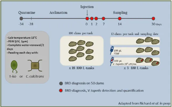
Two year oldVenerupis philippinarumwere provided by Fabien Fonteneau in Marennes-Oleron (“Les Claires de Bonsonge”®, EARL, brood stock producer). A first health diagnostic was performedin situon 50 clams to ensure absence of BRD, prior to the sampling effort for the bacterial challenge. They were transferred to the Ifremer’s facilities (Laboratoire de Physiologie des Invert´ebr´es, LEMAR, Plouzan´e) for six days quarantine with chloramphenicol (8 mg/L; Sigma Aldrich, St. Louis, MO, USA) at 13◦C.
For the duration of the experiment, clams were split randomly into eighteen 100 L-tanks with air-lift systems and a complete water-renewal every two days. During the whole experiment, clams were daily fed an algal ration (maintenance ratio from FAO, 2004) of two algae commonly used in aquaculture (Isochrysis affinis galbanaandChaetoceros calcitrans) (Figs. 1andS1A).
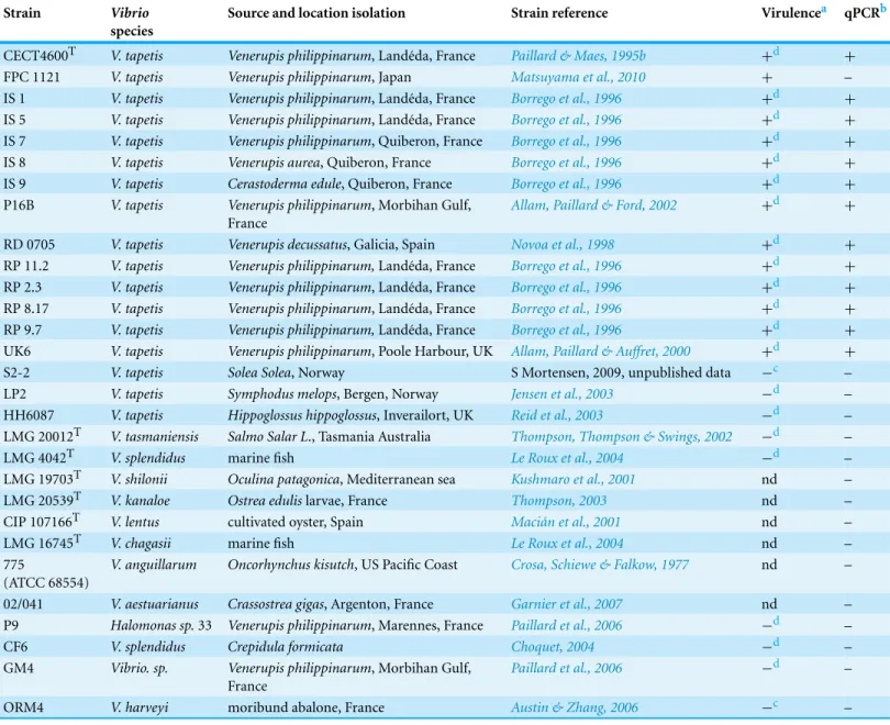
Table 1 Bacterial strains used in this study isolated from different hosts and origins, and specificity of Taqman qPCR method for the detection ofvirB4gene in strains pathogenic for clams.
Strain Vibrio
species
Source and location isolation Strain reference Virulencea qPCRb
CECT4600T V. tapetis Venerupis philippinarum, Land´eda, France Paillard & Maes, 1995b +d +
FPC 1121 V. tapetis Venerupis philippinarum, Japan Matsuyama et al., 2010 + –
IS 1 V. tapetis Venerupis philippinarum, Land´eda, France Borrego et al., 1996 +d +
IS 5 V. tapetis Venerupis philippinarum, Land´eda, France Borrego et al., 1996 +d +
IS 7 V. tapetis Venerupis philippinarum, Quiberon, France Borrego et al., 1996 +d +
IS 8 V. tapetis Venerupis aurea, Quiberon, France Borrego et al., 1996 +d +
IS 9 V. tapetis Cerastoderma edule, Quiberon, France Borrego et al., 1996 +d +
P16B V. tapetis Venerupis philippinarum, Morbihan Gulf, France
Allam, Paillard & Ford, 2002 +d +
RD 0705 V. tapetis Venerupis decussatus, Galicia, Spain Novoa et al., 1998 +d +
RP 11.2 V. tapetis Venerupis philippinarum,Land´eda, France Borrego et al., 1996 +d +
RP 2.3 V. tapetis Venerupis philippinarum, Land´eda, France Borrego et al., 1996 +d +
RP 8.17 V. tapetis Venerupis philippinarum, Land´eda, France Borrego et al., 1996 +d +
RP 9.7 V. tapetis Venerupis philippinarum,Land´eda, France Borrego et al., 1996 +d +
UK6 V. tapetis Venerupis philippinarum, Poole Harbour, UK Allam, Paillard & Auffret, 2000 +d +
S2-2 V. tapetis Solea Solea, Norway S Mortensen, 2009, unpublished data −c –
LP2 V. tapetis Symphodus melops, Bergen, Norway Jensen et al., 2003 −d –
HH6087 V. tapetis Hippoglossus hippoglossus, Inverailort, UK Reid et al., 2003 −d – LMG 20012T V. tasmaniensis Salmo Salar L., Tasmania Australia Thompson, Thompson & Swings, 2002 −d –
LMG 4042T V. splendidus marine fish Le Roux et al., 2004 −d –
LMG 19703T V. shilonii Oculina patagonica, Mediterranean sea Kushmaro et al., 2001 nd –
LMG 20539T V. kanaloe Ostrea edulislarvae, France Thompson, 2003 nd –
CIP 107166T V. lentus cultivated oyster, Spain Maci´an et al., 2001 nd –
LMG 16745T V. chagasii marine fish Le Roux et al., 2004 nd –
775
(ATCC 68554)
V. anguillarum Oncorhynchus kisutch, US Pacific Coast Crosa, Schiewe & Falkow, 1977 nd –
02/041 V. aestuarianus Crassostrea gigas, Argenton, France Garnier et al., 2007 nd – P9 Halomonas sp.33 Venerupis philippinarum, Marennes, France Paillard et al., 2006 −d –
CF6 V. splendidus Crepidula formicata Choquet, 2004 −d –
GM4 Vibrio. sp. Venerupis philippinarum, Morbihan Gulf, France
Paillard et al., 2006 −d –
ORM4 V. harveyi moribund abalone, France Austin & Zhang, 2006 −c –
Notes.
aVirulencein vivoonVenerupis philippinarum. bReal-time PCR results.
cUnpublished.
Figure 1 Schematic view of the infection procedure.
Seventy two clams were injected, in the extrapallial cavity, with 100µL of Filtered Sea Water (FSW), 72 were injected with 100µL ofV. tapetisCECT4600Tfresh suspension, at a 107 cells per mL density and the last 72 clams were not injected (Fig. 1).
Twelve samples from each three different conditions (a total of 36 clams) were sampled at six different time points (216 clams sampled in total): 0d, 1d, 2d, 7d, 14d and 30d (respectively 0—not injected, 1, 2, 7, 14 and 30 days post injection). For each clam, 500µL of extrapallial fluids were collected close to the shell under mantle, using a syringe fitted with a 25-G needle. The fluids were immediately flash frozen in liquid nitrogen and stored at−80◦C until DNA extraction.
Extrapallial fluids from an additional 40 clams were withdrawn and pooled to constitute the bacteria dilution range. Fifteen mL of the resulting pool were filtered from 80µm to 0.22µm, with intermediary filtrations at 10µm, 1µm and 0.45µm, to obtain extrapallial fluids free of bacteria.
The presence ofV. tapetisCECT4600Twas determined and quantified in collected samples by real-time PCR using the appropriate standard curve.
BRD diagnostic method
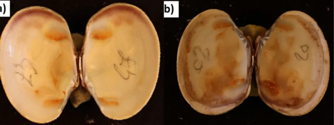
Figure 2 Photography of (A) BRD- clam and (B) BRD+clam.From Richard et al., 2015, unpublished data.
Total DNA extraction from bacterial culture and from EF
DNA extraction was performed using the QIAamp DNA mini kit (Qiagen) for both bacterial cultures and total extrapallial fluids. 450µL were centrifuged at 10,000 g at 4◦C for 10 min. Pellets were deproteinized in a hot dry bath at 56◦C by addition of 180
µL of ATL Buffer supplemented by 20µL of proteinase K during one hour. DNA extractions were then performed according to supplier instructions. Finally, DNA were eluted in 200µL of ultra-pure water and stored at−20◦C until use. DNA yield and purity were determined
by spectrophotometry (Quantifluor dsDNA kit; Promega, Madison, WI, USA; and POLARstar Omega microplate spectrophotometer; BMG Labtech, Orgenberg, Germany).
Enumeration ofV. tapetisby spectrophotometry
In order to accurately inoculate CECT4600T at the target dilution of 107 cells/mL during the experiment, enumeration of bacteria was performed to measure the ab-sorbance at 492 nm on a Multiskan spectrophotometer (Fisher Scientific, Hampton, NH, USA).Vibrio tapetisculture density was calculated according to the formula 1.3 109×DO−3.6 107CFU/mL (Choquet, 2004;Le Bris et al., 2015).
For the dilution ranges of bacteria (described below), an early stationary phase culture of CECT4600Twas enumerated, and was first diluted in Filtered SeaWater (FSW) to obtain a 108cells/mL suspension. Bacteria were then serially diluted in FSW in a final volume of 500µL from 2.25 107to 0.565 101cells/mL (ten-fold dilutions until 2.25 101and half dilutions for the last two) to generate the standard curve of bacteria. DNA was extracted from 450µL of each diluted suspension. For the second standard curve in extrapallial fluids, FSW was replaced by filtered EF prepared previously. DNA extractions were also performed on 450µL of sterile EF in the same conditions in order to establish a negative control for the standard curve.
PCR primer and fluorogenic probe design
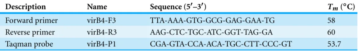
Table 2 Nucleotide sequences and melting temperatures(Tm)of primers and probe designed for real-time PCR reaction, targeting thevirB4gene.
Description Name Sequence (5′–3′) T
m(◦C)
Forward primer virB4-F3 TTA-AAA-GTG-GCG-GAG-GAA-TG 58
Reverse primer virB4-R3 AAG-CTC-TGC-ATC-GGT-TAG-GA 60
Taqman probe virB4-P1 CGA-GTA-CCA-ACA-TGC-CTT-CCC-GT 53.7
Vibrio tapetisCECT4600T(Genbank accession number:KT382306). Using Primer-3 software, several primers pairs were designed and tested with DNA samples of different species and strains of theVibriogenus available as positive and negative controls on a 9700 ABI®thermocycler (Applied Biosystems, Foster City, CA, USA) (Table 1). The choice for the best primer pair was determined, with optimal concentration and reaction conditions for PCR amplification compatible with the hydrolysis Taqman probe (Table 2). ThevirB4 probe was dually labeled with 5′-reporter dye 6-FAM (wavelength emission at 502 nm) and a downstream 3′-quencher dye TAMRA.
The selectivity of the primers was checked using the BLAST algorithm of the NCBI database (http://www.ncbi.nlm.nih.gov/), assuring their specificity for thevirB4region, without homology to other known sequences described in GenBank and EMBL databases. Oligonucleotides were also aligned to the CECT4600Tand LP2 genomes to ensure that they selectively amplified only thevirB4gene. Primers and probe were purchased from Eu-rogentec (Angers, France). The expected length of the amplicon deduced from nucleotide sequence was 173 bp, and the selectivity was also theoretically assessed by BLAST.
Quantitative real-time PCR (qPCR)
Real-time PCR was performed on a LightCycler 480 Instrument (Roche Diagnostics, Mannheim, Germany) using LightCycler 480 Probe Master Mix based on Taqman detection (Roche Diagnostics). Each real-time PCR experiment included technical triplicates, in a final volume of 15µL. Each reaction contained 5µL of DNA template, 0.5µM of each primer, 0.1µM of hydrolysis probe virB4-P1 and 7.5µL of LC480 Probe Master Mix 2X. A single initial denaturation step of 10 min at 95◦C was followed by 45
cycles of 95◦C for 10 s (denaturation), 54◦C for 20 s (annealing) and 72◦C for 1 s to measure the fluorescence signal. Finally, a cooling step at 40◦C during 10 s was included.
The results were analyzed with Roche LightCycler 480 software. Threshold cycle (Ct) value corresponds to the PCR cycle number at which an increase in reporter fluorescence above a baseline signal was first detected, after background subtraction. Negative controls with molecular biology grade water as template were performed in each run. For quantitation, dilution ranges ofV. tapetiswere tested in triplicates across multiple orders of magnitude described above. Results were analyzed by linear regression to calculate the slope. The PCR amplification efficiency(E)was measured according toE= [101/(−slope)] −1, using
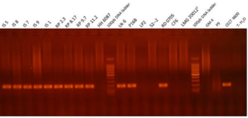
Figure 3 Visualization of the PCR product in agarose gel obtained with qPCRvirB4assay for repre-sentative strains ofVibrio, i.e., which were tested positive and negative for BRD development after an infection experiment.Lanes MT corresponds to the BenchTop DNA ladder (Promega, Madison, WI, USA). T-H2O represents the water negative control.
the limit of detection (LOD) was calculated in accordance with 95% of 20 tested replicates giving positive amplification results (Bustin et al., 2009). The repeatability (intra-assay variance) was estimated using triplicates of each template to assess the precision of the method. The reproducibility (inter-assay variance) was calculated using standard deviation (SD) of twovirB4gene standard concentrations used for each run to assess the variation between runs.
RESULTS
Selectivity of the real-time PCR protocol
Sequence alignments of the real-time Taqman PCR amplicon showed no cross reactivity with others species when compared using the BLAST analysis program. Only one theoretical partial cross reactivity withV. tasmaniensis(KP795691.1, 78% of identity with differences in 3′end terminals of both forward primer and Taqman hydrolysis
toV. tapetis, among tested strains (V. tasmaniensis,V. spendidus,V. lentus) (Thompson, Thompson & Swings, 2002;Le Roux et al., 2004;Sawabe et al., 2013;Al-saari et al., 2015), in agreement with the empirical and theoretical results generated by BLAST.
Positive signals obtained by real-time PCR experiments for bacterial strains were checked by electrophoresis. The amplicon observed at 173 bp, corresponds to the expected size, calculated from the nucleotide sequence of thevirB4gene (Fig. 3).
Sensitivity of the real-time PCR assay
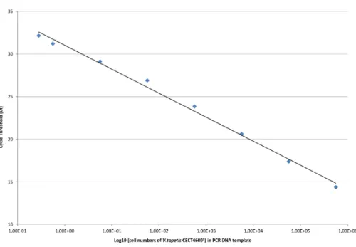
Standard curves forvirB4gene quantification were generated in parallel in filtered sterile water (FSW) and total extrapallial fluids (EF) with pure bacterial suspension ofV. tapetis CECT4600Tstrain of known concentrations, determined by spectrophotometry and checked by the Malassez counting method. Ten-fold bacterial dilutions ranging from 2.25 107to 2.25 102cells mL−1, and two last half dilutions to 0.565 101cells mL−1were prepared in these two diluents. Since only one copy of the chromosomalvirB4gene is present per bacteria, the threshold cycle (Ct) values deduced from real-time PCR amplifications on purified DNA extracts were plotted to the number of bacteria initially present in PCR templates.
Because similar slopes were achieved for quantification curves of each diluent in this study (data not shown), the standard curve in EF was chosen for analyzing the experimental infection samples as this diluent corresponded to thein situconditions of the samples, namelyV. tapetisin EF.
The standard curve reliably showed linearity across 8 orders of magnitude, from 2.25 107to 1.125 101cells mL−1. The final dilution (0.565 101cells mL−1) produced less consistent results, and was not retained for the calculation of the standard curve. After extractions, genomic DNA was checked by spectrophotometry and corresponded to values ranging from 900 pgµL−1to 156 pgµL−1. According to the Ct values obtained in triplicates, quantification curve exhibited an excellent linear regression with anr2 correlation coefficient of 0.99 and a PCR efficacy of 103% (Fig. 4) calculated in the linear zone according to the MIQE guidelines (Bustin et al., 2009). The intra-assay variance was evaluated from 0.01 and 0.32 from all the samples tested in triplicate. The reproducibility, calculated using the standard deviation of Ct values generated from two standard concentrations of 2.25 102cells mL−1and 2.25 106cells mL−1from different runs, corresponded to 0.14 and 0.10 respectively.
The threshold sensitivity of this method in targeting the presence ofvirB4gene and quantification is given by the lower bacterial concentration detected in the linear zone (at least 95% of 20 tested replicates), and corresponds to the limit of detection (LOD) of V. tapetisof 1.125 101bacteria mL−1.
Specificity of the method: kinetics of infection during Manila clams challenge
Figure 4 Standard curve for the detection and quantification of the virB4 gene by Taqman real-time PCR, in dilution range of EF samples artificially spiked with CECT4600Tbacterial strain.Standard curve was generated by plotting the log cell number of bacteria present in PCR DNA template against Ct values.
previously established for EF. The limit of detection established previously corresponded to a cell density of 1.125 101bacteria per mL of extrapallial fluids.
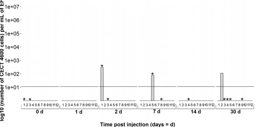
Overall during the experiment, each sampling time of CECT4600Tinjected clams exhibited at least two positive individuals by the real-time PCR assay (Fig. 5). Only 24 h after injection, nine individuals among twelve were spotted with thevirB4detection protocol. At the same time, all of them were BRD−. It was noticeable that at 7 days post-injection only two real-time PCR positive animals were detected, but with a high V. tapetisload, at nearly 107cells per mL in EF (6.53 106cells mL−1). As regards the BRD among CECT4600T-injected clams identified as positive by real-time PCR, one individual at 2 days and one at 7 days were classified as BRD+. At 14 and 30 days post-injection, four clams at each time were positive according to the qPCR assay and they were all BRD+. All theseV. tapetis-positive individuals showed a bacterial load above 102cells per mL in extrapallial fluids, with a maximum of 1.11 104cells per mL in extrapallial fluids.
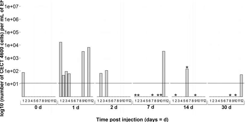
ThevirB4gene was also detected in several non-injected individuals: three animals on the day of infection; and one each at 2, 7 and 30 days during the experiment (Fig. 6and the points 0d onFigs. 5and7). The animals were BRD−at 0 and 30 days of the experiment whereas the two positivevirB4clams (2d and 7d onFig. 6) displayed clinical signs of BRD.
Figure 5 Kinetics of clam infection by CECT4600TV. tapetisstrain byvirB4real-time PCR in extrapallial fluids sampled at 0, 1, 2, 7, 14 and 30 days post-injection.0d means not injected. * corresponds to BRD+clam.
DISCUSSION
In this study, we developed a rapid and accurate real-time PCR process for the detection and quantification ofV. tapetispathogens for clams in extrapallial fluids of the Manila clam. This protocol was designed using a pair of primers and a Taqman probe targeting thevirB4gene ofV. tapetis, encoding a protein engaged in a large complex of type IV secretion systems.
Several publications dealing with rapid and specific molecular identification ofVibrio species use PCR techniques. Various PCR assays have been published (Thompson et al., 2005;Paillard et al., 2006;Rodr´ıguez et al., 2006) that have investigatedVibrio tapetis. These methods, based on 16S rRNA sequences are less reliable and can be time-consuming when associated with the BRD diagnostic necessary to achieveVibrio tapetisidentification (Drummond et al., 2006). Moreover, these protocols do not allow quantification of the bacteria.
Figure 6 Kinetics of non-injected clams byvirB4real-time PCR in extrapallial fluids sampled at 0, 1, 2, 7, 14 and 30 days of sampling during the experiment.* corresponds to BRD+clam.
The specificity of primers and Taqman probe have been demonstrated here with the real-time PCR assays carried out on DNA samples extracted from pure cultures of various bacterial strains belonging to theV. tapetisgroup. No signal was obtained with other species close related toV. tapetis(belonging toV. splendidusorV. tasmaniensis), even at high threshold cycle values (Ct>45). Positive fluorescence signals were acquired with allV. tapetisstrains virulent forV. philippinarum, except the FPC1121 Japanese strain (Table 1). We suggest that this strain lacks thevirB4gene and we could speculate that this Japanese strain uses a different secretion system from the type IV to translocate substrate for its virulence (G Dias, 2015, unpublished data).
Figure 7 Kinetics of FSW-injected clams byvirB4real-time PCR in extrapallial fluids sampled at 0, 1, 2, 7, 14 and 30 days of sampling during the experiment. 0d means not injected.* corresponds to BRD+clam.
We used the real-time PCR developed in this study to monitor the kinetics ofV. tapetis experimental infection in clams. During bacterial challenge, early stages of the infection showed a high number of individuals infected and high bacteria load (until 7 days post-injection), confirming that extrapallial fluids are involved in the first steps of infection (Allam, Paillard & Maes, 1996), where hemocytes contribute to defense against V. tapetisby phagocytosis (Allam & Paillard, 1998). At 7 days post-injection, only two individuals were detected as positive by the real-time PCR assay, suggesting that animals had efficiently defended against the pathogen. This hypothesis is corroborated by Richard and collaborators (2015, unpublished data). This result also agrees withPaillard & Maes (1994), who showed that the first BRD symptoms appeared in almost all the clams seven days post-injection, bearing out the reaction of hosts facing a pathogen injection. Indeed, the conchiolin matrix was developed to trap encompassing bacteria into the inner surface of the shell, inducing a decrease of circulating bacteria in the EF, and thus a lower concentration detected by the Taqman assay. Then, 14 and 30 days after injection, CECT4600T was again found in extrapallial fluids but the load was less than 7 days post-injection. Paillard and collaborators (2014) argue that an increase ofV. tapetisin the host’s fluids in advanced stages of the disease is due to the weakening of the clam, despite food intake and shell repair.
an effect of injection on animals which were supposedly infected byV. tapetis, present in their environment before sampling, despite the quarantine stage. It has already been shown that injection or handling could stimulate bacterial proliferation (Le Bris et al., 2015;Jean et al., in press). Focusing on non-injected clams, i.e., at 0d forV. tapetis- and FSW-injected clams and all the sampling times for non-injected clams, the interpretation thatV. tapetiswas present in individuals before sampling is corroborated: 3 individuals among 36 were already carrying bacteria on the day of injection. During the remaining time, 3 non-injected animals were detected as positive by real-time PCR protocol, while 11 animals displayed clinical signs of BRD among 72 sampled (15%), suggesting that the host has already mounted a defense response against pathogens before the experimental challenge. This corresponds to the natural prevalence of the disease described in literature (Paillard et al., 2014).
To conclude, we developed in this study a rapid, specific and individual method to detect and quantifyV. tapetis. This protocol is based on a real-time PCR assay and is suitable for field and hatchery animals. Indeed, thisvirB4real-time PCR assay is easy to implement because it does not require crushing and grinding and allows individual assays considering inter-individual variability. This protocol allows for early detection of the disease, especially to assess visibly healthy clams (BRD−), bearing in mind the threshold of detection, and will be very useful in helping prevent massive infection in clams, notably in clam aquaculture.
ACKNOWLEDGEMENTS
We would like to thank the company SATMAR for providing us with clams during the first steps of optimization of the method, and “Les Claires de Bonsonge” for supplying us clams for the experimental challenge. We are thankful to Val´erie Barbe and Claudine M´edigue from LABGeM and the National Infrastructure “France Genomique” for sequencing CECT4600Tstrain and Annick Jacq for expert annotation. We thank Didier Mazel and Fr´ed´erique Leroux, who were in charge of the Vibrioscope project within MaGe. We also thank Prof. Vianney Pichereau for his critical reading and advice on the manuscript, and Ewan Harney, a native speaker, for English corrections.
ADDITIONAL INFORMATION AND DECLARATIONS
Funding
This work was supported by the “Laboratoire d’Excellence” LabexMer (ANR-10-LABX-19) and co-funded by a grant from the French government under the program “Investisse-ments d’Avenir,” and by a grant from the Regional Council of Brittany. This work was also funded by the University of Western Brittany. The funders had no role in study design, data collection and analysis, decision to publish, or preparation of the manuscript.
Grant Disclosures
French government.
Regional Council of Brittany. University of Western Brittany.
Competing Interests
The authors declare there are no competing interests.
Author Contributions
• Adeline Bidault conceived and designed the experiments, performed the experiments, analyzed the data, wrote the paper, prepared figures and/or tables.
• Ga¨elle G. Richard performed the experiments, analyzed the data, prepared figures and/or tables, reviewed drafts of the paper.
• C´edric Le Bris performed the experiments.
• Christine Paillard contributed reagents/materials/analysis tools, reviewed drafts of the
paper.
Patent Disclosures
The following patent dependencies were disclosed by the authors: V. tasmaniensisgenbank accession numberKP795691.1.
DNA Deposition
The following information was supplied regarding the deposition of DNA sequences: GenBank accession number:KT382306.
Data Availability
The following information was supplied regarding data availability: This work did not generate any raw data.
Supplemental Information
Supplemental information for this article can be found online athttp://dx.doi.org/ 10.7717/peerj.1484#supplemental-information.
REFERENCES
Allam B. 1998.R ˆole des fluides extrapall´eaux des bivalves dans la d´efense immunitaire. Cas de la maladie de l’anneau brun chez la palourde d’´elevage,Ruditapes philippinarum. Th`ese de Doctorat de Biologie Thesis. Brest: Universit´e de Bretagne occidentale-Brest.
Allam B, Ford SE. 2006.Effects of the pathogenicVibrio tapetison defence factors of susceptible and non-susceptible bivalve species: I. Haemocyte changes followingin vitrochallenge.Fish & Shellfish Immunology20:374–383DOI 10.1016/j.fsi.2005.05.012.
Allam B, Paillard C. 1998.Defense factors in clam extrapallial fluids.Diseases of Aquatic Organisms 33:123–128DOI 10.3354/dao033123.
Allam B, Paillard C, Auffret M. 2000.Alterations in hemolymph and extrapallial fluid parameters in the manila clam,Ruditapes philippinarum, challenged with the pathogenVibrio tapetis.
Allam B, Paillard C, Ford SE. 2002.Pathogenicity ofVibrio tapetis, the etiological agent of brown ring disease in clams.Diseases of Aquatic Organisms48:221–231DOI 10.3354/dao048221.
Allam B, Paillard C, Maes P. 1996.Localization of the pathogenVibrioP 1 in clams affected by brown ring disease.Diseases of Aquatic Organisms27:149–155DOI 10.3354/dao027149.
Al-saari N, Gao F, Rohul AAKM, Sato K, Sato K, Mino S, Suda W, Oshima K, Hattori M, Ohkuma M, Meirelles PM, Thompson FL, Thompson C, Filho AGM, Gomez-Gil B, Sawabe T, Sawabe T. 2015.Advanced microbial taxonomy combined with genome-based-approaches reveals thatVibrioastriarenae sp. nov., an agarolytic marine bacterium, forms a new clade inVibrionaceae.PLoS ONE10:e0136279DOI 10.1371/journal.pone.0136279.
Austin B, Zhang X-H. 2006.Under the microscope.Vibrio harveyi: a significant pathogen of marine vertebrates and invertebrates. Letter of Applied Microbiology43:119–124
DOI 10.1111/j.1472-765X.2006.01989.x.
Balboa S, Dieguez AL, Doce A, Barja JL, Romalde JL. 2012.Evaluation of different culture media for the isolation and growth of the fastidiousVibrio tapetis, the causative agent of brown ring disease.Journal of Invertebrate Pathology111:74–81DOI 10.1016/j.jip.2012.06.007.
Borrego JJ, Castro D, Luque A, Paillard C, Maes P, Garcia MT, Ventosa A. 1996.Vibrio tapetis
sp. nov., the causative agent of the brown ring disease affecting cultured clams.International Journal of Systematic Bacteriology46:480–484DOI 10.1099/00207713-46-2-480.
Bustin SA, Benes V, Garson JA, Hellemans J, Huggett J, Kubista M, Mueller R, Nolan T, PfafflMW, Shipley GL, Vandesompele J, Wittwer CT. 2009.The MIQE guidelines: minimum information for publication of quantitative real-time PCR experiments.Clinical Chemistry 55:611–622DOI 10.1373/clinchem.2008.112797.
Casadevall A, Pirofski L. 1999.Host-pathogen interactions: redefining the basic concepts of virulence and pathogenicity.Infection and Immunity67:3703–3713.
Choquet G. 2004.Caract´erisation et pathog´enie des isolats deVibrio tapetis, bact´erie responsable de la maladie de l’anneau brun chez la palourde japonaise. Doctorat Microbiologie Thesis. Brest: Universit´e de Bretagne occidentale-Brest.
Choquet G, Soudant P, Lambert C, Nicolas J-L, Paillard C. 2003. Reduction of adhesion properties ofRuditapes philippinarumhemocytes exposed toVibrio tapetis.Diseases of Aquatic Organisms57:109–116DOI 10.3354/dao057109.
Christie PJ, Whitaker N, Gonz´alez-Rivera C. 2014.Mechanism and structure of the bacterial type IV secretion systems.Biochimica et Biophysica Acta (BBA)—Molecular Cell Research 1843:1578–1591DOI 10.1016/j.bbamcr.2013.12.019.
Crosa JH, Schiewe MH, Falkow S. 1977.Evidence for plasmid contribution to the virulence of fish pathogenVibrio anguillarum.Infection and Immunity18:509–513.
Drummond LC, O’reilly P, Mulcahy MF, Culloty SC. 2006.Comparison of techniques for diagnosis of Brown Ring Disease and detection ofVibrio tapetisin the Manila clam,Venerupis
(Ruditapes)philippinarum.Journal of Shellfish Research25:1043–1049
DOI 10.2983/0730-8000(2006)25[1043:COTFDO]2.0.CO;2.
Flassch J-P, Leborgne Y. 1994.Introduction in Europe, from 1972 to 1980, of the Japanese Manila clam (Tapes philippinarum) and the effects on aquaculture production and natural settlement In:Introductions and transfers of aquatic species: selected papers from a symposium held in Halifax, Nova Scotia, 12–13 June 1990. Cogenhagen: International Council for the Exploration of the Sea.Available athttp://archimer.ifremer.fr/doc/00037/14871/.
Frans I, Michiels CW, Bossier P, Willems KA, Lievens B, Rediers H. 2011.Vibrio anguillarum
Fronzes R, Christie PJ, Waksman G. 2009.The structural biology of type IV secretion systems.
Nature Reviews Microbiology7:703–714DOI 10.1038/nrmicro2218.
Garnier M, Labreuche Y, Garcia C, Robert M, Nicolas JL. 2007.Evidence for the involvement of pathogenic bacteria in summer mortalities of the Pacific oysterCrassostrea gigas.Microbial Ecology53:187–196DOI 10.1007/s00248-006-9061-9.
Garrido-Maestu A, Chapela M-J, Pe˜naranda E, Vieites JM, Cabado AG. 2014.In-house
validation of novel multiplex real-time PCR gene combination for the simultaneous detection of the main human pathogenicvibrios(Vibrio cholerae,Vibrioparahaemolyticus, andVibrio vulnificus).Food Control37:371–379DOI 10.1016/j.foodcont.2013.09.026.
Gosling E. 2008.Bivalve molluscs: biology, ecology and culture. Hoboken: John Wiley & Sons.
Gubala AJ. 2006.Multiplex real-time PCR detection ofVibrio cholerae.Journal of Microbiological Methods65:278–293DOI 10.1016/j.mimet.2005.07.017.
Jean F, Flye-Sainte-Marie J, Oudard C, Paillard C. 2011.Handling enhances the development of brown ring disease signs inRuditapes philippinarum.Journal of Shellfish ResearchIn Press.
Available athttp://hal.univ-brest.fr/hal-00568169/document.
Jensen S, Samuelsen OB, Andersen K, Torkildsen L, Lambert C, Choquet G, Paillard C, Bergh Ø. 2003.Characterization of strains ofVibrio splendidusandV. tapetisisolated from corkwing wrasse Symphodus melops suffering vibriosis.Diseases of Aquatic Organisms53:25–31
DOI 10.3354/dao053025.
Juhas M, Crook DW, Hood DW. 2008.Type IV secretion systems: tools of bacterial horizontal gene transfer and virulence.Cellular Microbiology10:2377–2386
DOI 10.1111/j.1462-5822.2008.01187.x.
Kushmaro A, Banin E, Loya Y, Stackebrandt E, Rosenberg E. 2001.Vibrioshiloi sp. nov., the causative agent of bleaching of the coralOculina patagonica.International Journal of Systematic and Evolutionary Microbiology51:1383–1388DOI 10.1099/00207713-51-4-1383.
Le Bris C, Richard G, Paillard C, Lambert C, Seguineau C, Gauthier O, Pernet F, Gu´erard F. 2015.Immune responses of phenoloxidase and superoxide dismutase in the manila clam
Venerupis philippinarumchallenged withVibrio tapetis—Part I: spatio-temporal evolution of enzymes’ activities post-infection.Fish & Shellfish Immunology42:16–24
DOI 10.1016/j.fsi.2014.10.021.
Le Roux F, Gay M, Lambert C, Nicolas J-L, Gouy M, Berthe FCJ. 2004.Phylogenetic study and identification ofVibrio splendidus-related strains based on gyrB gene sequences.Diseases of Aquatic Organisms58:143–150DOI 10.3354/dao058143.
Low HH, Gubellini F, Rivera-Calzada A, Braun N, Connery S, Dujeancourt A, Lu F, Redzej A, Fronzes R, Orlova EV, Waksman G. 2014.Structure of a type IV secretion system.Nature 508:550–553DOI 10.1038/nature13081.
Maci´an MC, Ludwig W, Aznar R, Grimont PA, Schleifer KH, Garay E, Pujalte MJ. 2001.Vibrio lentussp. nov., isolated from Mediterranean oysters.International Journal of Systematic and Evolutionary Microbiology51:1449–1456DOI 10.1099/00207713-51-4-1449.
Matsuyama T, Sakai T, Kiryu I, Yuasa K et al.2010.First isolation ofV. tapetis, the etiological agent of brown ring disease (BRD), in manila clamRuditapes philippinarumin Japan.Fish Pathology45:77–79DOI 10.3147/jsfp.45.77.
McCleary S, Henshilwood K. 2015.Novel quantitative TaqMan®MGB real-time PCR for sensitive detection ofVibrio aestuarianusinCrassostrea gigas.Diseases of Aquatic Organisms114:239–248
Mehrabadi JF, Morsali P, Nejad HR, Imani Fooladi AA. 2012.Detection of toxigenicVibrio cholerae with new multiplex PCR. Journal of Infection and Public Health 5:263–267
DOI 10.1016/j.jiph.2012.02.004.
Novoa B, Luque A, Castro D, Borrego JJ, Figueras A. 1998.Characterization and infectivity of four bacterial strains isolated from brown ring disease-affected clams.Journal of Invertebrate Pathology71:34–41DOI 10.1006/jipa.1997.4704.
Paillard C, Allam B, Oubella R. 2004.Effect of temperature on defense parameters in Manila clamRuditapes philippinarumchallenged withVibrio tapetis.Diseases of Aquatic Organisms 59:249–262DOI 10.3354/dao059249.
Paillard C, Gausson S, Nicolas JL, Le Pennec JP, Haras D. 2006.Molecular identification ofVibrio tapetis, the causative agent of the brown ring disease ofRuditapes philippinarum.Aquaculture 253:25–38DOI 10.1016/j.aquaculture.2005.03.047.
Paillard C, Jean F, Ford SE, Powell EN, Klinck JM, Hofmann EE, Flye-Sainte-Marie J. 2014.
A theoretical individual-based model of Brown Ring Disease in Manila clams,Venerupis philippinarum.Journal of Sea Research91:15–34DOI 10.1016/j.seares.2014.03.005.
Paillard C, Maes P. 1990.Etiologie de la maladie de l’anneau brun chez Tapes philippinarum: pathog´enicit´e d’unVibriosp.Comptes Rendus de l’Acad´emie des Sciences.S´erie 3, Sciences de la vie310:15–20.
Paillard C, Maes P. 1994.Brown ring disease in the Manila clamRuditapes philippinarum: establishment of a classification system.Diseases of Aquatic Organisms19:137–146
DOI 10.3354/dao019137.
Paillard C, Maes P. 1995a.The Brown Ring Disease in the Manila clamRuditapes philippinarum, II: microscopic study of the brown ring syndrome.Journal of Invertebrate Pathology65:101–110
DOI 10.1006/jipa.1995.1016.
Paillard C, Maes P. 1995b.The Brown Ring Disease in the Manila clamRuditapes philippinarum, I: ultrastructural alterations of the periostracal lamina.Journal of Invertebrate Pathology65:91–100
DOI 10.1006/jipa.1995.1015.
Reid HI, Duncan HL, Laidler LA, Hunter D, Birkbeck TH. 2003.Isolation ofVibrio tapetis
from cultivated Atlantic halibut (Hippoglossus hippoglossusL.).Aquaculture 221:65–74
DOI 10.1016/S0044-8486(03)00060-7.
Rodr´ıguez JM, L ´opez-Romalde S, Beaz R, Alonso MC, Castro D, Romalde JL. 2006.Molecular fingerprinting ofVibrio tapetisstrains using three PCR-based methods: ERIC-PCR, REP-PCR and RAPD.Diseases of Aquatic Organisms69:175–183DOI 10.3354/dao069175.
Romalde JL, Castro D, Magari˜nos B, Lopez-Cortes L, Borrego JJ. 2002.Comparison of Ribotyping, randomly amplified polymorphic dna, and pulsed-field gel electrophoresis for molecular typing ofVibrio tapetis.Systematic and Applied Microbiology25:544–550
DOI 10.1078/07232020260517689.
Saulnier D, De Decker S, Haffner P. 2009. Real-time PCR assay for rapid detection and quantification ofVibrio aestuarianusin oyster and seawater: a useful tool for epidemiologic studies.Journal of Microbiological Methods77:191–197DOI 10.1016/j.mimet.2009.01.021.
Schikorski D, Renault T, Paillard C, Bidault-Toffin A, Tourbiez D, Saulnier D. 2013.
Development of TaqMan real-time PCR assays for monitoringVibrio harveyiinfection and a plasmid harbored by virulent strains in European abalone Haliotis tuberculata aquaculture.
Aquaculture392–395:106–112DOI 10.1016/j.aquaculture.2013.02.005.
Sparling PF. 1983.Bacterial virulence and pathogenesis: an overview.Review of Infectious Diseases 5:S637–S646DOI 10.1093/clinids/5.Supplement 4.S637.
Thompson FL. 2003.Vibriokanaloae sp. nov.,Vibriopomeroyi sp. nov. andVibrio chagasiisp. nov., from sea water and marine animals.International Journal of Systematic and Evolutionary Microbiology53:753–759DOI 10.1099/ijs.0.02490-0.
Thompson FL, Gevers D, Thompson CC, Dawyndt P, Naser S, Hoste B, Munn CB, Swings J. 2005.Phylogeny and molecular identification ofVibrioson the basis of multilocus sequence analysis.Applied and Environmental Microbiology71:5107–5115
DOI 10.1128/AEM.71.9.5107-5115.2005.
Thompson FL, Thompson CC, Swings J. 2002.Vibrio tasmaniensis, sp. nov., isolated from Atlantic salmon (Salmo salarL.).Systematic and Applied Microbiology26:65–69
DOI 10.1078/072320203322337326.
Vallenet D, Belda E, Calteau A, Cruveiller S, Engelen S, Lajus A, Le Fevre F, Longin C, Mornico D, Roche D, Rouy Z, Salvignol G, Scarpelli C, Thil Smith AA, Weiman M, Medigue C. 2013.MicroScope–an integrated microbial resource for the curation and comparative analysis of genomic and metabolic data.Nucleic Acids Research41:D636–D647
DOI 10.1093/nar/gks1194.