241 www.scielo.br/rsbmt
Short Communication
Address to: Dra. Maisa Silva de Sousa. Lab. de Biologia Molecular e Celular/ NMT/UFPA. Av. Generalíssimo Deodoro 92, Umarizal, 66055-240 Belém, PA, Brasil.
Phone/Fax: 55 91 3241-4681 e-mail: maisasousa@ufpa.br Received 17 February 2011 Accepted 12 January 2012
Revista da Sociedade Brasileira de Medicina Tropical 46(2):241-243, Mar-Apr, 2013 http://dx.doi.org/10.1590/0037-8682-981-2013
Occurrence of strongyloidiasis among patients with
HTLV-1/2 seen at the outpatient clinic of the
Núcleo de
Medicina Tropical
, Belém, State of Pará, Brazil
Karen Cristini Yumi Ogawa Furtado
[1],[2], Carlos Araújo da Costa
[2], Louise de Souza Canto Ferreira
[3],
Luisa Carício Martins
[2], Alexandre da Costa Linhares
[2],[4], Edna Aoba Yassui Ishikawa
[2],
Evander de Jesus Oliveira Batista
[2]and Maisa Silva de Sousa
[2][1]. Faculdade de Farmácia, Instituto de Ciências da Saúde, Universidade Federal do Pará, Belém, PA. [2]. Programa de Pos-Graduação em Doenças Tropicais, Núcleo de Medicina Tropical, Universidade Federal do Pará, Belém, PA. [3]. Faculdade de Farmácia, Centro Universitário do Pará, Belém, PA. [4]. Seção de Virologia, Instituto Evandro Chagas, Ministério da Saúde, Ananindeua, PA.
ABSTRACT
Introduction: This study investigated the occurrence of Strongyloides stercoralis infestation and coinfection with HTLV-1/2 in Belém, Brazil. Methods:S. stercoralis was investigated in stool samples obtained from individuals infected with HTLV-1/2 and their uninfected relatives. Results: The frequency of S. stercoralis was 9% (9/100), including six patients infected with HTLV-1 (14.3%), two patients infected with HTLV-2 (11.1%), and one uninfected relative. Two cases of hyperinfestation by S. stercoralis
were characterized as HTLV-1. Conclusions: These results support the need for the routine investigation of S. stercoralis in patients with HTLV-1, in an attempt to prevent the development of severe forms of strongyloidiasis.
Keywords: Strongyloides stercoralis. HTLV. Coinfection.
Strongyloides stercoralis is an intestinal nematode that infects about 60 million people in tropical and subtropical regions1,2. This parasite is the only helminth able to complete its life cycle inside a single host3. Most infections caused by
S. stercoralis are asymptomatic. However, the severe form is
characterized by dissemination of the parasite to virtually any organ and may be associated with a high mortality rate1,4-6.
Severe forms of strongyloidiasis and a low therapeutic response of this infestation have been reported in patients infected with human T-cell lymphotropic virus type 1 (HTLV-1). Epidemiological studies have shown an association between infection with this virus and helminthic infestations, especially by S.stercoralis7,8.
HTLV-1/2 and S. stercoralis are considered to be endemic in the state of Pará, Brazil6,9; however, there are no reports of coinfection by these agents in the region. The Núcleo de Medicina Tropical (NMT) is an academic unit of the
Universidade Federal do Pará (UFPA) that has a local referral
center for the care and outpatient laboratory investigation of HTLV-infected patients. Therefore, the objective of the present study was to assess the relationship between S. stercoralis
infestation and HTLV infection in Belém, Pará, Brazil.
A total of 41 patients with HTLV who visited the Center for Tropical Medicine, Federal University of Pará, for the annual re-evaluation between March 2008 and November 2009, underwent tests for the identiication of Strongyloides stercoralis in stools. All patients’ relatives (spouses, mothers and children) were also invited to undergo these tests as well as screening for HTLV. Overall, 59 relatives voluntarily agreed to participate in this study, including those individuals identiied as being HTLV-reactive (n = 19), as well as those found to be negative for HTLV antibody (n = 40). All participants reported having had no recent anthelmintic treatment.
Anti-HTLV-1/2 antibodies were investigated in plasma samples obtained from the relatives of HTLV-infected patients through an enzyme immunoassay (ELISA) using the HTLV-1/2 kit (Ortho, USA), according to manufacturer’s instructions. Samples with positive serology and negative samples whose values were close to the cut-off were subjected to molecular analysis for the detection of proviral HTLV DNA. For this purpose, a 159-bp fragment of the HTLV pX gene was ampliied by nested PCR, followed by restriction fragment length polymorphism (RFLP) analysis using the TaqI endonuclease as described by Tuke et al.10,11.
Three methods were used for parasitological examination of one stool sample from each participant: direct method, Baermann-Moraes method, and Lutz method (also called Hoffman method) for the detection of S. stercoralis eggs and larvae12. All participants were instructed to collect the stool samples on the day of the test and not to store them in a refrigerator.
242
www.scielo.br/rsbmt
Furtado KCYO et al - Occurrence of strongyloidosis among patients with HTLV-1/2
REFERENCES
The authors declare that there is no conlict of interest.
CONFLICT OF INTEREST ACKNOWLEDGMENTS
FINANCIAL SUPPORT
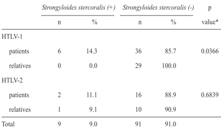
TABLE 1 - Frequency of Strongyloides stercoralis in HTLV-1 and HTLV-2 patients and their relatives
Strongyloides stercoralis (+) Strongyloides stercoralis (-) p
n % n % value*
HTLV-1
patients 6 14.3 36 85.7 0.0366
relatives 0 0.0 29 100.0
HTLV-2
patients 2 11.1 16 88.9 0.6839
relatives 1 9.1 10 90.9
Total 9 9.0 91 91.0
*Fisher´s exact test. +: positive; -: negative; Source:Núcleo de Medicina Tropical. frequency was calculated using Fisher’s exact test (for two samples) and G test (for one sample). Ages were compared using the Mann-Whitney test. The level of signiicance was set at 5% for all analyses.
Each participant signed a free informed consent form elaborated according to Resolution 196/96 from the Brazilian Health National Council. The study protocol was approved by the Ethics Committee on Human Research of NMT.
The frequency of strongyloidiasis was 9% (9/100) in the study, including six patients infected with HTLV-1, two patients infected with HTLV-2, and one uninfected relative (Table 1). Six (14.3%) of the 42 stool samples from patients infected with HTLV-1 were positive for S. stercoralis infestation. Strongyloidiasis was not diagnosed among the 29 relatives not infected with HTLV-1. The frequency of S. stercoralis
was 11.1% (2/18) among patients infected with HTLV-2 and 9.1% (1/11) among their relatives not infected with HTLV-2.
Four (66.7%) of the six HTLV-1-infected patients with
S. stercoralis were 50 years or older. Two cases of hyperinfestation by S. stercoralis were characterized as HTLV-1 and had dificulty in responding to treatment with Ivermectin. The mean age of patients with HTLV-1 was 41.99 years, and the mean age of their relatives was 30.57 years (p = 0.001). The mean ages of patients with HTLV-2 and their relatives were 49.94 and 51.63 years, respectively (p = 0.7049).
The association between S. stercoralis and HTLV-1 was irst reported in Okinawa, Japan14. In the present study, the rate of infection with S. stercoralis was 14.3% among patients infected with HTLV-1. This result was consistent with two reports from Brazil, speciically from the Cities of São Paulo (12.1%)15 and Salvador (15.7%)1.
Most of those infected with HTLV-1 were older than 50 years. According to Hirata et al.8 and Nakada et al.14, this increased rate among patients older than 50 years was likely due to the cumulative risk of infection over time.
In the present study, the frequency of S. stercoralis was signiicantly higher among patients infected with HTLV-1 than
among their seronegative relatives. Clearly, the frequency of
S. stercoralis among HTLV-1 patients was essential for this
signiicance. It should be noted that the control group, consisted of household relatives of patients infected with HTLV-1, may probably be exposed to the same risk factors for strongyloidiasis like the infected patients, as their living and hygiene conditions are similar.
The signiicant difference in mean ages between HTLV-1-infected patients and their relatives warns that increased age may be a risk factor for seroconversion and for coinfection among relatives of patients with HTLV-1.
In contrast, the frequency of S. stercoralis between patients infected with HTLV-2 was not signiicant, a inding that does not sustain such association. As there were no reports of an association of S. stercoralis and HTLV-2 carriers and the frequency of S. stercoralis in this group was not so different from that observed in patients with HTLV-1, a study to clarify the association between S. stercoralis and HTLV-2 could be made on larger sample sizes, and evaluation of its clinical manifestations is needed.
In addition, the coinfection of S. stercoralis and HTLV-1 can develop chronic strongyloidiasis and a disseminated form of the disease; it may alter the clinical course of diseases related to increased proliferation of T cells in diseases, such as T-cell leukemia/lymphoma (ATL)1. Thus, the prevalence of S. stercoralis observed in patients with HTLV-1 in this study, associated with cases of hyperinfestation, underscores the need for routine investigation of this nematode in patients infected with this virus to help prevent the development of severe forms of diseases.
The authors would like to thank the laboratory technicians, Maria de Fátima de Lima Martins and Maria Edir Guilherme Rodrigues, for assisting in the parasitological stool exams.
This study was supported by Fundação de Amparo
à Pesquisa do Estado do Pará (FAPESPA)
(PPSUS-PA/2006-2007_PROJ_428_9577372) in collaboration with
Universidade Federal do Pará (UFPA).
1. Carvalho EM, Porto AF. Epidemiological and clinical interaction between HTLV-1 and Strongyloides stercoralis. Parasite Immunol 2004; 26:487-497. 2. Grove DI. Historical introduction. In: Grove DI, editor. Strongyloidiasis: A Major
Roundworm Infection of Man. London: Taylor and Francis; 1989. p. 1-11. 3. Montes M, Sanchez C, Verdonck K, Lake JE, Gonzalez E, Lopez G, et al.
243 www.scielo.br/rsbmt
Rev Soc Bras Med Trop 46(2):241-243, Mar-Apr, 2013
4. Porto MAF, Muniz A, Oliveira Júnior J, Carvalho EM. Implicações clínicas e imunológicas da associação entre o HTLV-1 e a estrongiloidíase. Rev Soc Bras Med Trop 2002; 35:641-649.
5. Veloso MGP, Porto AS, Moraes M. Hiperinfecção por Strongyloides stercoralis: relato de caso autopsiado. Rev Soc Bras Med Trop 2008; 41:413-415. 6. Farthing M, Fedail S, Savioli L, Bundy DAP, Krabshuis JH. World
Gastroenterology Organisation Practice Guidelines: Management of Strongyloidiasis [Internet]. Milwaukee (USA): World Gastroenterology Organisation; 2004. [Cited 2010 November 18] 13 p. Available from: http://www. worldgastroenterology.org/assets/downloads/pt/pdf/guidelines/strongyloidiasis_ management_pt.pdf/.
7. Satoh M, Toma H, Sato Y, Takara M, Shiroma Y, Kiyuna S, et al. Reduced eficacy of treatment of strongyloidiasis in HTLV-1 carriers related to enhanced expression of IFN-ã and TGF-â1. Clin Exp Immunol 2002; 127:354-359. 8. Hirata T, Uchima N, Kishimoto K, Zaha O, Kinjo N, Hokama A, et al. Impairment
of host immune response against Strongyloides stercoralis by Human T cell Lymphotropic Virus type 1 infection. Am J Trop Med Hyg 2006; 74:246-249.
9. Catalan-Soares B, Carneiro-Proietti ABF, Proietti FA. Heterogeneous geographic distribution of Human T-cell Lymphotropic Viruses I and II (HTLV-I/II):
serological screening prevalence rates in blood donors from large urban areas in Brazil. Cad Saude Publica 2005; 21:926-931.
10. Tuke PW, Luton P, Garson JA. Differential diagnosis of I and HTLV-II infections by restriction enzyme analysis of nested PCR products. J Virol Methods 1992; 40:163-174.
11. Ferreira LSC, Costa JHG, Costa CA, Melo MFC, Andrade ML, Martins LC, et al. Soroprevalência do vírus linfotrópico de células T humanas em comunidades ribeirinhas da região nordeste do Estado do Pará, Brasil. Rev Pan-Amaz Saude 2010; 1:103-108.
12. Rocha MO. Exame parasitológico das Fezes. In: Neves DP, editor. Parasitologia Humana. 11th ed. Rio de Janeiro: Atheneu; 2005. p. 457-459.
13. Ayres M, Ayres Junior M, Ayres DL, Santos AS. Bio Estat 5.0: Aplicações estatísticas nas áreas das ciências biológicas e médicas. 5th ed. Belém: Instituto de Desenvolvimento Sustentável Mamiraua; 2007.
14. Nakada K, Kohakura M, Komoda H, Hinuma Y. High incidence of HTLV antibody in carriers of Strongyloides stercoralis. Lancet 1984; 1 (8377):633. 15. Chiefi PP, Chiattone CS, Feltrim EN, Alves RC, Paschoalotti MA. Coinfection